basic pulmonary anatomy the upper and lower airways
TRANSCRIPT

Basic Pulmonary Anatomy
The Upper and Lower Airways
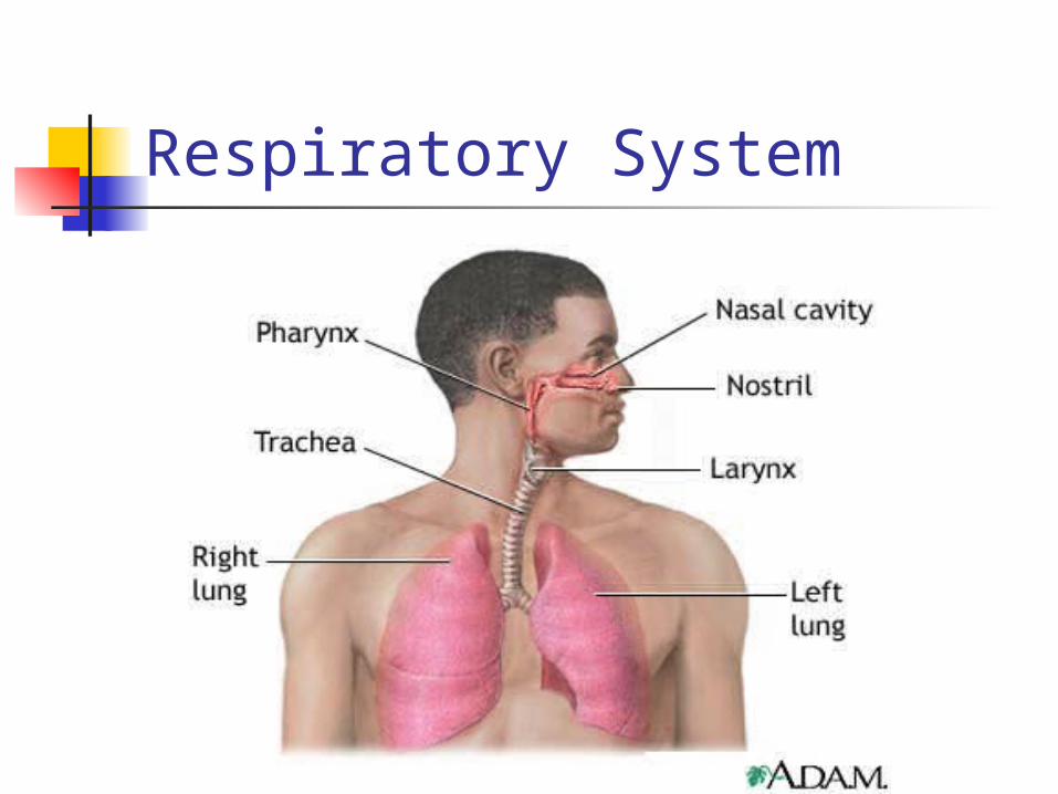
Respiratory System
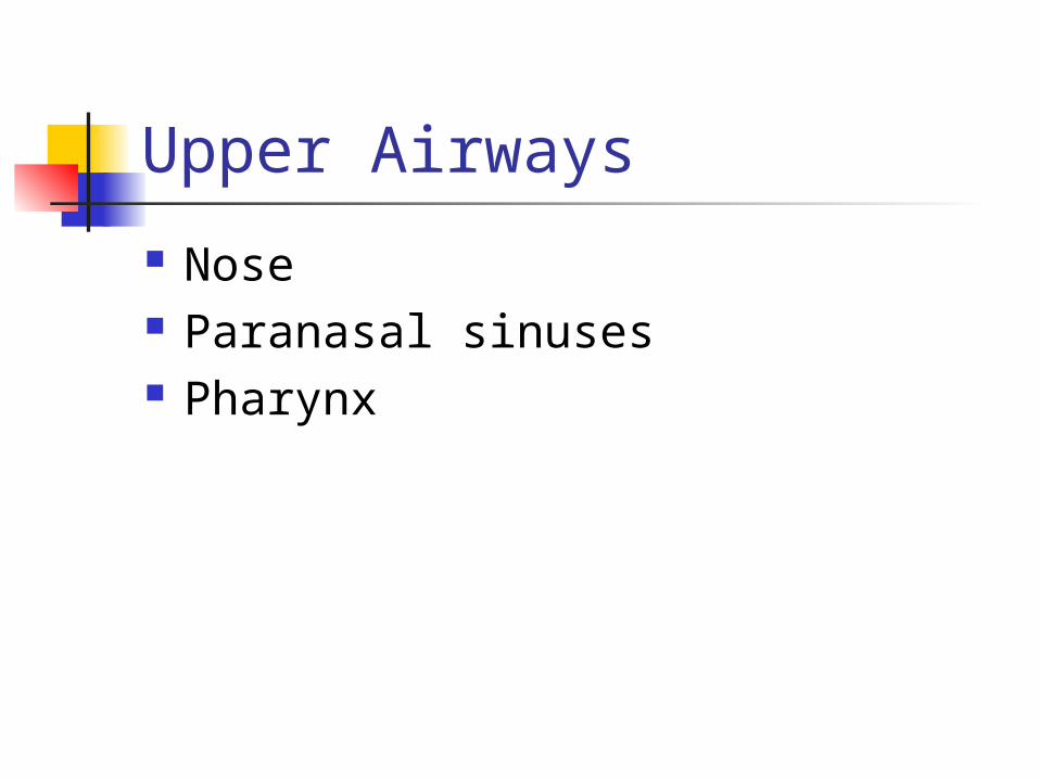
Upper Airways
Nose Paranasal sinuses Pharynx
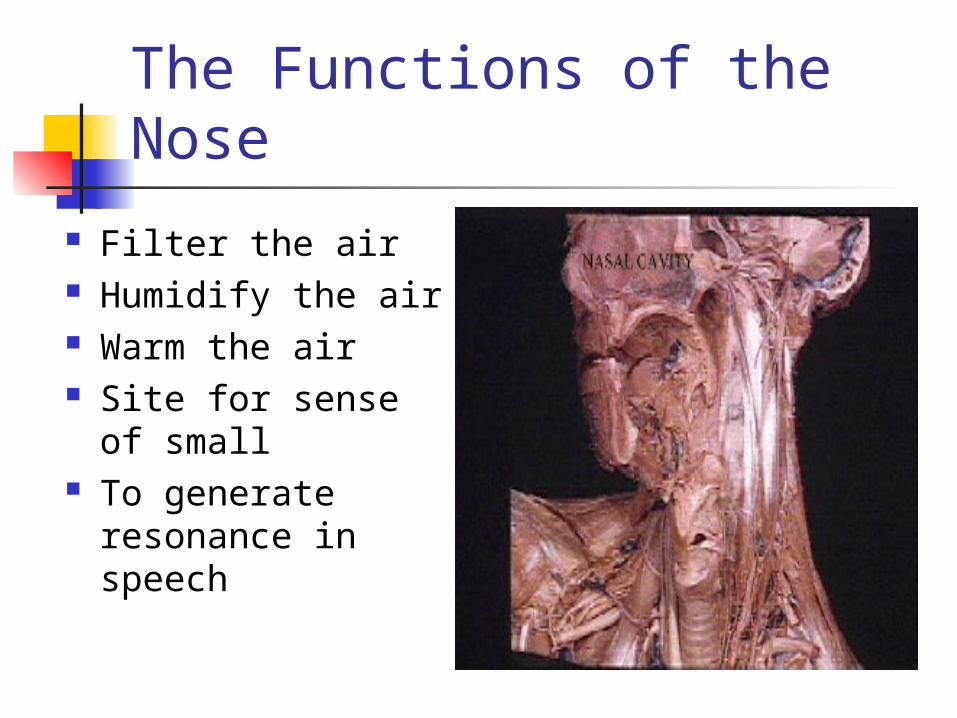
The Functions of the Nose
Filter the air Humidify the air Warm the air Site for sense of
small To generate
resonance in speech
The Nose
Rigid structure composed of cartilage and bone
Septal cartilage divides nasal cavity into two nasal fossae
Palate divides nasal cavity and oral cavity
Nose divided into 3 regions

Nasal structure

Nares and Nasal Cavity

Regions of the nose Nares or nostrils serve as opening
for the nasal fossae—two cavities in middle of the face
Vestibule/vestibular regionVestibule/vestibular region Lined with stratified squamous
epithelium Contain vibrissae-nasal hair, first
line of defense, function to filter inspired air

Sagital section of the head
Nasal regions
Vestibular area contains sebaceous glands; secrete sebum
Keeps vibrissae soft and filter gases
Olfactory regionOlfactory region: pseudostratified columnar epithelium and olfactory cells

Regions of the nose Respiratory—highly vascular;
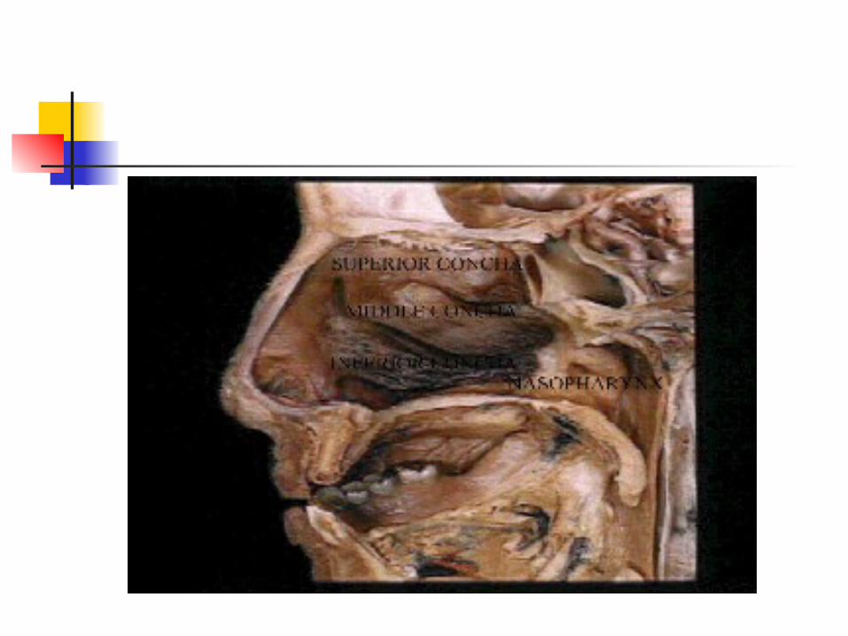
ciliated, pseudostratified columnar epithelium Contains turbinates or conchae;
increase surface area (166 cm2) for humidification, heating/cooling and filtering of air
Mucous membranes provide up to 650-1000 ml of water/day to humidify air

Respiratory region of nose
Goblet cells in mucus membrane secrete 100 ml/day of mucous; aids in trapping inspired particles and prevents them from entering lower respiratory trace
Each columnar cell contains 200-250 cilia; beat in waves toward oropharynx (mouth), 2cm/min

Sinuses
Air-filled cavities within the skull (cranium)
Aka paranasal sinuses (four pairs) Function not clear, lighten head and
provide voice resonance Lied with pseudostratified cliated
columnar epithelium and goblet cells
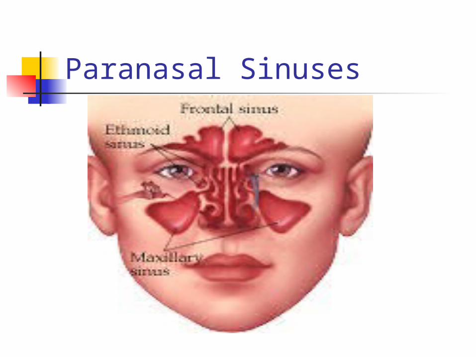
Paranasal Sinuses
Oral Cavity
Alternate respiratory passage Anterior 2/3 of tongue located in
oral cavity Another “respiratory” muscle Lined with stratified squamous
epithelium
+Pharaynx (Throat), hollow, upper portion of
the airway and the digestive tract Subdivided into: nasopharynx,
oropharynx, laryngopharynx
Nasopharynx Pseudostratified ciliated columnar
epithelium

Pharynx

Nasopharynx
Filters bacteria and foreign particles from inspired air
Carries this to the stomach Eustachian tube and auditory tube
open into lateral surfaces, connect nasopharynx to middle each, equalizes pressure of middle ear
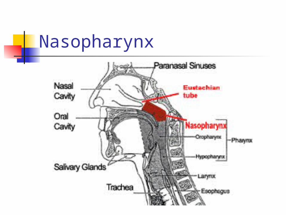
Nasopharynx

Oropharynx
Between soft palate above and base of tongue below
From tip of uvula to epiglottis Stratified squamous epithelium Gas conduction, filtering of air Defense mechanism: gag reflex

Oropharynx

Laryngopharynx
Stratified squamous epithelium Gas conduction Connecting zone between upper
and lower airway (vocal cords and below)
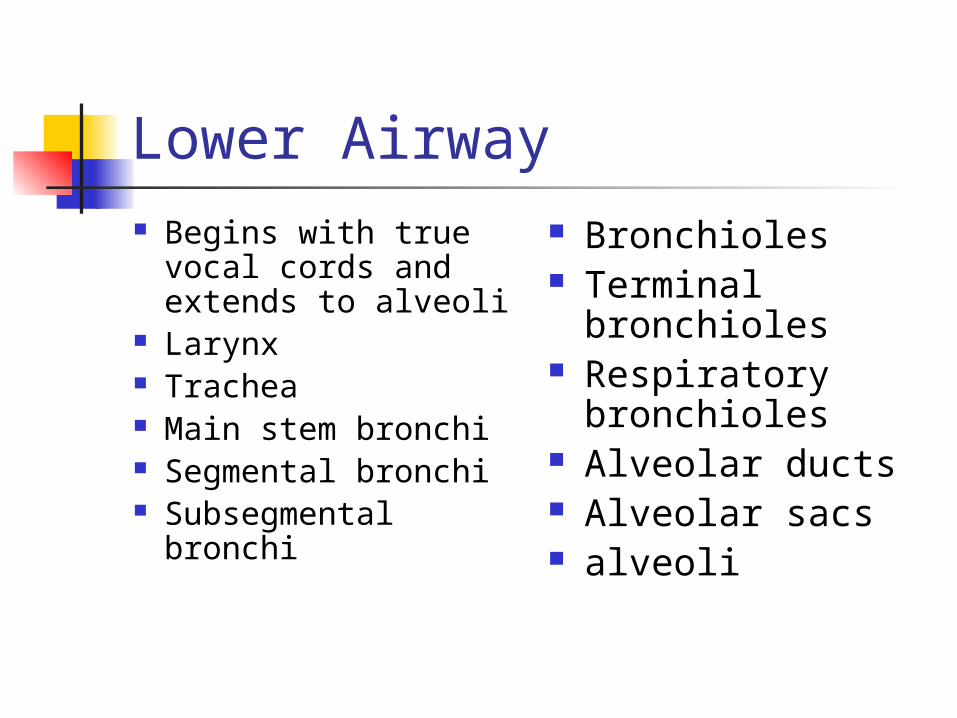
Lower Airway Begins with true
vocal cords and extends to alveoli
Larynx Trachea Main stem bronchi Segmental bronchi Subsegmental
bronchi
Bronchioles Terminal
bronchioles Respiratory
bronchioles Alveolar ducts Alveolar sacs alveoli

Larynx Lies between base of tongue and
trachea Protrusion is the thyroid cartilage,
aka “Adam’s apple.” Houses the vocal cords, primary
use is vocalization Connection point-upper and lower
airways

Larynx

Larynx Extends from C3
to C6 Pseudostratified
ciliated columnar epithelium
Functions: Free flow of air to
the lungs
During inspiration, vocal folds abduct, move apart, and widen glottis
Valsalva maneuver and Muller maneuver
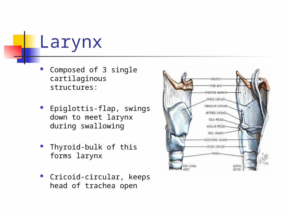
Larynx Composed of 3 single
cartilaginous structures:
Epiglottis-flap, swings down to meet larynx during swallowing
Thyroid-bulk of this forms larynx
Cricoid-circular, keeps head of trachea open
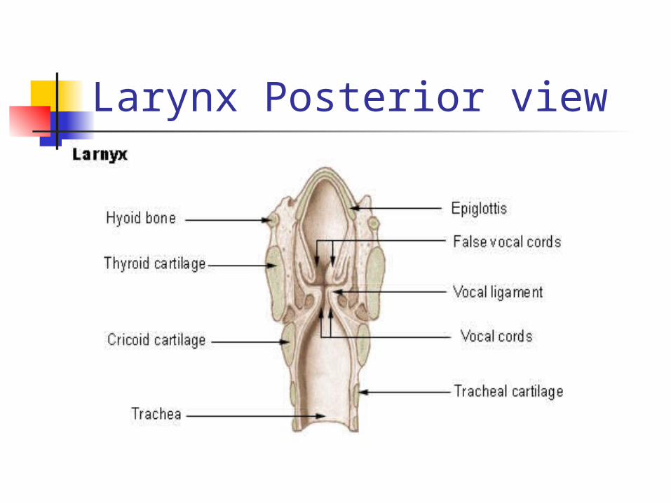
Larynx Posterior view

Epiglottis
Covers the rima glottidis during swallowing (glottis=cords & space)
Larynx has poor lymphatic drainge, prone to edema
Epiglottitis is a life-threatening condition (supraglottic croup), bacterial origin
Narrowest part of lower airway in adult

Glottis


Thyroid
Largest of the laryngeal cartilages Primary housing the vocal cords Inflammation below the vocal cords
known as laryngotracheobronchitis Croup Subglottic croup Viral orgins
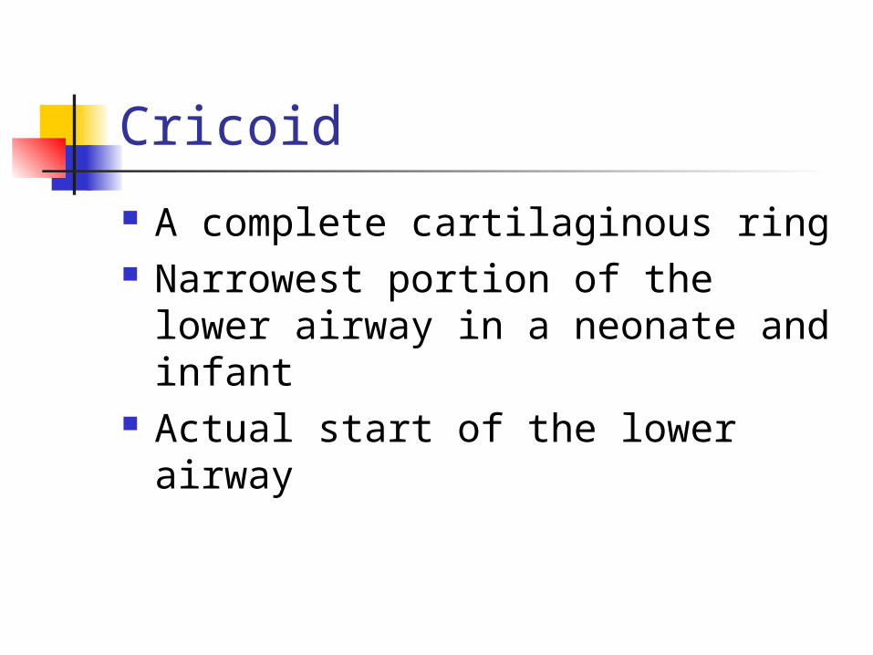
Cricoid
A complete cartilaginous ring Narrowest portion of the lower
airway in a neonate and infant Actual start of the lower airway

Larynx – superior view
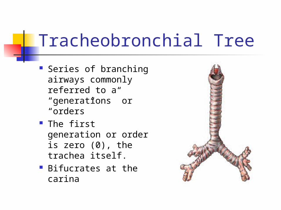
Tracheobronchial Tree Series of branching
airways commonly referred to a “generations” or “orders”
The first generation or order is zero (0), the trachea itself.
Bifucrates at the carina
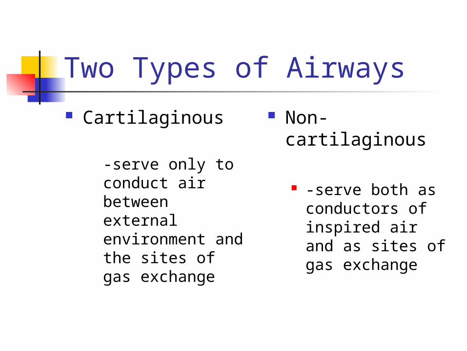
Two Types of Airways Cartilaginous
-serve only to conduct air between external environment and the sites of gas exchange
Non-cartilaginous
-serve both as conductors of inspired air and as sites of gas exchange

Tracheobronchial Tree Dichotomous branching (daughter
branches) Airways become progressively
narrower, shorter, and more numerous
Cross-sectional area enlarges Common histology (at the nose) and
throughout until the bronchiole generation

Histology
Three major layers
Epithelial lining
Lamina propria
Cartilaginous layer

Histology

Tracheal lining
Pseudostratified columnar epithelium with cilia; goblet cells, serous cells, and specialized submucosal bronchial glands
200+ cilia per cell, 5-7 microns long
Beat cephalid (head) toward oropharynx

Tracheal Lining
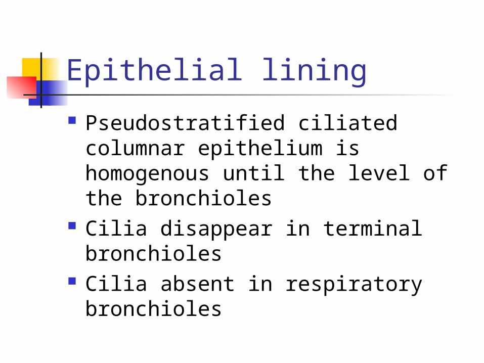
Epithelial lining
Pseudostratified ciliated columnar epithelium is homogenous until the level of the bronchioles
Cilia disappear in terminal bronchioles
Cilia absent in respiratory bronchioles


Histology

Mucous blanket
Covers the epithelial lining Composed of
-95% water -glycoproteins Carbohydrate lipids DNA Cellular debris
Mucous
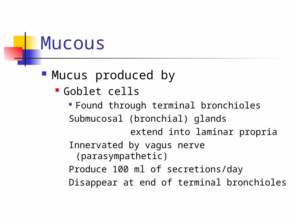
Mucus produced by Goblet cells
Found through terminal bronchiolesSubmucosal (bronchial) glands
extend into laminar propriaInnervated by vagus nerve
(parasympathetic)Produce 100 ml of secretions/dayDisappear at end of terminal bronchioles

Mucous Blanket Two distinct
layers Sol layer Gel layer
Cilia move through sol layer and strike gel layer propelling it toward mouth
At a rate of 2 cm/minute

Mucocilliary Escalator
Defense mechanism of lower airways
Mucus propelled up airway to larynx
Cough mechanism moves secretions into oropharynx via sheering forces

Mucociliary transport & cough

Factors Which Slow Mucocilliary Transport Cigarette smoke Dehydration Positive pressure
ventilation Endotracheal
suctioning High inspired
oxygen concentrations
Hypoxia Atmospheric
pollutants General
anesthesia Parasympatholyti
c drugs
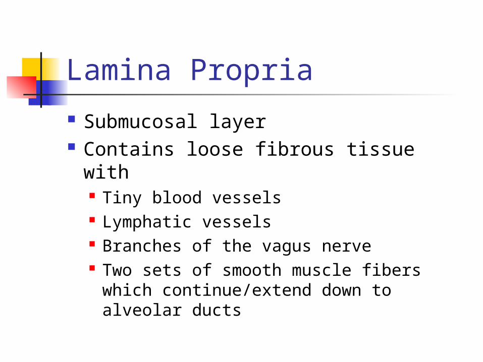
Lamina Propria
Submucosal layer Contains loose fibrous tissue with
Tiny blood vessels Lymphatic vessels Branches of the vagus nerve Two sets of smooth muscle fibers
which continue/extend down to alveolar ducts

Lamina propria

Mast cells Found in lamina propria near
Branches of vagus nerve and blood vessels
Scattered throughout smooth muscle Loose connective tissue of skin and
intestinal mucosa Cell constituents of submucosal glands
Important part of humoral immune response (circulating antibodies) which defend against antigens
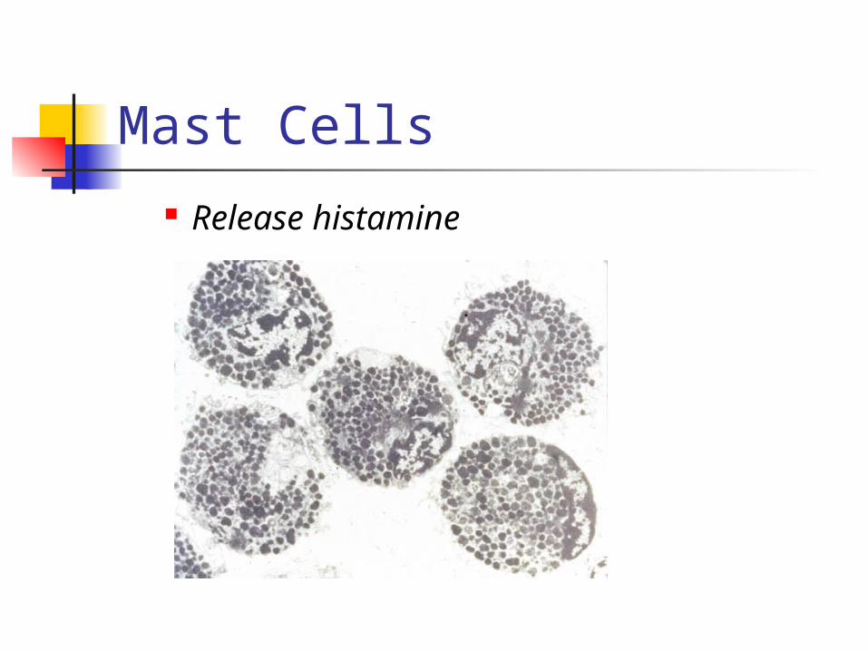
Mast Cells Release histamine

Cartilaginous Layer Outermost layer of
tracheobronchial tree Consist of
Trachea Mainstem bronchi Lobar bronchi Segmental bronchi Subsegmental bronchi
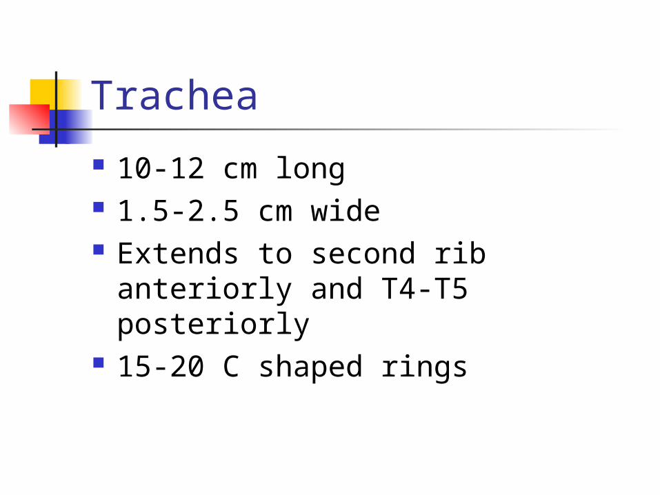
Trachea
10-12 cm long 1.5-2.5 cm wide Extends to second rib anteriorly
and T4-T5 posteriorly 15-20 C shaped rings

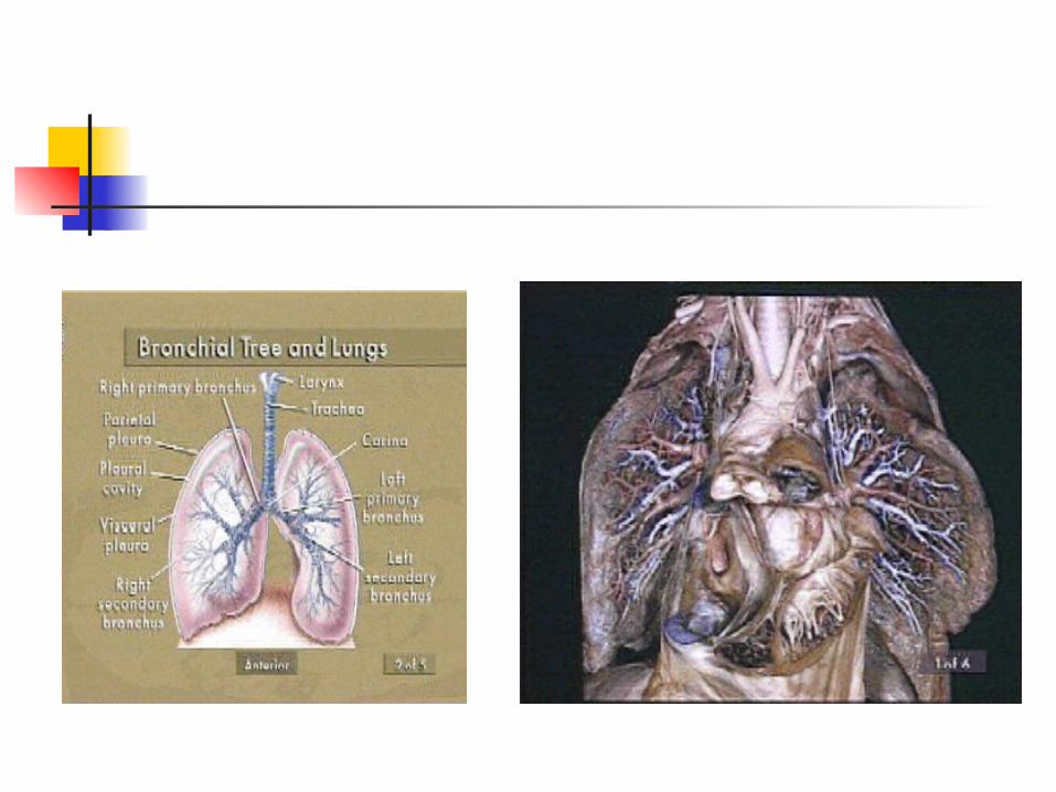
Main Stem Bronchi Right bronchus
Wider More vertical 5 cm shorter Supported by C
shaped cartilages 20-30 degree
angle First generation
Left bronchus Narrower More angular Longer Supported by C
shaped cartilages 40-60 degree
angle First generation
Lobar Bronchi R main stem
divides into: Upper lobar
bronchus Middle lobar
bronchus Lower lobar
bronchus
L main stem divides into: Upper lobar
bronchus
Lower lobar bronchus


Segmental Bronchi 3rd generation
R lobar divides into Segmental bronchi 10 segments on
right
L lobar divides into Segmental bronchi 8 segments on left

Subsegmental Bronchi
4th to 9th generations Progressively smaller airways 1-4 mm diameter At 1 mm diameter connective
tissue sheath disappears


Noncartilagenous Airways Bronchioles
10-th to 15th generation
Cartilage is absent Lamina propria is
directly connected with lung parenchyma
Surrounded by spiral muscle fibers
Epithelial cells are cuboidal
Less goblet cells and cilia
With no cartilage, airway remains open due to pressure gradients
Terminal Bronchioles
16th to 19th generation Average diameter is 0.5 mm Cilia and mucous glands begin to
disappear totally End of the conducting airway Canals of Lambert-interconnect this
generation,provide collateral ventilation

Gas exchange zone
Respiratory bronchioles Acinus (aka primary acinus; aka
primary lobule)—respiratory bronchioles to the alveoli
Ducts, sacs, alveolar Squamous epithelium
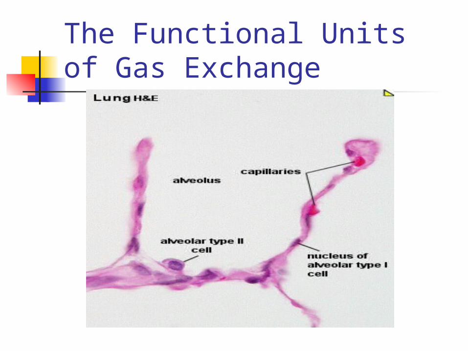
The Functional Units of Gas Exchange
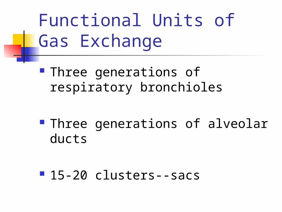
Functional Units of Gas Exchange
Three generations of respiratory bronchioles
Three generations of alveolar ducts
15-20 clusters--sacs

Gas exchange terminology
All of the structures arising from a single terminal bronchiole are called Primary lobule Acinus Terminal respiratory unit Lung parenchyma Functional units

Acinus/Primary lobule
Respiratory bronchioles with some alveoli arising from their walls
Alveolar ducts arise from respiratory bronchioles--alveoli whose septal wall contain smooth muscle
Alveoli
Ca. 300 million alveoli Between 75 µ to 300 µ in
diameter
Most gas exchange takes place at alveolar-capillary membrane

Anatomic Arrangement of Alveoli
85-95% of alveoli covered by small pulmonary capillaires
The cross-sectional area or surface area is approximately 70m2
Acinus or Lobule
Each acinus (unit) is approximately 3.5 mm in diameter
Each contains about 2000 aveloli
Approximately 130,000 primary lobules in the lung

Acinus


Alveolar epithelium
Two principle cell types:
Type I cell, squamous pneumocyte
Type II cell, granular pneumocyte

Type I Cell
95% of the alveolar surface is made up of squamous pneumocyte cells
Between 0.1 µ and 0.5µ thick
Major site of gas exchange
Type II Cell
5% of the surface of alveoli composed of granular pneumocyte cells
Cuboidal in shape with microvilli Primary source of pulmonary
surfactant Involved with reabsorption of fluids
in the dry, alveolar spaces

Type II Pneumocyte

Pore of Khon
Small holes in the walls of adjoining alveoli (alveaolar septa)
Between 3 to 13 µ in diameter Formation of pores may be due to:
Desquamation due to disease Normal degeneration due to aging Movement of macrophages leaving
holes

Canals of Lambert/Pores of Kohn
Provide for collateral ventilation of difference acinii or primary lobules
Additional ventilation of blocked units
May explain why diseases spread so quickly at the lung tissue (paremchymal) level
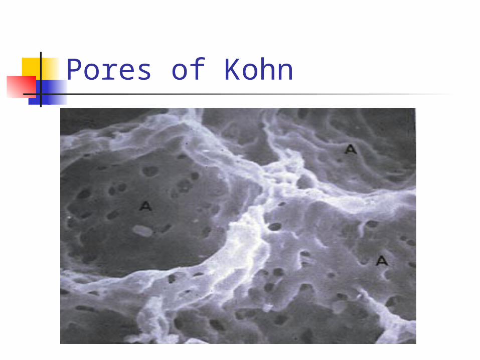
Pores of Kohn

Alveolar macrophages
So-called Type III cell Remove bacteria and foreign
particles May originate as
Stem cells precursors in bone marro Migrate as monocytes through the
blood and into the lungs
Intersitium/interstial space
Surround, supports, and shapes the alveoli and capillaries
Composed of a gel like substance and collagen fibers
Contains tight space and loose space areas

Alveolar-Capillary Site--Interstitial Space


Interstitium Water content in loose space can
increase by 30% before there is a significant change in pulmonary capillary pressure
Lymphatic drainage easily exceeded Collagen limits alveolar distensibility Area of scarring, making for stiffer or
non-compliant lungs

Blood Supply to the Pulmonary System
Two Systems
Bronchial Blood Supply
Bronchial arteries From aorta to temrinal bronchioles
Merge with pulmonary arteries and capillaries
1% of total cardiac output (left ventricle)

Bronchial arteries
Also nourish Mediastinal lymph nodes Pulmonary nerves Some muscular pulmonary arteries
and veins Portions of the esophagus Visceral pleura

Bronchial venous system
1/3 blood returns to right heart Azygous Hemiazygous Intercostal veins
This blood comes form the first two or three generations of bronchi

Bronchial venous return 2/3 of blood flowing to terminal
bronchioles drains into pulmonary circulation via “bronchopulmonary anastomoses”
Then flows to left atrium via pulmonary veins
Contributes to “venous admixture” or “anatomic shunt” (ca. 5% of C.O.)

Pulmonary Vascular System
The second source of blood to the lungs
Primary purpose is to deliver blood to lungs for gas exchange
Also delivers nutrients to cells distal to terminal bronchioles
Composed of arteries, arterioles, capllaries, venules, and veins
Pulmonary Capillaries
Walls are les than 0.1µ thick Total external thickness is about
10µ Selective permeability to water,
electrolytes, sugars Produce and destroy biologically
active substances
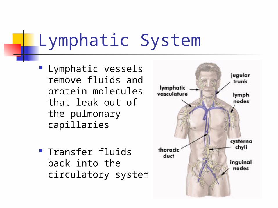
Lymphatic System Lymphatic vessels
remove fluids and protein molecules that leak out of the pulmonary capillaries
Transfer fluids back into the circulatory system
Lymphatics
Lymphatic vessels arise within loose spaces of connective tissue
Vessels then follow bronchial airways, pulmonary airways, pulmonary arteries and veins to the hilum
Lymphatics
Vessels end in pulmonary and bronchopulmonary lymph nodes within and outside of lung parenchyma
Nodes acts as filters to keep particles and bacteria from entering the blood

Lymphatics Lymphatic vessels are not
in the walls of the alveoli.
Within the interstial spaces to help drain fluids and foreign materials.
More lymphatic vessels on surface of lower lobes than on upper and middle lobes (dependent portions)
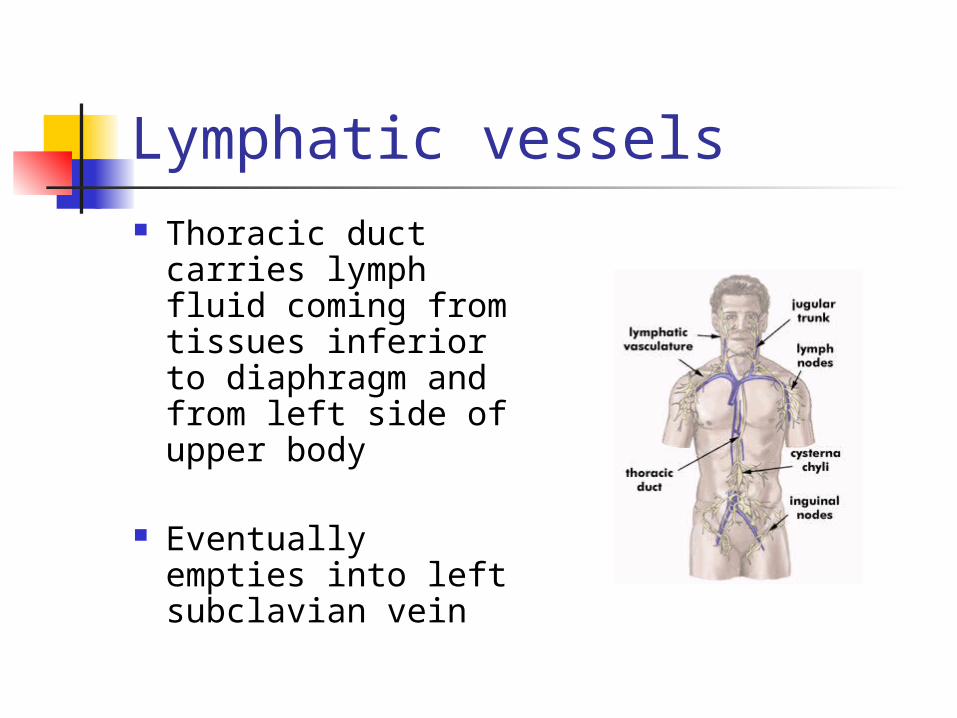
Lymphatic vessels Thoracic duct
carries lymph fluid coming from tissues inferior to diaphragm and from left side of upper body
Eventually empties into left subclavian vein
Lymphatic vessels
Right lymphatic duct drains the right half of the body superior to the diaphragm
Empties into the right subclavian vein