basic low vision rehabilitation for - coavision.org -ot and comp approach to lv.pdf · orientation...
TRANSCRIPT
1
Rebecca Kammer, OD, F.A.A.O., Diplomate
•Associate Professor
•Western University College of Optometry
Co-author of revised California Central
Visual Field Test
What exactly is modern low vision and
what is the optometrist’s role?
R Kammer, C Sell, R Jamara, E Kollbaum, 2010
Goal: to explore the low vision rehabilitation
practices of optometrists who prescribe devices
for the moderately impaired AMD patient.
136 responses Approx 69% were private practice ODs Balance were rehab, education, or other
Who completes training? 54% of respondents performed the training
themselves 15% used an OT 18% hired a technician The rest used a rehab teacher or didn’t train at
all Training is not performed in the patients home
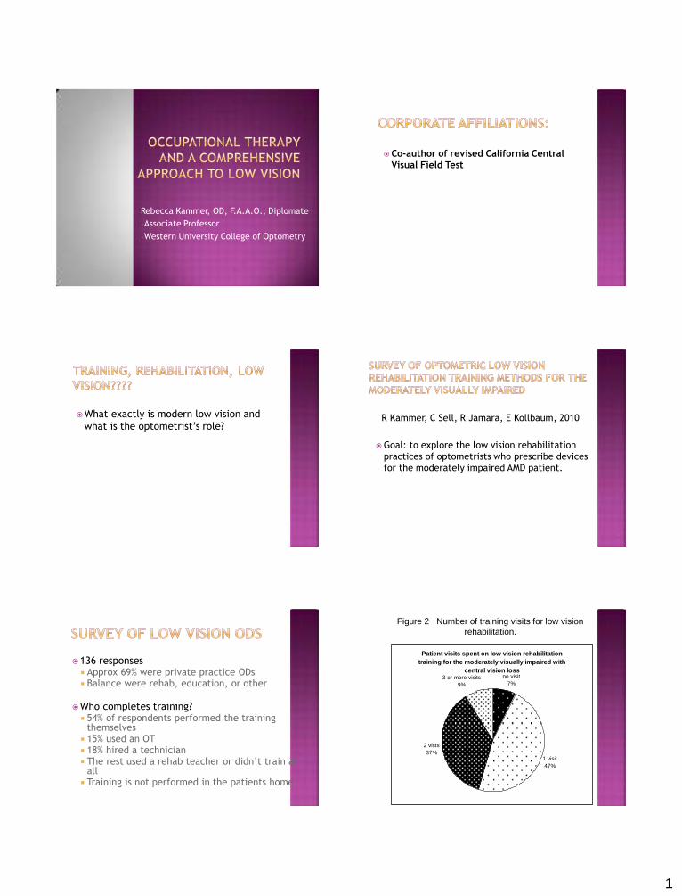
Figure 2 Number of training visits for low vision
rehabilitation.
Patient visits spent on low vision rehabilitation
training for the moderately visually impaired with
central vision lossno visit
7%
1 visit
47%
2 vists
37%
3 or more visits
9%

2
Training involves practice with eccentric viewing strategies.
○ All the time ○ Most of the time ○ Some of the time ○ Never
Training includes use of the device in spotting activities (i.e. mail, bills, medicine labels, phone books, and/or food packaging labels).
○ All the time ○ Most of the time ○ Some of the time ○ Never
Low vision optometric practitioners were
rarely prescribing reading rehabilitation
training longer than 2 visits for moderately
visually impaired AMD patients.
Most ODs provide a high level of optical
device training but incorporation of other
training strategies was less frequent.
OT
term “rehabilitation”
safety and falls
home assessments
emotional status
referral for resources
for transportation,
counseling
OD
Terms not consistent
optical device training
– Lighting, Focal point, Optics
eccentric viewing…
maybe?
Home Safety
Eccentric viewing
Device adaptation
Near-point functions
Distance/driving functions
Daily living skills
Teaching Independence/Social integration
Orientation and Mobility
Psych support/referral
Patients who have difficulty completing ADL’S (activities of daily living) due to visual impairment.
Potential for improved functioning.
For Medicare.....
Vision 20/70 or worse in the better eye.
Or use central scotoma code instead of VA
Occupational therapy is skilled treatment
that helps individuals achieve independence
in all facets of their lives. It gives people the
"skills for the job of living" necessary for
independent and satisfying lives

3
Individualized treatment programs to
improve one's ability to perform daily
activities
Comprehensive home and job site
evaluations with adaptation
recommendations
Performance skills assessments and
treatment
Adaptive equipment recommendations and
usage training
Guidance to family members and caregivers
Trained in disability and aging.
Able to address physical, psychological, cognitive and social needs.
Focus is positive---on improving daily functioning.
Not a replacement for the blindness system.
Reimbursed by Medicare and other ins.
Low Vision Evaluation, OD
History, Distance Acuities, Refraction, Central
Scotoma, Contrast, Reading Acuity, Response to
Magnification
Refer for OT evaluation
OT Assessment
OT Training Sessions (2-6 sessions)
Update OD on progress
Refer back to OD for prescribing decisions
Finalize OT Training with prescribed devices
Patient’s history, prior level of function in ADL, work and productive activities.
Functional activities that patient can and cannot perform due to visual impairment.
Reading and writing assessment. Contrast test (if OD doesn’t perform) and
lighting Central field assessment (functional) based
on OD field Patient’s Physical status Patient’s Cognitive status
Developed with patient and family members
to determine goals and outcomes (in-line
with OD evaluation).
Short term goals-updated monthly.
Long term goals-functional outcomes.
Needs to be signed by the referring MD or
OD – state laws. (A few states do not allow
OD to sign)
Average of 4-6 sessions 1½ to 2 hours.
Sessions in patient’s homes or in-office
Example of First session:
Education regarding contrast and lighting.
Lighting assessment.
Education regarding use of remaining vision.--- Central field loss vs. Peripheral field loss

4
OD refers/writes orders (signs referral)
OT carries out the orders Usually only OT’s are licensed to carry out
therapy Training by optician, OTA’s, CLVTs, technicians
will not be reimbursed
Billed by OD
OT is employed by OD (incident to –
most tx done in office)
OT is independent contractor, paid fee
for service
OT is employed by OD, salaried, use OT
MCR number for billing (advantage,
home visits)
Billed by OT (referral relationship only)
OD receives no compensation
OT Evaluation – about an hour - $100
Billed according to time – 15 minute increments
Reimbursed between $25-35/unit
Utilize OT codes 97530 – Therapeutic Activities (Eccentric
Viewing)
97353 – Self Care & home management
From 6 to 12 hours (depends upon region)
Must show progress connected to goals LL
OD and OT
If conservative arrangement of 70/30 split with
30% to you (OT as per diem)
$120/hr paid by MCR
$36 per hour for each of 4-6 visits
For each patient who works with OT,
If 1 of the 4 patients need OT services, total
additional NET revenue = $216 per half day
Partnership
Trust
3 patients a day for the OT is minimum to
pay salary and bring in extra revenue
Offer both in-home and in-office visits
Pick a driving radius
Salary range

5
Purpose of Case history
Identify realistic visual goals
Guides the prescribing process
OD is always thinking ahead
Assists in determining what level of
care is needed
Near Vision needs/abilities
Distance needs/abilities
Activities of Daily living issues
Social History
Illumination and Glare needs
Mobility needs
Job related needs
Distance Low Vision Chart Range greater than 20/200 Flip charts can be portable and easy to use
3 meter test distance (10 feet) Some ODs use 1 meter
M notation is easy to use linear (follows logMar) 2M is twice the size of 1M
Example for recording near acuity: 0.4/2M
Retinoscopy is critical
Trial Frame Refraction
Large lens changes
JND rule of thumb: use denominator of 20 foot
VA and move decimal point (e.g. 20/200, 2D JND,
so use +/- 1.00D lenses)

6
Cylinder screening (if not sure of ret findings)
Hold up a -2D Cylinder with a +1.00D sphere
Effectively this is a EDS = Plano
Hold both lenses at major meridians (show with and
without lenses)
If patient prefers with, then accepts cylinder at that
axis)
Automated central screener
Scanning Laser Ophthalmoscope
Amsler Grid?????
CCVFT

7
CCVFT
FAST
Inexpensive
EASY!
CCVFT has special value…
Maps Scotomas (blind spots)
Requires practice
Test distance 57cm (1cm = 1 degree)
Laser pointer(s)
Have patient view or attempt to view center
of spokes
Can do OU and then monocularly if needed
“The process of aligning the image into a new retinal viewing area is referred to as eccentric viewing (EV)”
Patients may spontaneously develop EV for an area near the fovea, a preferred retinal locus or area (PRL)
PRL location and utilization
Functional implications
Magnification needs
8
Schuchard RA, Naseer S, de Castro K. Characteristics of AMD patients with low vision receiving visual rehabilitation. J Rehabil Res Dev, 1999; Oct:36(4)294-302.
Cummings RW, Whittaker SG, Watson GR, Budd JM. Scanning characters and reading with a central scotoma. Am J Optom Physiol Opt, 1985;Dec:62(12)833-43.
Timberlake GT, Mainster MA, Peli E, Augliere RA, Essock EA, Arend LE. Reading with a macular scotoma. I. Retinal location of scotoma and fixation area. Invest Ophthalmol Vis Sci, 1986;Jul:27(7)1137-47.
Timberlake GT, Peli E, Essock EA, Augliere RA. Reading with a macular scotoma II. Retinal locus for scanning text. Invest Ophthalmol Vis Sci, 1987;Aug:28:1268-74.
Schuchard RA, Fletcher DC, Maino J. A scanning laser ophthalmoscope (SLO) low-vision rehabilitation system. Clin Eye Vision Care, 1994;6(3)101-7.
Fletcher DC, Schuchard RA. Preferred retinal loci relationship to macular scotomas in a low-vision population. Ophthalmol, 1997;104(4)632-8.
Frennesson C, Jakobsson P, Nilsson UL. A computer and video display based system for training eccentric viewing in macular degeneration with an absolute central scotoma. Doc Ophthalmol, 1995;91:9-16.
Nilsson UL, Frennesson C, Nilsson SEG. Location and stability of a newly established eccentric retinal locus suitable for reading, achieved through training of patients with a dense central scotoma. Opt Vis Sci, 1998;Dec;75(12)873-8.
Fletcher DC, Schuchard RA, Watson G. Relative locations of macular scotomas near the PRL: effect on low vision reading. J Rehabil Res Dev, 1999;Oct;36(4) 356-64.
http://www.lowvisionproject.org/vaevs.htm
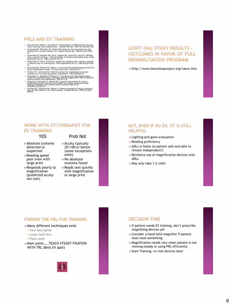
Absolute scotoma detected or suspected
Reading speed poor even with large print
Responds poorly to magnification (predicted acuity not met)
Acuity typically 20/100 or better (some exceptions exist)
No absolute scotoma found
Reads text quickly with magnification or large print
YES Prob Not Lighting and glare evaluation
Reading proficiency
ADLs in home (is patient safe and able to
remain independent?)
Reinforce use of magnification devices with
ADLs
May only take 1-2 visits
Many different techniques exist
Face description
Large clock face
Flash cards
Main point…..TEACH STEADY FIXATION
WITH TRL (Best EV spot)
If patient needs EV training, don’t prescribe
magnifying devices yet
Consider a hand held magnifier if patient
must have something
Magnification needs vary when patient is not
viewing steady or using PRL efficiently
Start Training, re-visit devices later

9
6) Response to Magnification
7) Device Evaluation
After OT visit(s) and patient returns
Perform more comprehensive Magnification evaluation
Keep goals in mind (e.g. read newspaper)
Identify magnification necessary to read newspaper
Introduce High addition lenses that will produce desired acuity
Introduce Telescopic Magnification that will produce desired goal acuity
A Common Method = Reciprocal of
Vision (ROV)
Denominator of Best Corrected Distance Acuity
--------------------------------------
Denominator of Goal Visual Acuity
Assumes Distance Acuity = Near Acuity
Goal acuity = 20/40 (meets most visual
demands)
Ex: patient has BCVA = 20/120
ROV = 120/40 = 3X
Prescribing a high add at near can create desired
magnification (just by patient holding print closer
or at the focal point of the lens)
Multiply 2.5D and the ROV to obtain Equivalent
power for near (High add power)
20/200 patient will require about a +10 D add
20/100 patient will require about a +5.00 D add
This is a very rough starting point without calculations

10
Hand Magnifiers
Stand Magnifiers
Dome Magnifiers
Used for viewing targets further than 10 feet
Two Types
Galilean (2X to 3X)
Keplerian (3X to 10X)
Children use for seeing whiteboard in
classroom
Adults use for seeing faces, signs, driving
Hand Held
Bioptics
Sportoculars –
sustained distance
viewing
3X and 4x
Consider refraction
Does the patient need the Rx to be
corrected?
Plo-2.50X180 20/60
Most likely requires correction prior to
magnification
Sunlight
Fluorescent Light
Modified Fluorescent
Incandescent Light
Neodymium Light
Halogen
Led (Light Emitting Diode)

11
Color
Transmission
Transition (photochromaticity)
Polarization
Selective Filtration
UV Coating
Example: 2% Dark Amber, 40% yellow, 20%
plum
Range of Magnification so rarely need
calculations
Great contrast enhancement
Require steady hands
Many options
Range of Magnification so never need
calculations
Great contrast enhancement
Require steady hands
Screen Magnification software
Keyboard Magnification Control (Control-plus
or mouse roller ball)
Accessibility Menu Options

12
Case History
Acuities
Refraction
Contrast Sensitivity
Fields
Response to Magnification
Prescribing for Near
Prescribing for Distance
Prescribing for Glare and lighting
Prescribing Technology
Multidisciplinary Evaluation & Care
CentraSight™ Program
Step Action Practitioner
1 Diagnosis Retina Specialist
2 Candidate Screening Retina Specialist
Low Vision Optometrist
3 Implantation Surgeon
4 Visual Rehabilitation/Training Occupational Therapist
69
Telescope Prosthesis Candidacy
•Diagnosis
•Refer
• Cornea health
•AC depth,
other
1 Retina
OT
•Functional Goals
•Rehab Potential
Optom
•Low Vision Eval
•Eye Selection
2 Low
Vision
3 Surgeon
71
Visit Overview
VISITS Med
Preop 1
Optom
Preop1
Optom
Preop1
Wk 1 Wk 2 Wk 3 Wk 4 Wk 5 Wk 6 Wk 7 Wk 8 Wk 9 Wk 10 Wk 11 Wk 12 Mo 6 Mo 12
Medical
Optom
Rehab
Implantation
72

13
Basis of Guide
73
Wide-angle micro-optics
Sits in capsular bag after lens extraction
Near and Distance
Standard spectacle Rx
Technology
74
Field of View
3X External Telescope = 8° 3X Wide Angle Implant = 20°
3X implant field of view 625% of external telescope (1,111% if mounted on
spectacles)
75
Functional Factors
• Both eyes scan together
• Avoids vestibular conflict
• Available on-demand for
dynamic & social activities
• Hands-free use
• Compatible with
interpersonal interaction
‒ Eye contact; face recognition
Implantable Telescope
Peli E. (2002) The optical functional advantages of an intraocular low vision telescope. Optometry and Vision Science 79(4): 225-233
76
End-Stage AMD
Disciform Scar or GA
No Active CNV
Bilateral Scotomas
20/160 – 20/800 BCVA
Patient Population
77
CAT CAT
CAT
CAT
Natural Lens / IOL
Telescope Prosthesis (enlarged retinal image)
Move eye, image too small for
perimacular retina
Vision with Macular Scar
78

14
Scarred Macula Central Visual Field Projection (Natural Lens/IOL)
CAT CAT
Telescope Implant Central Visual Field Projection
79
Vision with Macular Scar Clinical Trial Efficacy (IMT002)
20/326 Baseline
20/200 20/127
Source: * Stevenson, **Ebert,† Erber/Osborn
Ability to care for self &
others*
More able to perform
activities of daily living**
Face-face communication†
Mean Improvement with 3X model
Statutory Blindness
80
Realistic Goals
Watch TV
Recognize people, expressions
Enjoy hobbies (e.g., fly-fishing, card games, gardening,
painting)
Enjoy social events: parties, movies/plays, sporting events,
dining
Self-care
Read large print books, mail/email, medicine; write checks,
letters
Unrealistic Goals
Clear immediate vision
Driving
Playing tennis, seeing golf ball in flight
Never need to use low vision aids
This process will be easy! This is a cure…
81
Telescope Implant Screening Evaluation
Low Vision Exam can be same as IMT
screening visit
Eye Selection is the Key
Green Light for Implant
OT role can be part of initial eval or after
implant