bacteriology a case report on disseminated rhodococcus
TRANSCRIPT
819
*Correspondence to: Kakuda, T.: [email protected]©2018 The Japanese Society of Veterinary Science
This is an open-access article distributed under the terms of the Creative Commons Attribution Non-Commercial No Derivatives (by-nc-nd) License. (CC-BY-NC-ND 4.0: https://creativecommons.org/licenses/by-nc-nd/4.0/)
NOTEBacteriology
A case report on disseminated Rhodococcus equi infection in a Japanese black heiferRyoko NAKAGAWA1), Hiroaki MOKI1), Kazuhide HAYASHI1), Kaname OONIWA2), Kyori TOKUYAMA2), Tsutomu KAKUDA2)*, Kazuki YOSHIOKA3) and Shinji TAKAI2)
1)Yokkaichi City Health Center Food Sanitation Inspection Station, Yokkaichi, Mie 510-0064, Japan2)Laboratory of Animal Hygiene, School of Veterinary Medicine and Animal Sciences, Kitasato University,
Towada, Aomori 034-8628, Japan3)Laboratory of Veterinary Anatomy, School of Veterinary Medicine and Animal Sciences, Kitasato University,
Towada, Aomori 034-8628, Japan
ABSTRACT. Rhodococcus equi was isolated from the granulomatous lesions of the lung, kidney, liver, and hepatic, mesenteric, and abomasum lymph nodes of a Japanese black heifer. R. equi isolates were analyzed by polymerase chain reaction for virulence-associated protein genes. The vapN gene was detected in all the isolates examined. This is the first report in which vapN-positive R. equi was isolated from cattle in Japan.
KEY WORDS: cattle, Rhodococcus equi, VapN, virulence plasmid
Rhodococcus equi is a Gram-positive pathogen that causes severe pulmonary abscesses and purulent enterocolitis in foals [13]. R. equi is widespread in the environment in grazing farms and common in the feces of farm animals including cattle, swine, horses, and others [16]. R. equi is also the opportunistic pathogen that causes a disease similar to tuberculosis in human patients with AIDS [18, 22, 23].
The pathogenicity of R. equi is associated with the presence of a large virulence plasmid [17]. R. equi causes pyogranulomatous pneumonia and secondary ulcerative enterocolitis accompanied by a high mortality rate in young foals worldwide [20]. Isolates from the lesions of infected equine hosts possess a circular plasmid (pVapA), encoding virulence-associated protein A (VapA), a 15–17 kDa surface protein [10, 11, 17, 21]. R. equi has also been isolated from the porcine submaxillary lymph nodes with granulomatous lesions as well as from apparently healthy pigs. The strains isolated from the pig and wild boar often carry another type of virulence plasmid (pVapB) encoding VapB, a VapA variant [7–9, 19].
Recently, a novel linear virulence plasmid encoding VapN (pVapN) was characterized in cattle, goat, and human isolates [1, 14, 15, 24]. In cattle, R. equi has low pathogenicity and lesions are mostly confined to a single lymph node [5]. Herein, we report a case of disseminated infection with R. equi carrying pVapN in cattle.
A 19-month-old Japanese black heifer was brought to a slaughterhouse and inspected on December 14, 2015. The heifer appeared depressed and had anorexia while at the fattening farm. There was no particular abnormality in ante-mortem inspection. The biochemical measurement of a blood sample was done using SPOTCHEM™ EZ analyzer (ARKRAY Co., Ltd., Kyoto, Japan). Serum total bilirubin was 0.6 mg/dl. Blood urea nitrogen (BUN) was 16 mg/dl. To detect antibody against bovine leukemia virus, passive hemagglutination reaction (PHA) was performed according to the manufacture’s instruction using the Bovine Leucosis Antibody Assay Kit (Nisseiken Co., Ltd., Tokyo, Japan), and the result was negative.
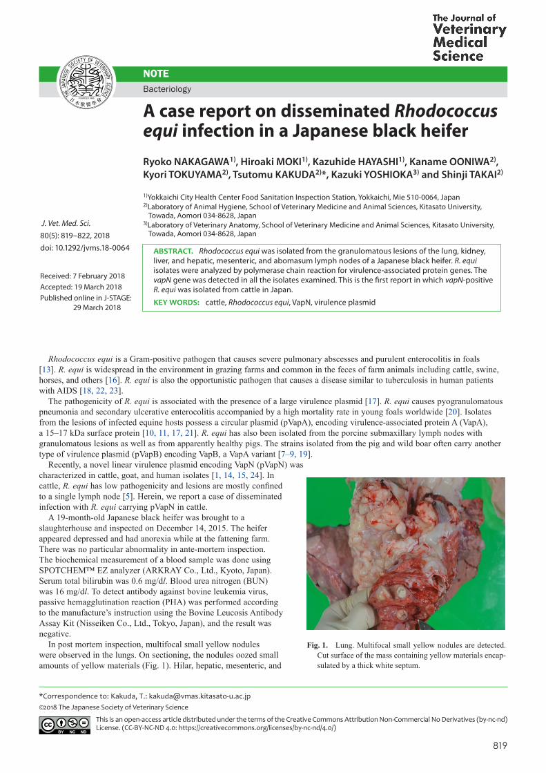
In post mortem inspection, multifocal small yellow nodules were observed in the lungs. On sectioning, the nodules oozed small amounts of yellow materials (Fig. 1). Hilar, hepatic, mesenteric, and
Received: 7 February 2018Accepted: 19 March 2018Published online in J-STAGE: 29 March 2018
J. Vet. Med. Sci. 80(5): 819–822, 2018doi: 10.1292/jvms.18-0064
Fig. 1. Lung. Multifocal small yellow nodules are detected. Cut surface of the mass containing yellow materials encap-sulated by a thick white septum.

R. NAKAGAWA ET AL.
820doi: 10.1292/jvms.18-0064
abomasum lymph nodes were markedly enlarged. Hepatitis, rumenitis, reticulitis, omasitis, abomasitis, enteritis, and colonitis were observed. The hilar lymph nodes were enlarged to about 11 cm by 7 cm; the hilar and other enlarged lymph nodes were solid and carneous. The mesenteric lymph nodes were liquefied in the central part, and other lymph nodes were observed to be white, gritty foci on a cut surface.
Stamp samples of the hilar, hepatic, mesenteric, and abomasum lymph nodes were stained with Modified Giemsa, Gram and Ziehl-Neelsen stainings. There were a few mature lymphocytes without atypia in stamp samples stained with Modified Giemsa staining. Gram-positive rod-shaped bacteria were identified in all the stamp samples. The results of Ziehl-Neelsen staining were negative in all the stamp samples. In Hematoxylin and Eosin staining samples, the hilar, hepatic, and abomasum lymph nodes contained many foci of necrosis and multiple variably-sized granulomas in the medulla (Fig. 2A). The boundary between cortex and medulla was unclear. The central zones of granulomas sometimes contained irregular mineralized foci and were delimited by neutrophils, numerous epithelioid macrophages, and occasionally Langhans giant cells surrounded by fibrous connective tissue (Fig. 2B). Scattered lymphocytes and plasma cells were identified in the marginal zones of the granulomas. Macrophages and Langhans giant cells that had abundant and foamy cytoplasms occasionally contained coccobacilli positively stained with Gram staining. The mesenteric lymph nodes exhibited severe necrosis.
Samples of the liver, kidney, lung, and hilar, hepatic, mesenteric, and abomasum lymph nodes were plated on 5% sheep blood agar plates and cultured under anaerobic and aerobic conditions. Salmon pink colonies were visible about 4 days after aerobic incubation at 37°C. Colonies were mucoid and coalescing in teardrop form on the agar. The colony morphology was identical in the isolates from the liver, kidney, lung, and each lymph node. The organism was a Gram-positive, pleomorphic coccobacillus, varying from coccoid to bacillary depending on growth conditions.
The isolated strains were catalase-positive, oxidase-negative, urease-positive, and nitrate reduction-positive. The strains produced phosphatase and α-glucosidase, failed to oxidize or ferment any carbohydrates, and were non-proteolytic. Since these results suggested the strains could be R. equi, we performed the CAMP test and PCR amplification of the R. equi-specific choE gene. The CAMP test was performed on sheep blood agar plates and Staphylococcus aureus was used as the indicator strain. The primer sequences and PCR conditions are described elsewhere [12]. The isolates were all CAMP test-positive and had a 959 bp DNA amplicon in PCR of the choE gene (data not shown). Therefore, we concluded that the isolates derived from the cattle in this study were R. equi.
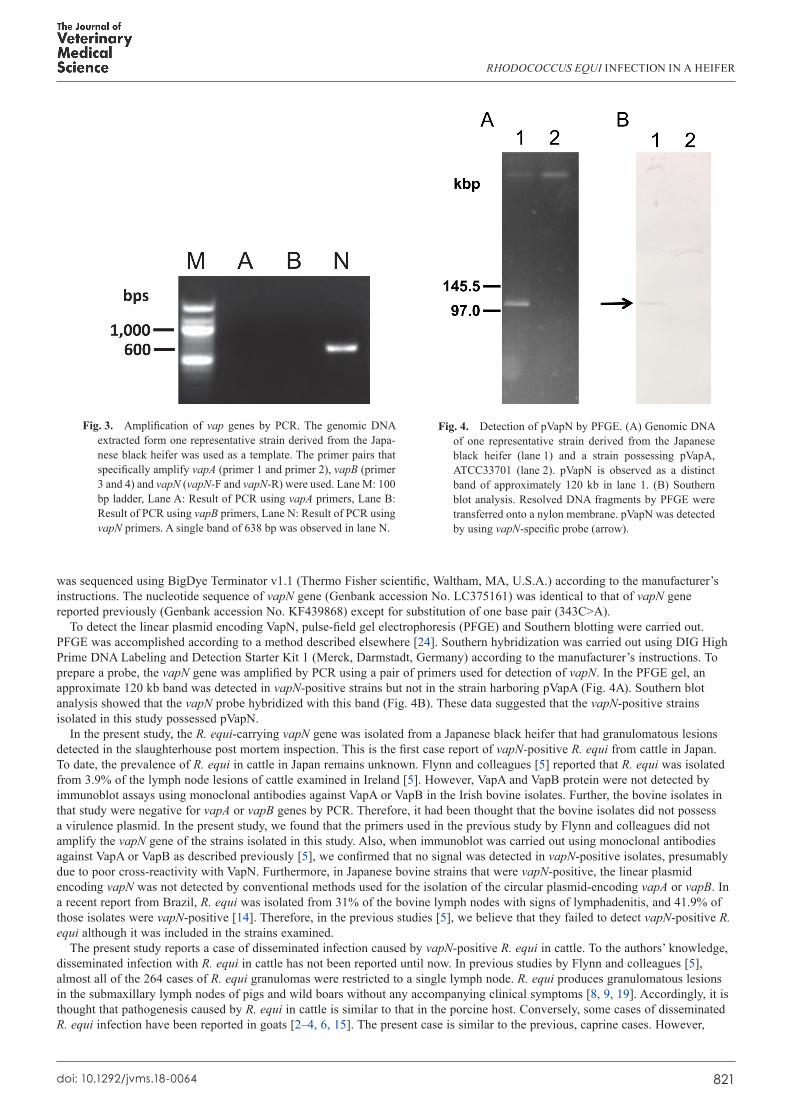
The presence of virulence plasmid specific markers, vapA, vapB, and vapN were examined by PCR using primer pairs shown in Table 1. PCR thermal cycler steps for each gene were identical: an initial denaturation at 94°C for 5 min, followed by 35 cycles of denaturation at 94°C for 30 sec, annealing at 55°C for 30 sec, extension at 72°C for 1 min, and a final extension at 72°C for 5 min. All the isolates were vapN-positive but vapA- and vapB-negative (Fig. 3). The DNA fragment amplified by vapN primers
Fig. 2. (A) Hepatic portal lymph nodes. Multiple variably sized caseating granulomas contain areas of necrosis in medulla. HE stain. Bar=100 µm. (B) Hepatic portal lymph nodes. Granuloma is composed of epithelioid cells and multinucleated giant cells. HE stain. Bar=50 µm
Table 1. Primers used in this study
Primer Sequence (5′→3′) ReferencePrimer1 GACTCTTCACAAGACGGT [23]Primer2 TAGGCGTTGTGCCAGCTA [23]Primer3 AACGTAGTCGCGGTGAGAA [23]Primer4 ACCGAGACTTGAGCGACTA [23]vapN F CGCTTTTATCGAGGGCACTC This studyvapN R TTTGCCAGGTCTTGCGAATG This study
RHODOCOCCUS EQUI INFECTION IN A HEIFER
821doi: 10.1292/jvms.18-0064
was sequenced using BigDye Terminator v1.1 (Thermo Fisher scientific, Waltham, MA, U.S.A.) according to the manufacturer’s instructions. The nucleotide sequence of vapN gene (Genbank accession No. LC375161) was identical to that of vapN gene reported previously (Genbank accession No. KF439868) except for substitution of one base pair (343C>A).
To detect the linear plasmid encoding VapN, pulse-field gel electrophoresis (PFGE) and Southern blotting were carried out. PFGE was accomplished according to a method described elsewhere [24]. Southern hybridization was carried out using DIG High Prime DNA Labeling and Detection Starter Kit 1 (Merck, Darmstadt, Germany) according to the manufacturer’s instructions. To prepare a probe, the vapN gene was amplified by PCR using a pair of primers used for detection of vapN. In the PFGE gel, an approximate 120 kb band was detected in vapN-positive strains but not in the strain harboring pVapA (Fig. 4A). Southern blot analysis showed that the vapN probe hybridized with this band (Fig. 4B). These data suggested that the vapN-positive strains isolated in this study possessed pVapN.
In the present study, the R. equi-carrying vapN gene was isolated from a Japanese black heifer that had granulomatous lesions detected in the slaughterhouse post mortem inspection. This is the first case report of vapN-positive R. equi from cattle in Japan. To date, the prevalence of R. equi in cattle in Japan remains unknown. Flynn and colleagues [5] reported that R. equi was isolated from 3.9% of the lymph node lesions of cattle examined in Ireland [5]. However, VapA and VapB protein were not detected by immunoblot assays using monoclonal antibodies against VapA or VapB in the Irish bovine isolates. Further, the bovine isolates in that study were negative for vapA or vapB genes by PCR. Therefore, it had been thought that the bovine isolates did not possess a virulence plasmid. In the present study, we found that the primers used in the previous study by Flynn and colleagues did not amplify the vapN gene of the strains isolated in this study. Also, when immunoblot was carried out using monoclonal antibodies against VapA or VapB as described previously [5], we confirmed that no signal was detected in vapN-positive isolates, presumably due to poor cross-reactivity with VapN. Furthermore, in Japanese bovine strains that were vapN-positive, the linear plasmid encoding vapN was not detected by conventional methods used for the isolation of the circular plasmid-encoding vapA or vapB. In a recent report from Brazil, R. equi was isolated from 31% of the bovine lymph nodes with signs of lymphadenitis, and 41.9% of those isolates were vapN-positive [14]. Therefore, in the previous studies [5], we believe that they failed to detect vapN-positive R. equi although it was included in the strains examined.
The present study reports a case of disseminated infection caused by vapN-positive R. equi in cattle. To the authors’ knowledge, disseminated infection with R. equi in cattle has not been reported until now. In previous studies by Flynn and colleagues [5], almost all of the 264 cases of R. equi granulomas were restricted to a single lymph node. R. equi produces granulomatous lesions in the submaxillary lymph nodes of pigs and wild boars without any accompanying clinical symptoms [8, 9, 19]. Accordingly, it is thought that pathogenesis caused by R. equi in cattle is similar to that in the porcine host. Conversely, some cases of disseminated R. equi infection have been reported in goats [2–4, 6, 15]. The present case is similar to the previous, caprine cases. However,
Fig. 3. Amplification of vap genes by PCR. The genomic DNA extracted form one representative strain derived from the Japa-nese black heifer was used as a template. The primer pairs that specifically amplify vapA (primer 1 and primer 2), vapB (primer 3 and 4) and vapN (vapN-F and vapN-R) were used. Lane M: 100 bp ladder, Lane A: Result of PCR using vapA primers, Lane B: Result of PCR using vapB primers, Lane N: Result of PCR using vapN primers. A single band of 638 bp was observed in lane N.
Fig. 4. Detection of pVapN by PFGE. (A) Genomic DNA of one representative strain derived from the Japanese black heifer (lane 1) and a strain possessing pVapA, ATCC33701 (lane 2). pVapN is observed as a distinct band of approximately 120 kb in lane 1. (B) Southern blot analysis. Resolved DNA fragments by PFGE were transferred onto a nylon membrane. pVapN was detected by using vapN-specific probe (arrow).

R. NAKAGAWA ET AL.
822doi: 10.1292/jvms.18-0064
judging from the rarity of reported cases of disseminated R. equi infection in cattle, some predisposing factors such as stress and immunosuppression might affect the host’s susceptibility.
In this study, vapN-positive R. equi was isolated from cattle in Japan. Although it is thought that R. equi generally has low pathogenicity in cattle [5], exposure to some predisposing factors could result in disseminated infection. Further investigation is necessary to examine the pathogenesis and prevalence of bovine rhodococcosis in Japan.
REFERENCES
1. Bryan, L. K., Alexander, E. R., Lawhon, S. D. and Cohen, N. D. 2018. Detection of vapN in Rhodococcus equi isolates cultured from humans. PLOS ONE 13: e0190829. [Medline] [CrossRef]
2. Carrigan, M. J., Links, I. J. and Morton, A. G. 1988. Rhodococcus equi infection in goats. Aust. Vet. J. 65: 331–332. [Medline] [CrossRef] 3. Davis, W. P., Steficek, B. A., Watson, G. L., Yamini, B., Madarame, H., Takai, S. and Render, J. A. 1999. Disseminated Rhodococcus equi infection
in two goats. Vet. Pathol. 36: 336–339. [Medline] [CrossRef] 4. Fitzgerald, S. D., Walker, R. D. and Parlor, K. W. 1994. Fatal Rhodococcus equi infection in an Angora goat. J. Vet. Diagn. Invest. 6: 105–107.
[Medline] [CrossRef] 5. Flynn, O., Quigley, F., Costello, E., O’Grady, D., Gogarty, A., Mc Guirk, J. and Takai, S. 2001. Virulence-associated protein characterisation of
Rhodococcus equi isolated from bovine lymph nodes. Vet. Microbiol. 78: 221–228. [Medline] [CrossRef] 6. Jeckel, S., Holmes, P., King, S., Whatmore, A. M. and Kirkwood, I. 2011. Disseminated Rhodococcus equi infection in goats in the UK. Vet. Rec.
169: 56. [Medline] [CrossRef] 7. Letek, M., Ocampo-Sosa, A. A., Sanders, M., Fogarty, U., Buckley, T., Leadon, D. P., González, P., Scortti, M., Meijer, W. G., Parkhill, J., Bentley,
S. and Vázquez-Boland, J. A. 2008. Evolution of the Rhodococcus equi vap pathogenicity island seen through comparison of host-associated vapA and vapB virulence plasmids. J. Bacteriol. 190: 5797–5805. [Medline] [CrossRef]
8. Makrai, L., Takayama, S., Dénes, B., Hajtós, I., Sasaki, Y., Kakuda, T., Tsubaki, S., Major, A., Fodor, L., Varga, J. and Takai, S. 2005. Characterization of virulence plasmids and serotyping of rhodococcus equi isolates from submaxillary lymph nodes of pigs in Hungary. J. Clin. Microbiol. 43: 1246–1250. [Medline] [CrossRef]
9. Makrai, L., Kobayashi, A., Matsuoka, M., Sasaki, Y., Kakuda, T., Dénes, B., Hajtós, I., Révész, I., Jánosi, K., Fodor, L., Varga, J. and Takai, S. 2008. Isolation and characterisation of Rhodococcus equi from submaxillary lymph nodes of wild boars (Sus scrofa). Vet. Microbiol. 131: 318–323. [Medline] [CrossRef]
10. Martens, R. J., Takai, S., Cohen, N. D., Chaffin, M. K., Liu, H., Sakurai, K., Sugimoto, H. and Lingsweiler, S. W. 2000. Association of disease with isolation and virulence of Rhodococcus equi from farm soil and foals with pneumonia. J. Am. Vet. Med. Assoc. 217: 220–225. [Medline] [CrossRef]
11. Morton, A. C., Begg, A. P., Anderson, G. A., Takai, S., Lämmler, C. and Browning, G. F. 2001. Epidemiology of Rhodococcus equi strains on Thoroughbred horse farms. Appl. Environ. Microbiol. 67: 2167–2175. [Medline] [CrossRef]
12. Navas, J., González-Zorn, B., Ladrón, N., Garrido, P. and Vázquez-Boland, J. A. 2001. Identification and mutagenesis by allelic exchange of choE, encoding a cholesterol oxidase from the intracellular pathogen Rhodococcus equi. J. Bacteriol. 183: 4796–4805. [Medline] [CrossRef]
13. Prescott, J. F. 1991. Rhodococcus equi: an animal and human pathogen. Clin. Microbiol. Rev. 4: 20–34. [Medline] [CrossRef] 14. Ribeiro, M. G., Lara, G. H. B., da Silva, P., Franco, M. M. J., de Mattos-Guaraldi, A. L., de Vargas, A. P. C., Sakate, R. I., Pavan, F. R., Colhado,
B. S., Portilho, F. V. R., Motta, R. G., Kakuda, T. and Takai, S. 2018. Novel bovine-associated pVAPN plasmid type in Rhodococcus equi identified from lymph nodes of slaughtered cattle and lungs of people living with HIV/AIDS. Transbound. Emerg. Dis. 65: 321–326. [Medline] [CrossRef]
15. Stranahan, L. W., Plumlee, Q. D., Lawhon, S. D., Cohen, N. D. and Bryan, L. K. 2017. Rhodococcus equi Infections in goats: characterization of virulence plasmids. Vet. Pathol. 55: 273–276. [Medline]
16. Takai, S. and Tsubaki, S. 1985. The incidence of Rhodococcus (Corynebacterium) equi in domestic animals and soil. Nippon Juigaku Zasshi 47: 493–496. [Medline] [CrossRef]
17. Takai, S., Sekizaki, T., Ozawa, T., Sugawara, T., Watanabe, Y. and Tsubaki, S. 1991. Association between a large plasmid and 15- to 17-kilodalton antigens in virulent Rhodococcus equi. Infect. Immun. 59: 4056–4060. [Medline]
18. Takai, S., Imai, Y., Fukunaga, N., Uchida, Y., Kamisawa, K., Sasaki, Y., Tsubaki, S. and Sekizaki, T. 1995. Identification of virulence-associated antigens and plasmids in Rhodococcus equi from patients with AIDS. J. Infect. Dis. 172: 1306–1311. [Medline] [CrossRef]
19. Takai, S., Fukunaga, N., Ochiai, S., Imai, Y., Sasaki, Y., Tsubaki, S. and Sekizaki, T. 1996. Identification of intermediately virulent Rhodococcus equi isolates from pigs. J. Clin. Microbiol. 34: 1034–1037. [Medline]
20. Takai, S. 1997. Epidemiology of Rhodococcus equi infections: a review. Vet. Microbiol. 56: 167–176. [Medline] [CrossRef] 21. Takai, S., Murata, N., Kudo, R., Narematsu, N., Kakuda, T., Sasaki, Y. and Tsubaki, S. 2001. Two new variants of the Rhodococcus equi virulence
plasmid, 90 kb type III and type IV, recovered from a foal in Japan. Vet. Microbiol. 82: 373–381. [Medline] [CrossRef] 22. Takai, S., Tharavichitkul, P., Sasaki, C., Onishi, Y., Yamano, S., Kakuda, T., Tsubaki, S., Trinarong, C., Rojanasthien, S., Sirimalaisuwan, A.,
Tesaprateep, T., Maneekarn, N., Sirisanthana, T. and Kirikae, T. 2002. Identification of virulence-associated antigens and plasmids in Rhodococcus equi from patients with acquired immune deficiency syndrome and prevalence of virulent R. equi in soil collected from domestic animal farms in Chiang Mai, Thailand. Am. J. Trop. Med. Hyg. 66: 52–55. [Medline] [CrossRef]
23. Takai, S., Tharavichitkul, P., Takarn, P., Khantawa, B., Tamura, M., Tsukamoto, A., Takayama, S., Yamatoda, N., Kimura, A., Sasaki, Y., Kakuda, T., Tsubaki, S., Maneekarn, N., Sirisanthana, T. and Kirikae, T. 2003. Molecular epidemiology of Rhodococcus equi of intermediate virulence isolated from patients with and without acquired immune deficiency syndrome in Chiang Mai, Thailand. J. Infect. Dis. 188: 1717–1723. [Medline] [CrossRef]
24. Valero-Rello, A., Hapeshi, A., Anastasi, E., Alvarez, S., Scortti, M., Meijer, W. G., MacArthur, I. and Vázquez-Boland, J. A. 2015. An invertron-like linear plasmid mediates intracellular survival and virulence in bovine isolates of Rhodococcus equi. Infect. Immun. 83: 2725–2737. [Medline] [CrossRef]