bacteria, fungus, or virus?
TRANSCRIPT

Bacteria, Fungus, or Virus?An Update on Infections of the Skin
Skin, Bones, Hearts, and Private Parts
Kara N. Roman, MMS, PA-C
Associate Director & Assistant Professor
Midwestern University PA Program, Downers Grove, IL
Disclosures and Images
• No conflicts to disclose
• Images – all images obtained and used within the guidelines provided from DermNetNZ.org unless otherwise attributed
• Reproduction or copying of content or images prohibited
• Thank you

Learning Objectives:
• Distinguish between the various types of bacterial, viral, and fungal skin and soft tissue infections utilizing a patient’s clinical presentation and risk factors
• Develop a plan to manage common skin infectious using appropriate medications and diagnostic/therapeutic procedures
• Discuss prevention measures for skin and soft tissue infections where appropriate

Picture goes herePatient presents for evaluation of the following skin lesions.
Image source: https://dermnetnz.org/

Bacteria, Fungus, or Virus?
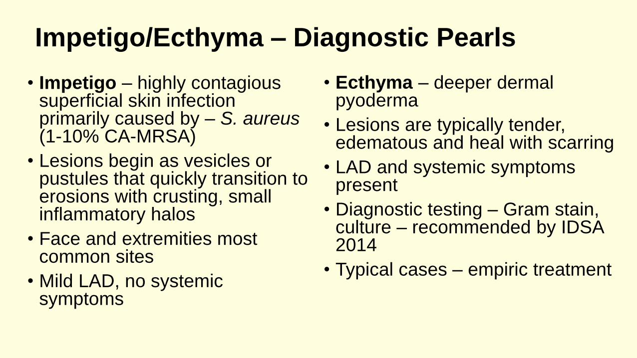
Impetigo/Ecthyma – Diagnostic Pearls
• Impetigo – highly contagious superficial skin infection primarily caused by – S. aureus (1-10% CA-MRSA)
• Lesions begin as vesicles or pustules that quickly transition to erosions with crusting, small inflammatory halos
• Face and extremities most common sites
• Mild LAD, no systemic symptoms
• Ecthyma – deeper dermal pyoderma
• Lesions are typically tender, edematous and heal with scarring
• LAD and systemic symptoms present
• Diagnostic testing – Gram stain, culture – recommended by IDSA 2014
• Typical cases – empiric treatment

Impetigo/Ecthyma – Treatment
• Practice Guidelines for Diagnosis and Management of Skin and Soft Tissue Infections: 2014 Update by IDSA https://www.idsociety.org/practice-guideline/skin-and-soft-tissue-infections/
• Topical treatment for limited impetigo• Mupirocin, Retapamulin – BID for 5 days
• Soak to remove crust
• Oral for numerous impetigo, in outbreaks, and for all ecthyma• CA-MRSA – Doxycycline, Clindamycin, TMP/SMX – 7 days
• MSSA - Dicloxacillin, Cephalexin – 7 days

Picture goes here Patient presents for evaluation of the following skin lesions.
Image source: https://dermnetnz.org/

Bacteria, Fungus, or Virus?

Purulent Infections – Diagnostic Pearls
• Folliculitis – single follicle
• Furuncle – deeper, yet still one follicle
• Carbuncle – larger, often with fever, LAD, fatigue
• Gram stain and culture are ideal
• Mild, moderate, severe classifications
• Most purulent infections = Staphylococcus aureus (MSSA and MRSA)
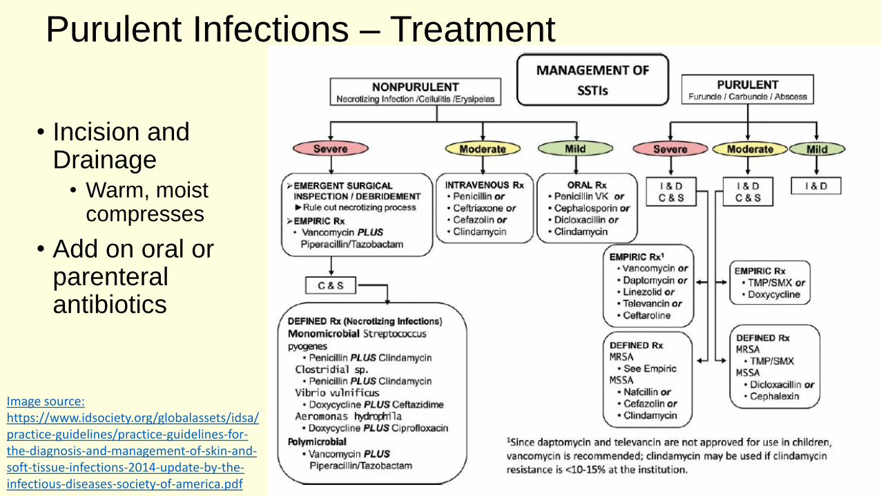
Purulent Infections – Treatment
• Incision and Drainage
• Warm, moist compresses
• Add on oral or parenteral antibiotics
Image source: https://www.idsociety.org/globalassets/idsa/practice-guidelines/practice-guidelines-for-the-diagnosis-and-management-of-skin-and-soft-tissue-infections-2014-update-by-the-infectious-diseases-society-of-america.pdf

Traditional I&D and Loop Drainage Technique
https://aneskey.com/cutaneous-abscess-or-pustule/
https://vimeo.com/19580472

Addition of Antibiotics in Purulent Skin Infections per IDSA 2014
• Temp > 38 degrees C or < 36 degrees C
• Tachypnea > 24/min
• Tachycardia > 90 bpm
• WBC > 12,000 or < 4,000
• Immunosuppression
• Hypotension

Guidelines vs Actual Management of Skin and Soft Tissue Infections in the EDKamath RS, et al. OFID 2017
• 214 cases of SSTI in ED retrospectively analyzed at Michael E. DeBakey Veterans Affairs Medical Center, Houston
• Total number that were managed in accordance with IDSA 2014 guidelines in all 4 categories (site of treatment, choice of antibiotic, I&D of abscess, ordering cultures)
43/214 = 20.1%

Systemic Antibiotics – New DataGottlieb, M et at. A Systematic Review and Meta-Analysis. Annals of Emergency Medicine Vol 73, No 1, January 2019.
• All randomized controlled trials comparing systemic antibiotics (MRSA coverage) vs placebo in the treatment of skin abscesses after I&D
• 4 studies, 16 clinical sites in US, ED and one outpt setting = 2,406 patients (4-44 yo)
• Most used TMP/SMZ, one used clindamycin
• Overall cure rate for abscesses after I&D was high in both groups
• Nearly 2-fold improvement in cure rates and a NNT of 14 for antibiotic groups
• Reduced number of return visits and need for painful repeat I&D, return to work sooner, decreased incidence of new lesions, decreased rate of infections in household members

To Pack or Not to Pack
NO
• 5 cm or less
• Immunocompetent pt
• Less pain
• No change in cure rate
• No change in secondary interventions
Maybe or YES
• Larger abscesses
• Consider the area
• Immunocompromised pts have not been studied
Source: List M et al. Treatment of Skin Abscesses: A Review of Wound Packing and Post-Procedural Antibiotics. South Dakota Medicine, March 2016.
Recurrent Skin Abscesses
• Consider 5 day decolonization regimen:• Twice daily intranasal mupirocin
• Daily chlorhexidine washes
• Daily decontamination of personal items such as towels, sheets, and cloths for recurrent S. aureus
• Evaluate adult patients for neutrophil disorder if recurrent abscesses began in childhood
• Search for local causes – pilonidal cyst, hidradenitis suppurativa, foreign body
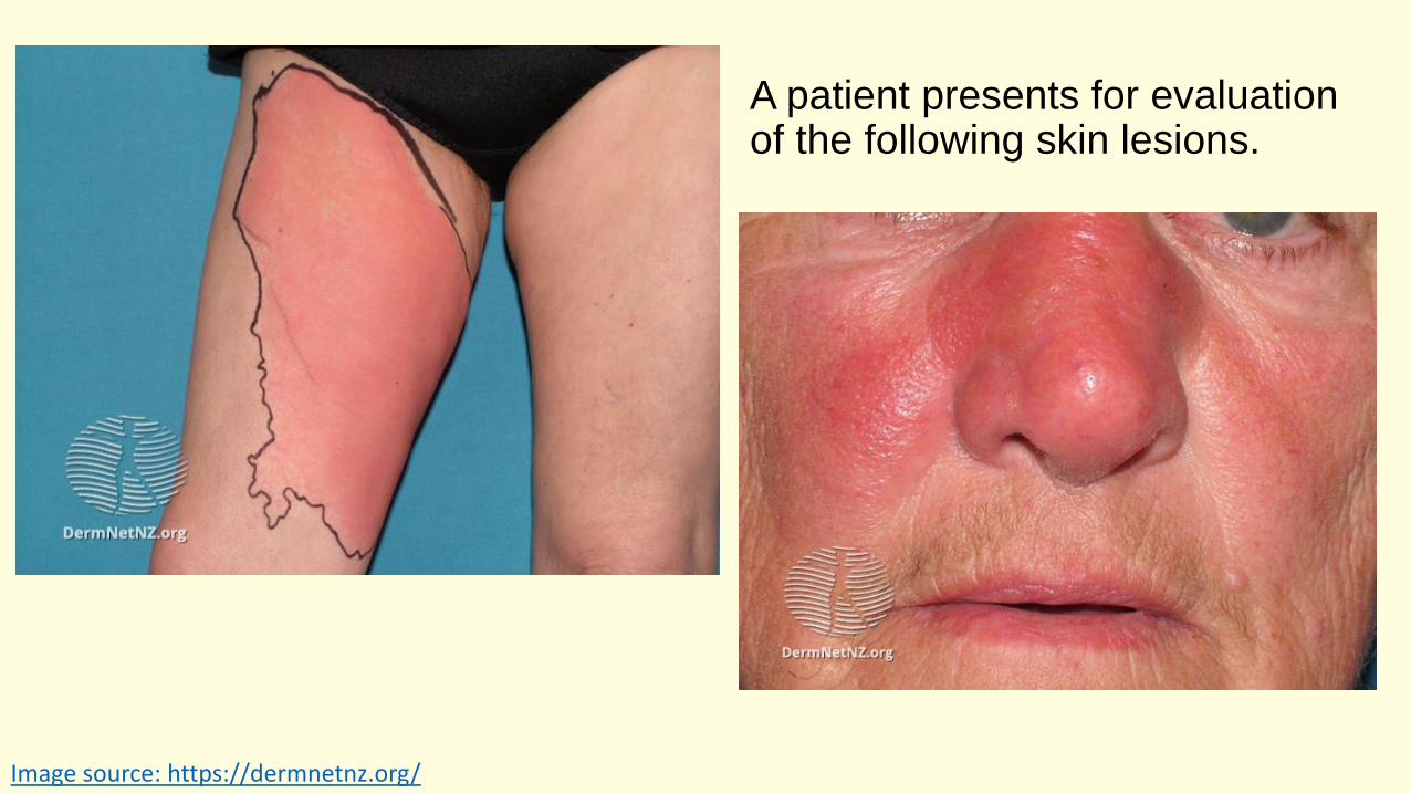
Picture goes here A patient presents for evaluation of the following skin lesions.
Image source: https://dermnetnz.org/

Bacteria, Fungus, or Virus?

Cellulitis/Erysipelas – Diagnostic Pearls
• Erysipelas – bright red, more superficial, raised border, well-demarcated margin, preceded by flu-like symptoms, burning at site
• Cellulitis – deeper location to subcutaneous tissues, non-elevated, poorly defined margins,
• Usually caused by StreptococcusA, B, C, G
• Bedside ultrasound best option for differentiating abscess from cellulitis
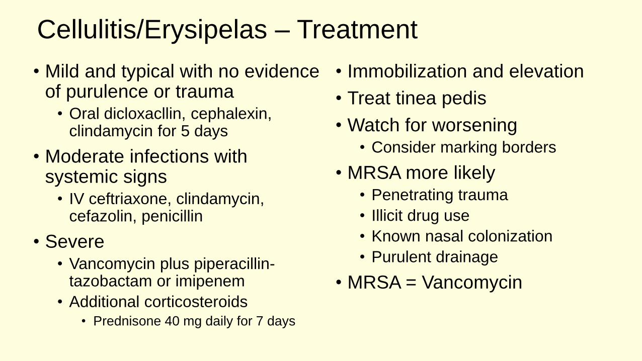
Cellulitis/Erysipelas – Treatment
• Mild and typical with no evidence of purulence or trauma
• Oral dicloxacllin, cephalexin, clindamycin for 5 days
• Moderate infections with systemic signs
• IV ceftriaxone, clindamycin, cefazolin, penicillin
• Severe• Vancomycin plus piperacillin-
tazobactam or imipenem
• Additional corticosteroids• Prednisone 40 mg daily for 7 days
• Immobilization and elevation
• Treat tinea pedis
• Watch for worsening• Consider marking borders
• MRSA more likely• Penetrating trauma
• Illicit drug use
• Known nasal colonization
• Purulent drainage
• MRSA = Vancomycin

Recurrent Cellulitis
• Risk factors
• Tinea pedis
• Obesity
• Venous insufficiency
• Lymphedema
• Prophylactic abx if 3-4 episodes per year despite treating predisposing factors
• Pen VK or erythromycin orally twice daily for 4-52 weeks

Risk for Atypical Organisms
• Immunosuppression
• Animal or human bites
• Sea or freshwater exposure to broken skin
• Exposure to animals, fish, or reptiles
• IVD use

Cat/Dog Bites – Identify pts at high risk
• Immunocompromised
• Asplenic
• Advanced liver disease
• Pre-existing or resultant edema of the affected area
• Moderate to severe injuries, especially to hand or face
• Injuries that penetrate the periosteum or joint capsule
• Deep puncture wounds
• 3-5 days of preemptive therapy
• Augmentin 875 mg twice daily
• Alternatives• Cefuroxime plus
clindamycin/metronidazole
• Imipenem/meropenem
• Moxifloxacin
• Doxycycline

FDA approves 5 new antibiotics for SSTI (2013)
• Ceftaroline – Beta-lactam
• MRSA and Enterbacteriaceae coverage
• IV, 5-14 days
• Dalbavancin - Lipoglycopeptide
• Once weekly dosing (2 total doses)
• Oritavancin - Lipoglycopeptide
• Single dose formulation
• Tedizolid
• Oral and parenteral forms
• Telavancin
• Black boxes – renal impairment, QT

Bacterial Infections
• Impetigo/Ecthyma
• Furuncle/Carbuncle/Abscess
• Cellulitis/Erysipelas
• Staphylococcal scalded skin/Scarlet fever/TSS
• Lyme
• Secondary syphilis
• Erythrasma
• Infectious Diseases Society of America (IDSA). Guidelines for skin and soft tissue infections, 2014.https://www.idsociety.org/practice-guideline/skin-and-soft-tissue-infections/

Picture goes here Patient presents for evaluation of the following skin lesions.

Bacteria, Fungus, or Virus?
HPV – Diagnostic Pearls
• HPV 1, 2, 3, 4, 10, 27, 57
• Most common in children and young adults (nearly 50% affected), increasingly seen in patients with atopic dermatitis and decreased cell-mediated immunity
• Types – verruca vulgaris, plantaris, plana, filiform
• Clinical diagnosis
• Thrombosed capillaries
• Altered dermatoglyphics

HPV – Treatment
• Spontaneous remission in 50% by 1 year, 2/3 of patients in 2 years, especially children
• Treatment indicated
• Pain/discomfort, functional impairment
• Concern for cosmesis or social stigma
• Persistent warts
• Immunosuppression
HPV- Treatment• Chemical or physical destruction
• Salicylic acid – irritation and exfoliation, paints or plasters, applied daily after paring with occlusion for 3-4 months
• Cryotherapy – every 2-3 weeks for 3 months, keep wart frozen for 15-30s
• Combo of both
• Enhancement of local immune response
• Imiquimod
• Contact or intralesional immunotherapy
• Antiproliferative therapy
• 5-FU
• Bleomycin
• Tretinoin

An Option for Recalcitrant and Extensive Warts?
• Intralesional or intramuscular HPV vaccine• Nofal A et al. Intralesional versus intramuscular bivalent human papillomavirus
vaccine in the treatment of recalcitrant common warts, J Am Acad Dermatol vol82, No 1, July 2019.
• Waldman, Abigail MD; Whiting, Dennis PA-C; Rani, Monica MD; Alam, Murad MD, MSCI, MBA, HPV Vaccine for Treatment of Recalcitrant Cutaneous Warts in Adults A Retrospective Cohort Study. Dermatologic Surgery: December 2019 - Volume 45 - Issue 12 - p 1739–1741

Picture goes here Patient presents for evaluation of the following skin lesions.
Image source: https://dermnetnz.org/

Bacteria, Fungus, or Virus?

Molluscum Contagiosum – Diagnostic Pearls
• Benign, self-limited disease
• Caused by a pox virus
• Most common ages 1-4
• More common in pts with atopic dermatitis and with swimming
• In adolescents and adults, consider STIs and immunocompromised states
• Clinical diagnosis
• Small, firm, pearly papules with a central depression
• Core may be expressed, producing a white cheesy material
• The lesions average 2 to 5 mm in size and are usually painless, but may become inflamed, red, and swollen
• Distribution typically face, truck, limbs

MCV – Treatment• Treatment is for cosmesis and to prevent
spreading
• Destructive therapies most common
• Curettage – most successful, least number of visits, greatest parent satisfaction
• Cantharidin
• Cryotherapy
• Patience
• 50% completely resolved 1 year
• 70% at 18 months
• BOTE sign “Beginning of the end”
• Clinical erythema and swelling of the lesion when regression phase begins
• Other therapies not proven superior to destructive therapies
• Imiquimod
• KOH
• Salicylic acid
• Retinoids
https://childhood101.com/kids-health-molluscum-contagiosum/

https://ars.els-cdn.com/content/image/1-s2.0-S0190962213004106-gr1_lrg.jpg https://www.dermatologytimes.com/article/verrica-develops-solution-common-warts
https://dermnetnz.org/topics/cantharidin/

Verrica develops a solution for common warts
Del Rosso JQ, Kircik L. Topical Cantharidin in the Management of Molluscum Contagiosum: Preliminary Assessment of an Ether-free, Pharmaceutical-grade Formulation. J Clin AesthetDermatol. 2019;12(2):27–30.
Source: https://www.ncbi.nlm.nih.gov/pmc/articles/PMC6415708/

14-year-old male presents for evaluation of red circular rash on his body that he noticed after soccer practice yesterday
• Pt states he doesn’t feel sick, but the rash might be a little itchy
• He is unaware of any family or friends that are ill or have rashes
• The soccer coach will not let him play or practice until he gets “checked out”
Image source: https://dermnetnz.org/

Bacteria, Fungus, or Virus?

Pityriasis Rosea – Diagnostic Pearls• Self-limiting skin condition
• Reactivation of HHV-6/7
• Typically presents in 10-35-year-old pts
• Herald patch appears first on truck in 90% of cases (lasts for 2 weeks in isolation)
• Prodromal symptoms – malaise, nausea, headache, URI, concentration difficulty, ST, body aching
• Secondary eruption on trunk in Langer lines and to proximal extremities (lasts for up to 12 weeks)
• All lesions have scaling and are typically pruritic
• Some medications associated with PR-like eruption – ACEI, NSAID
Image source: https://dermnetnz.org/topics/pityriasis-rosea-images/

PR – Treatment
• Self-limited = watchful waiting, patience
• Treat options
• Oral antihistamines
• Oral or topical corticosteroids
• Acyclovir (maybe)
• Phototherapy also hastens resolution
• No benefit to macrolides

Viral Infections
• HPV
• Molluscum
• Viral exanthems
• Erythema infectiousum
• Roseola
• Herpangina/HFM
• Pityriasis rosea
• Herpes
• Simplex
• Zoster

6-year-old male presents for evaluation of this pruritic eczematous rash on his torso
• It developed over the past week as an initial small circular lesion that has spread
http://advancedskinmd.com/tinea-corporis-ringworm/
http://www.crestingthehill.com.au/2015_04_01_archive.html

Bacteria, Fungus, or Virus?

Tinea Corporis – Diagnostic Pearls
• Usually presents as annular, scaly plaques
• At risk groups• Contact sports
• Domestic animal contact
• Warm, humid climates
• DM, immunodeficiency
• Typical organisms• M. canis
• T. rubrum, mentagrophytes, tonsurans
• Diagnostic options• KOH
• Wood’s lamp
• Culture
• Biopsy

Tinea Corporis – Treatment
• Topical antifungals• Azoles
• Econazole
• Oxiconazole – comes in a lotion form for more hairy areas
• Allylamines• Terbinafine
• Naftifine
• Oral antifungals• Terbinafine
• Itraconazole
• Twice a day application of topicals for 4-6 weeks generally
• Keep skin cool and dry
• Avoid combination products with steroids and antifungals
• Topical nail lacquers modestly effective
• Efinaconazole
• Tavaborole

Majocchi’s granuloma
• Perifollicular lesions
• T. rubrum, T. mentagrophytes
• Systemic therapy needed
Image source: https://dermnetnz.org/topics/majocchi-granuloma/

Picture goes here Patient presents for evaluation of the following skin lesions.
https://dermnetnz.org/

Bacteria, Fungus, or Virus?

Onychomycosis – Diagnostic Pearls
• Most often occurs in adults• Nail injury increases risk
• T. rubrum most frequent dermatophyte
• Common clinical manifestations• Nail discoloration
• Subungual hyperkeratosis
• Onycholysis
• Nail plate splitting and destruction
• Diagnostic testing• KOH - screen
• Fungal culture & PCR – identify the organism before treatment

Onychomycosis – Treatment
• Mild to moderate disease (<50% nail involvement) with no matrix involvement
• Ciclopirox
• Efinaconazole
• Tavaborole
• Established and severe disease• Terbinafine – preferred
• 250 mg orally once a day for 6 weeks for fingernails and 12 weeks for toenails
• Measure transaminases (and maybe CBC) before initiating therapy, no repeat testing in healthy <65 yo pts
• Itraconazole• 200 mg per day for 12 weeks for
toenails
• 200 mg twice a day for 1 week with second “pulse” 3 weeks later for fingernails
Devices to Treat Nail Fungus
• Laser
• Drilling
• Photodynamic therapy
• Plasma therapy

12-year-old AA male patient presents for evaluation of the following skin lesions.
• 4 week duration
• Non-pruritic
• Coincided with summer sports activities and increased perspiration
• No other family members with rash
• No recent travel or exposure to pets
Image source: https://adc.bmj.com/content/96/4/392

Bacteria, Fungus, or Virus?

Pityriasis Versicolor – Diagnostic Pearls
• Superficial fungal infection caused by Malassezia species of yeast in stratum corneum
• Flourishes in hot and humid environments
• Increased sebum production facilitates growth
• Hyper or hypo pigmented circles or ovals with fine scale
• Evoked scale sign
• Trunk, neck and upper arms
• KOH• Sticks and stones
• Spaghetti and meatballs
• Wood’s light• Greenish-yellow fluorescence

After Stretch
Vivian S Shi, and Peter A Lio Arch Dis Child 2011;96:392-393
Copyright © BMJ Publishing Group Ltd & Royal College of Paediatrics and Child Health. All rights reserved.
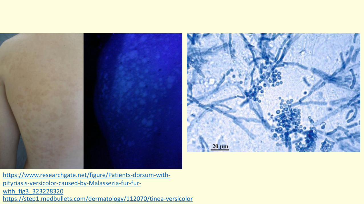
https://www.researchgate.net/figure/Patients-dorsum-with-pityriasis-versicolor-caused-by-Malassezia-fur-fur-with_fig3_323228320https://step1.medbullets.com/dermatology/112070/tinea-versicolor

Pityriasis Versicolor – Treatment
• Topical treatments effective and well-tolerated
• Shampoos and lotions – apply to affected area for 5-10 minutes then wash off, twice a day for 7-14 days, then once a month
• Zinc pyrithione
• Selenium sulfide
• Ketaconazole
• May take considerable time and maintenance
• Recalcitrant or recurring consider oral therapy
• Fluconazole 300 mg weekly for 2-4 weeks
• NOT terbinafine
• NOT ketaconazole

Fungal Infections
• Dermatophytoses• Tinea corporis
• Onychomycosis
• Pityriasis versicolor
• Candidiasis
• Majocchi granuloma (Pronunciation: mah-yok′ē)
• Sporotrichosis

Selected References/Resources
• Practice Guidelines for Diagnosis and Management of Skin and Soft Tissue Infections: 2014 Update by IDSA https://www.idsociety.org/practice-guideline/skin-and-soft-tissue-infections/
• Gupta, AK, et al. Fungal Skin Infections. Pediatrics in Review 2017;38;8.
• Lipner, SR, Scher, RK, Onychomycosis Treatment and Prevention of Recurrence, J Am Acad Dermatol, Vol 80, No 4, April 2019.
• Rush, J, Dinulos, JG, Childhood skin and soft tissue infections: new discoveries and guidelines regarding the management of bacterial soft tissue infections, molluscum contagiosum, and warts, CurrOpin Pediatr 201, 28:250-257.
• Ibrahim, F, Khan R, Pujalte, GA, Bacterial Skin Infections, Prim Care Clin Office Pract 42 (2015) 485-499.