bacteria: evaluating exposure and health risks · bacteria: evaluating exposure and health risks...
TRANSCRIPT

Joseph Manfrida, Ph.D.
Bacteria:Evaluating Exposure
and Health RisksAuthors:
Harriet Burge, Ph.D.,Michael Berg, Ph.D.,
Dave Gallup
Copyright © 2009-2010 EMLab P&K. All rights reserved.2
Outline
• Introduction to bacteria and bacterial classification
• Distribution and role of the bacteria in the natural environment
• Overview of the health effects of bacteria
• Overview of sampling and analytical methods for bacteria
• Legionella and Legionnaires' disease
• Mycobacteria
• MRSA in health care settings
• Sewage: evaluating hazards
Copyright © 2009-2010 EMLab P&K. All rights reserved.3
Introduction To Bacteria
• Naming bacteria
• Morphology– Macroscopic
– Microscopic
• Classification
• Physiology
Copyright © 2009-2010 EMLab P&K. All rights reserved.4
Bacterial Diversity
3 Distinct Domains, separated on the basis of biochemistry, genetics and cellular structure.
Eukarya
Archaea
Bacteria

Copyright © 2009-2010 EMLab P&K. All rights reserved.5
Naming Bacteria
The U.S. Food and Drug Administration is warning consumers in Puerto Rico that two hand sanitizers – "Bee-Shield Hand Sanitizer”with Aloe Vera (10 fl. oz. or 1 gallon bottles) and “MD Quality Hand Sanitizer” with Aloe Vera (10 fl oz. bottles) – contain high levels of a bacteria, Burkholderia cepacia, that can cause serious infections in humans. March 3, 2010
Binomials:– Genus: Escherichia– Species: coli
– Genus: Staphylococcus– Species: aureus
Copyright © 2009-2010 EMLab P&K. All rights reserved.6
Morphology
Size– 0.1 to about 600 µm over a
single dimension – Escherichia coli: 1.1 to 1.5 µm
by 2.0 to 6.0 µm– On surfaces, usually present
as colonies with a few or millions of cells
– When airborne, usually on rafts (e.g., skin scales) (>10µm) or in droplet nuclei (one or more bacteria surrounded by dried mucous) (>2µm).
Source: http://en.wikipedia.org/wiki/File:Relative_scale.svg
Copyright © 2009-2010 EMLab P&K. All rights reserved.7
Morphology (cont’d)
Shape– Cocci: Spherical
• Staphylococcus aureus
– Bacilli: Rod shaped • Escherichia coli• Pseudomonas aeruginosa
– Spirilli: Spiral rods; • Borrelia burgdorfii• Helicobacter pylori
– Filamentous: long branching strands• Thermoactinomyces vulgaris
Source: http://en.wikipedia.org/wiki/File:Bacterial_morphology_diagram.svg
Copyright © 2009-2010 EMLab P&K. All rights reserved.8
Morphology (cont’d)
Appendages– Flagellae– Pili
• Type IV • Reproductive
– Fimbriae
According to Live Science the bacteria found in yogurt can come to your teeth's rescue as the bacteria that has been used in the making of yogurt has been found to help prevent plaque from sticking to teeth and scientists are currently working on introducing it into toothpaste.

Copyright © 2009-2010 EMLab P&K. All rights reserved.9
Bacterial Cell Structure
• DNA: loosely organized, no membrane bound nucleus
• RNA: in cytoplasm• Ribosomes: make protein
from amino acids under instruction from RNA
• Plasma membrane: lipid/protein layer with selective permeability
Source: http://en.wikipedia.org/wiki/File:Average_prokaryote_cell-_en.svg
• Cell wall:– Peptidoglycan– Lipopolysaccharide (Gram negative)
• Capsule (polysaccharides)Copyright © 2009-2010 EMLab P&K. All rights reserved.10
Biofilms: A Larger Structure
• In nature, most bacteria are bound to surfaces in biofilms• Few microns to half a meter (yes, meter) in depth.• Complex arrangement of cells & extracellular components
including networks of channels to diffuse nutrients.
Source: wikipedia.org/wiki/Biofilm
Copyright © 2009-2010 EMLab P&K. All rights reserved.11
Practical Classification
BacillusCorynebacterium
StaphylococcusStreptococcus
StreptomycesMicropolyspora
E. ColiPseudomonas Neisseria Vibrio
Rods Cocci
Gram positive
Commonbacteria
Filaments Rods Cocci
Gram negative
Spirals
Copyright © 2009-2010 EMLab P&K. All rights reserved.12
The Gram Stain
• Add Crystal Violet; wait 1 minute; rinse with water
• Add Iodine; wait 1 minute; rinse with water
• Add Acetone Alcohol; wait 10-15 sec.; rinse with water
• Flood slide with Safranin; wait 1 minute; rinse with water
• Gently blot the slide dry. • View under oil immersion
(1000x) with a bright-field compound microscope.
Source: http://en.wikipedia.org/wiki/Gram_staining

Copyright © 2009-2010 EMLab P&K. All rights reserved.13
Gram Negative (Pink)
Cells with a thin lipopolysaccharide cell wall do not retain the violet dye
– E. coli– Legionella pneumophila– Pseudomonas aeruginosa– Neisseria gonorrhea– Etc.
Source: http://en.wikipedia.org/wiki/Gram_staining
Copyright © 2009-2010 EMLab P&K. All rights reserved.14
Gram Positive
Cells with a thick peptidoglycan cell wall do retain the violet dye
– Staphylococcus– Bacillus– Corynebacterium– Actinomycetes
Copyright © 2009-2010 EMLab P&K. All rights reserved.15
Acid Fast
Cells with mycolic acid in outer wall: retain a dye when treated with acid. All acid fast bacteria are Gram positive.
– Mycobacterium– Nocardia– Some amoebal cysts (not a bacteria)
Copyright © 2009-2010 EMLab P&K. All rights reserved.16
Physiology
• Psychrophiles: Grow best at cold temperature. <10°C
• Mesophiles: Grow best at medium temperature. All pathogenic bacteria are mesophiles. 10-40°C
• Thermophiles: Grow best at hot temperature. 40-80°+C

Copyright © 2009-2010 EMLab P&K. All rights reserved.17
Physiology (cont’d)
• Obligate Anaerobes: anaerobic fermentation; cannot survive in the presence of oxygen.
• Obligate Aerobes: strictly oxidative respiration and require oxygen for survival.
• Facultative Anaerobes: fermentation in the absence of O2, or respiration in its presence.
• Aerotolerant Anaerobes: never undergo oxidative respiration but can tolerate the presence of oxygen.
• Microaerophilic: facultative anaerobes, but they prefer low O2-concentration conditions.
Copyright © 2009-2010 EMLab P&K. All rights reserved.18
Physiology (cont’d)
• Professor Alan Parsons and Dr. Richard Heal of QinetiQ Ltd, claim to have shown that physically separated colonies of bacteria can transmit signals conferring resistance to commonly used antibiotics
• (volatile organic compounds)?
Copyright © 2009-2010 EMLab P&K. All rights reserved.19
Distribution and Role of the Bacteria in the Natural Environment
• Ubiquity: – “We live now in the ‘Age of Bacteria.’ Our planet has always
been in the Age of Bacteria, ever since the first fossils—bacteria, of course—were entombed in rocks more than 3 billion years ago. Bacteria are—and always have been—the dominant forms of life on Earth” Steven J Gould
– All surfaces on earth and all water contain bacteria– Approximately five nonillin (5x1030) bacteria on Earth, forming
much of the world's biomass.
Copyright © 2009-2010 EMLab P&K. All rights reserved.20
Environmental Bacteria
How many bacteria are acceptable/normal?Mean Concentrations of Total Airborne Culturable Bacteria (Sum of Mesophilicand Thermophilic Bacteria) (CFU/m3), by Location (Indoors/Outdoors) and Season.
Summer Winter Summer WinterTotal Gram+ rods 10.6 11.4 33.6 43.6
(Actinomycetes) (2) (1.2) (6.4) (3.4)(Bacillus species) (6.9) (6.6) (19.9) (23.4)(Other Gram+ rods) (1.7) (3.5) (7.3) (16.9)
Gram+ cocci 48.3 28.7 26.2 21.8Gram– rods 3.5 2.6 14.9 11Gram– cocci 1.6 1.3 1.1 3.3Unknown 51.8 42.6 89.1 114.7Total bacteria (All groups) 116 86.7 165 194.5
Indoor Outdoor
Tsai, F.C.; Macher, J.M. 2005. Concentrations of airborne culturable bacteria in 100 large U.S. office buildings from the BASE study. Indoor Air 15(Suppl 9):71-81.

Copyright © 2009-2010 EMLab P&K. All rights reserved.21
Environmental Bacteria: A Few Standards
Drinking water (EPA):• HPC (no more than 500 bacterial
colonies per ml)• Coliform baceria (no more than
5.0% samples total coliform-positive in a month)
• Legionella (no limit, but EPA believes that if Giardia and viruses are removed/inactivated, Legionella will also be controlled)
Copyright © 2009-2010 EMLab P&K. All rights reserved.22
Water
• A million (106) bacterial cells in a milliliter of fresh water• 5x10⁸ in an 8 ounce glass of water
– Pseudomonas, Flavobacterium and Acinetobacter
• Old, dirty water filters seem to make water taste better. Bacteria that thrive on dirty water filters can reduce the distasteful earthy tinge in tap water.
Copyright © 2009-2010 EMLab P&K. All rights reserved.23
E. coli
• E. coli is a gut organism, but is common in soil and water where animal feces are present.
• Approximately 0.1% of the total bacteria within an adult's intestines (on a Western diet) is represented by E. coli.
• E. coli and other bacteria in our intestines are essential and provide Vitamin K and B-complex vitamins.
• E. coli O157:H7 is a specialized rare strain that causes serious infections. It is native to cows, not people.
Copyright © 2009-2010 EMLab P&K. All rights reserved.24
Soil
• A teaspoon of productive soil generally contains between 100 million and 1 billion bacteria. That is as much mass as two cows per acre. (Elaine R. Ingham)
• Decomposers: can break down pesticides and pollutants in soil
• Mutualists form partnerships with plants. The most well-known of these are the nitrogen-fixing bacteria (ex. Rhizobium)
• Plant pathogens: xymomonas and erwinia species, and species of Agrobacterium that cause gall formation in plants

Copyright © 2009-2010 EMLab P&K. All rights reserved.25
Plant Surfaces
• Bacteria multiply on the surface of plants and are aerosolized by wind and rain action.
• In the air, water clumps around bacteria forming condensation nuclei, leading to rainfall.
• Precipitation returns bacteria to the ground. Even if one bacterium lands on a plant, it can multiply and form groups, thus causing the cycle to repeat.– Pseudomonas syringae
• Epiphytic bacteria can increase water permeability of leaf cuticles, which increases the availability of water and dissolved compounds.
Copyright © 2009-2010 EMLab P&K. All rights reserved.26
Air
• Bacteria are globally distributed in the atmosphere and are believed to play a large role in formation of snow and rain.
• Over 2,000 different kinds of bacteria may be present in the air on any given day (Gary Anderson).
• Air bubbles breaking at the Air-water interface remove bacteria that concentrate at the interface. Bubbles eject the bacteria into the atmosphere. Bacterial concentrations in the drops may range from 10 to 1,000 times that of the water.
• Bacteria may reproduce within airborne droplets• Diarrhea-causing Arcobacter and ulcer-inducing
Heliobacter have been recovered from air.
Copyright © 2009-2010 EMLab P&K. All rights reserved.27
Overview of the Health Effects of Bacteria
• Infection• Allergy• Toxicosis• Symbiosis
Copyright © 2009-2010 EMLab P&K. All rights reserved.28
Infection
Invasion by, and multiplication of, pathogenic microorganisms in a bodily part or tissue, which may produce subsequent tissue injury and progress to overt disease through a variety of cellular or toxic mechanisms.

Copyright © 2009-2010 EMLab P&K. All rights reserved.29
Overview of Bacterial Infections
http://en.wikipedia.org/wiki/File:Bacterial_infections_and_involved_species.png
Copyright © 2009-2010 EMLab P&K. All rights reserved.30
Environmental Bacteria – Pathogens
Pathogens• Obligate pathogens
– must cause disease in order to be transmitted from one host to another (e.g. Mycobacterium tuberculosis)
• Opportunistic pathogens– can be transmitted from one host to another without having to
cause disease– a host whose immune system is not functioning properly, the
bacteria can cause an infection that leads to a disease (e.g. Pseudomonas aeruginosa)
Copyright © 2009-2010 EMLab P&K. All rights reserved.31
Opportunistic Pathogens
Opportunistic pathogens are often hospital acquired. • Bacteria
– Legionella– Staphylococcus aures– Serratia marcescens– Pseudomonas aeruginosa
• Fungi (Eukarya, not bacteria)– Candida albicans– Aspergillus species (Aspergillosis)– Mucor/Rhizopus/Absidia (Mucormycosis)– Cryptococcus neoformans
Copyright © 2009-2010 EMLab P&K. All rights reserved.32
The Infection Process
• Invasion or reactivation and growth of the organism• Overwhelming communities of bacteria• Immune reactions leading to disease• Release of bacterial toxins that cause the symptoms of
disease

Copyright © 2009-2010 EMLab P&K. All rights reserved.33
Pathways To Infection
• Airborne infection: contracted by inhalation of microorganisms or spores suspended in air, on water droplets, or dust particles, or in droplet nuclei (dried droplets).
• Droplet infection: contracted by inhalation of respiratory pathogens suspended for a brief time on liquid particles exhaled by someone already infected.
• Direct contact: infection contracted by touching an infected person or contaminated surface.
• Endogenous infection: due to reactivation of organisms present in a dormant focus, as occurs in tuberculosis, etc.
• Tunnel infection: subcutaneous infection of an artificial passage into the body that has been kept open.
Copyright © 2009-2010 EMLab P&K. All rights reserved.34
Virulence
• Virulent infection: infection by an organism that can infect anyone without specific antibodies.
• Opportunistic infection: infection by an organism that does not ordinarily cause disease but becomes pathogenic under certain circumstances (e.g., impaired immune responses).
Copyright © 2009-2010 EMLab P&K. All rights reserved.35
Risk Factors For Infection
Poorly Developed or Impaired Immunity• Age. Neonates and the elderly are at increased risk of
bacterial infections. • Nutritional status. Malnutrition results in a depressed
immune system • Genetic predisposition. The Human Genome Project
increased our ability to locate specific genes related to infectious disease susceptibility ( Bentley, DR, 2000).
• Immunosuppression via disease or medications• Lack of induced specific antibodies
Copyright © 2009-2010 EMLab P&K. All rights reserved.36
Risk Factors For Infection (cont’d)
Exposure• Virulence and immune status determines how many
organisms are needed to initiate infection• Route of exposure is important

Copyright © 2009-2010 EMLab P&K. All rights reserved.37
Routes of Exposure
• Exogenous: those that originate outside the body– Food, water, air, surfaces, other people– Ingestion, inhalation, other entry points
• Endogenous: caused by bacteria within the body that cause disease when the body's resistance is lowered
Copyright © 2009-2010 EMLab P&K. All rights reserved.38
Respiratory Infections
• Upper respiratory tract infections (URI)– Leading cause of time lost from work and school– Bacteria account for up to 25 percent of URI (the rest are viral)– Streptococcus and Haemophilus influenzae
• Otitis media– Middle ear infection: most common bacterial infection in U.S.
children– Streptococcus pneumoniae
Copyright © 2009-2010 EMLab P&K. All rights reserved.39
Respiratory Infections (cont’d)
• Lower respiratory tract infections (LRI)– Acute, chronic pneumonia and bronchitis– LRI occur in both healthy and immunocompromised individuals– Streptococcus pneumoniae
• Tuberculosis (TB)– Affects 15 million people in the U.S. Fewer develop disease
which depends on nutritional status, age, HIV. incarceration– Mycobacterium tuberculosis
Copyright © 2009-2010 EMLab P&K. All rights reserved.40
Gastrointestinal Infection
• Infectious diarrhea is a leading cause of morbidity and mortality worldwide.
• In the US, 100 million people are affected every year. • Most diarrhea is viral but bacteria also important.
– 50% restrict activities– 10% consult physicians– 250,000 require hospitalization– approximately 3000 die– Campylobacter, Salmonella, Shigella, and E. coli O157:H7

Copyright © 2009-2010 EMLab P&K. All rights reserved.41
Campylobacter jejuni
• Most common cause of bacterial diarrhea in the US. • Over 1 million Americans are affected yearly.• Antibiotics in poultry- and cattle-feed linked to the
increasing incidence of drug-resistant C. jejuni• Transmission via contaminated food (especially chicken)
and water, or contact with infected animals (especially cats and puppies)
Copyright © 2009-2010 EMLab P&K. All rights reserved.42
Salmonella
• Second most frequent cause of bacterial disease in U.S.• In 2002, more than 44,000 cases were reported to the
CDC. Incidence may be 30 or more times greater than reported.
• Diarrhea, fever, and abdominal cramps.• The elderly, infants, and people with impaired immune
systems are at greater risk of severe disease.• Transmission is via exposure to contaminated food
(especially eggs) or water, or contact with infected animals (reptiles).
Copyright © 2009-2010 EMLab P&K. All rights reserved.43
Escherichia coli O157:H7
• Severe diarrheal disease called hemolytic uremic syndrome.
• An estimated 73,000 cases are reported in the United States annually.
• Transmission is through contaminated hamburger meat, apple cider, and fruits and vegetables.
Copyright © 2009-2010 EMLab P&K. All rights reserved.44
Shigella
• Most common symptoms are diarrhea, vomiting, stomach cramps, fever, flatulence, nausea, and constipation.
• An estimated 448,240 cases occur in the U.S. yearly.• Groups at highest risk in the U.S. are children in child
care centers, individuals in custodial institutions, and international travelers.

Copyright © 2009-2010 EMLab P&K. All rights reserved.45
Helicobacter pylori
• Most common chronic infection in humans causing chronic gastritis, peptic ulcer disease, and some types of stomach cancer.
• Half of the world's population is infected.• Drinking coffee or alcohol and smoking increase your risk
for an ulcer from H. pylori.• Impairs absorption of nutrients, altering the balance of iron,
vitamin B12, folic acid, alpha-tocopherol, vitamin C, and beta-carotene.
• Acute infection causes abdominal pain, weight loss, nausea, and vomiting.
• Has been found in river, creek, and lake water in central Pennsylvania.
Copyright © 2009-2010 EMLab P&K. All rights reserved.46
Skin Infection
• Skin infections include:– Impetigo, boils, carbuncles, cellulitis, and complications from
burns,– Staphylococcus aureus,– group A streptococci,– Pseudomonas aeruginosa,– Impetigo, a skin infection caused mostly by group A streptococci,
can cause severe kidney inflammation, sometimes resulting in kidney failure.
Copyright © 2009-2010 EMLab P&K. All rights reserved.47
Contagious Bacterial Infection
• Exogenous sources• Highly virulent• Risks depend on type of organism
– Direct contact– Droplet contact– Airborne
Copyright © 2009-2010 EMLab P&K. All rights reserved.48
Allergy
• IgE allergy– Most patients with nasal polyposis and/or chronic sinusitis
possess bacteria-specific IgE, while subjects with only allergic rhinitis do not;
– Multiple bacterial species isolated from chronically infected sinuses are capable of inducing IgE-mediated sensitization. (Calenoff et al 1993)
• IgG/cell mediated allergy– Machining coolant aerosols– Thermophilic actinomycetes

Copyright © 2009-2010 EMLab P&K. All rights reserved.49
Endotoxins
General definitions• Endotoxins: Toxic compounds
found inside bacteria and other pathogens, lipopolysaccharides, cell-associated.
• Exotoxins: Secreted in soluble form, extracellular, diffusible.
Copyright © 2009-2010 EMLab P&K. All rights reserved.50
Endotoxins (cont’d)
Endotoxins (in particular)• Component of cell walls from gram-
negative bacteria (E. coli, Salmonellaetc.)
• Lipopolysaccharide (LPS) – Potent stimulator of the immune system
Detection with Limulus Amebocyte Lysate
Copyright © 2009-2010 EMLab P&K. All rights reserved.51
Endotoxin (cont’d)
• Cell wall of Gram negative bacteria• Induces fever, irritant, immune stimulant• Geometric mean endotoxin concentrations: (in EU/mg):
– Bedroom floors, 35.3 (5th–95th percentile, 5.0–260); – Bedding, 18.7 (2.0–142);– Family room floors, 63.9 (11.5–331); – Sofas, 44.8 (6.4–240); – Kitchen floors, 80.5 (9.8–512).
• Significant relationships between increasing endotoxinlevels and diagnosed asthma, asthma symptoms in the past year, current use of asthma medications, and wheezing among adult residents.
Copyright © 2009-2010 EMLab P&K. All rights reserved.52
Endotoxin (cont’d)
• Airborne endotoxin concentration: 0.49 ± 3.49 EU/m3
• Doubling of the air endotoxin concentration was associated with an increase of 0.32 illness episodes per year (p = 0.0003).
• Short-term exposure in the air at levels > 45 EU/m3
linked with decreases in lung function over the course of a single day. Longer-term exposures to endotoxin levels as low as 10-28 EU/m3 may be linked with chronic decreases in lung function.

Copyright © 2009-2010 EMLab P&K. All rights reserved.53
Endotoxin (cont’d)
• The ACGIH recommends that exposures more than ten times background levels be considered a concern if there are complaints of respiratory symptoms, and that exposures of 100 times background be avoided at all times.
• The Dutch Expert Committee on Occupational Standards of the National Health Council proposed a health-based recommended limit value for workers of 50 EU/m3 over an eight-hour exposure period.
Copyright © 2009-2010 EMLab P&K. All rights reserved.54
Endotoxins – Exposure Limits
• Recommendation of ~100 EU/m3 as maximum exposure limit. Background levels of >10 EU/m3 are of concern and >30 EU/m3 should be avoided.
• Normal concentrations indoors: <1 EU/m3
• Higher concentrations may indicate water damage.• Concentrations increase up to 100-fold in rooms of
smokers.
Copyright © 2009-2010 EMLab P&K. All rights reserved.55
Endotoxins – Exposure Limits (cont’d)
When and where are endotoxins a problem?• Occupational environment:
– Waste collectors– Organic household composting facilities– Cotton mills– Power plants with biomass as biofuel– Biotech Industry– Metal grinding (metal working fluids)
Copyright © 2009-2010 EMLab P&K. All rights reserved.56
Endotoxins – Asthma
• Relationship between endotoxin and asthma is still unclear: Some studies indicate that exposure to endotoxins may protect against allergic asthma but is a risk factor for non-allergic asthma.
• Smoking, presence of furred pets and cleaning regime influence endotoxin levels.
• Health effects are significant both in short and long term but depend on dose

Copyright © 2009-2010 EMLab P&K. All rights reserved.57
Endotoxins – Sampling
• Preferred sample type is air (endotoxin-free filter cassettes)
• 250 – 1000 Liter sample volume• Dust can also be used as sample type
Copyright © 2009-2010 EMLab P&K. All rights reserved.58
Endotoxins – Control and Prevention
• Controlling water reduces possibility of Gram-negative bacteria (and endotoxin)
• Removal of contaminated materials and HEPA vacuuming can reduce endotoxin levels
• Do not smoke
Copyright © 2009-2010 EMLab P&K. All rights reserved.59
Lethality of Bacterial Protein Toxins
2x10³Guinea pig6x10-5Diphtheria toxin
1x10⁶Rabbit2.3x10-6Shiga toxin
1x10⁶Mouse4x10-8Tetanus toxin
3x10⁶Mouse0.8x10-8Botulinum toxin
Compared to Strychnine Host
Toxic dose (mg)Toxin
Copyright © 2009-2010 EMLab P&K. All rights reserved.60
Symbiosis
• Lactobacillus acidophilus is a harmless bacterium that resides in your intestines.
• Lactobacillus acidophilus helps you digest food, destroys some disease-causing organisms, and provides nutrients to your body.
• May also help prevent asthma.• E. coli provides vitamins.

Copyright © 2009-2010 EMLab P&K. All rights reserved.61
Sampling and Analysis
• Type of sample collection and choice of analytical methods depends on: – Hypothesis or goals (monitoring)– Expected concentrations– Agent(s) of concern– Standards/guidelines
Copyright © 2009-2010 EMLab P&K. All rights reserved.62
Types of Samples and Relevant Hypotheses or Goals
• Water– This water contains sewage organisms– This water contains Legionella pneumophila
• Surface– Potential pathogens are falling into wounds during surgery– Residual contamination is present on these surfaces
• Bulk– This slime contains Legionella pneumophila– This humidifier water contains Thermophilic actinomycetes
• Air– Exposure is occurring to this agent– This activity produces aerosols containing this agent
Copyright © 2009-2010 EMLab P&K. All rights reserved.63
Analytical Methods: Bacteria
• Culture• Microscopy• Stains• DNA methods• Bioassays• Immunoassays• HPLC• GCMS
Copyright © 2009-2010 EMLab P&K. All rights reserved.64
Culture
• Requires organism to be alive• Recovers only organisms that can reproduce under the
provided conditions– Best to use a broad spectrum medium which allows damaged
bacteria to recover
• Always underestimates concentrations and diversity• Hypotheses:
– Potential pathogens are falling into wounds during surgery– Legionella pneumophila is growing in this humidifier

Copyright © 2009-2010 EMLab P&K. All rights reserved.65
Microscopy
• Vital Staining– Use of stain that differentiates living from dead bacteria– All cells can be counted and % viable calculated
• Fluorescence staining– Allows microscopic or flow cytometric counting of cells
• Fluorescent antibody staining– Allows counting of specific organisms
• Can be used on all types of samples• Hypotheses:
– The bacterial aerosol has xx living and xx non-living organisms– This treatment kills bacteria
Copyright © 2009-2010 EMLab P&K. All rights reserved.66
Other Kinds of Staining
• All require 1000x oil immersion microscopy
• Gram stain• Acid fast staining
– Mycobacterium cells are present in this sample
• Acridine orange (a fluorescent stain)– Total concentration of
bacterial cells in this sample is xx
Mycobacterium tuberculosis (stained red) in tissue (blue).Source: http://en.wikipedia.org/wiki/acid-fast
Copyright © 2009-2010 EMLab P&K. All rights reserved.67
DNA Methods
• For identification• For monitoring populations
– This specific strain of Legionella pneumophila is present in this cooling tower
– Track composition of bacterial populations are present in this aerosol
– The bacterial population in this biofilm is the same as or different than the one in a different biofilm.
Copyright © 2009-2010 EMLab P&K. All rights reserved.68
Bioassays
• Limulus assay for endotoxin– Depends on the horseshoe crab– Quantitative only within each batch of lysate– Internal controls essential for every assay
• This sample contains more endotoxin than the outdoor air

Copyright © 2009-2010 EMLab P&K. All rights reserved.69
Immunoassays For Specific Bacteria
• There are immunoassays for surrogates of bacteria considered possible biowarfare agents (e.g., Bacillus globigii as a surrogate for Bacillus anthracis).
• Obviously it is possible since bacteria can stimulate an antibody (immune system) response. Such methods have not been widely used.
Copyright © 2009-2010 EMLab P&K. All rights reserved.70
HPLC, GCMS
• Chemical methods used for measurement of bacterial and other biological chemicals.
• HPLC: high pressure liquid chromatography• GCMS: gas chromatography mass spectroscopy• Fatty acid analysis used for identification of bacteria in
bulk samples
Copyright © 2009-2010 EMLab P&K. All rights reserved.71
Legionella and Legionnaires' Disease
• Nature of Legionella• Natural reservoirs• Human exposure• Sampling strategies
– Hypotheses– Sampling plans– Data interpretation
Copyright © 2009-2010 EMLab P&K. All rights reserved.72
Nature and Ecology
• Gram negative rod-shaped bacterium• Widely distributed natural inhabitants of waters. • Significant multiplication restricted to 20°C to 45°C • Growth promoted by other micro organisms: amoebae
amplify Legionellae multiplication • Other bacteria and algae provide nutrients • Low concentrations of metals such as iron, zinc and
potassium enhance proliferation• The constituents of certain types of rubbers used in
rubber fittings in water and cooling systems can also support the multiplication of L. pneumophila.

Copyright © 2009-2010 EMLab P&K. All rights reserved.73
Legionella
Legionella• Gram negative bacteria
common in many environments
• Approx. 50 species and 70 serogroups have been described
• Legionella is the causative agent of Legionellosis(Legionnaires’ disease and Pontiac fever)
Copyright © 2009-2010 EMLab P&K. All rights reserved.74
Legionellosis
• The source was identified as the Legionella bacterium and found in the cooling tower of a hotel’s air conditioning system.
• Over 90% of Legionellosis are caused by Legionellapneumophila Skyline Philadelphia
Legionnaires’ disease• The first recognized outbreak of the disease occurred
1976 in Philadelphia• As many as 221 people were treated and 34 deaths
occurred.
Copyright © 2009-2010 EMLab P&K. All rights reserved.75
Legionellosis (cont’d)
Legionellosis takes two distinct forms:1. Pontiac fever: respiratory illness without pneumonia,
symptoms resemble acute influenza2. Legionnaires’ disease: symptoms include fever, chills,
cough, muscle aches, headache, tiredness, loss of appetite, loss of coordination(ataxia), and occasionally diarrhea and vomiting.• L. pneumophila infections
may be subclinical.• Antibodies present in up to
25% of adults tested.
Copyright © 2009-2010 EMLab P&K. All rights reserved.76
Legionellosis (cont’d)
• 2-10 day incubation• One of the top three causes of
community-acquired pneumonia• 8,000 to 18,000 people get
legionellosis in U.S. each year • Many cases go undiagnosed• Transmission is not person to person• Worldwide distribution, although outbreaks of Legionnaires’
Disease are more common in the northeast U.S., England, Australia, the Netherlands
• Treatable with antibiotics if diagnosed early• Diagnosed with chest x-rays and laboratory confirmation

Copyright © 2009-2010 EMLab P&K. All rights reserved.77
Legionellosis (cont’d)
Community and Hospital AcquiredRisk factors:• Age
– Highest risk in elderly >65– Not common in people <50– Very rare in people <20
• Smoking• Pre-existing chronic
obstructive pulmonary disease (COPD), diabetes
• Compromised immune system
Copyright © 2009-2010 EMLab P&K. All rights reserved.78
Epidemiology
Infection and Transmission• Legionellosis infection occurs
after inhaling water droplets that originated from a water source contaminated with Legionella.
• Typical manmade water sources include cooling towers,evaporative coolers, hot water systems, showers, whirlpool spas, architectural fountains, room-air humidifiers, ice-making machines, misting equipment.
• Environmental sources for Legionella are freshwater ponds, rivers and creeks.
• Legionella survives in the environment as intracellular parasites of freshwater protozoa.
Copyright © 2009-2010 EMLab P&K. All rights reserved.79
Epidemiology (cont’d)
Temperature requirements for growth:
• Legionella bacteria will grow in water at temperatures 20C to 50C (68F to 122F).
• Ideal growth conditions are in stagnant water (95F to 115F)
Copyright © 2009-2010 EMLab P&K. All rights reserved.80
Sampling – Swabs and Air
Wear Respiratory Protection: • Wear appropriate respiratory protection during the examination
of water systems if a significant potential exists for exposure to high concentrations of contaminated aerosols (e.g. operating spray humidifier).
• Swabs: Sampling of biofilm (slime) or on water outlets (e.g. inside of shower heads). Use sterile swab and keep moist.
• Air: Air samples collected on special culture plates with an Andersen-type sampler rarely demonstrate the presence of Legionella in the air. Not recommended.

Copyright © 2009-2010 EMLab P&K. All rights reserved.81
Sampling – CDC Method
Water samples:Non-potable water source• Examples: cooling towers, chillers, condensate pans, surface water
in reservoirs, sprinklers.• Collect 250 mL water from the bottom or side of the vessel or
reservoir.
Potable water source• Use 1 liter bottles containing thiosulfate to neutralize chlorine.• Collect a 250-mL to 1-Liter “pre-flush” sample of the first water drawn
from bottom drains and outlet valves of storage tanks, sumps, and water heaters as will as faucets and showerheads.
• Run water until temperature stabilizes and collect a second “post-flush” sample when water temperature is constant (after ~60 sec.).
Copyright © 2009-2010 EMLab P&K. All rights reserved.82
Sampling – Shipping
Shipping• Samples should be protected from temperature extremes such
as sunlight or other external heat or cold sources during transport and storage, for example, temperatures below 3C (37F) and above 30C (86F).
Source: http://en.wikipedia.org/wiki/File:Fedexgroundtruck.jpg
• Use non-leaking sealed containers and overnight shipping.
• Label sample clearly and include Chain of Custody.
Copyright © 2009-2010 EMLab P&K. All rights reserved.83
Sampling / Analysis
• Impinger or a six-stage microbial impactor for detecting legionellae in air around a cooling tower contaminated with L. pneumophila (1.2±0.3×10⁵ CFU/100 ml).
• L. pneumophila SG 6 detected in the air around the cooling tower by the impinger (0.09 CFU/I. air).
• No legionellae were detected by the impactor with Legionella-selective agar plates (WYOa) because the plates were overgrown with fungi.
• PCR (rep-PCR, AP-PCR) were used to assess relationships among Legionella isolates from the air and the cooling tower water. L. pneumophila SG 6 isolated from the aerosols produced rep-PCR and AP-PCR fingerprints identical to those of L. pneumophila SG 6 strains from the cooling tower water.
Copyright © 2009-2010 EMLab P&K. All rights reserved.84
Water Treatment
Water treatment options to eradicate Legionella• Thermal Eradication• Copper-Silver Ionization (ionization unit)-best long term treatment• Chlorination• Ozonation• Chlorine Dioxide• Ultraviolet Irradiation (point of delivery treatment)
Heat treatment:70-80 C (158-176 F): Disinfection rangeAt 66 C (151 F): Legionellae die within 2 minutesAt 60 C (140 F): Legionellae die within 32 minutesAt 55 C (131 F): Legionellae die within 5 to 6 hours

Copyright © 2009-2010 EMLab P&K. All rights reserved.85
Legionella – Analysis
Legionella testing:• CDC and ISO method are commonly used.• Culture analysis: 10 – 14 days. Culture analysis is
considered the “gold standard.”• Detection of several species and serotypes of Legionella
can be done by culture on selective media followed by species- or type-specific staining.
• PCR test for L. pneumophila can be performed in 1-2 days and is helpful in outbreak situations.
Copyright © 2009-2010 EMLab P&K. All rights reserved.86
Legionella – Thresholds
CFU of Legionellaper Liter Action< 1000 System under control
1000 – 10,000 Review program operation. Conduct re-sampling. Review of control measures and risk assessment should be carried out to identify any remedial actions.
> 10,000 Implement corrective action. The system should immediately be re-sampled.
• No concrete threshold and action limits for Legionella.• The European Working Group for Legionella Infections
(EWGLI) published the following guidelines and action limits for cooling towers.
Copyright © 2009-2010 EMLab P&K. All rights reserved.87
Legionella Action Levels (cfu/ml)
101001,0002
1101001
HumidifierDomestic WaterCooling TowerAction
The OSHA Technical Manual offers the following guidelines for interpreting Legionella analysis.
• Action 1: Prompt cleaning and/or biocide treatment of the system.
• Action 2: Immediate cleaning and/or biocide treatment. Take prompt steps to prevent employee exposure.
Copyright © 2009-2010 EMLab P&K. All rights reserved.88
Legionella – Thresholds
• Threshold limits for potable water especially in hospitals and nursing homes should be considerably lower.
• Goal for “sensitive locations” is a zero count for Legionella (detection limits are typically around 100 cfu/liter.

Copyright © 2009-2010 EMLab P&K. All rights reserved.89
Legionella – More Information
More information and literature can be found at:• http://en.wikipedia.org/wiki/Legionnaire%27s_Disease• http://www.cdc.gov/legionella/index.htm• http://www.ewgli.org/
Copyright © 2009-2010 EMLab P&K. All rights reserved.90
TEM Micrograph or Mycobacterium tuberculosisSource: Wikipedia, the free encyclopedia (online)
• Aerobic non-motile bacteria (curved rods)• Acid-alcohol fast• Gram-positive (taxonomically, not in the actual staining
procedure)• Unusually thick cell wall
containing mycolic acids• Slow growing
Mycobacteria
Copyright © 2009-2010 EMLab P&K. All rights reserved.91
Mycobacteria cell wall1. outer lipids 2. mycolic acid 3. polysaccharides
(arabinogalactan) 4. peptidoglycan5. plasma membrane 6. lipoarabinomannan
(LAM) 7. phosphatidylinositol
mannoside8. cell wall skeleton
Source: Wikipedia, the free encyclopedia (online)
Mycobacteria – Cell Wall
Copyright © 2009-2010 EMLab P&K. All rights reserved.92
• Runyon classification: Characterization of environmental mycobacteria based on the rate of growth, production of pigment
• Runyon I: Photochromogens (slow growing, produce a yellow-orange pigment when exposed to light)
• Runyon II: Scotochromogens (slow growing, produce a yellow-orange pigment in light or in the dark )
• Runyon III: Nonchromogenic (slow growing, and do not produce pigment)
• Runyon IV: Rapid Growers (colonies visible in 5 days)
Mycobacteria – Classification
Copyright © 2009-2010 EMLab P&K. All rights reserved.93
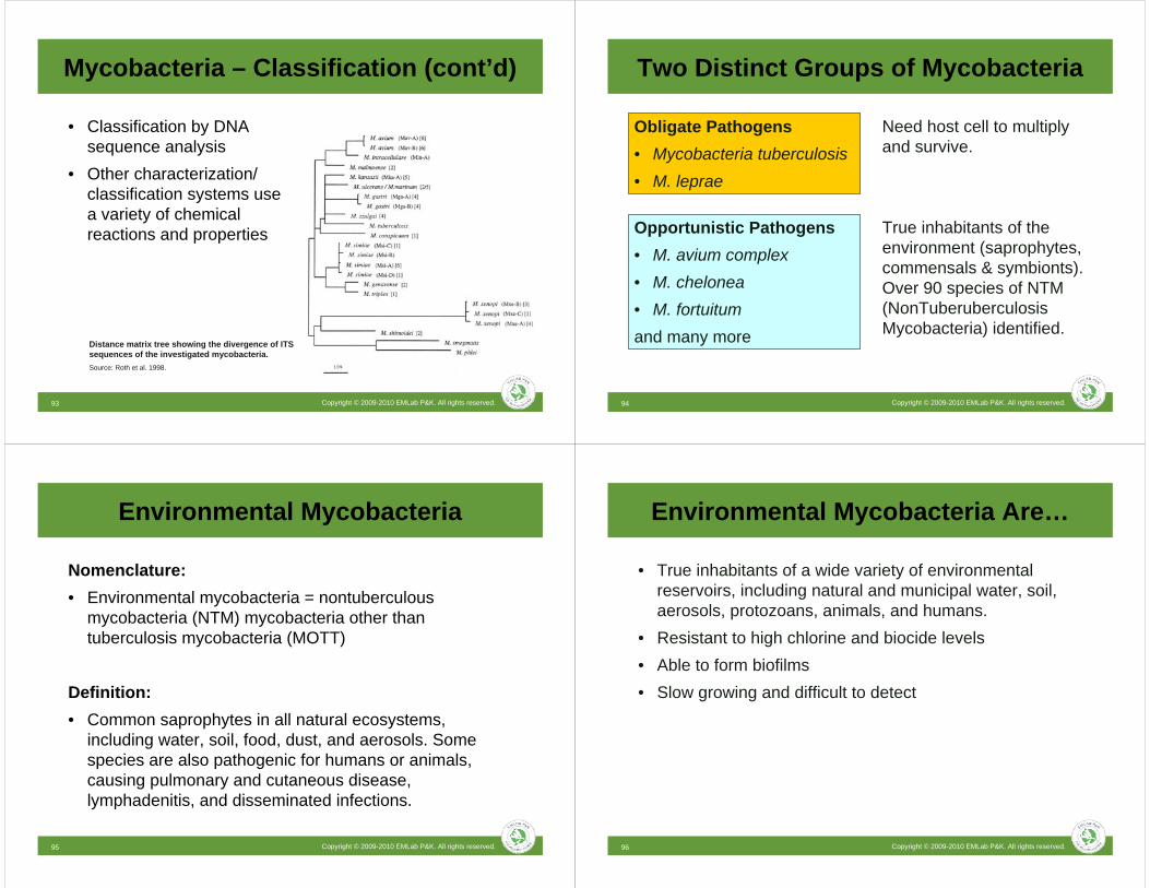
Distance matrix tree showing the divergence of ITS sequences of the investigated mycobacteria.Source: Roth et al. 1998.
• Classification by DNA sequence analysis
• Other characterization/ classification systems use a variety of chemical reactions and properties
Mycobacteria – Classification (cont’d)
Copyright © 2009-2010 EMLab P&K. All rights reserved.94
Obligate Pathogens• Mycobacteria tuberculosis• M. leprae
Opportunistic Pathogens• M. avium complex• M. chelonea• M. fortuitumand many more
Need host cell to multiply and survive.
True inhabitants of the environment (saprophytes, commensals & symbionts). Over 90 species of NTM (NonTuberuberculosisMycobacteria) identified.
Two Distinct Groups of Mycobacteria
Copyright © 2009-2010 EMLab P&K. All rights reserved.95
Environmental Mycobacteria
Nomenclature:• Environmental mycobacteria = nontuberculous
mycobacteria (NTM) mycobacteria other than tuberculosis mycobacteria (MOTT)
Definition:• Common saprophytes in all natural ecosystems,
including water, soil, food, dust, and aerosols. Some species are also pathogenic for humans or animals, causing pulmonary and cutaneous disease, lymphadenitis, and disseminated infections.
Copyright © 2009-2010 EMLab P&K. All rights reserved.96
• True inhabitants of a wide variety of environmental reservoirs, including natural and municipal water, soil, aerosols, protozoans, animals, and humans.
• Resistant to high chlorine and biocide levels• Able to form biofilms• Slow growing and difficult to detect
Environmental Mycobacteria Are…

Copyright © 2009-2010 EMLab P&K. All rights reserved.97
Diseases Caused byEnvironmental Mycobacteria
• Pulmonary disease (hot-tub lung)• Lymphadentitis (inflammation of lymph nodes)• Infections of soft tissue/skin• Disseminated disease (e.g. AIDS patients)• Associated with Crohn’s disease (chronic bowel disease)
Copyright © 2009-2010 EMLab P&K. All rights reserved.98
Transmission and Infection
• Transmission of environmental mycobacteria is not from human-to human or animal-to-human. Disease results from environmental exposure
• Aerosol associated infections (hypersensitivity pneumonitis)– Occupational: metal working fluids, life guards (swimming pools)– Home: aerated hot tubs, spas, humidifiers, water damaged
building material
Copyright © 2009-2010 EMLab P&K. All rights reserved.99
Source: Current Opinion in Allergy and Clinical Immunology 2009, 9:97–102
Mycobacteria in Metalworking Fluids
• Hypersensitivity pneumonitis (HP) has been associated with exposure of workers to metalworking fluids
• Recent work has focused on the presumed relationship between the microbiological contamination of MWF with M. immunogenum as the cause of hypersensitivity pneumonitis
Copyright © 2009-2010 EMLab P&K. All rights reserved.100
Source: WHO – Mycobacteria in Water
Mycobacteria in Water
• Mycobacteria are commonly found in municipal water.
• A number of mycobacteriainfections were reported and documented since the 1970s.
• Mycobacteria infections are often misdiagnosed and likely underreported.

Copyright © 2009-2010 EMLab P&K. All rights reserved.101
• 1.8 cases of nontuberculous diseases per 100,000 persons in the U.S. were estimated in the early 1980s. No current data for the U.S. available.
• Recent analysis from Ontario (Canada) found average annual increase of 8.4% for the isolation prevalence of NTM.
• Detection of NTM has improved significantly.
Epidemiology
Copyright © 2009-2010 EMLab P&K. All rights reserved.102
*NTM, nontuberculous mycobacteria; HCUP, Healthcare Cost and Utilization Project; SID, state inpatient databases; ICD-9, International Statistical Classification of Diseases, Revision 9.
Source: http://www.cdc.gov/eid/content/15/10/1562.htm
7,959 (48.3)Other primary diagnosis <1% of population
176 (1.07)Aspiration pneumonia caused by inhalation of food/vomitus5070
186 (1.13)Postinflammatory pulmonary fibrosis515
196 (1.19)Volume depletion2765
216 (1.31)Bronchiectasis with acute exacerbation4941
225 (1.37)Congestive heart failure, unspecified4280
392 (2.38)Acute respiratory failure51881
821 (4.98)Obstructive chronic bronchitis with acute exacerbation49121
1,156 (7.01)Pneumonia482
5,148 (31.25)Pulmonary NTM310
No. (%)Primary diagnosisICD-9 code
Primary diagnoses, non-AIDS pulmonary NTM-associated hospitalizations, HCUP-SID, USA, 1998–2005*
Epidemiology (cont’d)
Copyright © 2009-2010 EMLab P&K. All rights reserved.103
Source: http://www.cdc.gov/eid/content/15/10/1562.htm
Average annual prevalence of non-AIDS pulmonary nontuberculousmycobacteria-associated hospitalizations by age group and sex, Healthcare
Cost and Utilization Project state inpatient databases, USA, 1998–2005.
Epidemiology (cont’d)
Copyright © 2009-2010 EMLab P&K. All rights reserved.104
Source: http://www.cdc.gov/eid/content/15/10/1562.htm
Age-adjusted prevalence of non-AIDS pulmonary nontuberculousmycobacteria-associated hospitalizations among men, California (CA), Florida (FL), and New York (NY), USA, Healthcare Cost and Utilization Project state
inpatient databases, 1998–2005.
Epidemiology (cont’d)

Copyright © 2009-2010 EMLab P&K. All rights reserved.105
• Non-tuberculosis mycobacteria (NTM) is a significant and increasing cause of pulmonary illnesses in the United States.
• Prevalence of pulmonary NTM-associated hospitalizations is increasing in selected geographic areas.
• It has been estimated that in the U.S., 25% to 50% of individuals with AIDS will develop NTM diseases, primarily attributable to MAC.
• Waterborne NTM have been associated with hospital (nosocomial) infections worldwide.
Mycobacteria Summary
Copyright © 2009-2010 EMLab P&K. All rights reserved.106
Mycobacteria Summary (cont’d)
• In particular metalworking fluids and water in hospital transplant units should be monitored for NTM.
• Environmental mycobacteria (nontuberculousmycobacteria) are distinctly different from the obligate pathogens of the M. tuberculosis complex.
• NTM have been identified in numerous environmental sources, including water.
• NTM are not thought to be transmitted by the human to human route, but are instead thought to be transmitted from environmental sources.
Copyright © 2009-2010 EMLab P&K. All rights reserved.107
MRSA and Staphylococci – Overview
Staphylococcus aureus• Gram-positive bacteria• 0.5 – 1 m diameter• Commonly found on the skin
and in the nose of humans
Copyright © 2009-2010 EMLab P&K. All rights reserved.108
MRSA and Staphylococci – Overview
• Approximately one third of the world’s population has S. aureus bacteria on their body at any given time.
• About 1% of people carrying S. aureus have MRSA (CDC estimated).
• Spread from one person to another through casual contact or contaminated objects.
• Estimated 94,000 cases of MRSA infections in the U.S. per year and nearly 19,000 deaths.

Copyright © 2009-2010 EMLab P&K. All rights reserved.109
Staphylococci – Symptoms
Skin infections• Folliculitis (hair roots)• Impetigo (blisters)• Skin abscesses• Cellulitis• Necrolysis
Copyright © 2009-2010 EMLab P&K. All rights reserved.110
S. aureus – Symptoms
• Staphylococci tend to infect skin but can travel through the bloodstream and involve almost any site in the body, particularly the heart and the bones.
• May infect the respiratory tract, mainly in people with chronic lung disease or influenza and lead to staphylococcal pneumonia.
• Other severe and life-threatening infections with staphylococci include victims of severe burns and heart infections.
Copyright © 2009-2010 EMLab P&K. All rights reserved.111
MRSA – Resistance
• MRSA is a resistant variation of the common bacterium Staphylococcus aureus. It has evolved an ability to survive treatment with beta-lactamantibiotics, including penicillin, methicillin, and cephalosporins– Hospital acquired (HA)– Community acquired (CA)
Copyright © 2009-2010 EMLab P&K. All rights reserved.112
Hospital Acquired MRSA
• Most infections with MRSA occur in hospitals and healthcare facilities (HA-MRSA) including:– Surgical would infections– Urinary tract infections– Blood stream infections and pneumonia

Copyright © 2009-2010 EMLab P&K. All rights reserved.113
Risk factors for HA-MRSA:• Current or recent hospitalization• Residing in a long-term care facility• Invasive devices• Recent antibiotic use
Prevention:• Ask hospital staff to wash their hands• Wash your own hands frequently• Make sure invasive devices are kept sterile
Hospital Acquired MRSA (cont’d)
Copyright © 2009-2010 EMLab P&K. All rights reserved.114
Community acquired MRSA recently gained much attention in the news media.
Risk factors:• Young age (children)• Participating in contact sports• Sharing towels and athletic equipment• Weakened immune system• Living in crowded and/or unsanitary conditions• Association with health care workers
Community Acquired MRSA
Copyright © 2009-2010 EMLab P&K. All rights reserved.115
Prevention• Washing hands frequently• Keep personal items personal• Keep wounds covered• Shower after athletic games or practices• Do not participate in athletic games or practices if you
have infected wounds• Sanitize items• Get tested if you have a skin infection• Use antibiotics appropriately
Community Acquired MRSA (cont’d)
Copyright © 2009-2010 EMLab P&K. All rights reserved.116
Clinical testing• Sample from an infected site or a nasal swab• Clinical laboratory performs the testing
Environmental testing• Collect swab samples from items that are frequently
touched such as door knobs, keyboards, athletic equipment etc.
• Environmental laboratory can perform the testing for MRSA
Diagnosis and Environmental Testing

Copyright © 2009-2010 EMLab P&K. All rights reserved.117
Sewage – Evaluating Hazards
• Nature of Sewage• Microorganisms• Human exposure
– Direct contact– Ingestion– Aerosols
• Sampling Strategies– Hypotheses– Sampling plans– Data Interpretation
Copyright © 2009-2010 EMLab P&K. All rights reserved.118
Nature of Sewage
• Sewage is the water found in sewers. • “Used” water from homes, workplaces, surface runoff.• Contains waste products of human, animal, vegetable
and mineral origin. – Suspended solids – Solutes– Bacteria and other sewage microorganisms
• On average each of us generates between 135 and 180 liters of sewage each day.
• This sewage is over 99.9% liquid, with less than 0.1% being solid.
Copyright © 2009-2010 EMLab P&K. All rights reserved.119
Who Is At Risk?
• Employees involved in sewer inspection and maintenance work
• Construction workers who repair or replace live sewers• Water company employees who work with sewage
treatment plants• Agricultural and forestry workers who may be exposed to
sewage sludge• Sludge tanker drivers/operators and associated
maintenance staff• Plumbers• Employees who clean and maintain the underside of
railway carriages and empty aircraft sewage compartments and other types of portable lavatories
Copyright © 2009-2010 EMLab P&K. All rights reserved.120
Aerosol Exposure
Wastewater Treatment • Average concentrations of 17000 cfu/ml of mesophilic,
2100 cfu/ml of TSA-SB bacteria (bacteria associated with certain waterborne virulence factors) in the water.
• In the aerosol of the fixed-film reactor 3000 cfu/m3
mesophilic and 730 cfu/m3 TSA-SB bacteria.

Copyright © 2009-2010 EMLab P&K. All rights reserved.121
What Are The Health Risks?
• Gastroenteritis: cramping stomach pains, diarrhea and vomiting
• Weil's disease: a flu-like illness with persistent and severe headache; damage to liver, kidneys and blood may occur and the condition can be fatal
• Hepatitis: inflammation of the liver, and jaundice • Occupational asthma: attacks of breathlessness, chest
tightness and wheezing; produced by the inhalation of living or dead organisms
• Infection of skin or eyes• Rarely, allergic alveolitis: (inflammation of lung) with fever,
breathlessness, dry cough, and aching muscles and jointsCopyright © 2009-2010 EMLab P&K. All rights reserved.122
How Do Sewage Micro-organismsEnter The Body?
• Hand-to-mouth contact during eating, drinking and smoking,
• Wiping the face with contaminated hands or gloves, or by licking splashes from the skin.
• Skin contact, through cuts, scratches, or penetrating wounds, i.e. from discarded hypodermic needles.
• Aerosols landing on surfaces of the eyes, nose and mouth.
• By breathing them in, as either dust, aerosol or mist.
Copyright © 2009-2010 EMLab P&K. All rights reserved.123
Coliforms
• Rod-shaped Gram-negative non-spore forming organisms that ferment lactose with the production of acid and gas when incubated at 35-37°C.
• Coliforms are abundant in the feces of warm-blooded animals, but can also be found in the aquatic environment, in soil and on vegetation.
• TAXA– Citrobacter– Enterobacter– Escherichia– Hafnia
– Klebsiella– Serratia– Yersinia
Copyright © 2009-2010 EMLab P&K. All rights reserved.124
Water Quality Coliform Guidelines
• Culture based– 200 colonies of fecal coliform bacteria /100ml for primary contact
recreation– 1000 colonies of fecal coliform bacteria /100ml for secondary
contact recreation
• Swimming beaches:– geometric mean of 126 E.coli bacteria per 100 ml of water (fresh
water)

Copyright © 2009-2010 EMLab P&K. All rights reserved.125
Enterococcus
• May provide a higher correlation than fecal coliforms with many human pathogens.
• In 2004, Enterococcusspp. took the place of fecal coliforms as the new federal standard for water quality at public beaches.
Enterococcus infection in pulmonary tissue.Source: wikipedia.org
• The acceptable level of contamination is very low, – Hawaii: 7 colony forming units per 100 ml of water– Geometric mean of 35 / 100 ml of water for five samples over 30 days
and an instantaneous (single sample) standard of 104 / 100 ml of water (salt water).
Copyright © 2009-2010 EMLab P&K. All rights reserved.126
Products
• Air pumps and samplers
• Water bottles
• Swabs
• Media
http://www.emlabpk.com/store
Copyright © 2009-2010 EMLab P&K. All rights reserved.127
Thank you for your time.
Questions?