b cell–intrinsic tlr7 signaling is essential for the … cell–intrinsic tlr7 signaling is...
TRANSCRIPT

of June 2, 2018.This information is current as
Germinal Centersfor the Development of Spontaneous
Intrinsic TLR7 Signaling Is Essential−B Cell
RahmanKhan, Takashi Satoh, Shizuo Akira and Ziaur S. M. Chetna Soni, Eric B. Wong, Phillip P. Domeier, Tahsin N.
http://www.jimmunol.org/content/193/9/4400doi: 10.4049/jimmunol.1401720September 2014;
2014; 193:4400-4414; Prepublished online 24J Immunol
MaterialSupplementary
0.DCSupplementalhttp://www.jimmunol.org/content/suppl/2014/09/24/jimmunol.140172
Referenceshttp://www.jimmunol.org/content/193/9/4400.full#ref-list-1
, 33 of which you can access for free at: cites 71 articlesThis article
average*
4 weeks from acceptance to publicationFast Publication! •
Every submission reviewed by practicing scientistsNo Triage! •
from submission to initial decisionRapid Reviews! 30 days* •
Submit online. ?The JIWhy
Subscriptionhttp://jimmunol.org/subscription
is online at: The Journal of ImmunologyInformation about subscribing to
Permissionshttp://www.aai.org/About/Publications/JI/copyright.htmlSubmit copyright permission requests at:
Email Alertshttp://jimmunol.org/alertsReceive free email-alerts when new articles cite this article. Sign up at:
Print ISSN: 0022-1767 Online ISSN: 1550-6606. Immunologists, Inc. All rights reserved.Copyright © 2014 by The American Association of1451 Rockville Pike, Suite 650, Rockville, MD 20852The American Association of Immunologists, Inc.,
is published twice each month byThe Journal of Immunology
by guest on June 2, 2018http://w
ww
.jimm
unol.org/D
ownloaded from
by guest on June 2, 2018
http://ww
w.jim
munol.org/
Dow
nloaded from

The Journal of Immunology
B Cell–Intrinsic TLR7 Signaling Is Essential for theDevelopment of Spontaneous Germinal Centers
Chetna Soni,*,1 Eric B. Wong,*,1 Phillip P. Domeier,* Tahsin N. Khan,*,†
Takashi Satoh,‡ Shizuo Akira,‡ and Ziaur S. M. Rahman*
Spontaneous germinal center (Spt-GC) B cells and follicular helper T cells generate high-affinity autoantibodies that are involved in
the development of systemic lupus erythematosus. TLRs play a pivotal role in systemic lupus erythematosus pathogenesis. Although
previous studies focused on the B cell–intrinsic role of TLR-MyD88 signaling on immune activation, autoantibody repertoire, and
systemic inflammation, the mechanisms by which TLRs control the formation of Spt-GCs remain unclear. Using nonautoimmune
C57BL/6 (B6) mice deficient in MyD88, TLR2, TLR3, TLR4, TLR7, or TLR9, we identified B cell–intrinsic TLR7 signaling as
a prerequisite to Spt-GC formation without the confounding effects of autoimmune susceptibility genes and the overexpression of
TLRs. TLR7 deficiency also rendered autoimmune B6.Sle1b mice unable to form Spt-GCs, leading to markedly decreased
autoantibodies. Conversely, B6.yaa and B6.Sle1b.yaa mice expressing an extra copy of TLR7 and B6.Sle1b mice treated with
a TLR7 agonist had increased Spt-GCs and follicular helper T cells. Further, TLR7/MyD88 deficiency led to compromised B cell
proliferation and survival after B cell stimulation both in vitro and in vivo. In contrast, TLR9 inhibited Spt-GC development. Our
findings demonstrate an absolute requirement for TLR7 and a negative regulatory function for TLR9 in Spt-GC formation under
nonautoimmune and autoimmune conditions. Our data suggest that, under nonautoimmune conditions, Spt-GCs initiated by
TLR7 produce protective Abs. However, in the presence of autoimmune susceptibility genes, TLR7-dependent Spt-GCs produce
pathogenic autoantibodies. Thus, a single copy of TLR7 in B cells is the minimal requirement for breaking the GC-tolerance
checkpoint. The Journal of Immunology, 2014, 193: 4400–4414.
Upon T cell–dependent antigenic stimulation, B cellsdifferentiate into either preplasma IgM+ recirculatingmemory B cells or extrafollicular Ab-forming cells
(AFCs) or they form germinal centers (GCs) within primary fol-licles (1, 2). Extrafollicular AFCs are generally short-lived andsecrete IgM and/or low-affinity class-switched Abs. In contrast,rapidly dividing GC B cells undergo class-switch recombinationand site-directed somatic hypermutation of their Ig V region genes(3, 4). Clones selected for increased Ag affinity survive and dif-ferentiate into Ab-producing long-lived plasma cells or memoryB cells (4). Many of the GC-derived, long-lived plasma cells hometo the bone marrow (BM) and produce high-affinity Abs, confer-ring lasting humoral immunity (5–7).
B cells have emerged as key players in TLR-mediated systemicautoimmune responses, especially in systemic lupus erythematosus
(SLE). Exposure ofmurine B cells to TLR4, TLR7, andTLR9 agonists
increases the expression of B cell costimulatory factors and induces
B cells to proliferate, produce cytokines, differentiate into APCs,
switch Ig classes, and secrete Igs (8, 9). Consensus views drawn from
studies on TLR7- and TLR9-dependent MyD88 signaling in auto-
immune lupus-prone mouse models propose that TLR7 promotes
inflammation and lupus pathogenesis, whereas TLR9 plays a nega-
tive-regulatory role to dampen the disease process (10–14). However,
most of these studies focused primarily on the involvement of MyD88
or the two TLRs in B cell activation, autoantibody specificity, and the
development of glomerulonephritis (11, 12, 15–20).The GC is an important B cell–tolerance checkpoint in the
periphery (21, 22). Several autoimmune-prone mice develop
spontaneous GCs (Spt-GCs) in the spleen by 1–2 mo of age (23).
Dysregulated GC B cell and follicular (FO) helper T cell (Tfh cell)
responses make decisive contributions to the generation of class-
switched autoantibodies and to the development of lupus in mouse
models (22, 24–26), as well as to human SLE (27, 28). We also
recently reported a strong correlation between autoantibody
production and Spt-GCs in B6.Sle1b mice harboring the lupus-
associated SLAM genes derived from the autoimmune NZM2410
strain (29). Understanding altered regulation of both the FO GC
and extrafollicular pathways by TLRs in autoimmune diseases
will help to develop treatment options for the heterogeneous
population of SLE patients in whom either or both pathways may
be affected. Earlier studies extensively investigated the involve-
ment of TLRs in modulating autoimmune responses using MRL/
lpr mice (11, 15). This model allows for the extrafollicular dif-
ferentiation of B cells (15, 30). Recently, using different TLR-
overexpression and knockout (KO) autoimmune mouse models,
several groups suggested B cell–intrinsic and/or -extrinsic roles
*Department of Microbiology and Immunology, Pennsylvania State University Col-lege of Medicine, Hershey, PA 17033; †Department of Molecular Microbiology andImmunology, Oregon Health and Science University, Portland, OR 97239; and‡Department of Host Defense, Research Institute for Microbial Diseases, OsakaUniversity, Osaka 565-0871, Japan
1C.S. and E.B.W. contributed equally to this work.
Received for publication July 8, 2014. Accepted for publication August 28, 2014.
This work was supported by National Institutes of Health Grant AI091670 (to Z.S.M.R.).
Address correspondence and reprint requests to Dr. Ziaur Rahman, Department ofMicrobiology and Immunology, H107, Pennsylvania State University College ofMedicine, 500 University Drive, Hershey, PA 17033-0850. E-mail address:[email protected]
The online version of this article contains supplemental material.
Abbreviations used in this article: AFC, Ab-forming cell; ANA, anti-nuclear Ab; AP,alkaline phosphatase; B6, C57BL/6; BAFF, B cell activating factor; BM, bone mar-row; DC, dendritic cell; FDC, FO dendritic cell; FO, follicular; GC, germinal center;IMQ, imiquimod; KO, knockout; MZ, marginal zone; PI, propidium iodide; PNA,peanut agglutinin; SA, streptavidin; SLE, systemic lupus erythematosus; Sm/RNP,Smith Ag/ribonucleoprotein; Spt-GC, spontaneous GC; Tfh cell, follicular helperT cell.
Copyright� 2014 by TheAmericanAssociation of Immunologists, Inc. 0022-1767/14/$16.00
www.jimmunol.org/cgi/doi/10.4049/jimmunol.1401720
by guest on June 2, 2018http://w
ww
.jimm
unol.org/D
ownloaded from

for TLR-MyD88 signaling in the GC-differentiation pathway ofautoantibody production and autoimmune inflammatory responses(20, 31–33). However, the mechanisms and the requirement forphysiological levels of individual TLRs in controlling the for-mation of Spt-GCs and Tfh cell development remain unclear.In this study, we first addressed the requirement for TLRs in the
development of Spt-GCB cells and Tfh cells in the steady-state. Thesestudies were performed under nonautoimmune conditions, without theconfounding effects of TLR overexpression, exogenous TLR stimu-lation, or purposeful immunizations. We found that B cell–intrinsicTLR7-MyD88 signaling was required for the formation of Spt-GCsand that TLR9 signaling negatively regulated the magnitude of theTLR7-mediated response. In agreement with our observations innonautoimmune mice, TLR7-deficient autoimmune B6.Sle1b mice(Sle1b/TLR7-KO) also were unable to initiate GC formation, even by6 mo of age, and they had diminished total Ab and autoantibodyresponses. Our ex vivo and in vivo studies indicated suboptimal B cellsurvival and proliferation in the absence of TLR7. These resultshighlight the absolute requirement for TLR7 and the negative-regulatory function of TLR9 in Spt-GC responses in nonautoimmuneand autoimmune environments.
Materials and MethodsMice
C57BL/6 (B6) mice, 3 mo of age (for particular experiments), were pur-chased from The Jackson Laboratory (Bar Harbor, ME), Taconic (Hudson,NY), Charles River (Wilmington, MA), or the National Cancer Institute(Bethesda, MD). Spleens from B6 mice, housed at The Rockefeller Uni-versity (New York, NY) germ-free facility and specific pathogen–free fa-cility, were kindly provided by Dr. Daniel Mucida. MyD88fl/fl, CD11c-Cre+/2-MyD88fl/fl, and LysM-Cre+/2-MyD88fl/fl mice were a kind gift fromDr. Milena Bogunovic (Pennsylvania State University College of Medicine,Hershey, PA). Breeding pairs for B6, B6.mMT (B6.129S2-Ighmtm1Cgn/J),Rag1-KO, B6.SB-yaa/J (B6.yaa), MyD88-KO [B6.129P2(SJL)-Myd88tm1.1Defr/J], and OT-II–transgenic [B6.Cg-Tg (TcraTcrb)425Cbn/J]mice were originally purchased from The Jackson Laboratory and bredin-house. TLR7-KO (34) and TLR9-KO (35) mice backcrossed to B6 micefor 10 generations were bred in-house. The derivation of B6 mice congenicfor the Sle1b sublocus (named B6.Sle1b) was described previously (36).B6.Sle1b.yaa mice were generated by breeding B6.yaa males with B6.Sle1b females. Sle1b/TLR7-KO and Sle1b/TLR9-KO mice were generatedby crossing B6.Sle1b mice with TLR7-KO and TLR9-KO lines, respec-tively. All animals were housed in the specific pathogen–free animal fa-cility at Pennsylvania State University College of Medicine, and allprocedures were performed in accordance with the guidelines approved bythe Pennsylvania State University Institutional Animal Care and UseCommittee.
Flow cytometry
The following Abs were used for flow cytometric analysis of mousesplenocytes or BM cells. PacBlue–anti-B220 (RA3-6B2), Alexa Fluor 700–anti-CD4 (RM4-5), PE–anti-PD-1 (29F.1A12), PerCP–Cy5.5–anti-CD69(H1.2F3), allophycocyanin–anti-TCR Va2 (B20.1), allophycocyanin–Cy7–anti-CD25 (PC61), Cy5–anti-CD86 (GL1), PeCy7–anti-CD95 (FAS,Jo2), PeCy7–anti-MHC II (M5/114.15.2), allophycocyanin–anti-CD24(HSA) (M1/69), biotin–anti-Ly5.1 (BP-1) (6C3), FITC–anti-CD23 (B3B4),and PE-Cy5–streptavidin (SA) were from purchased from BioLegend(San Diego, CA). Biotin–anti-CXCR5 (2G8), FITC–anti-CD11c (HL3),FITC–anti-CD43 (S7) were from BD Pharmingen (San Diego, CA).FITC–peanut agglutinin (PNA) was from Vector Labs (Burlingame, CA).PE–anti-IgM (eB121-15F9), allophycocyanin–anti-CD93 (AA4.1), andFITC–anti-F4/80 (BM8) were from eBioscience (San Diego, CA).Stained cells were analyzed using a BD LSR II flow cytometer (BDBiosciences, Franklin Lakes, NJ). Data were acquired using FACSDivasoftware (BD Biosciences, San Jose, CA) and analyzed using FlowJosoftware (TreeStar, San Carlos, CA). Dead cells were quantified by flowcytometry using DAPI (Sigma-Aldrich, St. Louis, MO).
Histology, immunofluorescence, and anti-nuclear Ab staining
The following Abs and reagents were used for immunohistochemicalanalysis of mouse spleen sections: biotin-mouse anti-rat IgG (Jackson
ImmunoResearch, West Grove, PA); alkaline phosphatase (AP)-SA, VectorBlue AP Substrate Kit III, and Vector NovaRED Substrate Kit (all fromVector Labs); HRP-PNA (Sigma-Aldrich); and biotin–anti-IgD (11-26;Southern Biotechnology Associates). Immunofluorescence staining Abs,including anti-mouse Ki67 (16A8BL; BioLegend); PE–anti-CD4 (GK1.5),FITC-GL7 (RA3-6B2), purified rat anti-mouse FDC-M1, and allophyco-cyanin–anti-IgD, were from BD Biosciences (Franklin Lakes, NJ). FITC–anti-CD35 (eBio4E3) was from eBioscience. Biotin–anti-MFG-E8 (18A2-G10) was from MBL (Naka-ku-Nagoya, Japan). Kidney sections werestained for C3 using FITC–anti-C3 from Immunology Consultants Labo-ratory (Portland, OR). Anti-nuclear Abs (ANAs) were detected by indirectimmunofluorescence staining of Hep-2 culture slides (Antibodies, Davis,CA) using serum from mice at a 1:50 dilution in PBS, probed with FITC–rat anti-mouse k (H139-52.1; Southern Biotechnologies Associates, Bir-mingham, AL). For imaging spleen sections, we used a Leica DM4000microscope and Leica software (LAS-AF) (Leica Microsystems, BuffaloGrove, IL). The color intensity of images was enhanced slightly usingAdobe Photoshop CS4 (Adobe Systems, San Jose, CA). This was neces-sary for better visualization and was carried out consistently betweencontrol and test sections while maintaining the integrity of the data.
ELISPOT assays
ELISPOT assays were performed as described (37). Briefly, splenocytes in10% RPMI 1640 with antibiotics were plated at a concentration of 13 105
cells/well onto anti-IgM–coated or anti-IgG–coated (Invitrogen, GrandIsland, NY) multiscreen 96-well filtration plates (Millipore, Bedford, MA)or at 1 3 106 cells/well on dsDNA-, histone-, or nucleosome-coatedmultiscreen 96-well filtration plates. Serially diluted (1:2) cells were in-cubated for 6 h at 37˚C. IgM-producing AFCs were detected using bio-tinylated anti-mouse IgM (Jackson ImmunoResearch) and SA-AP (VectorLabs). IgG-producing AFCs were detected using AP-conjugated anti-mouse IgG (Molecular Probes, Grand Island, NY). dsDNA-, histone-,and nucleosome-specific AFCs were detected by biotinylated anti-k Ab(Invitrogen) and SA-AP (Vector Labs). Plates were developed using theVector Blue AP Substrate Kit III (Vector Labs). ELISPOTs were countedand analyzed using a computerized imaging/analysis system (CellularTechnology, Shaker Heights, OH).
Serology: Ig and autoantibody titers
Total serum IgM/IgG or IgG from in vitro B cell culture supernatants wasmeasured using standard ELISA protocols. Serum Ab titers were quanti-tated as described (29). Briefly, ELISA plates were coated with anti-IgM oranti-IgG capture Abs (Invitrogen) and detected using biotinylated anti-mouse IgM (Jackson ImmunoResearch) or AP-conjugated anti-mouseIgG (Molecular Probes). Total IgG autoantibody titers were measured inELISA plates coated with dsDNA, histone, nucleosome, Smith Ag/ribo-nucleoprotein (Sm/RNP), or cardiolipin and detected with biotinylatedanti-k Ab (Invitrogen). IgG subtype–specific autoantibody titers weredetected by biotinylated IgG1, biotinylated IgG2b, and AP-IgG2c Abs(Southern Biotech). Biotinylated Abs were detected by SA-AP (VectorLabs). The plates were developed by p-Nitrophenyl Phosphate, DisodiumSalt (Thermo Fisher Scientific, Rockford, IL) substrates for AP.
Generation of BM chimeric mice
Ten- to twelve-week-old female B6.mMT mice (recipients) were lethallyirradiated with 1000 rad x-rays (X-Rad 320iX Research Irradiator; Preci-sion X-Ray, North Branford, CT) prior to the transfer of BM cells. Eachmouse received 7–10 3 106 (T cell–depleted) BM cells (i.v.) isolated from8–10-wk-old female donor mice, with 80% cells from B6.mMT mice and20% cells from B6, TLR9-KO, or TLR7-KO mice. The recipient micewere analyzed after 3 mo for Spt-GC B cells and Tfh cells.
Adoptive transfer
CD4+ T cells were purified by negative selection from B6 mice using a mousePan T Cell Isolation Kit II (Miltenyi Biotec, Bergisch Gladbach, Germany).B cells from B6, TLR7-KO, or TLR9-KO mice were negatively selected usinganti-CD43 (Ly-48) MicroBeads (Miltenyi Biotec). Purified B and T cells weremixed at a 3:1 ratio and transferred (i.v.) into Rag1-KO mice. Two monthslater, mice were sacrificed, and spleens were analyzed for Spt-GCs.
In vitro B cell–proliferation assay
B cells were purified from naive 8–10-wk-old B6, TLR7-KO, TLR9-KO, orMyD88-KO mice with mouse anti-CD43 (Ly-48) MicroBeads. PurifiedB cells were stained with 3 mM CFSE (Sigma Aldrich) in PBS with 5%FBS for 15 min at room temperature. Stained B cells were cultured with 25mg/ml soluble anti-IgM (Jackson ImmunoResearch) and 20 mg/ml soluble
The Journal of Immunology 4401
by guest on June 2, 2018http://w
ww
.jimm
unol.org/D
ownloaded from

anti-CD40 Ab (BioLegend), with or without 10 ng/ml B cell activatingfactor (BAFF; PeproTech, Rocky Hill, NJ). After 72 or 96 h of stimulation,cells were surface stained (if required), DAPI stained or fixed, andsubjected to flow cytometry.
In vivo B cell proliferation
Eight- to ten-week-old female B6.mMT mice were immunized with 200 ml10% SRBCs in PBS, 2 d prior to cell transfers. Each mouse received (i.v.)5 3 106 CFSE-labeled purified B cells from female B6, TLR7-KO, orTLR9-KO mice. Recipients were analyzed for B cell proliferation on thefourth day of transfer.
Cell cycle analysis
B cells were cultured with anti-IgM (25 mg/ml) and anti-CD40 (20 mg/ml)for the indicated time periods. Subsequently, cells were harvested andwashed with chilled PBS and then fixed with chilled 70% ethanol over-night at 220˚C. Cells were centrifuged at 1000 3 g for 10 min at 4˚C andwashed with PBS. Propidium iodide (PI) staining solution containing 50mg/ml PI, 50 mg/ml RNase A (Roche Applied Sciences, Indianapolis, IN),and 100 mM EDTA in PBS was used to stain the samples for 1–2 h at 42˚C.Data were analyzed by flow cytometry.
RNA preparation and real-time RT-PCR
Total RNAwas isolated using TRIzol reagent (Ambion, Grand Island, NY),as per the manufacturer’s instructions, from total splenocytes or purifiedB cells of indicated mice. RNA was reverse transcribed using a high-capacity reverse transcription kit (Applied Biosystems, Grand Island,NY). Gene expression was quantified using a Power SYBR Green PCRMaster Mix Kit and the StepOnePlus Real-Time PCR System (both fromApplied Biosystems). Primers were designed using Primer3 software andsynthesized by IDT Technologies (Coralville, IA). Amplification con-ditions for all primer sets were one cycle of 95˚C for 10 min, followed by40 cycles of 95˚C for 15 s and 60˚C for 1 min. 18srRNA was used as thereference gene for sample normalization. PCR primer sequences are asfollows: PD-1 Forward 59-GAG CTC GTG GTA ACA GAG AGA A-39;PD-1 Reverse 59-ACA GGG ATA CCC ACT AGG GC-39: ICOS Forward59-CGG ATC CAG TGT GCA TGA CC-39; ICOS Reverse 59-AGC TTATGA GGT CAC ACC TGC-39: XBP-1 Forward 59-GGC CAA GGG GAGTGG AGT AAG-39; XBP-1 Reverse 59-GCT GCA GAG GTG CAC ATAGTC-39: IRF-4 Forward 59-CAC AGC TCATGT GGA ACC TCT-39; IRF-4 Reverse 59-TCA GGT AAC TCG TAG CCC CT-39: Bcl-6 Forward 59-AGA CAT TGG CAG AGT TCC AGA-39; Bcl-6 Reverse 59-CTG GCAGCG ATC ACA TTT GTA-39: AICDA Forward 59-CCT TCG CAA CAAGTC TGG CT-39; AICDA Reverse 59-GAA CCA GGT GAC GCG GTAA-39; 18srRNA forward 59-CAC TTT TGG GGC CTT CGT GT-39; and18srRNA Reverse 59-AGG CCC AGA GAC TCA TTT CTT C-39.
Statistical analysis
An unpaired, nonparametric, Mann–Whitney, Student t test was used tocompare two groups, whereas one-way ANOVA, followed by the Tukeymultiple-comparison test, was used to compare more than two groups.GraphPad Prism 6 software (La Jolla, CA) was used for all analyses. Errorbars reflect mean, unless otherwise indicated. If the statistical analysis isnot shown, nonsignificant differences were found.
ResultsTLR7 drives the formation of steady-state Spt-GCs and Abs inB6 mice
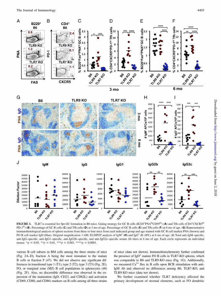
GCs are usually described in the context of T-dependent antigenicstimulation or under autoimmune conditions (23, 38, 39). However,we observed Spt-GCs in unmanipulated, nonautoimmune B6 miceat 3 mo of age and older, housed in a specific pathogen–free facility,albeit at a lower frequency compared with age-matched autoim-mune prone mice (29). We extended this observation to B6 micehoused in various facilities (The Jackson Laboratory, NationalCancer Institute, Charles River, and Taconic) and found that, irre-spective of the facility, all B6 mice developed well-defined Spt-GCswith comparable percentages of B220+PNAhiFashi GC B cells andCD4+CXCR5hiPD-1hi Tfh cells at ∼3 mo of age (data not shown).Self-Ags and/or endogenous microbial components might stim-
ulate TLRs, thereby driving the Spt-GC reaction (40–42). Wecompared Spt-GC B cell and Tfh cell percentages, in the absence
of exogenous TLR stimulation, in 3-mo-old TLR2-, TLR3-,TLR4-, TLR7-, or TLR9-deficient mice with those in age- andsex-matched B6 mice. The percentages of Spt-GC B cells and Tfhcells in TLR2- and TLR3-deficient mice were comparable to B6controls (Supplemental Fig. 1A, 1B). Although not statisticallysignificant, TLR4-KO mice had lower percentages of Spt-GCB cells and Tfh cells compared with B6, TLR2-KO, and TLR3-KO mice (Supplemental Fig. 1A, 1B). However, histologicalanalysis of 3-mo-old TLR4-KO spleens showed well-formedIgD2GL7+ GCs (Supplemental Fig. 1C), indicating that TLR4 isnot required for the formation of Spt-GCs. At 3 mo of age, TLR7-KO mice had the lowest percentage of GC B cells and Tfh cellscompared with B6 controls, although the difference was not sta-tistically significant (Fig. 1A–D). However, at 6 mo, the per-centage of GC B cells and Tfh cells was significantly reduced inTLR7-KO mice compared with B6 controls and TLR9-KO mice(Fig. 1E, 1F). Conversely, TLR9-KO mice had an increased per-centage of GC B cells and Tfh cells compared with B6 and TLR7-KO mice starting at 3 mo of age (Fig. 1A–D). At 6 mo, the per-centage of GC B cells in TLR9-KO mice was comparable to B6mice; however, the Tfh cell percentage remained significantlyelevated (Fig. 1E, 1F).Immunohistological analysis of spleen sections from 3- and 6-
mo-old mice revealed the complete absence of IgD2PNA+ Spt-GCB cells in TLR7-KO mice, whereas TLR9-KO mice had largerSpt-GCs than did B6 controls (Fig. 1G). Moreover, IgM- and IgG-producing AFCs were barely detected in TLR7-KO mice, whereasTLR9-KO mice exhibited a significant increase (Fig. 1H, 1I).TLR7-KO mice had significantly lower Ab titers of total serumIgG2b and IgG2c than did TLR9-KO mice (Fig. 1J). Averageserum IgG2b and IgG2c titers in TLR7-KO mice were lower thanin B6 controls, but the differences were not statistically significant(Fig. 1J). No significant differences were observed in IgM, totalIgG, and IgG1 serum Ab titers among these three strains of mice(Fig. 1J). To ascertain whether the loss of Spt-GCs in TLR7-KOmice involved signaling via MyD88, we analyzed 3-mo-oldMyD88-KO mice and found that they also were unable to de-velop Spt-GCs in the absence of antigenic stimulation comparedwith 3-mo-old B6 mice (Supplemental Fig. 1D).To investigate the source of the TLR ligands stimulating Spt-GC
formation in naive B6 mice, we treated B6 mice with antibiotics for6 wk and compared their Spt-GC profile with control mice. We didnot observe any effect of antibiotics treatment on the percentage ofSpt-GC B cells or Tfh cells (Supplemental Fig. 1E, 1F). We alsoanalyzed the Spt-GC profile of B6 germ-free mice and foundthat they also formed Spt-GCs, albeit to a reduced extent(Supplemental Fig. 1G), similar to TLR4-KO mice. These resultssuggest that TLR7 ligands (self-nucleic acids and/or retroviralelements) are the primary stimulators for the formation of Spt-GCs and Tfh cells (41, 43–45), whereas TLR4 ligands (endoge-nous microbial components) may have a role in maintaining GCs.These observations are consistent with the recent report by Giltiayet al. (46). They used mice expressing both TLR7 and RNasetransgenes to show that self-RNA is involved in the anti-RNA Abresponses.
Markedly reduced Spt-GCs and Ab responses in TLR7-KOmice are not the outcome of developmental defects in primaryB cells and FO dendritic cell network
Next, we tested whether markedly reduced Spt-GCs and Tfh cells inTLR7-KO mice resulted from a defect in primary B cell developmentand maturation in the absence of TLR7. Flow cytometric analysis ofBM cells and splenocytes from B6, TLR9-KO, and TLR7-KO micerevealed no significant differences in the distribution of cells among
4402 TLR7 DRIVES SPONTANEOUS GERMINAL CENTER FORMATION
by guest on June 2, 2018http://w
ww
.jimm
unol.org/D
ownloaded from
various B cell subsets in BM cells among the three strains of mice(Fig. 2A–D), fraction A being the most immature to the matureB cells in fraction F (47). We did not observe any significant dif-ferences in transitional type 1 (T1), type 2 (T2), type 3 (T3) (Fig. 2E),FO, or marginal zone (MZ) B cell populations in splenocytes (48)(Fig. 2F). Also, no discernible difference was observed in the ex-pression of the maturation (IgD, CD23, and CD62L) and activation(CD69, CD80, and CD86) markers on B cells among all three strains
of mice (data not shown). Immunohistochemistry further confirmedthe presence of IgD+ mature FO B cells in TLR7-KO spleens, whichwas comparable to B6 and TLR9-KO mice (Fig. 1G). Additionally,we measured Ca2+ flux in B cells upon BCR stimulation with anti-IgM Ab and observed no differences among B6, TLR7-KO, andTLR9-KO mice (data not shown).We further examined whether TLR7 deficiency affected the
primary development of stromal elements, such as FO dendritic
FIGURE 1. TLR7 is essential for Spt-GC formation in B6 mice. Gating strategy for GC B cells (B220+PNAhiCD95hi) (A) and Tfh cells (CD4+CXCR5hi
PD-1hi) (B). Percentage of GC B cells (C) and Tfh cells (D) at 3 mo of age. Percentage of GC B cells (E) and Tfh cells (F) at 6 mo of age. (G) Representative
immunohistological analysis of spleen sections from three or four mice from each indicated group and age stained with GC B cell marker PNA (brown) and
FO B cell marker IgD (blue). Original magnification 3100. ELISPOT analysis of IgM+ (H) and IgG+ (I) AFCs at 6 mo of age. (J) Total anti-IgM–specific,
anti-IgG–specific, anti-IgG1–specific, anti-IgG2b–specific, and anti-IgG2c–specific serum Ab titers at 6 mo of age. Each circle represents an individual
mouse. *p # 0.05, **p # 0.01, ***p # 0.001, ****p # 0.0001.
The Journal of Immunology 4403
by guest on June 2, 2018http://w
ww
.jimm
unol.org/D
ownloaded from

cells (FDCs), which are instrumental in the development of B cellfollicles and the formation of GCs. Spleen sections from 6-mo-oldB6, TLR9-KO, and TLR7-KO mice that were stained with anti-CD35 and anti-CD4 showed a comparable primary FO stromalnetwork or FDCs (Fig. 2G). However, markers expressed on“secondary” FDCs during active formation of GCs, like MFG-E8and FDC-M1, showed reduced expression in TLR7-KO micecompared with B6 and TLR9-KO mice, presumably due to theabsence of Spt-GC formation (Fig. 2G). Together, these datademonstrated that a defect in primary B cell and FDC networkdevelopment did not cause the complete absence of Spt-GCs inTLR7-KO mice.
TLR7 promotes and TLR9 suppresses Spt-GC formation inautoimmune B6.Sle1b mice
TLR7 has a pathogenic role and TLR9 has a protective function inmurine SLE disease (10, 11, 13–15, 17, 49). Overexpression of
TLR7 in mouse models suggested its role in the enhancement of
GC and plasmablast development (32), whereas, low copy number
of TLR7 expression did not have any effect on GC and plasma cell
development (31). Recently, Jackson et al. (20) suggested op-
posing regulatory roles for B cell–specific TLR7 and TLR9 in GC
and Tfh cell responses in a mouse model of systemic autoim-
munity driven by mutations in WASp. We recently showed that
FIGURE 2. BM and splenic B cell and FDC development are normal in TLR7-KO mice. Representative contour plots show the gating strategy for the
BM B cell developmental fractions A–C (A) and D–F (C). Scatter plots show the percentage of total BM cells from the indicated mice in fractions A–C (B)
and D–F (D). Each circle represents an individual mouse. (E) Representative contour plots show the gating strategy and the percentage of splenic tran-
sitional type 1 (T1), type 2 (T2), and type 3 (T3) B cells. (F) Representative contour plots show gating strategy and percentage of MZ and FO B cells in total
splenocytes of the indicated mice. (G) Representative immunofluorescence analysis of spleen sections from 6-mo-old mice stained for FDC markers (CD35,
MFGE-8, FDC-M1) plus CD4. Flow cytometry data are representative of three or four independent experiments with three or four mice/group. Immu-
nofluorescence data are representative of at least three or four mice/genotype. Original magnification 3100.
4404 TLR7 DRIVES SPONTANEOUS GERMINAL CENTER FORMATION
by guest on June 2, 2018http://w
ww
.jimm
unol.org/D
ownloaded from

B6.Sle1b mice carrying lupus-associated SLAM family geneshave significantly higher numbers of Spt-GC B cells and Tfh cells(29). However, the mechanism by which TLR7 and TLR9 mayregulate Spt-GC formation and Tfh cell development in B6.Sle1bmice is not defined. To investigate, we generated TLR7-deficient(Sle1b/TLR7-KO) or TLR9-deficient (Sle1b/TLR9-KO) mice onthe B6.Sle1b autoimmune genetic background. B6.Sle1b micecarry lupus-associated SLAM family genes (50), and B6.Sle1bfemales develop significantly higher numbers of Spt-GC B cellsand Tfh cells than do B6 controls, leading to increased AFCs andANAs (29).Six-month-old female Sle1b/TLR7-KO mice failed to form Spt-
GC B cells and Tfh cells compared with B6.Sle1b counterparts,which had an average of 3% Spt-GC B cells (Fig. 3A–D). Thisresult suggests that TLR7 is absolutely required for Spt-GC for-mation in B6.Sle1b mice. In contrast, Sle1b/TLR9-KO mice hada 2-fold higher percentage of Spt-GC B cells than did B6.Sle1bcontrols (Fig. 3A, 3C) and also had increased Tfh cells (Fig.3B, 3D). Immunofluorescence analysis yielded similar results.IgD2GL7+ Spt-GC B cells could not be detected in Sle1b/TLR7-KOmice, whereas Sle1b/TLR9-KO mice had more of these cells thandid B6.Sle1b controls (Fig. 3E). In accordance with the GC pro-files, the numbers of IgM+ AFCs or IgG+ AFCs were near back-ground levels in Sle1b/TLR7-KO mice, but they were significantlyhigher in Sle1b/TLR9-KO mice than in the B6.Sle1b controls(Fig. 3F, 3G). Additionally, the total IgG serum titers were sig-nificantly lower in Sle1b/TLR7-KO mice compared with B6.Sle1bmice. The difference was more pronounced in the titers of theIgG2b and IgG2c subclasses. However, the loss of TLR9 did notsignificantly affect any of the serum Ab titers in the B6.Sle1b mice(Fig. 3H).Consistent with previous reports (10, 11), both Sle1b/TLR9-KO
and Sle1b/TLR7-KO mice had significantly fewer anti-DNA, anti-histone, and anti-nucleosome AFCs than did B6.Sle1b mice, withthe lowest values observed in Sle1b/TLR7-KO mice (Fig. 4A). Weused a fluorescent ANA assay to directly measure RNA- andDNA-specific autoantibodies. Serum from B6.Sle1b mice showeda bright and uniform nuclear staining pattern. Very faint nuclearstaining was observed with serum from Sle1b/TLR7-KO mice,whereas a uniform, but faint, cytoplasmic staining was observedwith serum from Sle1b/TLR9-KO mice (Fig. 4B). Measurement ofthe amount of anti-Sm/RNP IgG subclasses in the sera showedsignificant decreases in IgG1 and IgG2c in Sle1b/TLR7-KO mice,whereas Sle1b/TLR9-KO mice had levels comparable to B6.Sle1bmice (Fig. 4C). A marked decrease in all subclasses of anti-cardiolipin Abs was observed in Sle1b/TLR7-KO mice. How-ever, a significant decrease was only noted in anti-cardiolipinIgG2c subclass in Sle1b/TLR9-KO mice compared with Sle1bmice (Fig. 4D).These data indicate that, in the absence of TLR7-mediated
Spt-GC B cell and Tfh cell responses, the production of autoanti-bodies against RNA-associated self-Ags (Sm/RNP), as well as withspecificities for nuclear Ags (dsDNA, histone and nucleosome)and nonnuclear Ags (cardiolipin), is markedly compromised. Theseresults emphasize the importance of the GC pathway and TLR7 ingenerating autoantibodies with diverse specificities.Although B6.Sle1b mice develop high titers of ANAs (29), they
do not show severe lupus nephritis and associated mortality (36).However, B6.Sle1b mice in combination with the yaa locus de-velop highly penetrant glomerulonephritis (51). Similarly, TLR7activation by imiquimod (IMQ) also induces glomerulonephritis inMRL/lpr mice (19). Conversely, loss of TLR7 in MRL/lpr miceameliorates kidney disease (11). To address the contribution ofTLR signaling in kidney pathology of B6.Sle1b mice, we evalu-
ated the glomerular immune complex and complement depositionby immunofluorescence staining. Consistent with the reportedliterature and the serum autoantibody titers, total IgG and C3deposition in the kidney sections of B6.Sle1b/TLR7-KO mice wassignificantly reduced (Supplemental Fig. 2). However, as opposedto other lupus-prone mouse models (11, 20), TLR9 deficiency inB6.Sle1b mice did not exacerbate glomerular IgG and C3 depo-sition compared with B6.Sle1b mice (Supplemental Fig. 2). In-stead, in conjunction with the reduced serum levels of ANAs,we observed reduced glomerular IgG deposition (SupplementalFig. 2).
Autoantibody production strongly correlates with elevatedSpt-GC B cell and Tfh cell responses in B6.Sle1b.yaa mice
A recent report by Hwang et al. (31) showed increased Tfh cell andautoantibody responses, but reduced MZ B cells, in B6.Sle1.yaamice (expressing the Sle1 interval and yaa locus bearing twocopies of TLR7). The effect of epistatic interaction between theSle1 and yaa loci on Spt-GCs was not examined in these mice.Sle1 is a long interval consisting of four subloci: Sle1a, Sle1FcR,Sle1b, and Sle1c. B6 mice congenic for each sublocus displayvarying autoimmune phenotypes (36), and the epistatic interactionof each sublocus with the yaa locus may have differential out-comes. Given our data that TLR7 deficiency eliminated Spt-GCs,we asked whether an extra copy of TLR7 could enhance the Spt-GC B cell and Tfh cell responses in B6.Sle1b mice. We crossedB6.Sle1b mice with the B6.yaa strain to generate B6.Sle1b.yaamice. The percentages of GC B cells and Tfh cells were strikinglyhigher in B6.Sle1b.yaa males compared with B6, B6.yaa, andB6.Sle1b males (Fig. 5A, 5B). These data indicate that the epis-tatic interaction of Sle1b and yaa affects the GC-tolerance path-way of autoantibody production.In addition to TLR7, the translocated Yaa locus from the
X chromosome contains 15 other genes (52). To ascertain whetherin vivo TLR7 stimulation with an agonist alone could enhance theSpt-GC responses, we treated age- and sex-matched naive B6 andB6.Sle1b mice with the TLR7 agonist IMQ. There was no sig-nificant difference observed in the percentages of Spt-GC B cellsand Tfh cells between IMQ- and PBS-treated B6 mice (Fig. 5C,5D). However, B6.Sle1b mice treated with IMQ had significantlyincreased percentages of GC B cells and Tfh cells compared withPBS-treated controls (Fig. 5C, 5D).Across the full array of responses, B6.Sle1b.yaa mice showed
levels that were significantly higher than in B6, B6.yaa, orB6.Sle1b mice. These responses included the production of AFCsagainst DNA, histone, and nucleosome (Fig. 5E–G); elevatedlevels of pathogenic IgG2b and IgG2c autoantibodies againstDNA, histone, and nucleosome (Fig. 5H–J); and IgM, IgG2b, andIgG2c Abs against Sm/RNP (Fig. 5K) or cardiolipin (Fig. 5L).These data indicate that the expression of an extra copy of TLR7in the Sle1b genetic background and the resulting increase in Spt-GC B cell and Tfh cell responses was sufficient to enhance a broadautoimmune signature.
B cell–intrinsic TLR7 expression controls steady-state Spt-GCformation and Tfh cell development
To determine the B cell–specific function of TLR7 in the forma-tion of Spt-GC B cells and Tfh cells, we chose the non-autoimmune B6 mouse model in which we could eliminate thecontribution of autoimmune-susceptibility genes and the over-expression of TLR7. We generated mixed BM chimeras byreconstituting lethally irradiated B6.mMT mice, which lack ma-ture B cells, with a mixture of BM cells. Eighty percent of thetransferred BM cells were derived from mMT mice, and 20% were
The Journal of Immunology 4405
by guest on June 2, 2018http://w
ww
.jimm
unol.org/D
ownloaded from

derived from B6, TLR9-KO, or TLR7-KO mice. The mice wererested for 3 mo and then analyzed. We found the complete absenceof IgD2GL7+ Spt-GC B cells in mMT mice that received BM cellsfrom TLR7-KO mice, whereas the chimeras that received B6 orTLR9-KO BM cells developed the typical staining pattern of Spt-GCs (Fig. 6A). Quantification by flow cytometry supported theimaging results (Fig. 6B, 6C). We confirmed our results by
adoptively transferring B cells from B6, TLR7-KO, or TLR9-KOmice along with T cells from B6 mice into Rag1-KO mice.Consistent with the BM chimeras, Rag1-KO mice that receivedTLR7-KO B cells did not develop any Spt-GCs, whereas well-formed GCs were observed in mice receiving B6 or TLR9-KOB cells (Fig. 6D). Similar results were obtained by flow cytometryanalysis of GC B cells (Fig. 6E, 6F). Additionally, Rag1-KO mice
FIGURE 3. Spt-GCs fail to develop in B6.Sle1b mice in the absence of TLR7. Representative contour plots show the gating strategy for GC B cells (A) and
Tfh cells (B). Scatter plots show the percentage of GC B cells (C) and Tfh cells (D). (E) Representative spleen sections from 6-mo-old mice stained with GL-7
(green), anti-CD4 (red), and anti-IgD (blue). Data are representative of at least three or four mice analyzed per group. Original magnification 3100. ELISPOT
analysis of IgM+ (F) and IgG+ (G) AFCs at 6 mo of age. (H) Total anti-IgG–specific, anti-IgG1–specific, anti-IgG2b–specific, and anti-IgG2c–specific serum Ab
titers in the indicated mice at 6 mo of age. Each circle represents an individual female mouse. *p # 0.05, **p # 0.01, ***p # 0.001, ****p # 0.0001.
4406 TLR7 DRIVES SPONTANEOUS GERMINAL CENTER FORMATION
by guest on June 2, 2018http://w
ww
.jimm
unol.org/D
ownloaded from

receiving TLR7-KO B cells had significantly fewer IgM+ AFCsand IgG+ AFCs than did mice populated with B6 or TLR9-KOB cells (Fig. 6G, 6H).
We also examined whether myeloid cell–intrinsic TLR-MyD88signaling is directly involved in controlling steady-state Spt-GC for-mation in nonautoimmune B6 mice. Three-month-old MyD88fl/fl,
FIGURE 4. TLR7 and TLR9 regulate autoantibody titers in B6.Sle1b mice. (A) ELISPOT analysis shows anti-dsDNA–specific, anti-histone–specific, and anti-
nucleosome–specific AFCs in splenocytes of the indicated mice. (B) ANA detection using Hep-2 culture slides stained with serum (1:50 dilution in PBS) from the
indicated mouse strains. Data are representative of at least 8–10 serum samples analyzed per mouse strain. (C) Anti-Sm/RNP–specific IgG1, anti-Sm/RNP–specific
IgG2b, and anti-Sm/RNP–specific IgG2c serum Ab titers in the indicated mice. (D) Anti-cardiolipin–specific IgM, anti-cardiolipin–specific IgG2b, and anti-cardiolipin–
specific IgG2c serum Ab titers in the indicated mice. Each circle represents an individual female mouse. *p # 0.05, **p # 0.01, ***p # 0.001, ****p # 0.0001.
The Journal of Immunology 4407
by guest on June 2, 2018http://w
ww
.jimm
unol.org/D
ownloaded from
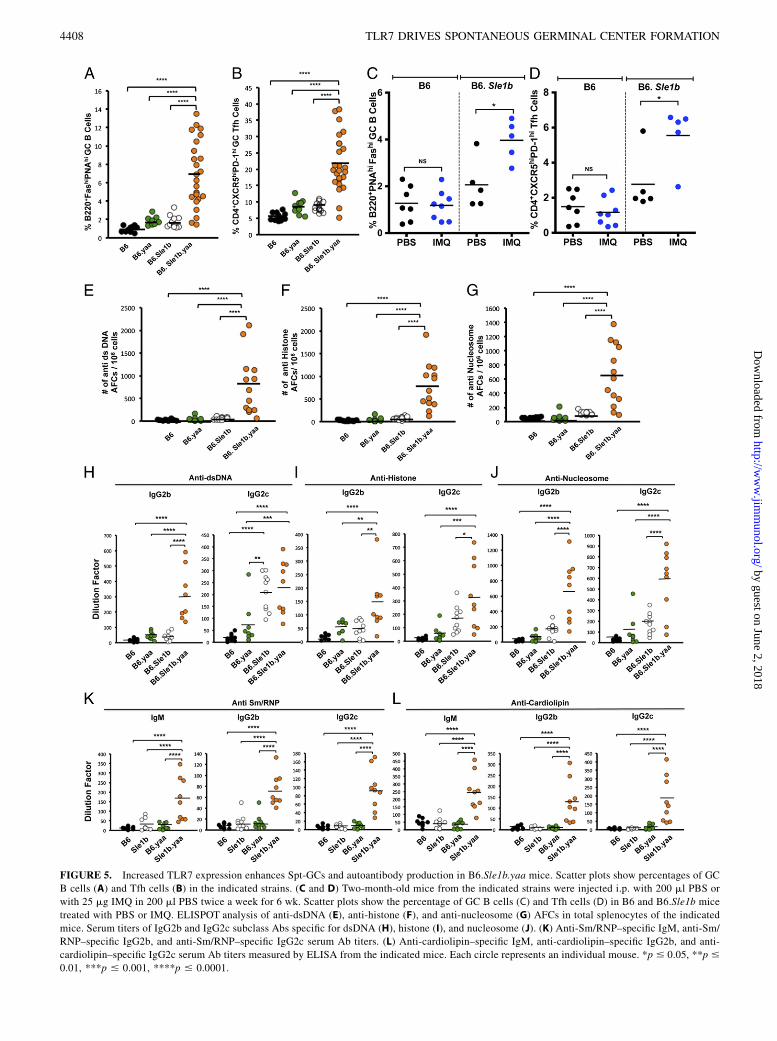
FIGURE 5. Increased TLR7 expression enhances Spt-GCs and autoantibody production in B6.Sle1b.yaa mice. Scatter plots show percentages of GC
B cells (A) and Tfh cells (B) in the indicated strains. (C and D) Two-month-old mice from the indicated strains were injected i.p. with 200 ml PBS or
with 25 mg IMQ in 200 ml PBS twice a week for 6 wk. Scatter plots show the percentage of GC B cells (C) and Tfh cells (D) in B6 and B6.Sle1b mice
treated with PBS or IMQ. ELISPOT analysis of anti-dsDNA (E), anti-histone (F), and anti-nucleosome (G) AFCs in total splenocytes of the indicated
mice. Serum titers of IgG2b and IgG2c subclass Abs specific for dsDNA (H), histone (I), and nucleosome (J). (K) Anti-Sm/RNP–specific IgM, anti-Sm/
RNP–specific IgG2b, and anti-Sm/RNP–specific IgG2c serum Ab titers. (L) Anti-cardiolipin–specific IgM, anti-cardiolipin–specific IgG2b, and anti-
cardiolipin–specific IgG2c serum Ab titers measured by ELISA from the indicated mice. Each circle represents an individual mouse. *p # 0.05, **p #
0.01, ***p # 0.001, ****p # 0.0001.
4408 TLR7 DRIVES SPONTANEOUS GERMINAL CENTER FORMATION
by guest on June 2, 2018http://w
ww
.jimm
unol.org/D
ownloaded from

LySM-Cre+/2-MyD88fl/fl, and CD11c-Cre+/2-MyD88fl/fl mice hadsimilar Spt-GC B cell and Tfh cell profiles (Fig. 7), arguing againsta myeloid cell–intrinsic TLR7-MyD88 signaling requirement for the
development of steady-state Spt-GCs. These data strongly suggestthat the expression of at least one copy of TLR7 specifically in B cellsis necessary for Spt-GC B cell and Tfh cell formation.
FIGURE 6. B cell–intrinsic TLR7 signaling is required for Spt-GC formation. Mixed BM chimeric mice were generated as described inMaterials and Methods.
(A) Representative spleen sections of the indicated chimeras stained with GL-7 (green), anti-CD4 (red), and anti-IgD (blue). Original magnification 3100. Rep-
resentative contour plots show GC B cells (B) and Tfh cells (C) from the indicated chimeras. (D) B and T cells were adoptively transferred into Rag1-KO mice, as
described in Materials and Methods. Spleen sections of Rag1-KO mice were stained as in (A). Original magnification 3100. (E) Representative dot plots show
gating strategy for GC B cells (in rectangles) in Rag1-KO mice transferred with B cells from B6 (upper panel), TLR9-KO (middle panel), or TLR7-KO (lower
panel) mice. (F) Scatter plots show the percentage of GC B cells in Rag1-KO mice transferred with B cells from the indicated strains. ELISPOT analysis of IgM+
(G) and IgG+ (H) AFCs in splenocytes of Rag1-KO mice transferred with B6, TLR9-KO, or TLR7-KO B cells. Four or five mice were analyzed per genotype for the
immunofluorescence analysis. Each circle represents an individual mouse. *p # 0.05, **p # 0.01.
The Journal of Immunology 4409
by guest on June 2, 2018http://w
ww
.jimm
unol.org/D
ownloaded from

Suboptimal B cell proliferation and survival in the absence ofTLR7 signaling
Next, we evaluated whether the absence of Spt-GCs in TLR7-KOmice resulted from a defect in B cell proliferation and survival.Sle1b/TLR9-KO mice had significantly higher IgD2Ki67+ pro-liferating GC B cells than did B6 or B6.Sle1b controls, but B cellfollicles of Sle1b/TLR7-KO spleens completely lacked Ki67+ cells(Fig. 8A). The deficiency in TLR7 did not affect the number ofKi67+ cells outside the follicles (Fig. 8A). We also performed anex vivo proliferation assay in which B cells were stimulated withanti-IgM and anti-CD40 (without any exogenous TLR ligands).As gauged by the CFSE dilution, a significantly reduced per-centage of B cells deficient in TLR7 or MyD88 underwent celldivisions compared with B6 and TLR9-KO B cells (Fig. 8B, 8C).Moreover, naive TLR7-KO B cells cultured or not with stimula-tion secreted reduced titers of IgG in the culture supernatantcompared with TLR9-KO and B6 B cells (Fig. 8D, 8E).To investigate whether this proliferative defect also occurred
in vivo, we transferred CFSE-labeled naive B cells from B6,TLR7-KO, or TLR9-KO mice into B6.mMT mice preimmunizedwith SRBCs. Once again, TLR7-KO B cells proliferated to alesser extent than did TLR9-KO or B6 B cells (Fig. 8F, 8G). Theresults described above prompted us to evaluate whether B cellsurvival was compromised upon BCR stimulation. We found a 2-foldreduction in the percentage of live TLR7-KO B cells compared withB6 and TLR9-KO B cells after .48 h of stimulation with anti-IgMand anti-CD40 (Fig. 9A).Cell cycle analysis by PI staining of B cells, cultured for 72 h
after stimulation with anti-IgM and anti-CD40, showed a signifi-cantly higher percentage of TLR7-KO B cells in the apoptotic sub-G1 population, but a lower percentage in the G0-G1 and S-G2/Mphase, compared with B6 B cells (Fig. 9B). The addition of BAFFto in vitro B cell–proliferation cultures was unable to rescueTLR7-KO B cells from cell death (Fig. 9C, 9D). These ex vivodata indicate that B cell proliferation and survival are suboptimalin the absence of TLR7 signaling via MyD88. Taken together, ourresults suggest that B cell–specific TLR7-MyD88 signaling and
B cell survival/proliferation are necessary for Spt-GC formationand Ab production.We also compared the transcript levels of key factors involved in
GC formation and maintenance. Consistent with elevated Spt-GC,Tfh cell, and AFC responses, splenocytes from TLR9-KO andB6.yaa mice expressed higher levels of PD-1, ICOS, and Xbp-1transcripts than did B6 and TLR7-KO mice (Supplemental Fig.3A–C). B6.yaa splenocytes expressed more Bcl-6 mRNA than didB6, TLR9-KO, or TLR7-KO splenocytes (Supplemental Fig. 3E),but the IRF-4 transcript level was comparable among all groups(Supplemental Fig. 3D). We found that Aicda mRNA levels weredirectly proportional to the dose of TLR7 and inversely propor-tional to the dose of TLR9. Therefore, Aicda mRNA expression inpurified B cells fell into the following hierarchy: B6.yaa . TLR9-KO . B6 . TLR7-KO (Supplemental Fig. 3F).
DiscussionSpt-GC B cells and Tfh cells play a significant role in generatinghigh-affinity pathogenic autoantibodies in murine models oflupus and human SLE (53). Previous studies elucidated therole of TLRs in B cell activation, autoantibody specificity, andlupus pathogenesis. However, the mechanisms underlying TLR-dependent differential regulation of Spt-GC B cell and Tfh cellformation are not clear. In nonautoimmune mice, the involve-ment of MyD88, TLR7, and TLR9 in the enhancement of B cellAb production has been discussed extensively in the context ofactive immunizations. The antigenic sources include variousT-dependent Ags (mixed with or conjugated to TLR ligands),RNA/DNA viruses, virus-like particles, or bacterial infections(inherently carrying natural TLR ligands) (54–58). However, theinvolvement of TLR signaling under steady-state conditions,with an antigenic repertoire comprising self-Ags derived fromapoptotic cells (59, 60) or endogenous microbial agents, has notbeen discussed.Our analysis of various TLR-deficient B6 mice revealed that
GCs that are spontaneously formed in the absence of exogenousstimuli are entirely dependent on TLR7 signaling via MyD88.
FIGURE 7. MyD88 deficiency in macrophages and DCs does not affect Spt-GC formation. Representative contour plots show the gating strategy (A) and
scatter plots show the percentage of GC B cells (B) in the indicated mouse strains. Representative contour plots show the gating strategy (C) and scatter
plots show the percentage of Tfh cells (D) in the indicated mouse strains. Each circle represents an individual mouse.
4410 TLR7 DRIVES SPONTANEOUS GERMINAL CENTER FORMATION
by guest on June 2, 2018http://w
ww
.jimm
unol.org/D
ownloaded from

Unlike B6 mice, TLR7-KO mice did not develop Spt-GCs, even bythe age of 6 mo. In contrast, TLR9-KOmice had increased numbersof Spt-GCs. However, upon immunization with T-dependent Ags,
TLR7-KO mice developed GC B cells and Tfh cells, although toa significantly lesser extent than did B6 mice (Supplemental Fig.4). Again, in the absence of TLR9, the GC responses to these
FIGURE 8. TLR7-MyD88 signaling is required for optimal B cell proliferation. (A) Representative immunofluorescence analysis of spleen sections from three
or four 6-mo-old mice stained for Ki67 (red) and IgD (blue). GCs are outlined. Original magnification 3100. (B) Representative graphs show the proliferation
profile of CFSE-labeled B cells from the indicated mice stimulated with anti-IgM/anti-CD40 for 72 h. Solid gray lines represent CFSE-labeled unstimulated cells,
and dashed gray lines represent unlabeled unstimulated B cells. (C) Bar graph shows the percentage of in vitro proliferating B cells in the indicated mouse strains
stimulated with anti-IgM/anti-CD40 for 72 h and assessed by flow cytometry. Error bars represent6SEM. Analysis of total IgG secreted in culture supernatants of
unstimulated B cells (D) or B cells stimulated with anti-IgM/anti-CD40 for 72 h (E), as assessed by ELISA. Error bars represent 6SD. In vitro data are rep-
resentative of four independent experiments with three or four sex-matched 8–10-wk-old mice/group. (F) Pseudocolor plots show the gating of in vivo–proliferating
B cells (labeled with CFSE) from the indicated strains, which were transferred into mMT mice preimmunized with SRBCs (upper panels). Profiles of in vivo–
proliferating transferred B cells, as assessed by decreasing CFSE fluorescence intensity (lower panels). (G) Percentage of in vivo–proliferating B cells [as described
for (F)] from the indicated strains. Error bars represent 6SD. Each circle represents an individual mouse. *p # 0.05, **p # 0.01, ***p # 0.001.
The Journal of Immunology 4411
by guest on June 2, 2018http://w
ww
.jimm
unol.org/D
ownloaded from

exogenous Ags were elevated (Supplemental Fig. 4). Thesestudies highlight the important stimulatory role of TLR7-MyD88signaling in the GC pathway. Our data also identify TLR9 asa repressor of TLR7 activity during a GC reaction under non-autoimmune conditions.B cell differentiation and autoantibody production primarily
proceed through the extrafollicular and FO GC pathways. MRL/lprmice have been used extensively to study the role of TLR7 andTLR9 in murine lupus, including the specificity of anti-nuclearAbs and kidney pathology (15). The autoreactive B cells in MRL/lpr mice proliferate and undergo somatic hypermutation outsidethe follicles in the T cell zone (30). Studies in the AM14 andMRL/lpr dual model systems suggested that both T-dependent andT-independent mechanisms are responsible for TLR7- or TLR9-mediated extrafollicular activation, expansion, and differentia-tion of the AM14 rheumatoid factor B cells (61). Similarly, inTLR7.1Tg mice, splenic red pulp is the primary site for the ac-tivation and proliferation of anti-RNA–specific transitional type 1B cells in the periphery (46). Additionally, in lupus-prone BAFF-Tg mice, autoantibody production involves MyD88-TLR–depen-dent expansion of autoreactive MZ and B1 B cells but not GCB cells (62). Together, these studies highlight the importance ofTLR7 or TLR9 in regulating spontaneously activated B cells in theextrafollicular regions but not in the FO GC pathway of B celldifferentiation.Many autoimmune mouse models, including (NZB/NZW) F1,
BXSB.yaa, and sanroque, spontaneously develop increased GCB cells, Tfh cells, and high titers of serum autoantibodies (22, 52,63, 64). Recently, Hua et al. (33) showed the requirement of GCsand MyD88-signaling in ANA production and nephritis in Lyn-deficient mice. These autoimmune mouse models highlight thesignificance of the FO GC pathway in B cell responses and au-toantibody production but do not delineate the contribution orrequirement for TLR7 and TLR9 in Spt-GC formation. A recentpublication by Rawlings and colleagues (20) focused on the rolesof TLR7 and TLR9 signaling in immune cell activation, autoan-tibody repertoire, systemic inflammation, and kidney pathologyusing the WASp autoimmune mouse model. Although this studyaddresses the opposing effects of B cell–intrinsic TLR7 and TLR9
expression on immune activation, including GC B cells and Tfhcells, it does not define the mechanisms by which TLRs controlthe formation of Spt-GC B cells and Tfh cells.Consistent with our observations in B6 mice and the results
reported using the WASp model (20), we found that TLR7 isabsolutely essential for Spt-GC formation in B6.Sle1b mice,whereas TLR9 signaling inhibited such responses. Supplementa-tion of an extra copy of TLR7 (yaa locus) in B6.Sle1b mice ortreatment of B6.Sle1b mice with a TLR7 agonist increased theSpt-GC B cell, Tfh cell, and autoantibody responses. These dataindicate that Spt-GCs can help to breach B cell tolerance at theGC checkpoint either in the presence of genetic predispositionalong with one copy of TLR7 or when TLR7 alone is overex-pressed. The role of TLR7 in promoting Spt-GC and Tfh cellresponses was examined previously using two mouse models: oneexpressed 15–18 copies of TLR7 on a B6 background (32), andthe other expressed two or three copies of TLR7 on the Sle1background (31). B cell–intrinsic expression of 15–18 copies ofTLR7 promoted Spt-GC and plasmablast development, whereasa 2-3–fold increase in B cell TLR7 expression resulted in en-hanced splenic B and T cell activation, MZ B cell reduction butdid not promote Spt-GC, Tfh cell, and plasmablast development.These two models allowed for the investigation of the role of TLR7overexpression in the enhancement of B and T cell responses;however, these studies did not determine whether TLR7 is re-quired for the initiation of Spt-GCs. Our study directlyaddresses the function of a single copy of the TLR7 gene in theinitiation of Spt-GCs under nonautoimmune and autoimmuneconditions.Our results also show that TLR7 has a major role in promoting
the generation of autoantibodies through the formation of Spt-GCs and that TLR9 inhibits the TLR7-mediated FO GC responsesin B6.Sle1b mice. This model addresses the potential epistatic inter-action between TLRs and the lupus-associated SLAM family genesin breaking the GC tolerance checkpoint. Recent studies showedthat the deficiency in nucleic acid–sensing TLR signaling due tomutations in Unc93b1 is sufficient to interfere with the generationof ANAs (65, 66). Additionally, other pathogenic nonnuclear au-toantibody specificities, including anti-cardiolipin, anti-RBC, and
FIGURE 9. TLR7 deficiency leads
to compromised B cell survival. (A)
Percentage of live cells assessed by
DAPI staining of purified B cells that
were stimulated with anti-IgM and
anti-CD40 for 72 or 96 h from the
indicated mouse strains. (B) Cell cycle
analysis of purified B cells from the
indicated strains at 72 h after stimu-
lation with anti-IgM and anti-CD40.
(C) Pseudocolor dot plots represent
the percentage of live and dead CFSE-
labeled B cells, as assessed by DAPI
staining, from the indicated strains
that were stimulated in vitro with anti-
IgM and anti-CD40, with or without
BAFF, for 72 h. (D) Percentage of
DAPI+ cells from three independent
experiments in (C). Error bars repre-
sent6SEM. Data are representative of
four independent experiments with
three or four sex-matched 8–10-wk-
old mice/group. *p # 0.05, ***p #
0.001.
4412 TLR7 DRIVES SPONTANEOUS GERMINAL CENTER FORMATION
by guest on June 2, 2018http://w
ww
.jimm
unol.org/D
ownloaded from

anti-myeloperoxidase, which are commonly found in SLE patients,also are reduced by Unc93b1 mutations (65, 66). Our work extendsthose observation and defines TLR7 as the key nucleic acid–sensingTLR for the production of nuclear and nonnuclear Ag-specificautoantibodies.B6 mice deficient in both TLR8 and TLR9 were reported to
develop a stronger autoimmune response than either TLR8-KO orTLR9-KO mice, implying that TLR8 and TLR9 can independentlysuppress TLR7-mediated autoimmunity (67). However, anotherstudy using 564Igi mice with knock-in genes encoding an auto-reactive anti-RNA Ab showed that, upon single deletions ofTLR7, TLR8, or TLR9 or combined deletion of TLR7 and TLR9,autoantibodies were still produced but removing TLR7 and TLR8together abolished the autoantibody production (68). These con-flicting results about the role of TLR8 have yet to be explained.Our adoptive-transfer and BM chimera studies demonstrated that
TLR7 signaling in B cells is essential for the formation of Spt-GCB cells and Tfh cells in B6 mice. Other investigators (40) proposedthat TLR-stimulated dendritic cells (DCs) can induce a strong Ag-dependent Th cell response, which, in turn, can promote extra-follicular and GC-mediated Ab responses. TLR7 signaling also isimplicated in the activation of plasmacytoid DCs (11). However,we did not observe any direct effect of TLR7 signaling in DCs onthe formation of steady-state Spt-GCs and Tfh cells.TLR7 overexpression was shown to promote the proliferation
of transitional type 1 B cells (46). Also, TLR7 signaling was shownto be important for GC B cell proliferation during chronic viralinfection (57). However, the effect of TLR7 and MyD88 signalingon B cell proliferation in the absence of TLR7 overexpression orinfection is not clear. We found significantly reduced proliferationof B cells in TLR7-KO and MyD88-KO mice compared with B6controls. Moreover, the total percentage of dead B cells after 48 hof activation was significantly higher in TLR7-KO mice comparedwith B6 and TLR9-KO mice. TLR-MyD88 signaling induces theexpression of genes that control cell survival and proliferation(69–71); hence, it is very likely that the prosurvival and/or pro-liferative signals transduced by TLR7-MyD88 signaling arecritical specifically within the Spt-GC microenvironment whereB cell antigenic stimulation is not overwhelming. It is conceivablethat during the formation of Spt-GCs, caused by mild B cell an-tigenic stimulation, the TLR7-MyD88–induced signals becomeequally necessary for the sustenance of B cells within GCs. Thecause of the disparity in outcomes between the signals transducedby TLR7 and TLR9, which are thought to use the same signalingintermediates, remains to be determined.Our finding that TLR7 signaling is required for the optimal
survival of B cells, thereby promoting GC formation and Ab/autoantibody production predominantly through the FO GC path-way, has therapeutic implications for the treatment of SLE andother humoral immune response–mediated autoimmune disorders.Modulating the FO GC pathway by targeting TLR7 signaling inB cells may be an effective strategy to decrease autoantibody pro-duction against diverse autoantigens.
AcknowledgmentsWe thank Drs. Aron Lukacher, David Spector, and Todd Schell for critical
reading of the manuscript and helpful discussions. We thank Dr. Daniel
Mucida for providing spleens from germ-free mice and Dr. Milena
Bogunovic for helpful discussions and providing spleens from MyD88fl/fl,
CD11c-Cre+/2-MyD88fl/fl, and LysM-Cre+/2-MyD88fl/fl mice. We also
thank Stephanie Schell for proofreading the manuscript. Finally, we
thank Melinda Elias for technical assistance, as well as the Pennsylvania
State University College of Medicine Flow Cytometry Core Facility.
DisclosuresThe authors have no financial conflicts of interest.
References1. Inamine, A., Y. Takahashi, N. Baba, K. Miyake, T. Tokuhisa, T. Takemori, and
R. Abe. 2005. Two waves of memory B-cell generation in the primary immuneresponse. Int. Immunol. 17: 581–589.
2. MacLennan, I. C. 1994. Germinal centers. Annu. Rev. Immunol. 12: 117–139.3. Jacob, J., G. Kelsoe, K. Rajewsky, and U. Weiss. 1991. Intraclonal generation of
antibody mutants in germinal centres. Nature 354: 389–392.4. Berek, C., A. Berger, and M. Apel. 1991. Maturation of the immune response in
germinal centers. Cell 67: 1121–1129.5. Blink, E. J., A. Light, A. Kallies, S. L. Nutt, P. D. Hodgkin, and D. M. Tarlinton.
2005. Early appearance of germinal center-derived memory B cells and plasmacells in blood after primary immunization. J. Exp. Med. 201: 545–554.
6. Benner, R., W. Hijmans, and J. J. Haaijman. 1981. The bone marrow: the majorsource of serum immunoglobulins, but still a neglected site of antibody forma-tion. Clin. Exp. Immunol. 46: 1–8.
7. Takahashi, Y., P. R. Dutta, D. M. Cerasoli, and G. Kelsoe. 1998. In situ studies ofthe primary immune response to (4-hydroxy-3-nitrophenyl)acetyl. V. Affinitymaturation develops in two stages of clonal selection. J. Exp. Med. 187: 885–895.
8. Genestier, L., M. Taillardet, P. Mondiere, H. Gheit, C. Bella, and T. Defrance.2007. TLR agonists selectively promote terminal plasma cell differentiation ofB cell subsets specialized in thymus-independent responses. J. Immunol. 178:7779–7786.
9. Rubtsov, A. V., C. L. Swanson, S. Troy, P. Strauch, R. Pelanda, and R. M. Torres.2008. TLR agonists promote marginal zone B cell activation and facilitate T-dependent IgM responses. J. Immunol. 180: 3882–3888.
10. Christensen, S. R., M. Kashgarian, L. Alexopoulou, R. A. Flavell, S. Akira, andM. J. Shlomchik. 2005. Toll-like receptor 9 controls anti-DNA autoantibodyproduction in murine lupus. J. Exp. Med. 202: 321–331.
11. Christensen, S. R., J. Shupe, K. Nickerson, M. Kashgarian, R. A. Flavell, andM. J. Shlomchik. 2006. Toll-like receptor 7 and TLR9 dictate autoantibodyspecificity and have opposing inflammatory and regulatory roles in a murinemodel of lupus. Immunity 25: 417–428.
12. Ehlers, M., H. Fukuyama, T. L. McGaha, A. Aderem, and J. V. Ravetch. 2006.TLR9/MyD88 signaling is required for class switching to pathogenic IgG2a and2b autoantibodies in SLE. J. Exp. Med. 203: 553–561.
13. Lartigue, A., P. Courville, I. Auquit, A. Francois, C. Arnoult, F. Tron, D. Gilbert,and P. Musette. 2006. Role of TLR9 in anti-nucleosome and anti-DNA antibodyproduction in lpr mutation-induced murine lupus. J. Immunol. 177: 1349–1354.
14. Yu, P., U. Wellmann, S. Kunder, L. Quintanilla-Martinez, L. Jennen, N. Dear,K. Amann, S. Bauer, T. H. Winkler, and H. Wagner. 2006. Toll-like receptor 9-independent aggravation of glomerulonephritis in a novel model of SLE. Int.Immunol. 18: 1211–1219.
15. Nickerson, K. M., S. R. Christensen, J. Shupe, M. Kashgarian, D. Kim, K. Elkon,and M. J. Shlomchik. 2010. TLR9 regulates TLR7- and MyD88-dependent au-toantibody production and disease in a murine model of lupus. J. Immunol. 184:1840–1848.
16. Pan, Z. J., S. Maier, K. Schwarz, J. Azbill, S. Akira, S. Uematsu, andA. D. Farris. 2010. Toll-like receptor 7 (TLR7) modulates anti-nucleosomalautoantibody isotype and renal complement deposition in mice exposed tosyngeneic late apoptotic cells. Ann. Rheum. Dis. 69: 1195–1199.
17. Santiago-Raber, M. L., I. Dunand-Sauthier, T. Wu, Q. Z. Li, S. Uematsu,S. Akira, W. Reith, C. Mohan, B. L. Kotzin, and S. Izui. 2010. Critical role ofTLR7 in the acceleration of systemic lupus erythematosus in TLR9-deficientmice. J. Autoimmun. 34: 339–348.
18. Silver, K. L., T. L. Crockford, T. Bouriez-Jones, S. Milling, T. Lambe, andR. J. Cornall. 2007. MyD88-dependent autoimmune disease in Lyn-deficientmice. Eur. J. Immunol. 37: 2734–2743.
19. Pawar, R. D., P. S. Patole, D. Zecher, S. Segerer, M. Kretzler, D. Schlondorff,and H. J. Anders. 2006. Toll-like receptor-7 modulates immune complex glo-merulonephritis. J. Am. Soc. Nephrol. 17: 141–149.
20. Jackson, S. W., N. E. Scharping, N. S. Kolhatkar, S. Khim, M. A. Schwartz,Q. Z. Li, K. L. Hudkins, C. E. Alpers, D. Liggitt, and D. J. Rawlings. 2014.Opposing impact of B cell-intrinsic TLR7 and TLR9 signals on autoantibodyrepertoire and systemic inflammation. J. Immunol. 192: 4525–4532.
21. Meffre, E., and H. Wardemann. 2008. B-cell tolerance checkpoints in health andautoimmunity. Curr. Opin. Immunol. 20: 632–638.
22. Vinuesa, C. G., M. C. Cook, C. Angelucci, V. Athanasopoulos, L. Rui,K. M. Hill, D. Yu, H. Domaschenz, B. Whittle, T. Lambe, et al. 2005. A RING-type ubiquitin ligase family member required to repress follicular helper T cellsand autoimmunity. Nature 435: 452–458.
23. Luzina, I. G., S. P. Atamas, C. E. Storrer, L. C. daSilva, G. Kelsoe,J. C. Papadimitriou, and B. S. Handwerger. 2001. Spontaneous formation ofgerminal centers in autoimmune mice. J. Leukoc. Biol. 70: 578–584.
24. Lee, S. K., D. G. Silva, J. L. Martin, A. Pratama, X. Hu, P. P. Chang, G. Walters,and C. G. Vinuesa. 2012. Interferon-g excess leads to pathogenic accumulationof follicular helper T cells and germinal centers. Immunity 37: 880–892.
25. Linterman, M. A., R. J. Rigby, R. K. Wong, D. Yu, R. Brink, J. L. Cannons,P. L. Schwartzberg, M. C. Cook, G. D. Walters, and C. G. Vinuesa. 2009. Fol-licular helper T cells are required for systemic autoimmunity. J. Exp. Med. 206:561–576.
The Journal of Immunology 4413
by guest on June 2, 2018http://w
ww
.jimm
unol.org/D
ownloaded from

26. Lamagna, C., Y. Hu, A. L. DeFranco, and C. A. Lowell. 2014. B cell-specificloss of Lyn kinase leads to autoimmunity. J. Immunol. 192: 919–928.
27. Simpson, N., P. A. Gatenby, A. Wilson, S. Malik, D. A. Fulcher, S. G. Tangye,H. Manku, T. J. Vyse, G. Roncador, G. A. Huttley, et al. 2010. Expansion ofcirculating T cells resembling follicular helper T cells is a fixed phenotype thatidentifies a subset of severe systemic lupus erythematosus. Arthritis Rheum. 62:234–244.
28. Cappione, A., III, J. H. Anolik, A. Pugh-Bernard, J. Barnard, P. Dutcher,G. Silverman, and I. Sanz. 2005. Germinal center exclusion of autoreactiveB cells is defective in human systemic lupus erythematosus. J. Clin. Invest. 115:3205–3216.
29. Wong, E. B., T. N. Khan, C. Mohan, and Z. S. Rahman. 2012. The lupus-proneNZM2410/NZW strain-derived Sle1b sublocus alters the germinal centercheckpoint in female mice in a B cell-intrinsic manner. J. Immunol. 189: 5667–5681.
30. William, J., C. Euler, S. Christensen, and M. J. Shlomchik. 2002. Evolution ofautoantibody responses via somatic hypermutation outside of germinal centers.Science 297: 2066–2070.
31. Hwang, S. H., H. Lee, M. Yamamoto, L. A. Jones, J. Dayalan, R. Hopkins,X. J. Zhou, F. Yarovinsky, J. E. Connolly, M. A. Curotto de Lafaille, et al. 2012.B cell TLR7 expression drives anti-RNA autoantibody production and exacer-bates disease in systemic lupus erythematosus-prone mice. J. Immunol. 189:5786–5796.
32. Walsh, E. R., P. Pisitkun, E. Voynova, J. A. Deane, B. L. Scott, R. R. Caspi, andS. Bolland. 2012. Dual signaling by innate and adaptive immune receptors isrequired for TLR7-induced B-cell-mediated autoimmunity. Proc. Natl. Acad.Sci. USA 109: 16276–16281.
33. Hua, Z., A. J. Gross, C. Lamagna, N. Ramos-Hernandez, P. Scapini, M. Ji,H. Shao, C. A. Lowell, B. Hou, and A. L. DeFranco. 2014. Requirement forMyD88 signaling in B cells and dendritic cells for germinal center anti-nuclearantibody production in Lyn-deficient mice. J. Immunol. 192: 875–885.
34. Hemmi, H., T. Kaisho, O. Takeuchi, S. Sato, H. Sanjo, K. Hoshino, T. Horiuchi,H. Tomizawa, K. Takeda, and S. Akira. 2002. Small anti-viral compounds ac-tivate immune cells via the TLR7 MyD88-dependent signaling pathway. Nat.Immunol. 3: 196–200.
35. Hemmi, H., O. Takeuchi, T. Kawai, T. Kaisho, S. Sato, H. Sanjo, M. Matsumoto,K. Hoshino, H. Wagner, K. Takeda, and S. Akira. 2000. A Toll-like receptorrecognizes bacterial DNA. Nature 408: 740–745.
36. Morel, L., K. R. Blenman, B. P. Croker, and E. K. Wakeland. 2001. The majormurine systemic lupus erythematosus susceptibility locus, Sle1, is a cluster offunctionally related genes. Proc. Natl. Acad. Sci. USA 98: 1787–1792.
37. Rahman, Z. S., B. Alabyev, and T. Manser. 2007. FcgammaRIIB regulates au-toreactive primary antibody-forming cell, but not germinal center B cell, activity.J. Immunol. 178: 897–907.
38. Shlomchik, M. J., and F. Weisel. 2012. Germinal centers. Immunol. Rev. 247: 5–10.39. Zotos, D., and D. M. Tarlinton. 2012. Determining germinal centre B cell fate.
Trends Immunol. 33: 281–288.40. DeFranco, A. L., D. C. Rookhuizen, and B. Hou. 2012. Contribution of Toll-like
receptor signaling to germinal center antibody responses. Immunol. Rev. 247:64–72.
41. Meyer-Bahlburg, A., and D. J. Rawlings. 2008. B cell autonomous TLR sig-naling and autoimmunity. Autoimmun. Rev. 7: 313–316.
42. Sun, X., A. Wiedeman, N. Agrawal, T. H. Teal, L. Tanaka, K. L. Hudkins,C. E. Alpers, S. Bolland, M. B. Buechler, J. A. Hamerman, et al. 2013. Increasedribonuclease expression reduces inflammation and prolongs survival in TLR7transgenic mice. J. Immunol. 190: 2536–2543.
43. Matzinger, P. 2002. The danger model: a renewed sense of self. Science 296:301–305.
44. Avalos, A. M., L. Busconi, and A. Marshak-Rothstein. 2010. Regulation of autore-active B cell responses to endogenous TLR ligands. Autoimmunity 43: 76–83.
45. Yu, P., W. L€ubben, H. Slomka, J. Gebler, M. Konert, C. Cai, L. Neubrandt,O. Prazeres da Costa, S. Paul, S. Dehnert, et al. 2012. Nucleic acid-sensing Toll-like receptors are essential for the control of endogenous retrovirus viremia andERV-induced tumors. Immunity 37: 867–879.
46. Giltiay, N. V., C. P. Chappell, X. Sun, N. Kolhatkar, T. H. Teal, A. E. Wiedeman,J. Kim, L. Tanaka, M. B. Buechler, J. A. Hamerman, et al. 2013. Overexpressionof TLR7 promotes cell-intrinsic expansion and autoantibody production bytransitional T1 B cells. J. Exp. Med. 210: 2773–2789.
47. Hardy, R. R., C. E. Carmack, S. A. Shinton, J. D. Kemp, and K. Hayakawa.1991. Resolution and characterization of pro-B and pre-pro-B cell stages innormal mouse bone marrow. J. Exp. Med. 173: 1213–1225.
48. Allman, D., R. C. Lindsley, W. DeMuth, K. Rudd, S. A. Shinton, andR. R. Hardy. 2001. Resolution of three nonproliferative immature splenic B cellsubsets reveals multiple selection points during peripheral B cell maturation. J.Immunol. 167: 6834–6840.
49. Wu, X., and S. L. Peng. 2006. Toll-like receptor 9 signaling protects againstmurine lupus. Arthritis Rheum. 54: 336–342.
50. Wandstrat, A. E., C. Nguyen, N. Limaye, A. Y. Chan, S. Subramanian,X. H. Tian, Y. S. Yim, A. Pertsemlidis, H. R. Garner, Jr., L. Morel, andE. K. Wakeland. 2004. Association of extensive polymorphisms in the SLAM/CD2 gene cluster with murine lupus. Immunity 21: 769–780.
51. Croker, B. P., G. Gilkeson, and L. Morel. 2003. Genetic interactions betweensusceptibility loci reveal epistatic pathogenic networks in murine lupus. GenesImmun. 4: 575–585.
52. Pisitkun, P., J. A. Deane, M. J. Difilippantonio, T. Tarasenko, A. B. Satterthwaite,and S. Bolland. 2006. Autoreactive B cell responses to RNA-related antigens dueto TLR7 gene duplication. Science 312: 1669–1672.
53. Celhar, T., R. Magalhaes, and A. M. Fairhurst. 2012. TLR7 and TLR9 in SLE:when sensing self goes wrong. Immunol. Res. 53: 58–77.
54. Hou, B., P. Saudan, G. Ott, M. L. Wheeler, M. Ji, L. Kuzmich, L. M. Lee,R. L. Coffman, M. F. Bachmann, and A. L. DeFranco. 2011. Selective utilizationof Toll-like receptor and MyD88 signaling in B cells for enhancement of theantiviral germinal center response. Immunity 34: 375–384.
55. Hou, B., B. Reizis, and A. L. DeFranco. 2008. Toll-like receptors activate innateand adaptive immunity by using dendritic cell-intrinsic and -extrinsic mecha-nisms. Immunity 29: 272–282.
56. Browne, E. P. 2011. Toll-like receptor 7 controls the anti-retroviral germinalcenter response. PLoS Pathog. 7: e1002293.
57. Clingan, J. M., and M. Matloubian. 2013. B Cell-intrinsic TLR7 signaling isrequired for optimal B cell responses during chronic viral infection. J. Immunol.191: 810–818.
58. Rookhuizen, D. C., and A. L. DeFranco. 2014. Toll-like receptor 9 signaling actson multiple elements of the germinal center to enhance antibody responses. Proc.Natl. Acad. Sci. USA 111: E3224–E3233.
59. Cline, A. M., and M. Z. Radic. 2004. Murine lupus autoantibodies identifydistinct subsets of apoptotic bodies. Autoimmunity 37: 85–93.
60. Neeli, I., M. M. Richardson, S. N. Khan, D. Nicolo, M. Monestier, andM. Z. Radic. 2007. Divergent members of a single autoreactive B cell cloneretain specificity for apoptotic blebs. Mol. Immunol. 44: 1914–1921.
61. Herlands, R. A., S. R. Christensen, R. A. Sweet, U. Hershberg, andM. J. Shlomchik. 2008. T cell-independent and toll-like receptor-dependentantigen-driven activation of autoreactive B cells. Immunity 29: 249–260.
62. Groom, J. R., C. A. Fletcher, S. N. Walters, S. T. Grey, S. V. Watt, M. J. Sweet,M. J. Smyth, C. R. Mackay, and F. Mackay. 2007. BAFF and MyD88 signalspromote a lupuslike disease independent of T cells. J. Exp. Med. 204: 1959–1971.
63. Fairhurst, A. M., S. H. Hwang, A. Wang, X. H. Tian, C. Boudreaux, X. J. Zhou,J. Casco, Q. Z. Li, J. E. Connolly, and E. K. Wakeland. 2008. Yaa autoimmunephenotypes are conferred by overexpression of TLR7. Eur. J. Immunol. 38:1971–1978.
64. Mohan, C., E. Alas, L. Morel, P. Yang, and E. K. Wakeland. 1998. Geneticdissection of SLE pathogenesis. Sle1 on murine chromosome 1 leads to a se-lective loss of tolerance to H2A/H2B/DNA subnucleosomes. J. Clin. Invest. 101:1362–1372.
65. Koh, Y. T., J. C. Scatizzi, J. D. Gahan, B. R. Lawson, R. Baccala, K. M. Pollard,B. A. Beutler, A. N. Theofilopoulos, and D. H. Kono. 2013. Role of nucleic acid-sensing TLRs in diverse autoantibody specificities and anti-nuclear antibody-producing B cells. J. Immunol. 190: 4982–4990.
66. Kono, D. H., M. K. Haraldsson, B. R. Lawson, K. M. Pollard, Y. T. Koh, X. Du,C. N. Arnold, R. Baccala, G. J. Silverman, B. A. Beutler, andA. N. Theofilopoulos. 2009. Endosomal TLR signaling is required for anti-nucleic acid and rheumatoid factor autoantibodies in lupus. Proc. Natl. Acad.Sci. USA 106: 12061–12066.
67. Desnues, B., A. B. Macedo, A. Roussel-Queval, J. Bonnardel, S. Henri,O. Demaria, and L. Alexopoulou. 2014. TLR8 on dendritic cells and TLR9 onB cells restrain TLR7-mediated spontaneous autoimmunity in C57BL/6 mice.Proc. Natl. Acad. Sci. USA 111: 1497–1502.
68. Umiker, B. R., S. Andersson, L. Fernandez, P. Korgaokar, A. Larbi,M. Pilichowska, C. C. Weinkauf, H. H. Wortis, J. F. Kearney, and T. Imanishi-Kari. 2014. Dosage of X-linked Toll-like receptor 8 determines gender differ-ences in the development of systemic lupus erythematosus. Eur. J. Immunol. 44:1503–1516.
69. Li, X., S. Jiang, and R. I. Tapping. 2010. Toll-like receptor signaling in cellproliferation and survival. Cytokine 49: 1–9.
70. Pone, E. J., J. Zhang, T. Mai, C. A. White, G. Li, J. K. Sakakura, P. J. Patel,A. Al-Qahtani, H. Zan, Z. Xu, and P. Casali. 2012. BCR-signalling synergizeswith TLR-signalling for induction of AID and immunoglobulin class-switchingthrough the non-canonical NF-kB pathway. Nat. Commun. 3: 767.
71. Kawai, T., and S. Akira. 2005. Toll-like receptor downstream signaling. ArthritisRes. Ther. 7: 12–19.
4414 TLR7 DRIVES SPONTANEOUS GERMINAL CENTER FORMATION
by guest on June 2, 2018http://w
ww
.jimm
unol.org/D
ownloaded from