azooxanthellate scleractinia … · ... kochi prefecture, japan by ... the author had the...
TRANSCRIPT

*1 Coastal Branch of Natural His tory Museum and Insti tute, Chiba
123 Yoshio, Katsuura, Chiba Prefecture, 299-5242, Japan e-mail : [email protected]
AZOOXANTHELLATE SCLERACTINIA
(HEXACORALLIA, ANTHOZOA, CNIDARIA)
COLLECTED FROM OTSUKI, KOCHI PREFECTURE, JAPAN
by
Hiroyuki TACHIKAWA* 1
Abstract
Specimens representing 35 species of azooxanthellate scleractinian coral collected
from Otsuki Town, Kochi Prefecture, Japan, are described and illustrated. Thirteen of the
species were collected by hand with SCUBA to depths of 45 m. The remaining 22 species
were collected by boat while dragging traditional precious coral tangle-nets at depths of
83-134 m. Twenty-seven species are new records for this region. Eight families are
represented in the collection, but more than half of the taxa (20 species) belong to the
family Dendrophylliidae. For several species with taxonomic problems, type specimens are
also redescribed and figured.
Introduction
The Pacific coast of southwestern Japan enjoys the strong influence of the warm
Kuroshio current, which allows it to display an abundant subtropical flora and fauna in
spite of the relatively high latitude. The southwestern part of Kochi Prefecture, where the
Biological Institution on Kuroshio is located, is a typical Kuroshio-influenced region well
known for its excellent subtropical underwater scenery. In this region, several Marine Park
zones have been designated as part of the Ashizuri-Uwakai National Park, due mainly to
their well developed coral communities (Iwase, 2005). Many zooxanthellate scleractinian
corals, which are tropical to subtropical components of the marine fauna, are known to
flourish in this region: Veron (1992) and Nishihira and Veron (1995) reported 147
species of zooxanthellate Scleractinia from Tosashimizu City, where some of the Marine
Park zones are located. However, the azooxanthellate Scleractinia of this region have
seldom been studied except by Yabe and Eguchi (1942), who reported on rather
deep-water species (see “Previous Studies” below).
In October, 2004, the author had the opportunity to visit the Biological Institution on
Kuroshio in Otsuki Town, Kochi Prefecture, to study the azooxanthellate scleractinian
corals of this region. In this report, the specimens collected are identified, described, and
figured, and taxonomic problems concerning several species are discussed. Type specimens
TACHIKAWA : AZOOXANTHELLATE SCLERACTIN IA COLLECTED FR OM OTSUKI 1Kuroshio Bio sphere
Vol . 2, De c. 2005, pp. 1- 27 + 13 p l s.

of some species are also redescribed and illustrated for the purpose of comparison.
Material and Methods
Specimens of azooxanthellate scleractinian corals were collected either by hand using
SCUBA or by tangle-nets. Four series of SCUBA dives were undertaken to a depth of 45
m around Komo, southwards off Tachibanaura, Otsuki, on 5 and 6 October 2004 (Fig. 1).
Alternatively, specimens were collected on board a coral-fishing vessel, which used a
traditional coral tangle-net to catch precious corals, Corallium spp. Such a net is composed
of a thick log, large stone sinkers attached to it at regular intervals, and tufts of fishing net
attached as streamers to the sinkers (see Kitahara, 1903 for traditional precious coral
fishing) . The vessel drags the net along the sea bottom to entangle the precious corals, and
many other bottom animals are caught at the same time. In this study, a locally operated
vessel was hired to drag a coral tangle-net at 4 stations off Nishidomari, Otsuki, on 7
October 2004. In both methods, all coral specimens were collected with the permission of
the Governor of Kochi Prefecture.
Fig. 1. Sampling stations of the present study.
The sampling stations are abbreviated as follows:
Komo: Off Komo, southwards off Tachibanaura, collected by H. Tachikawa, F. Iwase, and
T. Hayashi using SCUBA, to a depth of 45 m.
CN1: Coral tangle-net station 1, off Nishidomari, Otsuki, Kochi Pref., from 32 ° 37.66 ′ N,
132 ° 50.44 ′ E, 114 m to 32 ° 37.56 ′ N, 132 ゚ 47.88 ′ E. The depth at the end of the
operation was not recorded.
CN2: Coral tangle-net station 2, off Nishidomari, Otsuki, Kochi Pref., from 32 ° 34.14 ′ N,
132 ° 48.59 ′ E, 117 m to 32 ° 34.18 ′N, 132 ° 47.59 ′ E, 125 m.
2 Kur oshio Biosphere : BULL. BIOL. INST. KUROSHIO Vol. 2, 2005

TACHIKAWA : AZOOXANTHELLATE SCLERACTIN IA COLLECTED FR OM OTSUKI 3
CN3: Coral tangle-net station 3, off Nishidomari, Otsuki, Kochi Pref., from 32 ° 43.08 ′ N,
132 ° 48.06 ′ E, 85 m to 32 ° 43.12 ′ N, 132 ° 47.68 ′ E, 83 m.
CN4: Coral tangle net station 4, off Nishidomari, Otsuki, Kochi Pref., from 32 ° 40.68 ′ N,
132 ° 47.57 ′ E, 130 m to 32 ° 40.68 ′N, 132 ° 46.99 ′ E, 134 m.
Institutional abbreviations are as follows:
BIK: Biological Institution on Kuroshio, Kuroshio Biological Research Foundation, Otsuki.
CMNH: Coastal Branch of Natural History Museum and Institute, Chiba, Katsuura.
IGPS: Institute of Geology and Paleontology, Tohoku University, Sendai.
NSMT: National Science Museum, Tokyo (Showa Memorial Institute), Tsukuba.
Morphological terminology follows Cairns (1989, 1994, 2004), including the
following abbreviations: GCD, greater calicular diameter; GCD:LCD, ratio of gretaer to
lesser calicular diameter; PD:GCD, ratio of pedicel diameter to greater calicular diameter;
Sx, septa of cycle designated by numerical subscript.
Collected specimens were cleaned using sodium hypochlorite solution and stored as
dry specimens. To enhance the contrast in conventional black-and-white photographs, some
of the specimens were stained dark using cyanine blue solution and coated with ammonium
chloride prior to photography. SEM photographs were made using a JEOL JSM-5310LV at
CMNH in low vacuum mode. All the conventional and SEM photographs were made by
the author. Plate citations below each species heading pertain only to specimens from the
present collection; photographs of other material are cited separately in the text. Specimens
are deposited at CMNH (with a code of ZG) and BIK (with a code of C). Reference
specimens were studied in IGPS and NSMT.
Previous Studies
In this section, species are divided into two categories for the sake of convenience.
Species encountered by SCUBA divers are treated as shallow-water species, and species
from much deeper areas that are only accessible using fishing gear or research apparatus are
treated as deep-water species.
Shallow-water species. – Many field surveys have been conducted in the Marine Park
zones in Ashizuri-Uwakai National Park, and a number of reports have been published up
to now. Some of them include faunal lists of the region that mention many scleractinian
corals. Almost all of these have been zooxanthellate species, with azooxanthellate species
recorded only sporadically (Nature Conservation Society of Japan, 1965; Kochi Prefectural
Government and Marine Parks Center of Japan, 1971; Nature Conservation Bureau,
Environment Agency and Marine Parks Center of Japan, 1995). Shallow-water
azooxanthellates previously reported from the Ashizuri-Uwakai region are Rhizotrochus
typus (as Monomyces uchiuraensis), Tubastraea coccinea, Tubastraea micranthus
(commonly as Dendrophyllia micrantha) , and Flabellum pavoninum var. paripavoninum.
Of these, the former three species were recorded also in the present study. The last one is
cited by Nature Conservation Society of Japan (1965), but in a quote from a previously
published preliminary report on the establishment of Ashizuri National Park. Unfortunately,
the original report could not be accessed by the author and the occurrence of this species in
shallow water was not confirmed by the present study.
Deep-water species. – In the significant work on Japanese azooxanthellate Scleractinia of

4 Kur oshio Biosphere : BULL. BIOL. INST. KUROSHIO Vol. 2, 2005
Yabe and Eguchi (1942), a majority of the materials examined was collected by R/V
Soyo-maru of the Imperial Fisheries Experimental Station, which dredged extensively
around Japan during the years 1922-1930. Fourteen Soyo-maru stations (Sts. 231, 322-333,
and 336), ranging from 88 to 684 m in depth, were located off Okinoshima Island and off
Cape Ashizuri, Kochi Prefecture, the region including the sampling stations of the present
study. From these stations, 27 species and subspecies of azooxanthellate Scleractinia were
recorded by Yabe and Eguchi (1942) , although the names of some of the species in their
report should be amended to fit the present-day taxonomy (see Cairns, 1994; Cairns and
Zibrowius, 1997). Five of Yabe and Eguchi’s species were recorded again in the present
study: Bathyactis palifera (Fungiacyathus paliferus herein), Premocyathus compressus (P.
dentiformis herein), Citharocyathus conicus (Notocyathus conicus herein), Flabellum
distinctum (Flabellum pavoninum herein), and Balanophyllia fistula (Eguchipsammia
wellsi herein). In these cases, Yabe and Eguchi’s records were confirmed by reexamination
of their specimens deposited at IGPS. Regarding two other of their species records,
Stephanophyllia formosissima and Balanophyllia cf. rediviva, results of this reexamination
are discussed in the accounts of Letepsammia superstes and Balanophyllia sp. cf. B.
rediviva , respectively.
Result and Discussions
The specimens collected from the field survey included 35 species of azooxanthellate
Scleractinia. Thirteen of these were collected by SCUBA diving and 22 were collected by
coral tangle-net. As mentioned above, 8 (3 shallow-water and 5 deep-water) species were
previously known from the Ashizuri-Uwakai region: the other 27 species are regarded as
new records for this region. Eight families are represented, but more than half (20) of the
species belong to the family Dendrophylliidae. Especially , the overwhelming majority (11
of 13) of species collected by SCUBA are dendrophylliids, which indicates a high diversity
of this family in the shallow waters of this region. Among the species collected by
tangle-net, the proportion of Dendrophylliidae is also relatively high (9 of 22). This can be
attributed to the method of collection employed in this study. Many species of this family
have a colonial, branched growth form and a rough exterior, and are apt to be entangled by
the net. This method is less suitable for collecting relatively small, solitary, free-living
species that are not easily entangled. In fact, all 5 species of Fungiacyathidae,
Micrabaciidae, and Turbinoliidae collected in this study were each represented by just one
or two dead coralla adhering to dead gastropod shells or to dead colonies of Dendrophyllia
brought up together with bottom sediment. Field surveys by dredging aimed mainly at the
free-living soft-bottom species are needed to supplement the results of the present study. In
addition, in light of the rather narrow depth range of this study, researches extending to
much greater depths are necessary to fully clarify the azooxanthellate scleractinian fauna of
this region.
List of Species
Family POCILLOPORIDAE

TACHIKAWA : AZOOXANTHELLATE SCLERACTIN IA COLLECTED FR OM OTSUKI 5
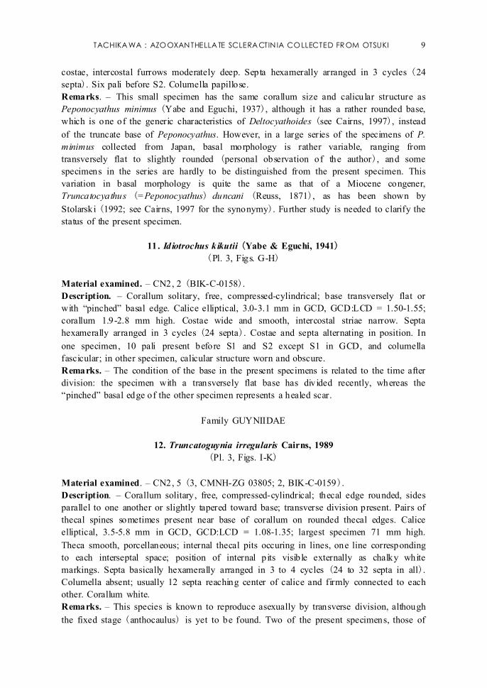
1. Madracis sp.
(Pl. 1, Figs. A-E)
Material examined. – Komo, 37 m, part of a colony and several branch fragments
(CMNH-ZG 03761; BIK-C-0150).
Description. – Colonial: colony attached to substrate, consisting of irregularly arborescent,
slender, attenuate branches; diameter of branch 3.5-4.0 mm at 10 mm from tip. Collected
specimen (part of a colony) 80 mm high and 115 mm across. Corallites 2.2-3.0 mm in
GCD; corallites near branch tips polygonal and closely spaced, separated by thin ridges
bearing unilinear row of conical teeth up to 0.5 mm high; corallites on middle and proximal
parts of branches elliptical and separated by 1-2.5 mm of coenosteum. Coenosteum
distinctly striated longitudinally and sparsely covered with pointed granules. Septa
decamerally arranged in 1 cycle (10 septa), but 12 or 14 septa in occasional corallites;
each septum bearing septal lobe as tall as intercalicular teeth. Columella a large, solid mass
forming circular platform of about half calicular diameter, with slightly protruding conical
central portion and laterally compressed apex. Living colony purplish brown; corallum
white.
Remarks. – The present specimen has uniquely striated coenosteum and appears to belog
to an undescribed species. In Japan, branching species of Madracis found in shallow waters
have been identified customarily as Madracis asanoi Yabe and Sugiyama, 1936 (e.g.,
Eguchi, 1968; Uchida, 1994; Nishihira and Veron, 1995). Living colonies figured as M.
asanoi in Uchida (1994) and Nishihira and Veron (1995) have growth forms similar to
the present specimen; however, it is necessary to compare skeletal characters before
concluding whether they are conspecific. The present specimen differs from M. asanoi not
only in the striated coenosteum but also in the finer, attenuate branches. From Japan,
another unidentified branching Madracis with two cycles of septa and a crown of distinct
paliform lobes was recorded by Cairns (1994) . It seems that the Madracis of Japan, and
those of the Indo-Pacific region as well, are in need of revision.
The holotype of M. asanoi, collected from 100 fathoms (i.e., 183 m) off the Palao
Islands (currently Republic of Palau, or Belau), is deposited at IGPS (Pl. 1, Figs. F-H). It
is part of a sparsely branching colony with a maximum dimension of 62 mm. Its branch
tips are rounded, the diameter of the terminal branches is 4-5.5 mm, and the diameter of the
basal branch is 6.5 mm. The corallites are almost circular even at the tips of the branches
and are characteristically variable in size. Larger corallites attain 2.5 mm in GCD. Each
corallite has 10 (in some large corallites 12) lobed septa, which protrude up to 0.7 mm
from the coenosteum, and no pali. The columella is a widely conical, solid mass of about
half the calicular diameter, with a laterally compressed, blunt apex. Adjacent calices are
separated by 0.5-1.0 mm of granular coenosteum.
Another branching species, Madracis palauensis Yabe and Sugiyama, 1936, was also
described from the Palao Islands, at 80 fathoms (i.e., 146 m: Pl. 1, Figs. I-J). Yabe and
Sugiyama (1936) distinguished it from M. asanoi by the broader, compressed branches,
larger calices, and relatively narrow columella. Reexamination of the holotype has revealed
that the colony was originally overgrowing an elongate substratum, presumably the axis of
a dead gorgonian colony, and consequently the proximal branch was widened.
Characteristics of the calices, including the variable calicular diameter, are very similar in

6 Kur oshio Biosphere : BULL. BIOL. INST. KUROSHIO Vol. 2, 2005
both species. Madracis palauensis has a slightly less protruded columella and, in a few
corallites, rudimentary second order septa, in which 1 or 2 very small spines comprise each
septum. These differences are not a sufficient basis enough for separating species, and I
agree with Cairns and Zibrowius (1997) in synonymizing M. asanoi and M. palauensis
under the former name.
Family FUNGIACYATHIDAE
2. Fungiacyathus (Fungiacyathus) paliferus (Alcock, 1902)
(Pl. 2, Figs. A-B)
Material examined. – CN2, 1 (BIK-C-0151).
Description. – Corallum solitary and unattached, flat and semicircular, 4.4 mm in calicular
diameter. Costae round, covered with low granules. Oral side worn but about 25 septa
present in 2 systems flanked by 2 half systems, in 4 cycles. Adjacent septa united by solid
synapticular plates. Columellar region broken.
Remarks. – This species is known to have two growth forms: a relatively large, circular
corallum and a small, semicircular to wedge-shaped corallum, the latter being a result of
longitudinal division. The present specimen is a long-dead, semicircular corallum,
considered to be a juvenile specimen of the latter growth form. The adult corallum of F.
paliferus has 5 cycles of septa hexamerally arranged, although coralla between 1.8 and 4.5
mm in calicular diameter have only 4 cycles of septa (Cairns, 1989), which is consistent
with the septal number of this specimen.
Family MICRABACIIDAE
3. Letepsammia superstes Ortmann, 1888
(Pl. 2, Figs. C-D)
Material examined. – CN2, 1 (BIK-C-0152).
Description. – Corallum solitary, discoid and unattached, 9.3 mm in GCD. Costae thin,
separated by widely open intercostal spaces and regularly connected by thin synapticulae,
forming a mesh-like appearance. Septa hexamerally arranged in micrabaciid fashion (ca. 80
septa). Septa and costae alternating in position. Columella worn, formed of entangled
trabeculae.
Remarks. – Most corals from Japan referred to Stephanophyllia formosissima Moseley,
1876, or Letepsammia formosissima, are actually this species; an exceptional case is a paper
by Ogawa and Takahashi (2004) , who figured true L. formosissima. Letepsammia superstes
differs from L. formosissima in having a papillose (vs. flat-topped and spongy) columella,
fewer septa (usually 96 vs. up to 144), and a smaller but relatively robust corallum. The
present specimen is a considerably worn corallum with a GCD of 9.3 mm but generally
agrees with the characteristics of L. superstes.
Specimens reported as Stephanophyllia formosissima from Soyo-maru St. 242 by
Yabe and Eguchi (1942) were reexamined and found to be several small specimens of
Letepsammia fissilis Cairns, 1995.

TACHIKAWA : AZOOXANTHELLATE SCLERACTIN IA COLLECTED FR OM OTSUKI 7
Family CARYOPHYLLIIDAE
4. Caryophyllia (Caryophyllia) rugosa Moseley, 1881
(Pl. 2, Figs. E-F)
Material examined. – CN1, 8 (CMNH-ZG 03798); CN2, 9 (5, CMNH-ZG 03802; 4,
BIK-C-0153) .
Description. – Corallum solitary, trochoid, attached by thick pedicel, PD:GCD = 0.42-0.87.
Calice circular to slightly elliptical, GCD:LCD = 1.00-1.16; largest corallum 6.7 mm in
GCD; height of corallum 4.2-11.7 mm. Costae low and indistinct, covered with fine
transverse sculpture. Septa octamerally arranged in 3 cycles (32 septa). All septa with
sinuous inner edges. Crown of 8 sinuous pali before S2. Columella fascicular, with a few
twisted elements. Corallum light brown to white.
5. Caryophyllia (Caryophyllia) hawaiiensis Vaughan, 1907
(Pl. 2, Figs. G-H)
Material examined. – CN1, 1 (BIK-C-0154).
Description. – Corallum solitary, ceratoid, attached by narrow (4.2 mm in diameter)
pedicel, PD:GCD = 0.44. Calice slightly elliptical, 9.5 x 7.7 mm in diameter; height of
corallum 18.6 mm. Costae indistinct, covered with low granules. Septa pentamerally
arranged in 4 cycles (40 septa). S1 and S2 exsert, forming calicular lancets. Crown of 10
pali before S3. Columella fascicular.
6. Caryophyllia (Caryophyllia) sp. cf. C. (C.) japonica Marenzeller, 1888
(Pl. 2, Figs. I-J)
Material examined. – CN2, 1 (CMNH-ZG 03803); CN3, 1 (CMNH-ZG 03813).
Description. – Corallum solitary, trochoid, and attached by thick pedicel, PD:GCD =
0.46-0.49. Calice slightly elliptical, 4.6 x 4.1 to 5.3 x 4.6 mm in diameter; height of
corallum 6.9-8.5 mm. Costae low, rounded, covered with low granules. Septa octamerally
arranged in 3 cycles (32 septa). Inner edges of S2 highly sinuous. Crown of 8 pali before
S2. Columella fascicular.
Remarks. – These small specimens resemble C. japonica in corallum shape, costal
ornamentation, and the highly sinuous inner edges of the penultimate cycle of septa ( in this
case S2), although the septal arrangement is not hexameral but octameral. More specimens
should be examined to clarify the identification.
7. Premocyathus dentiformis (Alcock, 1902)
(Pl. 2, Figs. K-L)
Material examined. – CN2, 1 (BIK-C-0155).
Description. – Corallum solitary, free, ceratoid, compressed. Corallum curved in plane of
GCD; base open, 2.5 x 2.0 mm in diameter, revealing 7 large and 7 small protosepta.

8 Kur oshio Biosphere : BULL. BIOL. INST. KUROSHIO Vol. 2, 2005
Calice elliptical, 3.4 x 2.7 mm in diameter; height of corallum 5.2 mm. Costae low,
rounded. Twelve large septa alternating with 12 small septa (24 septa in all); pali absent.
Columella a fascicular mass.
Remarks. – Only a small, dead specimen was collected. Mori (1987) demonstrated in a
Pleistocene fossil of Caryophyllia (Premocyathus) compressa (Yabe and Eguchi, 1942)
(= Premocyathus dentiformis; see Cairns and Zibrowius, 1997 for the synonymy) that the
septal plan is intrinsic and the number of pali is related to the insertion of the third-cycle
septa. The present specimen is considered to be a dodecameral specimen with the third
cycle of septa yet to be developed, and consequently, lacking pali.
8. Paracyathus sp.
(Pl. 3, Figs. A-B)
Material examined. – CN2, 1 (CMNH-ZG 03804); CN3, 1 (CMNH-ZG 03814).
Description. – Corallum solitary, trochoid to subcylindrical, attached by thick pedicel,
PD:GCD = 0.66-0.94. Calice slightly elliptical, 5.8 x 4.9 to 6.3 x 5.9 mm in diameter;
height of corallum 4.1-7.6 mm. Costae as low, rounded ridges covered with granules; most
of theca covered with calcareous deposits. Septa hexamerally arranged in 5 incomplete
cycles, with 3 to 5 pairs of S4 lacking (38-42 septa in all). Paliform lobes as twisted
papillae; 1 to 2 paliform lobes before each septum of all but last cycle in the half system;
in other words, in half system with S4, paliform lobes present before S1-S3, whereas in
half system without S4, before S1 and S2 only. Columella papillose, indistinguishable from
paliform lobes. Septa and upper theca mottled dark brown.
Remarks. – There are many “obscure” species of Paracyathus described from the
Indo-West Pacific region (see Cairns, 2004). At present I cannot convincingly identify the
two small specimens of Paracyathus collected in this study.
Family TURBINOLIIDAE
9. Notocyathus conicus (Alcock, 1902)
(Pl. 3, Figs. C-D)
Material examined. – CN2, 1 (BIK-C-0156).
Description. – Corallum solitary, free, conical; base pointed, thecal angle in plane of GCD
45 °. Calice slightly elliptical, 4.8 mm in GCD, GCD:LCD = ca. 1.20; corallum 5.4 mm
high. Theca covered with worn costae; intercostal furrows deep. Septa hexamerally
arranged in 4 cycles (48 septa). Twelve pali before S3: pair of pali in each system joined
before common S2, forming V-shaped structure. Columella papillose.
10. Deltocyathoides? sp.
(Pl. 3, Figs. E-F)
Material examined. – CN2, 1 (BIK-C-0157).
Description. – Corallum solitary, free, cylindrical, with rounded base. Calice circular, 3.0
mm in GCD, GCD:LCD = 1.05; corallum 2.7 mm high. Theca covered with rounded
TACHIKAWA : AZOOXANTHELLATE SCLERACTIN IA COLLECTED FR OM OTSUKI 9
costae, intercostal furrows moderately deep. Septa hexamerally arranged in 3 cycles (24
septa). Six pali before S2. Columella papillose.
Remarks. – This small specimen has the same corallum size and calicular structure as
Peponocyathus minimus (Yabe and Eguchi, 1937), although it has a rather rounded base,
which is one of the generic characteristics of Deltocyathoides (see Cairns, 1997), instead
of the truncate base of Peponocyathus. However, in a large series of the specimens of P.
minimus collected from Japan, basal morphology is rather variable, ranging from
transversely flat to slightly rounded (personal observation of the author), and some
specimens in the series are hardly to be distinguished from the present specimen. This
variation in basal morphology is quite the same as that of a Miocene congener,
Truncatocyathus (=Peponocyathus) duncani (Reuss, 1871), as has been shown by
Stolarski (1992; see Cairns, 1997 for the synonymy). Further study is needed to clarify the
status of the present specimen.
11. Idiotrochus kikutii (Yabe & Eguchi, 1941)
(Pl. 3, Figs. G-H)
Material examined. – CN2, 2 (BIK-C-0158).
Description. – Corallum solitary, free, compressed-cylindrical; base transversely flat or
with “pinched” basal edge. Calice elliptical, 3.0-3.1 mm in GCD, GCD:LCD = 1.50-1.55;
corallum 1.9-2.8 mm high. Costae wide and smooth, intercostal striae narrow. Septa
hexamerally arranged in 3 cycles (24 septa). Costae and septa alternating in position. In
one specimen, 10 pali present before S1 and S2 except S1 in GCD, and columella
fascicular; in other specimen, calicular structure worn and obscure.
Remarks. – The condition of the base in the present specimens is related to the time after
division: the specimen with a transversely flat base has divided recently, whereas the
“pinched” basal edge of the other specimen represents a healed scar.
Family GUYNIIDAE
12. Truncatoguynia irregularis Cairns, 1989
(Pl. 3, Figs. I-K)
Material examined. – CN2, 5 (3, CMNH-ZG 03805; 2, BIK-C-0159).
Description. – Corallum solitary, free, compressed-cylindrical; thecal edge rounded, sides
parallel to one another or slightly tapered toward base; transverse division present. Pairs of
thecal spines sometimes present near base of corallum on rounded thecal edges. Calice
elliptical, 3.5-5.8 mm in GCD, GCD:LCD = 1.08-1.35; largest specimen 71 mm high.
Theca smooth, porcellaneous; internal thecal pits occuring in lines, one line corresponding
to each interseptal space; position of internal pits visible externally as chalky white
markings. Septa basically hexamerally arranged in 3 to 4 cycles (24 to 32 septa in all).
Columella absent; usually 12 septa reaching center of calice and firmly connected to each
other. Corallum white.
Remarks. – This species is known to reproduce asexually by transverse division, although
the fixed stage (anthocaulus) is yet to be found. Two of the present specimens, those of

10 Kuroshio Biosphere : BULL. BIOL. INST. KUR OSHIO Vol. 2, 2005
3.3 and 3.7 mm in GCD, have an elongate-conical corallum with a small, open base of,
respectively, 2.4 x 1.6 and 1.5 x 1.5 mm in diameter. The small basal diameter of these
coralla (anthocyathi) suggests that the unknown anthocauli of this species have narrow
pedicel. Two of the other specimens have thecal spines of quite the same form as those in
species of Truncatoflabellum. The spines attain 3.4 mm in length and 0.8 mm in basal
diameter. The largest specimen (Pl.3, Fig. K) has 4 pairs of spines in the lower half of the
theca, although most of them are broken and only their scars are visible. Thecal spines have
not been known in this species previously; however, I have collected several “spined”
specimens of Truncatoguynia from various stations around Japan together with many
“unspined” specimens. These two forms cannot be distinguished except by the presence or
absence of the thecal spines, and this character is considered here to exhibit intraspecific
variation.
Family FLABELLIDAE
13. Flabellum pavoninum Lesson, 1831
(Pl. 4, Figs. A-B)
Material examined. – CN2, 1 (CMNH-ZG 03806); CN4, 2 (BIK-C-0160).
Description. – Corallum solitary, with narrow pedicel but free when collected. Corallum
compressed, its fan-shaped planar faces meeting at acute edges, and edges bearing low,
discontinuous crests; edge angle (excluding crests) 90 °-106 °, face angle 41 °-43 °.
Calicular edge smooth. Calice elongate, 15.7-27.0 mm in GCD; corallum 10.7-22.3 mm
high. Septa hexamerally arranged in 6 incomplete cycles (108-110 septa in larger two
specimens) . Columella rudimentary.
Remarks. – These specimens pertain to the “coalitum” form of the species, which is
distinguished from the typical form by having a smaller corallum, a lower edge angle, and
fewer septa (Cairns, 1994, 1999; Cairns and Zibrowius, 1997).
14. Javania insignis Duncan, 1876
(Pl. 4, Figs. C-D)
Material examined. – CN1, 1 (BIK-C-0161).
Description. – Corallum solitary, ceratoid, attached by tectura-reinforced pedicel, PD:GCD
= 0.27. Calicular edge weakly serrated. Calice elliptical, GCD:LCD = 1.50, 22.5 mm in
GCD, height of corallum 29.5 mm. Theca smooth. Septa hexamerally arranged in 5 cycles
(96 septa); lower-order septa moderately exsert. Fossa deep; columella absent.
15. Rhizotrochus typus Milne Edwards & Haime, 1848
(Pl. 4, Figs. E-H)
Material examined. – Komo, 42-45 m, 5 (2, CMNH-ZG 03762; 1, CMNH-ZG 03763; 1,
CMNH-ZG 03764; 1, BIK-C-0162).
Description. – Corallum solitary, turbinate to fan-shaped, fixed by narrow pedicel as well
as many rootlets. Calice elliptical to elongate, GCD:LCD = 1.21-2.33, largest corallum 105

TACHIKAWA : AZOOX ANTHELLATE SCLERACTINIA COLLECTED FROM OTSUKI 11
mm in GCD and 70 mm high. Theca thin, usually encrusted by various epifauna and/or
calcareous algae; calicular edge smooth. Many hollow rootlets connecting lower part of
theca and substrate; number of rootlets uncountable in most coralla due to encrusting
organisms. Septa hexamerally arranged in 6 cycles, with some additional S7 pairs in large
corallum (202 septa in a corallum of GCD 98 mm). Upper 4-6 mm of septa very narrow,
with each S1-3 forming large septal lobe below. Fossa deep; columella an elongate,
connected mass of thick trabeculae. Living polyps extending brilliantly colored tentacles,
usually orange, reddish orange, or pink.
Remarks. – Around Tachibanaura, this species is known only from several restricted areas
(M. Nakano, personal communication) . In one of these areas, Komo, several large
individuals are attached to the rocky bottom at about 45 m depth. One of the specimens
collected has a rather flabellate corallum with an elongate, slightly S-shaped calice 105 x
45 mm in diameter, presumably the largest corallum ever reported for this species.
DENDROPHYLLIIDAE
16. Balanophyllia (Balanophyllia) vanderhorsti Cairns, 2001
(Pl. 4, Figs. I-J)
Material examined. – Komo, 15-20 m, 4 coralla and 1 pseudocolony of 7 corallites (3,
CMNH-ZG 03765; 1, ZG 03766; 1, BIK-C-0163).
Description. – Corallum solitary or pseudocolonial; trochoid, attached by thick pedicel,
PD:GCD = 0.54-0.67. Calice elliptical, GCD:LCD = 1.19-1.76; largest specimen 25.1 mm
in GCD and 36.5 mm high. Costae narrow, each bearing unilinear row of acute teeth and
separated by porous intercostal striae. Lower half of corallum epithecate. Septa crowded,
hexamerally arranged in 5 cycles in weak Pourtalès plan; with some pairs of S6 in large
corallum (up to 108 septa in all). Columella an elongate mass of twisted lamellae.
Coenosarc of living corallum orange.
Remarks. – This species has been known as Balanophyllia ponderosa van der Horst, 1926,
but because this specific name is preoccupied by B. (Eupsammia) ponderosa Vaughan,
1900, a replacement name was proposed by Cairns (2001).
17. Balanophyllia (Balanophyllia) sp. cf. B. (B.) rediviva Moseley, 1881
(Pl. 4, Figs. K-L; Pl. 5, Fig. A)
Material examined. – CN2, 1 (CMNH-ZG 03807); CN4, 1 (CMNH-ZG 03820).
Description. – Corallum solitary, elongate-cylindrical, attached by thick pedicel. Larger
corallum 8.4 x 7.8 mm in calicular diameter, 5.6 mm in pedicel diameter, 37 mm high;
showing one episode of rejuvenescence. Costae flat, indistinct, covered with conical
granules and separated by porous intercostal striae. Epitheca extending up to 9 mm below
calicular edge. Septa hexamerally arranged in 4 cycles in Pourtalès plan, 1 to 4 half
systems lacking pairs of S4 (40-46 septa in all). Columella discrete, spongy.
Remarks. – Two coralla examined agree fairly well with the description of B. redivida, but
they differ in having non-ridged lower-order costae (C1-C3) and less exsert S1 (see
Cairns and Zibrowius, 1997) . In consideration of these differences, the identification of

12 Kuroshio Biosphere : BULL. BIOL. INST. KUR OSHIO Vol. 2, 2005
these specimens is provisional.
The specimen reported as B. cf. rediviva by Yabe and Eguchi (1942) from
Soyo-maru St. 325 (210 m depth) was reexamined. This specimen is an elongate-conical
corallum of 12.1 x 10.6 mm calicular diameter and 44.0 mm high, attached to the substrate
by a narrow pedicel only 2.9 mm thick. Although it has similar calicular and costal
characters to the specimens collected in this study, I cannot positively identify them all as
the same species for the present.
18. Eguchipsammia wellsi (Eguchi, 1968)
(Pl. 5, Fig. E; Pl. 6, Figs. A-C)
Material examined. – CN2, 21 (15, CMNH-ZG 03810; 6, BIK-C-0164).
Description. – Colonial, unattached and recumbent. Colony consisting of elongate axial
corallite and perpendicularly budded secondary corallites at sparse and irregular intervals,
with occurrence of tertiary and occasionally quaternary corallites. All corallites irregularly
bent. Corallum up to 72 mm high. Calice circular to elliptical, often slightly hexagonal,
GCD:LCD = 1.01-1.22, axial corallite up to 7.0 mm in GCD. Costae as low, porous ridges
covered with blunt to conical granules and separated by porous intercostal furrows.
Epitheca extending up to 2 mm from calicular edge. Septa hexamerally arranged in 3 to 4
cycles in Pourtalès plan. Septal face covered with sparse, minute granules; inner edges of
all septa entire, separated from columella by distinct fossa. Columella discrete, composed of
interconnected vertical lamellae. Coenosarc of living colony orange.
Remarks. – It seems that 4 morphologically very similar species are included in the
abundant material of Eguchipsammia collected from CN2. Eguchipsammia wellsi is
distinguished from its congeners by its almost flat septal faces, entire septal margins, and
discrete columella composed of interconnected lamellae. This species was previously
reported as lacking an epitheca (e.g., Cairns, 1994; Cairns and Zibrowius, 1997); however,
the present specimens have a thin epitheca, as do the other three congeners in the present
material.
The lectotype of E. wellsi designated by Cairns (1994) is deposited at IGPS (Pl. 5,
Figs. B-D). Characters of the present specimens generally agree well with the lectotype.
19. Eguchipsammia gaditata (Duncan, 1873)
(Pl. 5, Fig. F; Pl. 6, Figs. D-F)
Material examined. – CN2, ca. 80 (ca. 70, CMNH-ZG 03809; 10, BIK-C-0165).
Description. – Colonial, unattached and recumbent. Growth form similar to that of E.
wellsi. Intratentacular budding rarely occurring. Corallum up to 56 mm high. Calice circular
to elliptical, often slightly hexagonal, GCD:LCD = 1.01-1.24, axial corallite up to 4.2 mm
in GCD. Costae as low, porous ridges covered with blunt to conical granules and separated
by porous intercostal furrows. Epitheca extending up to 1 mm from calicular edge. Septa
hexamerally arranged in 3 cycles in weak Pourtalès plan. Septal face densely covered with
coarse, conical granules; interseptal region narrow. Inner edges of septa laciniate and
intermingled with the small, indistinct columella. Coenosarc of living colony orange.
Remarks. – Eguchipsammia gaditata is characterized by the coarse septal granulations and

TACHIKAWA : AZOOX ANTHELLATE SCLERACTINIA COLLECTED FROM OTSUKI 13
the laciniate inner edges of the septa that intermingle with the indistinct, nondiscrete
columella. The differences from E. sp. 2 are noted in the account of that species.
20. Eguchipsammia sp. 1
(Pl. 5, Fig. G; Pl. 6, Figs. G-I)
Material examined. – CN1, 4 (CMNH-ZG 03799); CN2, 10 (8, CMNH-ZG 03811; 2,
BIK-C-0166) ; CN4, 1 (CMNH-ZG 03821).
Description. – Colonial, unattached and recumbent. Growth form similar to that of E.
wellsi. Largest corallum 52 mm high. Calice circular to elliptical, GCD:LCD = 1.00-1.23,
axial corallite up to 7.0 mm in GCD. Costae as narrow, relatively high, irregular ridges
covered with conical granules; intercostal furrows wide and irregularly porous. Epitheca
extending up to calicular edge. Septa thin, hexamerally arranged in 3-4 cycles in Pourtalès
plan; septal face covered with sparse, conical granules; septal edges irregularly curled. Inner
edges of septa laciniate and continuous with columella composed of loosely connected
lamellae. Coenosarc of living colony orange.
Remarks. – Eguchipsammia sp. 1 is distinguished from its 3 congeners in the present
material by the widely spaced, irregularly curled septa, the nondeiscrete columella of
loosely connected lamellae, and the extremely coarse theca.
21. Eguchipsammia sp. 2
(Pl. 5, Fig. H; Pl. 6, Figs. J-L)
Material examined. – CN2, 12 (10, CMNH-ZG 03812; 2, BIK-C-0167).
Description. – Colonial, unattached and recumbent. Growth form similar to that of E.
wellsi. Largest corallum 67 mm high. Calice circular to elliptical, GCD:LCD = 1.01-1.24,
axial corallite up to 5.5 mm in GCD. Costae as low, porous ridges covered with conical
granules; lower order costae often slightly higher and narrower than others, each with
unilinear row of conical teeth; intercostal furrows porous. Epitheca extending up to 1.5 mm
below calicular edge. Septa hexamerally arranged in 3 to 4 cycles in weak Pourtalès plan;
septal face covered with sparse, conical granules; inner edges of septa laciniate. Columella
relatively large, composed of interconnected lamellae or a spongy mass, intermingled with
inner septal lace. Coenosarc of living colony orange.
Remarks. – Eguchipsammia sp. 2 is very similar to E. gaditata; however, it has less
granular, more widely spaced septa and a large columella of interconnected lamellae and is
here considered to be a discrete species.
22. Rhizopsammia sp. cf. R. verrilli van der Horst, 1922
(Pl. 7, Figs. A-J)
Material examined. – Komo, 15-25 m, 10 colonies (9, CMNH-ZG 03767-03775; 1,
BIK-C-0168) .
Description. – Colonial; colony bushy, consisting of cylindrical corallites budded from
common coenosteum or theca of other corallites as well as from spreading stolons. Stolons
costate, flat to semicircular in cross-section, up to 7 mm thick. Largest colony 60 mm high

14 Kuroshio Biosphere : BULL. BIOL. INST. KUR OSHIO Vol. 2, 2005
and 105 mm across. Calices large, almost circular, sometimes slightly scalloped, GCD:LCD
= 1.00-1.17, largest corallite 16.8 mm in GCD, and corallites exceeding 14 mm in GCD
common; individual corallites up to 42 mm high. Costae porous, covered with blunt
granules and separated by porous intercostal furrows. Septa hexamerally arranged in 5
cycles in pronounced Pourtalès plan; 5th cycle never complete but pairs of 6th cycles
developed in larger corallites (up to 102 septa altogether). Columella an elliptical, spongy
mass. Coenosarc of living colony orange.
Remarks. – Although the present specimens have a growth form similar to that of
Cladopsammia, they always have more or less distinct stolons near the base of the colony
and must be included in Rhizopsammia. They most closely resemble R. verrilli; however,
the corallites of the present specimens are considerably larger. The identification of these
specimens is only tentative at present.
23. Cladopsammia sp. cf. C. gracilis (Milne Edwards & Haime, 1848)
(Pl. 8, Figs. A-E)
Material examined. – Komo, 20 m, 1 colony (CMNH-ZG 03776).
Description. – Colonial; colony phaceloid, consisting of cylindrical corallites budded from
common coenosteum or lower theca of larger corallites. Colony 55 mm high and 70 mm
across. Calice elliptical, GCD:LCD = 1.08-1.33, largest corallite 13.6 mm in GCD. Costae
narrow but distinct, covered with pointed granules and separated by porous intercostal
furrows. Septa thin, hexamerally arranged in 5 cycles in Pourtalès plan. Columella
elliptical, coarsely spongy. Coenosarc of living colony orange.
Remarks. – The present specimen has a growth form similar to that of C. gracilis,
although differing slightly in septal configuration. In C. gracilis, the septa are arranged in a
“pronounced” Pourtalès Plan (see Cairns, 1994: pl. 38e), whereas in the Pourtalès plan of
the present specimens, S2 to S4 are not distinctly independent and S2 are wide and reach
the columella. Considering these differences, the identification of this specimen remains
tentative.
24. Cladopsammia eguchii (Wells, 1982)
(Pl. 8, Figs. F-H)
Material examined. – Komo, 10 m, 4 fragments from 1 colony (CMNH-ZG 03783).
Description. – Colonial, colony phaceloid, consisting of compressed corallites budded from
common coenosteum or lower theca of larger corallites. Calice elliptical to elongate,
GCD:LCD = 1.32-2.01, largest corallite 13.7 mm in GCD; height of corallite up to 28 mm.
Costae narrow and distinct, each bearing unilinear row of conical granules, and separated
by sparsely porous intercostal striae. Septa closely packed, hexamerally arranged in 4 to 5
cycles in weak Pourtalès plan, with additional S6 pairs in larger corallites (up to 104 septa
in largest corallite). Columella an elongate, spongy mass. Coenosarc of living colony
reddish pink.
Remarks. – The taxonomic relationship between this species and “Dendrophyllia
compressa” is discussed in the account of the next species.

TACHIKAWA : AZOOX ANTHELLATE SCLERACTINIA COLLECTED FROM OTSUKI 15
25. Cladopsammia? sp. cf. “Dendrophyllia compressa Ogawa & Takahashi, 1995”
(Pl. 8, Figs. I-K)
Material examined. – Komo, 15 m, 2 branches from one colony (CMNH-ZG 03785).
Description. – Colonial; colony bushy, consisting of slender corallites budded from
common base or theca of larger corallites. Calice elliptical, GCD:LCD = 1.20-1.22, largest
corallite 6.1 mm in GCD; height of corallite up to 28.5 mm. Costae narrow, covered with
minute granules; intercostal furrows wide and irregularly porous. Epitheca extending to
3.5-4.5 mm below calicular edge. Septa thin, crowded, hexamerally arranged in 4 cycles in
Pourtalès plan; 4 to 6 pairs of S5 as well in 2 larger corallites (56-60 septa altogether).
Columella spongy, continuous with inner septal lobes. Coenosarc of living colony pale
orange.
Remarks. – Only two fragments of a small colony were collected: each fragment consists
of a rather slender axial corallite and 2 to 3 secondary corallites budded from the theca of
the axial corallite. Although these fragments resemble species of Eguchipsammia, the whole
colony is bushy and firmly attached to the substratum (in situ observation), so the generic
placement should be Dendrophyllia or, with considerable likelihood, Cladopsammia . The
present specimens are presumably conspecific with Dendrophyllia compressa described by
Ogawa and Takahashi (1995). In 1973, Eguchi described Dendrophyllia arbuscula var.
compressa Eguchi and Sasaki in Eguchi, however, according to the International Code of
Zoological Nomenclature (ICZN) Article 15.2, Eguchi’s variation name, published after
1960, is excluded from zoological nomenclature (International Commission on Zoological
Nomenclature, 2000). Later, Ogawa and Takahashi (1995) briefly described D. compressa
based on 41 variously shaped colonies. Although they attributed the authorship of D.
compressa to Eguchi and Sasaki, the author of this species must be Ogawa and Takahashi,
who elevated the variation name to the rank of specific name (International Commission on
Zoological Nomenclature, 2000: ICZN Article 45.5.1). The 41 specimens mentioned by
Ogawa and Takahashi (1995) should be considered as syntypes and reexamination of these
specimens is urgently needed.
When Wells (1982) described Balanophyllia eguchii, he regarded “D. arbuscula var.
compressa Eguchi and Sasaki, 1973” as a synonym of his species, but he proposed a new
name presumably because he considered D. arbuscula var. compressa as a junior secondary
homonym of Balanophyllia compressa Seguenza, 1880. Cairns (1991, 1994) followed
Wells in treating Eguchi and Sasaki’s “compressa” as a junior synonym of Balanophyllia
(or Cladopsammia) eguchii. On the other hand, Ogawa and Takahashi (1995) and Ogawa
et al. (1998) considered two species to have been included in what had been called B.
eguchii, namely “B. eguchii” sensu stricto and “D. compressa”. Although I suspect that
“compressa” of Eguchi and Sasaki in Eguchi (1973) and that of Ogawa and Takahashi
(1995) are conspecific, I agree with Ogawa and Takahashi in recognizing the existence of
two similar but distinct species. These two species differ in corallite size and growth form
of the colony, and they often occur together in situ where they can readily be distinguished.
26. “Cladopsammia coccinea (Ehrenberg, 1834)”
(Pl. 9, Figs. A-E)

16 Kuroshio Biosphere : BULL. BIOL. INST. KUR OSHIO Vol. 2, 2005
Material examined. – Komo, 10-15 m, 2 colonies (1, CMNH-ZG 03779; 1, BIK-C-0169).
Description. – Colonial; colony phaceloid, consisting of bushy cluster of relatively long,
cylindrical corallites, these budded from common coenosteum or from theca of larger
corallites. Pairs of lateral corallites tending to bud simultaneously from opposite sides of
lower theca of parent corallite, with next lateral pair budding from both axils between
parent corallite and proximal lateral corallites; consecutive pairs of lateral corallites directed
alternately forward and backward. Largest colony 54 mm high and 87 mm across. Calice
circular to elliptical, GCD:LCD = 1.07-1.23, largest corallite 15.3 mm in GCD. Costae as
low ridges covered with irregular granules and separated by porous intercostal furrows.
Septa hexamerally arranged in 5 cycles in Pourtalès plan. Fossa very deep, depth to top of
columella up to 18.5 mm. Columella usually a small, loosely connected, fascicular mass.
Coenosarc of living colony orange.
Remarks. – This characteristic species was illustrated as Tubastraea coccinea (Ehrenberg)
by Eguchi (1968; in part: pl. C14, figs. 8-9; see remarks of next species) and as
Dendrophyllia coccinea by Ogawa and Takahashi (1995). Because Oculina coccinea
Ehrenberg, 1834 was once placed in Tubastraea by Eguchi (1968) , it should be considered
as a junior secondary homonym of Tubastraea coccinea Lesson, 1829 and must be replaced
by the oldest available junior synonym. It is necessary first to investigate carefully the
synonymy and identity of O. coccinea Ehrenberg. The new generic combination of the
name Cladopsammia coccinea was proposed by Ogawa and Takahashi (2000: 15).
27. “Cladopsammia coarctata (Duncan, 1889)” sensu Ogawa and Takahashi (1995)
(Pl. 9, Figs. F-J)
Material examined. – Komo, 10-25 m, 3 colonies (2, CMNH-ZG 03777-03778; 1,
BIK-C-0170) .
Description. – Colonial; colony basically plocoid, consisting of cylindrical corallites
budded from broad coenosteum; individual corallites protruding up to 20 mm. Largest
colony 44 mm high and 70 mm across. Calice elliptical, GCD:LCD = 1.19-1.33, largest
corallite 18.7 mm in GCD. Costae as coarse, low ridges, each with unilinear row of conical
teeth and separated by porous intercostal furrows. Septa hexamerally arranged in 5 cycles in
Pourtalès plan. Columella a large, elongate, spongy mass. Coenosarc of living colony
orange.
Remarks. – This is a well-defined species with a plocoid growth form similar to that of
Tubastraea coccinea Lesson, large and elliptical corallites with septa arranged in the
Pourtalès plan, and characteristically coarse costae. Variations of the growth form of this
species have been illustrated by Ogawa and Takahashi (1995) as Dendrophyllia coarctata.
I consider some of the specimens of Tubastraea coccinea of Eguchi (1968) to be this
species as well (e.g., pl. C14, figs. 4-5 and pl. C26, fig. 1). Ogawa and Takahashi (1995)
identified this species as “Dendrophyllia coarctata Duncan, 1876”; however, illustrations in
the original description of D. coarctata by Duncan (1889: pl. 1, figs. 27-28; not Duncan,
1876) indicate a phaceloid colony in which several cylindrical or slightly conical corallites
are budded from a rather small base, quite unlike the plocoid colony form of the present
species. The appropriate scientific name for this species seems to be in need of
reconsideration. The generic placement of this species in Cladopsammia was proposed by

TACHIKAWA : AZOOX ANTHELLATE SCLERACTINIA COLLECTED FROM OTSUKI 17
Ogawa and Takahashi (2000: 15).
28. Dendrophyllia sp. cf. D. ijimai Eguchi, 1965
(Pl. 10, Figs. A-E)
Material examined. – CN3, 4 colonies (3, CMNH-ZG 03815; 1, CMNH-ZG 03816).
Description. – Colonial; colony arborescent and attached by massive base, consisting of
main branch comprising long, tapered axial corallite and sparse lateral branches of similar
form. Lateral corallites on main and lateral branches aligned in 4 somewhat irregular rows
on each branch and protruding up to 6.5 mm. Largest colony 96 mm high. Calice circular,
axial coralllites 4.2-5.4 mm in GCD, lateral corallites 3.6-5.2 mm in GCD. Costae rounded,
each with unilinear row of blunt teeth or covered with pointed granules; intercostal striae
narrow, not very porous. Septa hexamerally arranged in 4 cycles in Pourtalès plan; 4th
cycle never complete. Columella small, consisting of tightly fused, thick trabeculae and
covered with coarse granules. Coenosarc of living colony bright orange.
Remarks. – The name D. ijimai was proposed by Eguchi (1935) for the unidentified
species of Dendrophyllia cited and illustrated in Ijima (1918), but he did not describe the
species at that time. This taxon was first described briefly by Eguchi (1965) in Japanese,
and three years later Eguchi (1968) described it again in both English and Japanese; in
both cases, the authorship was attributed to Yabe and Eguchi. According to the ICZN,
Eguchi (1965) satisfies the requirements to be available (Article 13.1.1) although the
authorship should be Eguchi instead of Yabe and Eguchi in Eguchi, because no indication
of Yabe’s involvement in the production of the description is present (Article 50.1;
International Commission on Zoological Nomenclature, 2000). As a result, the author and
published date of D. ijimai should be Eguchi (1965). The illustrated specimen in Eguchi
(1965), which is also figured in Eguchi (1968), is considered to be one of the syntypes,
and deposited at NSMT (Pl. 10, Figs. F-J). It is a branching colony 160 mm high with
axial corallites of 6.3-7.6 mm in GCD and lateral corallites of up to 7.5 mm in GCD. The
costae are irregularly porous and covered with sparse granules. The septa are hexamerally
arranged in 4 cycles in Pourtalès plan. The columellae are relatively large, tightly
connected, trabecular masses.
Although the specimens collected in the present study have a growth form similar to
that of the illustrated syntype of D. ijimai, they have considerably smaller corallites and
different costal ornamentation. These specimens agree well with the specimen described by
van der Horst (1922) as Dendrophyllia minuscula Bourne, 1905, especially in the size of
the corallites. Until the intraspecific variation of D. ijimai is better understood, the
identification of these specimens remains tentative.
29. Dendrophyllia subcornigera Eguchi, 1968
(Pl. 11, Figs. A-E)
Material examined. – Komo, 15-35 m, 3 colonies (2, CMNH-ZG 03781-03782; 1,
BIK-C-0171) .
Description. – Colonial; colony bushy, consisting of several straight primary branches
spreading from rather small base; each primary branch consisting of tall axial corallite and

18 Kuroshio Biosphere : BULL. BIOL. INST. KUR OSHIO Vol. 2, 2005
elongate, conical, lateral corallites sparsely budded at angles of ca. 60 °; 3rd order corallites
appearing on elongate lateral corallites. Largest colony 105 mm high and 160 mm across.
Primary branches up to 95 mm high, lateral corallites attaining 32 mm high before start of
3rd-order budding. Calice elliptical; axial corallites 9.4-11.4 mm in GCD, GCD:LCD =
1.07-1.17, diameter of primary branches just above base 12.5-14 mm; lateral corallites up to
9.5 mm in GCD. Costae as narrow ridges, each bearing unilinear row of conical teeth,
separated by porous intercostal furrows. Septa hexamerally arranged in 4 cycles in Pourtalès
plan; pairs of S5 occuring in a few axial corallites. Columella elliptical, formed as
interconnected trabeculae or spongy mass, often constricted at inner edges of lateral S1.
Coenosarc of living colony orange.
Remarks. – The holotype of D. subcornigera subcornigera is deposited at IGPS, and a part
of the colony (one branch) is deposited at NSMT (Pl. 11, Figs. F-H). Although the
holotype has an abnormally widened axial corallite and thicker bases of the primary
branches, the specimens of the present study generally agree well with it. The holotype of
another subspecies, D. subcornigera cylindrical Eguchi, 1968, is also deposited at IGPS and
one of its branch is deposited at NSMT (Pl. 11, Figs. I-J). Compared with the holotype of
the nominate subspecies, it has lateral corallites budded at slightly wider intervals and less
thickened primary branches; however, other characters are quite similar. Although Ogawa
and Takahashi (1995) regarded them as discrete species, it seems more appropriate to
consider the differences between these subspecies as intraspecific variation.
The year of publication of both D. subcornigera subcornigera and D. subcornigera
cylindrica is sometimes referred to 1934. In fact, Eguchi (1934) only proposed the names
of both D. subcornigera and D. subcornigera cylindrica in a distributional table of
eupsammid (=dendrophylliid) corals. The formal descriptions of this species and its
subspecies were not published until Eguchi (1968). The authorship of them should be
Eguchi as in the case discussed for D. ijimai.
This species may be conspecific with Dendrophyllia arbuscula van der Horst, 1922,
although the lectotype of D. arbuscula, designated by Cairns and Zibrowius (1997), has
considerably smaller corallites (5-6 mm in GCD) and densely divided branches (van der
Horst, 1922: pl. 8, fig. 6; Cairns and Zibrowius, 1997: fig. 29a). The differences between
D. arbuscula and D. subcornigera should be evaluated more thoroughly in the future.
30. Dendrophyllia boschmai van der Horst, 1926
(Pl. 12, Figs. A-C)
Material examined. – CN3, 3 fragments of worn colonies (CMNH-ZG 03818).
Description. – Colonial; colony arborescent, consisting of upright branches; each branch
composed of sympodially budded cylindrical corallites, in which terminal corallite replaced
by successive corallite. Largest specimen (part of a colony) 90 mm high. Calice circular to
elliptical, GCD:LCD = 1.00-1.30, largest corallite 9.0 mm in GCD; calicular margin not
flared nor lanceted. Costae as low ridges, covered with relatively large, conical granules
and separated by less porous, narrow striae. Septa hexamerally arranged in 4 cycles in
Pourtalès plan. Columella a small, elongate mass of fused trabeculae.
Remarks. – This species belongs to the “sympodial” group of Dendrophyllia defined by
Cairns (1994). It can be distinguished from other sympodial Dendrophyllia of Japan,

TACHIKAWA : AZOOX ANTHELLATE SCLERACTINIA COLLECTED FROM OTSUKI 19
namely, D. florulenta Alcock, 1902, D. cyathoheloides Eguchi, 1965, and possibly D. sp.
cf. D. incisa (Crossland, 1952), by having cylindrical corallites with generally smooth, not
flared nor lanceted, calicular margins.
31. Dendrophyllia sp. cf. Dendrophyllia incisa (Crossland, 1952)
(Pl. 12, Figs. D-G)
Material examined. – CN1, 4 worn colonies (CMNH-ZG 03801); CN2, 5 worn colonies
(CMNH- ZG 03808).
Description. – Colonial; colony small, clump of corallites budded in 1 plane, consisting of
relatively large, sympodially budded, short corallites and smaller, elongate-conical corallites
budded from axil or lower theca of larger corallites; larger corallites often immersed in
thick coenosteum within axil of adjacent corallites; base of colony extremely thickened.
Largest colony 70 mm high. Calice elliptical to compressed; GCD:LCD = 1.22-2.32, largest
corallite 17.4 x 7.5 mm in calicular diameter. Costae distinct near calicular edge, each
bearing unilinear row of conical teeth, separated by porous intercostal furrows. Septa
hexamerally arranged in 5 cycles in Pourtalès plan. S1 and S2 moderately exert, forming
short lancets. Columella distinctive, composed of interconnected trabeculae or vertical
lamellae.
Remarks. – Although all of the present specimens are considerably worn, they have a
distinctive growth form and are likely included in the sympodial group of the genus (see
Cairns, 1994). These specimens are presumably conspecific with D. incisa, which is known
only from its small holotype from unknown depth on the Great Barrier Reef, Australia
(Crossland, 1952; Cairns, 2004). Additional, better preserved specimens and reexamination
of the holotype of D. incisa are needed to confirm this identification.
Some of the specimens of Dendrophyllia boschmai cyathohelioides (sic!) of Eguchi
(1968: e.g., pl. C15, figs. 1-3) seem to be similar to the present specimens. As in the case
of D. ijimai, the authorship and date of publication of D. boschmai cyathoheloides should
be Eguchi (1965) . In the spelling of the subspecific name, Eguchi (1968) confused
cyathohelioides and cyathoheloides, the former being used most often in that work, but the
latter occurring once in the synonymy in the English part (p.C57) and again in the heading
for this taxon in the Japanese part (p.C35); however, the correct original spelling, from
Eguchi (1965), is cyathoheloides. Eguchi (1968) invalidly designated a specimen from
Kowa, Mie Prefecture (Reg. No. 57443: depository was not given but the number coincides
with a specimen at IGPS) as the holotype of this subspecies. Mie Prefecture was not one
of the localities for this subspecies mentioned in Eguchi (1965), so the designation of the
specimen from there as holotype is invalid in all respects; the specimen in question is not
even a syntype eligible to be named as lectotype. The illustrated syntype in Eguchi (1965)
could not be located either in IGPS or NSMT. Later, Ogawa and Takahashi (1995) elevated
this subspecies to specific rank.
32. Dendrophyllia sp.
(Pl. 12, Figs. H-K)
Material examined. – CN3, 1 colony (CMNH-ZG 03818).

20 Kuroshio Biosphere : BULL. BIOL. INST. KUR OSHIO Vol. 2, 2005
Description. – Colonial; colony consisting of gently bent, long axial corallite and sparsely
branching lateral corallites, these latter being elongate-conical and also gently bent in
irregular directions. Colony 80 mm high and 105 mm across, attached by pedicel 14.8 mm
in diameter. Calice elliptical, axial corallite 14.0 x 10.8 mm in calicular diameter, largest
lateral corallite 14.7 x 12.2 mm in calicular diameter. Costae flat and porous, separated by
narrow, less porous, slit-like intercostal striae. Septa thin, hexamerally arranged in 5 cycles
in Pourtalès plan, 5th cycle incomplete. Columella a discrete, spongy mass. Coenosarc of
living colony light brown.
Remarks. – This specimen seems to belong to the “bush-like” group of Dendrophyllia (see
Cairns, 1994). Although its growth form is unique, additional specimens are needed before
discussing its identification further.
33. Tubastraea coccinea Lesson, 1829
(Pl. 13, Figs. A-C)
Material examined. – Komo, 10-25 m, 3 colonies (2, CMNH-ZG 03786-03787; 1,
BIK-C-0172) .
Description. – Colonial; colony plocoid, flat to hemispherical, consisting of cylindrical
corallites budded from broad coenosteum, individual corallites protruding up to 11 mm.
Largest colony 75 mm in maximum dimension. Calice circular to slightly elliptical,
GCD:LCD = 1.04-1.22; largest corallite 15.5 mm in GCD. Costae as low ridges, covered
with minute granules and separated by sparsely porous striae; coenosteum covered with
irregularly curled, thin ridges separated by wide, highly porous furrows. Septa hexamerally
arranged in 4 cycles; S1 and S2 having straight inner edges, S3 laciniate or as rows of long,
irregular trabeculae; S4 rudimentary or as rows of irregular trabeculae often joined to
adjacent S3; Pourtalès plan absent. Columella variable in size, a trabecular or fascicular
mass. Coenosarc of living colony orange.
34. Tubastraea diaphna (Dana, 1846)
(Pl. 13, Figs. D-F)
Material examined. – Komo, 10-25 m, 2 colonies (1, CMNH-ZG 03787; 1, BIK-C-0173).
Description. – Colonial; colony phaceloid, consisting of bushy cluster of cylindrical
corallites budded from narrow common coenosteum or lower part of other corallites; height
of colony up to 61 mm. Calice circular to elliptical, GCD:LCD = 1.05-1.53; largest corallite
16.3 mm in GCD. Costae as low ridges, covered with minute granules and separated by
porous intercostal furrows. Septa hexamerally arranged in 4 cycles; S1 and S2 having
straight inner edges, S3 and S4 rudimentary; deep in fossa, inner edges of a few S4 (rarely
S3) produced into tall, vertical trabeculae as paliform lobes. Pourtalès plan absent.
Columella usually as small, elongate, fascicular mass. Coenosarc of living colony
black-green.
35. Tubastraea micranthus (Ehrenberg, 1834)
(Pl. 13, Figs. G-K)

TACHIKAWA : AZOOX ANTHELLATE SCLERACTINIA COLLECTED FROM OTSUKI 21
Material examined. – Komo, 10-25 m, 7 colonies (6, CMNH-ZG 03789-03794, 1,
BIK-C-0174) .
Description. – Colonial; colony large, arborescent, attached by massive base; consisting of
few stems of long, tapered axial corallites and sparse lateral branches. Lateral corallites of
distal branches arranged in 2 rows on opposite sides of branch; corallites of each row
directed alternately forward and backward. Corallites of mid to proximal branches gradually
immersed in thick coenosteum and linear arrangement obscured. Largest specimen (part of
a colony) 270 mm high. Calice circular to slightly elliptical; axial corallites 8.1-10.5 mm
in GCD, GCD:LCD = 1.02-1.26; lateral corallites 6.2-7.8 mm in GCD. Costae as low
ridges, covered with granules and separated by not very porous intercostal furrows. Septa
hexamerally arranged in 4 cycles. S1 and S2 having straight inner edges, S3 and S4
rudimentary or as irregularly fused trabecular rows. Pourtalès plan absent. Columella
various: fascicular, trabecular, or plate-like mass of fused trabeculae. Coenosarc of living
colony either black-green or bright orange.
Remarks. – Cairns and Zibrowius (1997) noted that “most corals referred to this species
(= T. micranthus) from Japan are Dendrophyllia.” Although the specimen figured as
Dendrophyllia micranthus by Eguchi (1968: pl. 24C, figs. 2-3) appears to be a species of
Dendrophyllia, T. micranthus is a rather common shallow-water coral in southwestern
Japan and occasionally observed by SCUBA divers. In growth form, D. ijimai is similar to
T. micranthus; however, D. ijimai lives deeper and is rarely encountered by divers.
Tubastraea micranthus has two distinct forms in respect to coenosarc coloration:
black-green and orange. This species is common in shallow water at Otsuki, where both the
black and orange color forms occur but black is dominant.
Acknowledgements
I am most grateful to Fumihito Iwase, Toru Hayashi, and staff of BIK for their
cooperation and generosity during my stay in Otsuki. I also thank Masao Nakano (Diving
Shop Seahorse) who guided and supported the SCUBA diving, and Harumi Tajima
(captain of Kiryo-maru) who skillfully operated the coral tangle-net fishing vessel.
Yukimitsu Imahara (Wakayama Prefectural Museum of Natural History) and Asako
Matsumoto (University of Tokyo) helped to collect specimens on board Kiryo-maru.
Hiroshi Namikawa (NSMT) and Jun Nemoto (IGPS) facilitated visits to their collections
and loaned me specimens. I express special gratitude to Stephen D. Cairns (National
Museum of Natural History, Smithsonian Institution) and Mark J. Grygier (Lake Biwa
Museum), who reviewed the manuscript and provided valuable comments. The Kuroshio
Biological Research Foundation supported this study financially .
Literature Cited
Cairns, S.D. 1989. A revision of the ahermatypic Scleractinia of the Philippine Islands and
adjacent waters, part 1: Fungiacyathidae, Micrabaciidae, Turbinoliinae, Guyniidae,
and Flabellidae. Smithsonian Contributions to Zoology, 486: 136 pages, 42 plates.
Cairns, S.D. 1991. A revision of the ahermatypic Scleractinia of the Galápagos and Cocos
Islands. Smithsonian Contributions to Zoology, 504: 44 pages, 12 plates.

22 Kuroshio Biosphere : BULL. BIOL. INST. KUR OSHIO Vol. 2, 2005
Cairns, S.D. 1994. Scleractinia of the temperate North Pacific. Smithsonian Contributions to
Zoology, 557: 150 pages, 42 plates.
Cairns, S.D. 1997. A generic revision and phylogenetic analysis of the Turbinoliidae
(Cnidaria: Scleractinia). Smithsonian Contributions to Zoology, 591: 55 pages, 10
plates.
Cairns, S.D. 1999. Cnidaria Anthozoa: Deep-water azooxanthellate Scleractinia from
Vanuatu, and Wallis and Futuna Islands. In A. Crosnier (ed.), Résultats des
Campagnes MUSORSTOM, vol. 20. Mémoires du Muséum National d’Histoire
Naturelle, 180: 31-167, 22 plates.
Cairns, S.D. 2001. A generic revision and phylogenetic analysis of the Dendrophylliidae
(Cnidaria: Scleractinia). Smithsonian Contributions to Zoology, 615: 75 pages, 14
plates.
Cairns, S.D. 2004. The azooxanthellate Scleractinia (Coelenterata: Anthozoa) of Australia.
Records of the Australian Museum, 56: 259-329.
Cairns, S.D. and H. Zibrowius. 1997. Cnidaria Anthozoa: Azooxanthellate Scleractinia from
the Philippine and Indonesian regions. In A. Crosnier and P. Bouchet (eds.),
Résultats des Campagnes MUSORSTOM, vol. 16. Mémoires du Muséum National
d’Histoire Naturelle, 172(2): 27-243, 29 plates.
Crossland, C. 1952. Madreporaria, Hydrocorallinae, Heliopora and Tubipora. Great Barrier
Reef Expedition 1928-29: Scientific Reports, 6(3): 85-257, 56 plates.
Duncan, P.M. 1876. Notices of some deep-sea and littoral corals from the Atlantic Ocean,
Caribbean, Indian, New Zealand, Persian Gulf, and Japanese &c. seas. Proceedings of
the Zoological Society of London, (1876): 428-442, plates 38-41.
Duncan, P.M. 1889. On the Madreporaria of the Mergui Archipelago collected for the
Trustees of the Indian Museum, Calcutta, by Dr. John Anderson, F.R.S.,
Superintendent of the Museum. The Journal of the Linnean Society, 21: 1-25.
Eguchi, M. 1934. Eupsammidae, a family of so called deep sea corals. Journal of the
Geological Society of Japan, 41(489): 365-369 (in Japanese).
Eguchi, M. 1935. On some names of stony corals. Botany and Zoology, 3(11): 2025-2027
(in Japanese).
Eguchi, M. 1965. Scleractinia. In T. Uchida et al. (eds.), New Illustrated Encyclopedia of
the Fauna of Japan, vol.1, pages 270-296. Hokuryukan Publ. Co. Ltd., Tokyo (in
Japanese).
Eguchi, M. 1968. The Hydrocorals and Scleractinian Corals of Sagami Bay. Maruzen Co.,
Ltd., Tokyo. 221 pages, 70 plates, 1 map.
Eguchi, M. 1973. On some new or little known corals from Japan and Australia.
Publications of the Seto Marine Biological Laboratory, 20 (Proceedings of the
Second International Symposium on Cnidaria): 81-87.
Horst, C.J. van der. 1922. The Madreporaria of the Siboga Expedition, part 3:
Eupsammidae. Siboga-Expeditie, 16c: 45-75, plates 7-8.
Ijima, I. 1918. A Manual of Zoology. Dainippon-tosho Co. Ltd., Tokyo. 950+30 pages (in
Japanese).
International Commission on Zoological Nomenclature. 2000. International Code of
Zoological Nomenclature, fourth edition, Japanese text. The Union of Japanese
Societies for Systematic Zoology, Sapporo, 133 pages (in Japanese).

TACHIKAWA : AZOOX ANTHELLATE SCLERACTINIA COLLECTED FROM OTSUKI 23
Iwase, F. 2005. Shikoku. In Ministry of the Environment and Japanese Coral Reef Society
(eds.), Coral Reefs of Japan, pages 258-269. Ministry of the Environment, Tokyo.
Kitahara, T. 1903. Report on jewel coral fishing. Journal of the Fisheries Bureau, Ministry
of Agriculture and Commerce, 13(3): 1-24, 4 plates (in Japanese).
Kochi Prefectural Government and Marine Parks Center of Japan. 1971. Scientific Report
on the Planning of the Marine Parks in Kochi Prefecture. 122 pages (in Japanese).
Mori, K. 1987. Intraspecific morphological variations in a Pleistocene solitary coral,
Caryophyllia (Premocyathus) compressa Yabe and Eguchi. Journal of Paleontology,
61(1): 21-31.
Nature Conservation Bureau, Environment Agency and Marine Parks Center of Japan. 1995.
Ecosystem Diversity Survey on Ashizuri-Uwakai Area. 207 pages (in Japanese).
Nature Conservation Society of Japan. 1965. Report on the research on marine parks
around Tatsukushi and Okinoshima, Kochi Prefecture. NACS-J Scientific Report, No.
14, 55 pages (in Japanese).
Nishihira, M. and J.E.N. Veron. 1995. Hermatypic Corals of Japan. Kaiyusha Publ. Co.,
Ltd. Tokyo, 439 pages (in Japanese).
Ogawa, K. and K. Takahashi. 1995. A revision of Japanese ahermatypic corals around the
coastal region with guide to identification. II. Genus Dendrophyllia. Nankiseibutu, 37
(1): 15-33, 7 plates (in Japanese with English abstract).
Ogawa, K. and K. Takahashi. 2000. Notes on Japanese ahermatypic corals. II. New species
of Dendrophyllia . Publications of the Seto Marine Biological Laboratory, 39(1):
9-46, 4 plates.
Ogawa, K. and K. Takahashi, 2004. A revision of Japanese ahermatypic corals around the
coastal region with guide to identification. X. Fungiacyathus, Letepsammia, and
Anthemiphyllia. Nankiseibutu, 46(1): 11-17, 2 plates (in Japanese with English
abstract).
Ogawa, K., K. Takahashi and J. Chiba. 1998. A revision of Japanese ahermatypic corals
around the coastal region with guide to identification. IV. Genus Balanophyllia.
Nankiseibutu, 37(1): 15-33, 7 plates (in Japanese with English abstract).
Stolarski, J. 1992. Transverse division in a Miocene scleractinian coral. Acta
Palaeontologica Polonica, 36(4): 413-426.
Uchida, H. 1994. Sea anemones and stony corals. In: T. Okutani (ed.), Seashore Animals
and Plants, pages 49-70. Yama-Kei Publ. Co., Ltd. Tokyo (in Japanese).
Veron, J.E.N. 1992. Hermatypic Corals of Japan. Australian Institute of Marine Sciences,
Monograph Series, 9, 234 pages.
Wells, J.W. 1982. Notes on Indo-Pacific scleractinian corals, part 9: New corals from the
Galápagos Islands. Pacific Science, 36(2) : 211-219.
Yabe, H. and M. Eguchi. 1942. Fossil and Recent simple corals from Japan. Scientific
Report of the Tohoku Imperial University, Series 2 (Geology), 22(2): 105-178,
plates 9-12.
Yabe, H. and T. Sugiyama. 1936. Some deep-water corals from the Palao Islands.
Proceedings of the Imperial Academy of Japan, 12: 436-439.

24 Kuroshio Biosphere : BULL. BIOL. INST. KUR OSHIO Vol. 2, 2005
EXPLANATION OF PLATES
PLATE 1
Madracis sp.
Figs. A-E. CMNH-ZG 03761, colony, branch, branch tip, mid branch, and proximal branch,
x0.6, x1.7, x3.9, x4.0, and x4.0, respectively.
Madracis asanoi
Figs. F-H. Holotype from Belau, IGPS No. 60632, colony, branch, and branch tip, x1.1,
x1.6, and x5.0, respectively; Figs. I-J. Holotype of Madracis palauensis from Belau, IGPS
No. 60634, colony and branch tip , x0.67 and x5.0, respectively.
PLATE 2
Fungiacyathus paliferus
Figs. A-B. BIK-C-0151, calicular and basal views, both x10.0.
Letepsammia superstes
Figs. C-D. BIK-C-0152, calicular and basal views, both x4.6.
Caryophyllia (Caryophyllia) rugosa
Figs. E-F. CMNH-ZG 03798, calicular and side views, x7.9 and x5.8, respectively.
Caryophyllia (Caryophyllia) hawaiiensis
Figs. G-H. BIK-C-0154, calicular and side views, x5.1 and x2.4, respectively
Caryophyllia (Caryophyllia) sp. cf. C. (C.) japonica
Figs. I-J. CMNH-ZG 03803, calicular and side views, x8.5 and x5.9, respectively.
Premocyathus dentiformis
Figs. K-L. BIK-C-0155, calicular and side views, x12.3 and x9.5, respectively
PLATE3
Paracyathus sp.
Figs. A-B. CMNH-ZG 03814, calicular and side views, x7.6 and x5.2, respectively.
Notocyathus conicus
Figs. C-D. BIK-C-0156, calicular and side views, x9.5 and x8.6, respectively.
Deltocyathoides? sp.
Figs. E-F. BIK-C-0157, calicular and side views, both x13.0.
Idiotrochus kikutii
Figs. G-H. BIK-C-0158, calicular and side views, x15.0 and x14.0, respectively.
Truncatoguynia irregularis
Fig. I. CMNH-ZG 03805-2 (left) and BIK-C-0159 (right), spined and tapered coralla,
x3.0; Fig. J. CMNH-ZG 03805-2, oblique calicular view showing internal thecal pits, x11.3;
Fig. K. CMNH-ZG 03805-1, elongate corallum, x1.3.

TACHIKAWA : AZOOX ANTHELLATE SCLERACTINIA COLLECTED FROM OTSUKI 25
PLATE 4
Flabellum pavoninum
Figs. A-B. CMNH-ZG 03806, calicular and side views, x1.7 and x1.6, respectively.
Javania insignis
Figs. C-D. BIK-C-0161, calicular and side views, x2.0 and x1.3, respectively.
Rhizotrochus typus
Figs. E-F. BIK-C-0162, oblique calicular and side views, x0.66 and x0.7, respectively; Figs.
G-H. CMNH-ZG 03764, calicular and oblique calicular views, x0.5 and x0.9, respectively.
Balanophyllia (Balanophyllia) vanderhorsti
Figs. I-J. BIK-C-0163, calicular and side views, x2.0 and x1.3, respectively.
Balanophyllia (Balanophyllia) sp. cf. B. (B.) rediviva
Figs. K-L. CMNH-ZG 03820, calice and costae, x5.2 and x3.6, respectively.
PLATE 5
Balanophyllia (Balanophyllia) sp. cf. B. (B.) rediviva
Fig. A. CMNH-ZG 03820, side view, x2.5.
Eguchipsammia wellsi
Figs. B-D. Lectotype from off Shionomisaki, Wakayama Pref., IGPS No. 58969, side view,
axial calice, and secondary calice, x2.0, x6.1, and x20.0 respectively; Fig. E. CMNH-ZG
03810, side views of five coralla, x0.67.
Eguchipsammia gaditata
Fig. F. CMNH-ZG 03809, side views of five coralla, x0.67.
Eguchipsammia sp. 1.
Fig. G. CMNH-ZG 03811, side views of five coralla, x0.67.
Eguchipsammia sp. 2.
Fig. H. CMNH-ZG 0312, side views of five coralla, x0.67.
PLATE 6
Eguchipsammia wellsi
Figs. A-C. CMNH-ZG03810, two calices and costae, x7.2, x8.0, and x5.5, respectively.
Eguchipsammia gaditata
Figs. D-F. CMNH-ZG03809, intratentacularly budding calice, calice and costae, x9.6, x9.1,
and x6.8, respectively.
Eguchipsammia sp. 1.
Figs. G-I. CMNH-ZG 03811, two calices and costae, x8.0, x8.0, x6.1 respectively.
Eguchipsammia sp. 2.
Figs. J-L. CMNH-ZG 03812, two calices and costae, x10.6, x9.0, x7.6 respectively.

26 Kuroshio Biosphere : BULL. BIOL. INST. KUR OSHIO Vol. 2, 2005
PLATE 7
Rhizopsammia sp. cf. R. verrilli
Figs. A, F, I. CMNH-ZG 03773, colony, stolons, and calice, x0.6, x1.2, and x3.1,
respectively; Fig. B. CMNH-ZG 03768, colony, x0.5; Fig. C. CMNH-ZG 03775, colony,
x0.9; Figs. D, E, J. CMNH-ZG 03767, stolons, colony, and calice, x1.4, x0.5, and x2.4,
respectively; Figs. G, H. CMNH-ZG 03771, costae and calice, x2.3, x3.0 respectively.
PLATE 8
Cladopsammia sp. cf. C. gracilis
Figs. A-E. CMNH-ZG 03776, two views of colony, two calices, and costae, x0.9, x1.0,
x4.0, x3.5, and x2.9, respectively.
Cladopsammia eguchii
Fig. F. CMNH-ZG 03783-1, colony fragment, x1.4; Figs. G-H. CMNH-ZG 03783-2, calice
and costae, x3.0 and x2.5, respectively.
Cladopsammia? sp. cf. “Dendrophyllia compressa”
Figs. I-K. CMNH-ZG 03785, side view of two colony fragments, calice, and costae, x1.4,
x7.2, and x5.3, respectively.
PLATE 9
“Cladopsammia coccinea”
Fig. A. CMNH-ZG 03815, top view of colony, x0.7; Figs. B-E. BIK-C-0169, colony, two
calices, and costae, x1.0, x3.0, x3.0, and x3.0, respectively.
“Cladopsammia coarctata” sensu Ogawa and Takahashi
Figs. F, H, J. CMNH-ZG 03815, colony, calice, and costae, x1.0, x2.6, and x2.5,
respectively; Figs. G, I. BIK-C-0170, colony and calice, x1.2 and x2.9, respectively.
PLATE 10
Dendrophyllia sp. cf. D. ijimai
Figs. A. C-E. CMNH-ZG 03815, colony, calice, oblique calice, and costae, x0.8, x7.7, x6.7,
and x6.7 respectively; Fig. B. CMNH-ZG 03816, colony with relatively long lateral
corallites, x0.67.
Dendrophyllia ijimai
Figs. F-J. Figured syntype from Kamegiho, Sagami Bay, NSMT-Co R 842, colony, branch
tip, lateral calice, axial calice, and costae, x0.45, x1.1, x5.8, x3.0, and x3.2 respectively.

TACHIKAWA : AZOOX ANTHELLATE SCLERACTINIA COLLECTED FROM OTSUKI 27
PLATE 11
Dendrophyllia subcornigera
Fig. A. CMNH-ZG 03781, large colony, x0.5; Figs. B-E. BIK-C-0171, colony, two calices,
and costae, x0.6, x4.0, x3.7, and x2.0 respectively; Figs. F-H. Holotype of D. subcornigera
subcornigera from Numazu (Enoura Bay), Shizuoka Pref., IGPS No.40859 and NSMT-Co
R 896 (dark-colored branch) conjoined, colony, calice, and abnormally widened axial
calice, x0.5, x5.6, and x1.5 respectively; Figs. I-J. Holotype of D. subcornigera cylindrica
from Shirahama (formerly Kanayama-mura), Wakayama Pref., IGPS No.39754 and
NSMT-Co R897 (dark-colored branch) conjoined, colony and calice, x0.4 and x5.2,
respectively.
PLATE 12
Dendrophyllia boschmai
Figs. A-C. CMNH-ZG 03817, colony, branch, and calice, x0.6, x1.3, and x5.4, respectively.
Dendrophyllia sp. cf. D. incisa
Figs. D-F. CMNH-ZG 03808-2, colony and two calices, x1.0, x3.2, and x3.3, respectively;
Fig. G. CMNH-ZG 03808-1, side view of colony, x0.9.
Dendrophyllia sp.
Figs. H-K. CMNH-ZG 03818, colony, two calices, and costae, x0.7, x3.5, x3.6, and x2.0,
respectively.
PLATE 13
Tubastraea coccinea
Figs. A-C. BIK-C-0172, colony and two calices, x1.2, x3.8, and x3.4, respectively.
Tubastraea diaphna
Figs. D-F. BIK-C-0173, colony and two calices, x0.9, x3.1, and x2.7, respectively.
Tubastraea micranthus
Fig. G. CMNH-ZG 03794, part of large colony, x0.22; Fig. H. CMNH-ZG 03791, colony
with short branches, x0.56; Figs. I-K. CMNH-ZG 03793, two side views of distal branch
and calice, x0.6, x1.1, and x5.0, respectively.
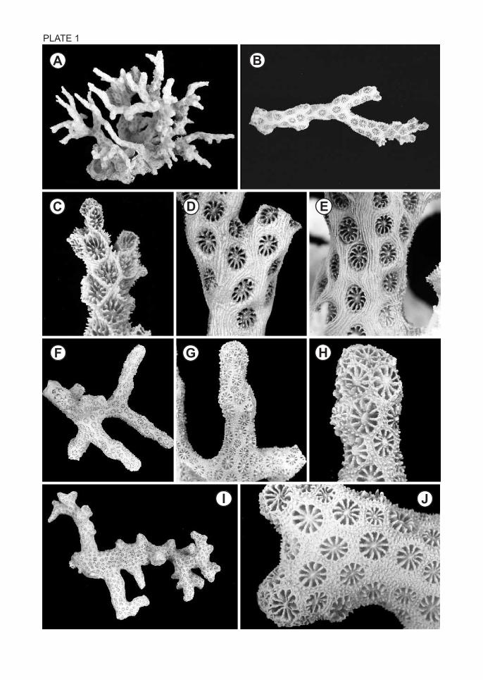
PLATE 1
A B
C D E
F G H
I J

PLATE 2
A
B
C
D
E
F
G
H
I
J
K
L

PLATE 3
A
B
C
D
E
F
G
H
I
J
K

PLATE 4
A
B
C
D
E
F
G
H
I
J
K
L

PLATE 5
A B C
D
E F
G H

PLATE 6
A B C
D E F
G H I
J K L

PLATE 7
A B
C D
E F G
H I J

PLATE 8
A B
C D E
F G H
I J K

PLATE 9
A B
C D E
F G
H I J

PLATE 10
A B
C D E
F G
H I J

PLATE 11
A B
C D E
F
G H
I
J

PLATE 12
A B C
D E F
G H
I J K
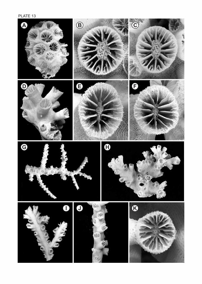
PLATE 13
A B C
D E F
G H
I J K