award number: w81xwh-12-1-0097 - apps.dtic.mil · 12 distinct alpha integrin subunits2, forming...
TRANSCRIPT
1
AD_________________
Award Number: W81XWH-12-1-0097 TITLE: “Targeting Alpha5 Beta1 Integrin to Prevent Metastatic Breast Cancer Cell Invasion: PhScN Target Site Definition and Plasma Stability” PRINCIPAL INVESTIGATOR: Phillip C.Andrew CONTRACTING ORGANIZATION: University of Michigan Ann Arbor, MI 48109-1274 REPORT DATE: September 2013 TYPE OF REPORT: Annual PREPARED FOR: U.S. Army Medical Research and Materiel Command Fort Detrick, Maryland 21702-5012 DISTRIBUTION STATEMENT: Approved for Public Release; Distribution Unlimited The views, opinions and/or findings contained in this report are those of the author(s) and should not be construed as an official Department of the Army position, policy or decision unless so designated by other documentation.

REPORT DOCUMENTATION PAGE Form Approved
OMB No. 0704-0188 Public reporting burden for this collection of information is estimated to average 1 hour per response, including the time for reviewing instructions, searching existing data sources, gathering and maintaining the data needed, and completing and reviewing this collection of information. Send comments regarding this burden estimate or any other aspect of this collection of information, including suggestions for reducing this burden to Department of Defense, Washington Headquarters Services, Directorate for Information Operations and Reports (0704-0188), 1215 Jefferson Davis Highway, Suite 1204, Arlington, VA 22202-4302. Respondents should be aware that notwithstanding any other provision of law, no person shall be subject to any penalty for failing to comply with a collection of information if it does not display a currently valid OMB control number. PLEASE DO NOT RETURN YOUR FORM TO THE ABOVE ADDRESS. 1. REPORT DATE September 2013
2. REPORT TYPE Annual
3. DATES COVERED 1 September 2012 - 31 August 2013
4. TITLE AND SUBTITLE
Targeting Alpha5 Beta1 Integrin to Prevent Metastatic Breast Cancer Cell Invasion: PhScN Target Site Definition and Plasma Stability
5a. CONTRACT NUMBER
5b. GRANT NUMBER
W81XWH-12-1-0097 5c. PROGRAM ELEMENT NUMBER
6. AUTHOR(S)
Phillip C. Andrews5d. PROJECT NUMBER
5e. TASK NUMBER
5f. WORK UNIT NUMBER 7. PERFORMING ORGANIZATION NAME(S) AND ADDRESS(ES)
University of Michigan Ann Arbor, MI 48109-1274
8. PERFORMING ORGANIZATION REPORT NUMBER
9. SPONSORING / MONITORING AGENCY NAME(S) AND ADDRESS(ES) 10. SPONSOR/MONITOR’S ACRONYM(S) U.S. Army Medical Research and Materiel Command
Fort Detrick, Maryland 21702-5012
11. SPONSOR/MONITOR’S REPORT
NUMBER(S)
12. DISTRIBUTION / AVAILABILITY STATEMENT
Approved for Public Release; Distribution Unlimited 13. SUPPLEMENTARY NOTES
14. ABSTRACT It has been suggested that PHSCN inhibits metastatic invasion by forming a covalent, disulfide bond with a cysteine residue on the beta1 (β1) subunit of alpha5 beta1 (α5β1) integrin1. However, these studies were performed with purified α5β1 integrin, which also produces evidence of covalent bond formation with the α5 subunit (tandem mass spectroscopy data not shown). Hence, the specificity of the reported interaction between PHSCN and the β1 subunit1 is suspect. Moreover, the cysteine–rich β1 subunit can heterodimerize with 12 distinct alpha integrin subunits2, forming integrins that function in many pathways. In contrast, the α5 subunit interacts uniquely with the β1 subunit2 to induce invasion and support adhesion3-5; hence it is a much more desirable target. 15. SUBJECT TERMS- none provided
16. SECURITY CLASSIFICATION OF:
17. LIMITATION OF ABSTRACT
18. NUMBER OF PAGES
19a. NAME OF RESPONSIBLE PERSON USAMRMC
a. REPORT
U b. ABSTRACT
U c. THIS PAGE
U
UU
19b. TELEPHONE NUMBER (include area
code)

Table of Contents
Page
INTRODUCTION 2
BODY 2
KEY RESEARCH ACCOMPLISHMENTS 10
REPORTABLE OUTCOMES 11
CONCLUSION 11
REFERENCES 12

2
INTRODUCTION: It has been suggested that PHSCN inhibits metastatic invasion by forming a covalent, disulfide bond with a cysteine residue on the beta1 (β1) subunit of alpha5 beta1 (α5β1) integrin1. However, these studies were performed with purified α5β1 integrin, which also produces evidence of covalent bond formation with the α5 subunit (tandem mass spectroscopy data not shown). Hence, the specificity of the reported interaction between PHSCN and the β1 subunit1 is suspect. Moreover, the cysteine–rich β1 subunit can heterodimerize with 12 distinct alpha integrin subunits2, forming integrins that function in many pathways. In contrast, the α5 subunit interacts uniquely with the β1 subunit2 to induce invasion and support adhesion3-5; hence it is a much more desirable target. Our funded research is designed to test definitively the hypothesis that both PHSCN and PhScN peptides inhibit α5β1 integrin fibronectin receptor–mediated invasion through noncovalent interaction with α5β1 receptors of SUM149PT and MDA-MB-231 metastatic human breast cancer cells, and to lay the foundation for detailed structure–activity relationship (SAR) studies for both peptides as inhibitors of α5β1–mediated metastatic breast cancer invasion by precisely defining their target site(s) on α5β1 integrin, and by developing an assay that can be used to evaluate in vitro degradation rates in plasma, and to monitor in vivo plasma levels. Objective/Hypothesis: Our preliminary data suggest that the invasion inhibitory potencies of the S–acetylated and the S–methylated PHSCN peptide derivatives are increased by over 1,000–fold, relative to PHSCN. In addition, the PhScN peptide is still more potent: preliminary results suggest that it is 104 to 105–fold more potent than PHSCN at blocking α5β1–mediated invasion. Thus, we hypothesize that the actual invasion–inhibitory interaction of PHSCN with α5β1 integrin involves a noncovalent interaction with the target site, which is promoted by including D–isomers of histidine and cysteine in the PhScN peptide. Thus, covalent, disulfide bond formation with α5β1 integrin is actually a side reaction that decreases potency. Because mammalian proteins do not contain D–amino acids, endoproteinses evolved to cleave between L–amino acids only. Since PhScN does not contain 2 linked L–amino acids, it cannot be degraded by endoproteases, usually present at high levels in tumors and increasing with malignancy6-8. Thus, in addition to promoting noncovalent interaction with its α5β1 integrin target, the inclusion of D–amino acids at alternate positions is expected to result in increased PhScN potency as a result of preventing endoproteolytic degradation. Our research is designed to achieve the following specific aims. Specific Aims: Specific Aim 1: To lay the foundation for determining their structure-activity relationships (SARs) as inhibitors of α5β1–mediated invasion3-5,9,10, we propose to assay PHSCN and PhScN peptide derivatives, having modifications of N– or C–termini, and/or cysteine side chain. Peptides will be tested in quantitative serum–free invasion assays with naturally occurring basement membranes3-5,9-15. Specific Aim 2: To identify the binding site for the PHSCN sequence on α5β1 integrin, which will aid in the development of potent PHSCN/PhScN analogs, biotinylated, S-BPM–derivatized PHSCN and PhScN peptides will be photocrosslinked to SUM149PT and MDA-MB-231 cells. Tagged cells and extracts will be screened by fluorescent antibody staining and Western blotting for biotin and α5β1 integrin. The identity of the BPM–modified subunit and sites(s) of modification will then be determined using tandem mass spectrometry to localize the PHSCN/PhScN binding site(s) on α5β1 integrin. Specific Aim 3: To develop a mass spectrometry–based quantitative assay for PHSCN sequence analogs, including PhScN, and use it to evaluate and enhance their stabilities in human and mouse plasma. This assay is crucial for future in vivo studies where bioavailability and clearance rates are to be monitored. BODY: Specific Aim 1 We proposed to test PHSCN and PhScN derivatives with modifications of the N– or C–termini, and/or cysteine side chain to determine the initial spatial constraints of the binding pocket of the ligand to its target site. This will aid in the selection of crosslinking agents, modifications, tag additions and strategies for determination of

3
the target site(s) performed in Aim 2 and lay the foundation for determining the structure-activity relationships (SARS) of these inhibitors with the target. Selection of peptides. Table 1 is an initial list of peptides for testing and the IC50’s determined using our in vitro invasion assay. Initial focus has been on modification of the cysteine side chain and the C-terminus. Modification of the cysteine side chain shows increased potency to the L-cysteine and no change to the D-cysteine activity. The addition of the biotin and the creation of a MAP (multi-antigenic peptide) on the C-terminus and the retention of activity suggest that the “tail” of the peptide has room for modification. These initial results suggest that the C–terminus and cysteine side chain are the logical place to start for placement of cross-linkers for work in Aim 2. Table 1. Invasion inhibitory potency of PHSCN and PhScN derivatives. Values expressed in Molarity to adjust for molecular weight of modifications. C, commercially synthesized; D, derivatization in house of the appropriate commercial parent compound; TBD, to be determined; bio, biotin tag; MAP, multiantigenic peptide; Me, S–methylated; OAc, S–acetylated; acm, S–acetamidomethyl; BPM, benzophenone-4-maleimide
Derivatization of the peptide ligands with a photoactivatable BPM cross-linker Under the direction of Dr. Phil Andrews, a protocol for the derivatization of the Ac-PHSCNGGK-bio and the Ac-PhScNGGK-bio with BPM (benzophenone-4-maleimide) was developed. It was determined that the peptide required a pretreatment of TCEP, a phosphine reducing agent, to reduce any dimer formation of the peptide and that the derivatization protocol required a higher concentration of organic solvent to maintain solubility of the cross-linker. Once derivatized the modified peptide is stable and can be cleaned by C18 column as necessary. The modified peptides were analyzed by MS/MS and were found to have acquired the BPM tag, the parent compound was detected but not determined to be significant. The modified peptides were then tested by our in vitro invasion assay to determine their potency and are listed in Table 1. Results of in vitro invasion assays for C–terminus and/or cysteine–modified peptide ligands. Figure 1 shows the invasion inhibitory affects in our standard SU-ECM serum-induced invasion assay for the BPM derivatized compounds that will be used in the crosslinking experiments in Aim 2. Functionally, it can be seen that the bench-top derivatization with BPM (benzophenone-4-maleimide) of the cysteine of PHSCN
Table 1
C or D
mw
MDA-MB-231 IC50 M
SUM149PT IC50 M
L or D isomer
Modification Region
Ac-PHSCN-NH2 C 598 2.8 E-8 1.5 E-8 L parent Ac-PHSCNGGK-bio C 1180 4.8 E-9 7.3 E-9 L tail Ac-PHSC(Me)N-NH2 C 612 5.3 E-13 4.0 E-13 L side Ac-PHSC(OAc)N-NH2 C 640 1.4 E-14 7.3 E-15 L side Ac-PHSC(acm)NGGK-bio C 1537 TBD TBD L Tail + side Ac- PHSC(BPM)GGK-bio D 1436 TBD 3.6 E-14 L Tail + side Ac-PHSCNGGK-MAP C 7575 2.0 E-11 2.0 E-11 L Tail (MAP) Ac-PhScN-NH2 C 598 2.2 E-14 1.3 E-13 D parent Ac-PhScNGGK-bio C 1180 1.3 E-13 4.4 E-14 D tail Ac- PhSc(BPM)GGK-bio D 1436 1.2 E-14 2.5 E-14 D Tail + side Ac-PhSc(acm)N-GGK-bio C 1537 TBD TBD D Tail + side Ac-PhScNGGK-MAP C 7575 1.7 E-20 5.8 E-21 D Tail (MAP)
4
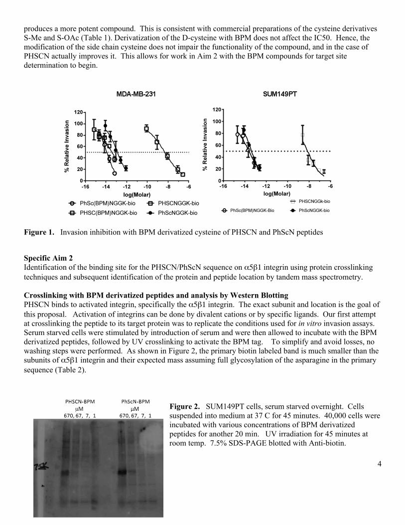
produces a more potent compound. This is consistent with commercial preparations of the cysteine derivatives S-Me and S-OAc (Table 1). Derivatization of the D-cysteine with BPM does not affect the IC50. Hence, the modification of the side chain cysteine does not impair the functionality of the compound, and in the case of PHSCN actually improves it. This allows for work in Aim 2 with the BPM compounds for target site determination to begin.
Figure 1. Invasion inhibition with BPM derivatized cysteine of PHSCN and PhScN peptides Specific Aim 2 Identification of the binding site for the PHSCN/PhScN sequence on α5β1 integrin using protein crosslinking techniques and subsequent identification of the protein and peptide location by tandem mass spectrometry. Crosslinking with BPM derivatized peptides and analysis by Western Blotting PHSCN binds to activated integrin, specifically the α5β1 integrin. The exact subunit and location is the goal of this proposal. Activation of integrins can be done by divalent cations or by specific ligands. Our first attempt at crosslinking the peptide to its target protein was to replicate the conditions used for in vitro invasion assays. Serum starved cells were stimulated by introduction of serum and were then allowed to incubate with the BPM derivatized peptides, followed by UV crosslinking to activate the BPM tag. To simplify and avoid losses, no washing steps were performed. As shown in Figure 2, the primary biotin labeled band is much smaller than the subunits of α5β1 integrin and their expected mass assuming full glycosylation of the asparagine in the primary sequence (Table 2).
Figure 2. SUM149PT cells, serum starved overnight. Cells suspended into medium at 37 C for 45 minutes. 40,000 cells were incubated with various concentrations of BPM derivatized peptides for another 20 min. UV irradiation for 45 minutes at room temp. 7.5% SDS-PAGE blotted with Anti-biotin.
5
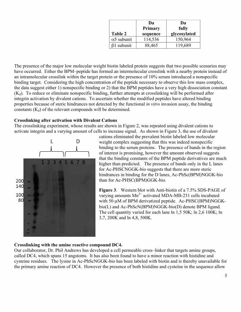
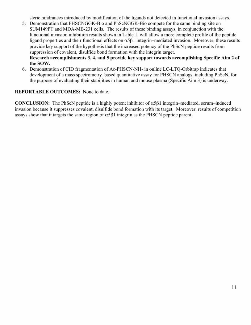
The presence of the major low molecular weight biotin labeled protein suggests that two possible scenarios may have occurred. Either the BPM–peptide has formed an intermolecular crosslink with a nearby protein instead of an intramolecular crosslink within the target protein or the presence of 10% serum introduced a nonspecific binding target. Considering the high concentration of the peptide necessary to observe this low mass complex, the data suggest either 1) nonspecific binding or 2) that the BPM peptides have a very high dissociation constant (Kd). To reduce or eliminate nonspecific binding, further attempts at crosslinking will be performed after integrin activation by divalent cations. To ascertain whether the modified peptides have altered binding properties because of steric hindrances not detected by the functional in vitro invasion assay, the binding constants (Kd) of the relevant compounds will be determined. Crosslinking after activation with Divalent Cations The crosslinking experiment, whose results are shown in Figure 2, was repeated using divalent cations to activate integrin and a varying amount of cells to increase signal. As shown in Figure 3, the use of divalent
cations eliminated the prevalent biotin labeled low molecular weight complex suggesting that this was indeed nonspecific binding to the serum proteins. The presence of bands in the region of interest is promising, however the amount observed suggests that the binding constants of the BPM peptide derivatives are much higher than predicted. The presence of bands only in the L lanes for Ac-PHSCNGGK-bio suggests that there are more steric hindrances in binding for the D lanes, Ac-PhSc(BPM)NGGK-bio than for Ac-PHSC(BPM)GGK-bio. Figure 3. Western blot with Anti-biotin of a 7.5% SDS-PAGE of varying amounts Mn2+ activated MDA-MB-231 cells incubated with 50 µM of BPM derivatized peptide. Ac-PHSC(BPM)NGGK-bio(L) and Ac-PhScN(BPM)NGGK-bio(D) denote BPM ligand. The cell quantity varied for each lane ln 1,5 50K; ln 2,6 100K; ln 3,7, 200K and ln 4,8, 500K.
Crosslinking with the amine reactive compound DC4. Our collaborator, Dr. Phil Andrews has developed a cell permeable cross–linker that targets amine groups, called DC4, which spans 15 angstoms. It has also been found to have a minor reaction with histidine and cysteine residues. The lysine in Ac-PhScNGGK-bio has been labeled with biotin and is thereby unavailable for the primary amine reaction of DC4. However the presence of both histidine and cysteine in the sequence allow
Table 2
Da Primary sequence
Da fully
glycosylated α5 subunit 114,536 150,964 β1 subunit 88,465 119,689

6
for a low level reaction with DC4. As before, cells were activated by divalent cations or FBS, followed by incubated with the biotin labeled peptide, Ac-PhScNGGK-bio. After incubation the cells were quickly spun and the excess peptide was removed, the cell pellet was then resuspended in the DC4 cross-linker. Crosslinking is rapid and complete in about 20-30 minutes. Since any lysine can react, the concentration of the DC4 and the available protein must be empirically determined. Figure 4 shows the results of the DC4 cross-linker as compared to crosslinking with BPM derivatives. In Panel 4A, it can be seen that DC4 has cross–linked the biotin labeled peptide Ac-PhScNGGK-bio, corresponding to molecular weights of approximately 120 KDa and 140 KDa, which are the mass of the α5β1 integrin subunits. By using DC4, the Ac-PhScNGGK-bio is able to bind at its normal binding Kd without steric interference, and the DC4 can react with either the histidine or the cysteine. Unlike reaction with lysine, reaction to histidine or cysteine is a reversible by using hydroxylamine. The ability to reverse and confirm this type of reaction may be useful in identifying the target site peptides with MS/MS. In Panel B, the L and D BPM derivatives behave differently, again suggesting steric interference of the BPM group. However, it is interesting to note that the labeled bands are of similar molecular weight as those in Panel A. This is encouraging and attempts to scale the experiment to levels where enrichment and protein detection can be observed with staining will continue.
Figure 4. Western blot with Anti-biotin primary antibody, of a 7.5% SDS-PAGE of activated SUM149PT cells using DC4 or the BPM peptides. The 100 K cells were activated using either divalent cation(Mn2+) or FBS. Panel A) The concentration of 100 µM Ac-PhScNGGK-bio was used, cells were spun to remove excess labeled peptide prior to resuspension in DC4 crosslinker. Panel B) 200 µM Ac-PHSC(BPM)NGGK-bio(L) and Ac-PhScN(BPM)NGGK-bio(D) were incubated with cells and then subjected to UV light. The samples were prepared at the same time and run on the same gel and developed together. However, different exposures were required to optimize each panel. Images were scanned and then overlaid. The cross-linking with the BPM peptides will continue; attempts will be made to scale the experiments for protein enrichment and identification by MS/MS. The DC4 cross-linker will complement this work, and will also continue. Preliminary evidence indicates that there are steric hindrances by modification of the cysteine,

7
therefore a new peptide, Ac-PhScNGGK(biotin)GK-NH2 , with be synthesized to allow for cross-linking via the C-terminal lysine residue with either DC4 or other commercially available amine reactive cross-linking agents. Determination of Binding Constants The presence of a biotin tag on the peptide allows for a direct determination of the binding constant (Kd) to live cells by detection with steptavadin-HRP. In Figure 5, the Kd of the Ac-PhScNGGK-bio was determined and compared to Ac-PHSCNGGK-bio; they were found to be similar, at approximately 0.03 µM for both MDA-MB-231 and SUM149PT cells. The Kd determination for the BPM-derivatized biotin peptides is in progress and will provide information on any steric hindrances with the BPM cysteine modification not detected in the functional assay. Figure 5. Determination of the Kd values for the underivatized biotinylated peptides with suspended MDA-MB-231 or SUM149PT cells. Binding characteristics of non-biotin labeled peptides can be assessed indirectly by competition binding assays. These assays require a constant concentration of labeled peptide near its Kd is and is dissociated by increasing amounts of an unlabeled competitor. This generates an inhibition constant (Ki) which will allow comparison of binding properties of the unlabeled peptides. This assay will also confirm whether ligands are competing for the same binding site. Figure 6 shows the competition assay for the parent compounds PHSCN and PhScN and confirms that the peptides compete for the same site. These binding assays in conjunction with the functional assay results in Table 1 will allow a more complete profile of the peptide ligand properties

8
Figure 6. Competition binding assays: Ac-PHSCNGGK-bio and dissociation with unlabeled Ac-PhScN-NH2. Specific Aim 3 Develop a mass spectrometry–based quantitative assay for PHSCN analogs, including PhScN, and evaluate their stabilities in human and mouse plasma. Experimental design for quantitative assay of PHSCN analogs An experimental approach based on mass spectrometric analysis to evaluate the stabilities of PHSCN analogs in human and mouse plasma has been designed as outlined in Scheme 1. A known amount of target peptide is mixed with plasma and incubated at 37 °C for up to 24 hours. At specific incubation time points an aliquot is transferred to a small tube and protease reactions were quenched by adding three volumes of 50% acetonitrile with 0.5%TFA. Precipitated plasma proteins were removed by centrifugations, and a known amount of an internal standard was spiked into the supernatant containing the peptides, in order to increase the accuracy of quantitative measurements of LC-MS by reducing variances associated with sample preparations. MRFA (Met-Arg-Phe-Ala, monoisotopic molecular mass = 523.247.), a stable synthetic peptide with similar molecular mass to the target peptide, Ac-PHSCN-amide (monoisotopic molecular mass = 597.2328), is added into samples prior to LC-MS analysis. Stabilities of target peptides in plasma were evaluated by LC-MS. Mass spectrometry The LTQ-Orbitrap XL(Thermo Finnegan) connected to a 2D nanoLC(Exigent) and an autosampler in Andrews laboratory provides high mass resolution and accuracy for analytes separated on a capillary column at a flow rate of 200 nL/min, which assures confident identification of target molecules. Extracted peptides from plasma are reconstituted in 0.1% TFA and loaded into a C18 trap by autosampler and washed for 6 min at 5 microL/min with 0.1% TFA in water delivered by the LC loading pumps. Trapped peptides were introduced by switching valve into a capillary column (75 micro-m x 15 cm) custom-packed with C18 resin (3 micron) and

9
separated over 60 min gradient at a flow rate of 200 nL/min. Analytes were introduced into the LTQ-Orbitrap via an electrospray device (Triversa Nanomate, Advion). MS and MSMS data were acquired in data-dependent mode for ions within 400-800 m/z. Figure 7 shows the total ion chromatogram (TIC) of LC-MSMS analysis of PHSCN and the internal standard MRFA.
Figure 7. TIC from an LC-MSMS of PHSCN and MRFA mixture. Quantitative assay of target peptides Peptide quantification can be achieved by ion intensities in MS or by MSMS spectral counts in data-dependent acquisition mode. These two approaches are well established label-free quantification methods for peptides and proteins by LC-MSMS, which can be readily applied to a bioanalytical assay of therapeutic peptides. We are currently testing a new assay method, an MRM-like analysis via data-independent acquisition mode in the LTQ-Orbirap, in which ion packets within a mass range of 5 or 10 Da accumulated for a set time are co-fragmented. One or multiple fragment ions specific to the target peptide can be monitored in MSMS based on highly accurate precursor ion mass and LC retention time. This MRM-like approach can provide a highly selective and sensitive bioanalytical assay under conditions of high chromatographic interference. Study of collision–induced dissociation (CID) fragmentation of Ac-PHSCN-NHs in online LC-LTQ-Orbitrap Ac-PHSCN-NH2 introduced into the mass spectrometer via a nanoESI device (Triversa Nanomate) was predominantly observed as singly protonated form at 598.2328 m/z. However, in online LC-MS, the peptide was observed in doubly charged dimeric forms at 597.23 m/z and 589.23 m/z. The peptide dimer ions were characterized by CID that revealed disulfide-bond formation between two peptides that was catalyzed on C18 LC, as shown in a CID spectrum of 597.23 m/z. This is a potential source of interference with the assay that can be easily addressed by incorporating reduction and alkylation of the Cys residues into the protocol prior to LCMSMS.

10
Figure 8. CID fragmentation of doubly–charged ion at 597.73 m/z (the second isotope to 597.23 m/z). Fragment ions B4, A2, Y2,Y3, and Y4 containing S-S cross-linked peptides confirm disulfide bond formation between two thiols of cysteine. KEY RESEARCH ACCOMPLISHMENTS:
1. Determination of invasion–inhibitory potencies for a total of 12 PHSCN and PhScN derivatives, targeting α5β1 integrin–mediated invasion of naturally serum–free basement membranes by 2 metastatic human breast cancer cell lines, SUM149PT and MDA-MB-231. Demonstration that covalent S–modification by several different moieties increases invasion–inhibitory potency by 100,000– to 1,000,000–fold, and that a 1,000,000–fold increase in potency is also obtained by substiution of D–His and D–Cys. These results demonstrate that the reason for the greatly increased invasion–inhibitory potency of PhScN is the suppression of covalent disulfide bond formation between the PHSCN sequence and its integrin target, and hence they are a key step in the determination of the PhScN target site on α5β1 integrin receptors of breast cancer cells.
2. Determination of invasion–inhibitory potencies of PHSCN and PhScN peptides, containing BPM–derivatized cysteine or D–cysteine. The greatly increased invasion–inhibitory potencies of the BPM–modified PHSCN and PhScN peptides also confirm the noncovalent nature of invasion inhibition by the PhScN peptide. Research accomplishments 1 and 2 complete Specific Aim 1 of the SOW.
3. Crosslinking with BPM–derivatized peptides, after integrin α5β1 activation with divalent cations, and analysis by Western blotting. Successful crosslinking with biotin labeled peptides, modified either at the cysteine or on the C–terminus, is anticipated to pave the way for protein enrichment and binding site identification by MS/MS.
4. Determination of binding constants for PHSCNGGK-Bio and PhScNGGK-Bio on MDA-MB-231 and SUM149PT cells, and demonstration of their similarity provides the basis for acquiring information on any
11
steric hindrances introduced by modification of the ligands not detected in functional invasion assays. 5. Demonstration that PHSCNGGK-Bio and PhScNGGK-Bio compete for the same binding site on
SUM149PT and MDA-MB-231 cells. The results of these binding assays, in conjunction with the functional invasion inhibition results shown in Table 1, will allow a more complete profile of the peptide ligand properties and their functional effects on α5β1 integrin–mediated invasion. Moreover, these results provide key support of the hypothesis that the increased potency of the PhScN peptide results from suppression of covalent, disulfide bond formation with the integrin target. Research accomplishments 3, 4, and 5 provide key support towards accomplishing Specific Aim 2 of the SOW.
6. Demonstration of CID fragmentation of Ac-PHSCN-NH2 in online LC-LTQ-Orbitrap indicates that development of a mass spectrometry–based quantitative assay for PHSCN analogs, including PhScN, for the purpose of evaluating their stabilities in human and mouse plasma (Specific Aim 3) is underway.
REPORTABLE OUTCOMES: None to date. CONCLUSION: The PhScN peptide is a highly potent inhibitor of α5β1 integrin–mediated, serum–induced invasion because it suppresses covalent, disulfide bond formation with its target. Moreover, results of competition assays show that it targets the same region of α5β1 integrin as the PHSCN peptide parent.
12
REFERENCES: 1. Donate, F., G.C. Parry, Y. Shaked, H. Hensley, et al., Pharmacology of the novel antiangiogenic peptide ATN-161 (Ac-
PHSCN-NH2): Observation of a U-shaped dose-response curve in several preclinical models of angiogenesis and tumor growth. Clin Cancer Res, 2008. 14: p. 2137-2144. PMID: 18381955
2. Takada, Y., X. Ye, and S. Simon, The integrins. Genome Biol, 2007. 8(5): p. 215. PMID: 17543136 3. Jia, Y., Z.Z. Zeng, S.M. Markwart, K.F. Rockwood, et al., Integrin fibronectin receptors in matrix metalloproteinase-1-
dependent invasion by breast cancer and mammary epithelial cells. Cancer Res, 2004. 64(23): p. 8674-81. PMID: 15574776
4. Livant, D.L., R.K. Brabec, K.J. Pienta, D.L. Allen, et al., Anti-invasive, antitumorigenic, and antimetastatic activities of the PHSCN sequence in prostate carcinoma. Cancer Res, 2000. 60(2): p. 309-320. PMID: 10667582
5. Zeng, Z.-Z., H. Yao, E.D. Staszewski, K.F. Rockwood, et al., (alpha)5(beta)1 integrin ligand PHSRN induces invasion and (alpha)5 mRNA in endothelial cells to stimulate angiogenesis. Transl Oncol., 2009. 2: p. 8-20. PMID: 19252747
6. Clevers, H., At the crossroads of inflammation and cancer. Cell, 2004. 118(6): p. 671-4. PMID: 15369667 7. Coussens, L.M. and Z. Werb, Inflammation and cancer. Nature, 2002. 420(6917): p. 860-7. PMID: 12490959 8. Scherer, R.L., J.O. McIntyre, and L.M. Matrisian, Imaging matrix metalloproteinases in cancer. Cancer Metastasis Rev,
2008. 27(4): p. 679-90. PMID: 18465089 9. Livant, D.L., R.K. Brabec, K. Kurachi, D.L. Allen, et al., The PHSRN sequence induces extracellular matrix invasion and
accelerates wound healing in obese diabetic mice. J Clin Invest, 2000. 105(11): p. 1537-1545. PMID: 10841512 10. Zeng, Z.-Z., Y.F. Jia, N.J. Hahn, S.M. Markwart, et al., Role of focal adhesion kinase and phosphatidylinositol 3'-kinase in
integrin fibronectin receptor-mediated, matrix metalloproteinase-1 dependent invasion by metastatic prostate cancer cells. Cancer Res, 2006. 66(16): p. 8091-8099. PMID: 16912186
11. van Golen, K.L., L. Bao, G.J. Brewer, K.J. Pienta, et al., Suppression of tumor recurrence and metastasis by a combination of the PHSCN sequence and the antiangiogenic compound tetrathiomolybdate in prostate carcinoma. Neoplasia, 2002. 4(5): p. 373-9. PMID: 12192595
12. Yao, H., D. Veine, K. Fay, E. Staszewski, et al., The PHSCN dendrimer as a more potent inhibitor of human breast cancer cell invasion, extravasation, and lung colony formation. Breast Cancer Research and Treatment, 2011. 125: p. 363-375. PMID: 20300829
13. Yao, H., D.M. Veine, Z.Z. Zeng, K.S. Fay, et al., Increased potency of the PHSCN dendrimer as an inhibitor of human prostate cancer cell invasion, extravasation, and lung colony formation. Clin Exp Metastasis, 2010. 27(3): p. 173-84. PMID: 20339907
14. Woods Ignatoski, K.M., N.K. Grewal, S. Markwart, D.L. Livant, and S.P. Ethier, p38MAPK induces cell surface alpha4 integrin downregulation to facilitate erbB-2-mediated invasion. Neoplasia, 2003. 5(2): p. 128-34. PMID: 12659685
15. Woods Ignatoski, K.M., D.L. Livant, S. Markwart, N.K. Grewal, and S.P. Ethier, The role of phosphatidylinositol 3'-kinase and its downstream signals in erbB-2-mediated transformation. Mol Cancer Res, 2003. 1(7): p. 551-60. PMID: 12754302