avds winter newsletter - australian veterinary association winter... · australian veterinary...
TRANSCRIPT
WELCOME to the winter edition of the AVDS Newsletter! The AVDS newsletter for this quarter is full of information and right on time for Pet Dental Health Month! We hope you enjoy reading the latest edition and look forward to hearing your feedback.
AVA National Conference Wrap Up Pet Dental Health Month – New theme! Looking for Ideas and Support? Free Webinar – 19 August Practical Tips for PDHM Horses : Smile, It's your birthday! Case Report: Unilateral Facial Swelling in a dog Further afield: ANZCVS Science Week report Anaesthesia free dentistry – Why is it an issue and what is the AVDS going to do about it?
Australian Veterinary Dental Society Newsletter
Winter 2014
AVA National Conference Wrap Up
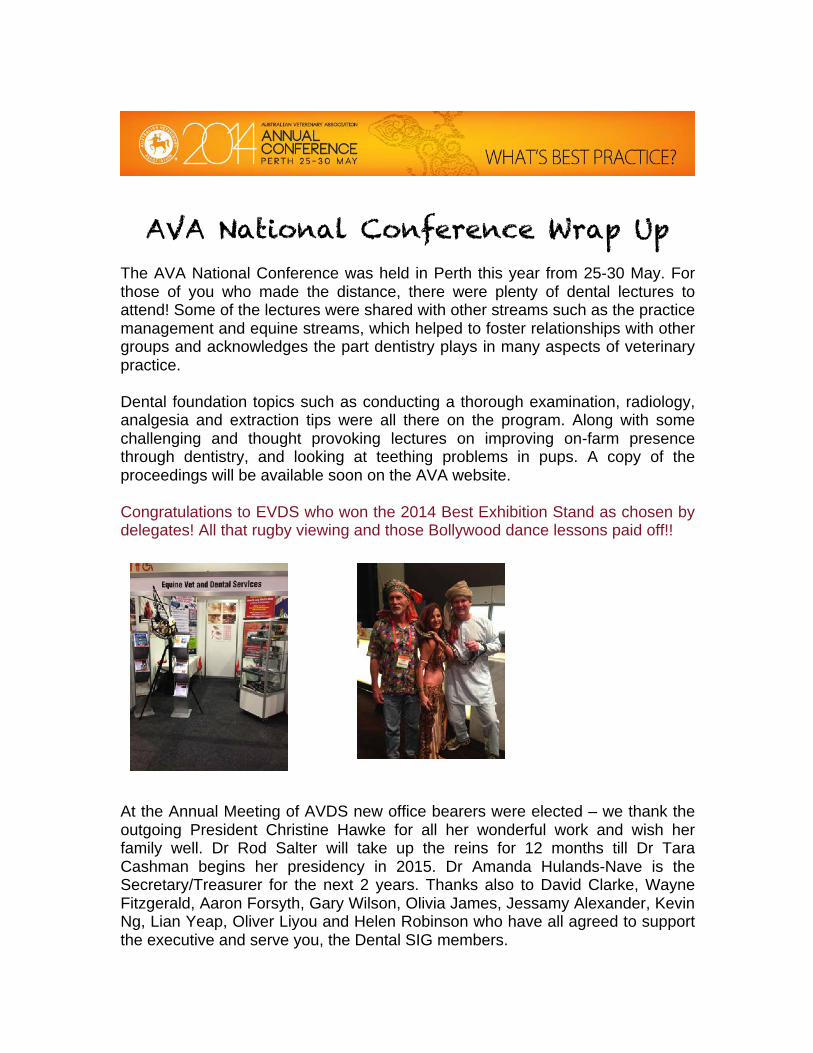
The AVA National Conference was held in Perth this year from 25-30 May. For those of you who made the distance, there were plenty of dental lectures to attend! Some of the lectures were shared with other streams such as the practice management and equine streams, which helped to foster relationships with other groups and acknowledges the part dentistry plays in many aspects of veterinary practice. Dental foundation topics such as conducting a thorough examination, radiology, analgesia and extraction tips were all there on the program. Along with some challenging and thought provoking lectures on improving on-farm presence through dentistry, and looking at teething problems in pups. A copy of the proceedings will be available soon on the AVA website. Congratulations to EVDS who won the 2014 Best Exhibition Stand as chosen by delegates! All that rugby viewing and those Bollywood dance lessons paid off!!
At the Annual Meeting of AVDS new office bearers were elected – we thank the outgoing President Christine Hawke for all her wonderful work and wish her family well. Dr Rod Salter will take up the reins for 12 months till Dr Tara Cashman begins her presidency in 2015. Dr Amanda Hulands-Nave is the Secretary/Treasurer for the next 2 years. Thanks also to David Clarke, Wayne Fitzgerald, Aaron Forsyth, Gary Wilson, Olivia James, Jessamy Alexander, Kevin Ng, Lian Yeap, Oliver Liyou and Helen Robinson who have all agreed to support the executive and serve you, the Dental SIG members.
PET DENTAL HEALTH MONTH
- what lurks beneath……
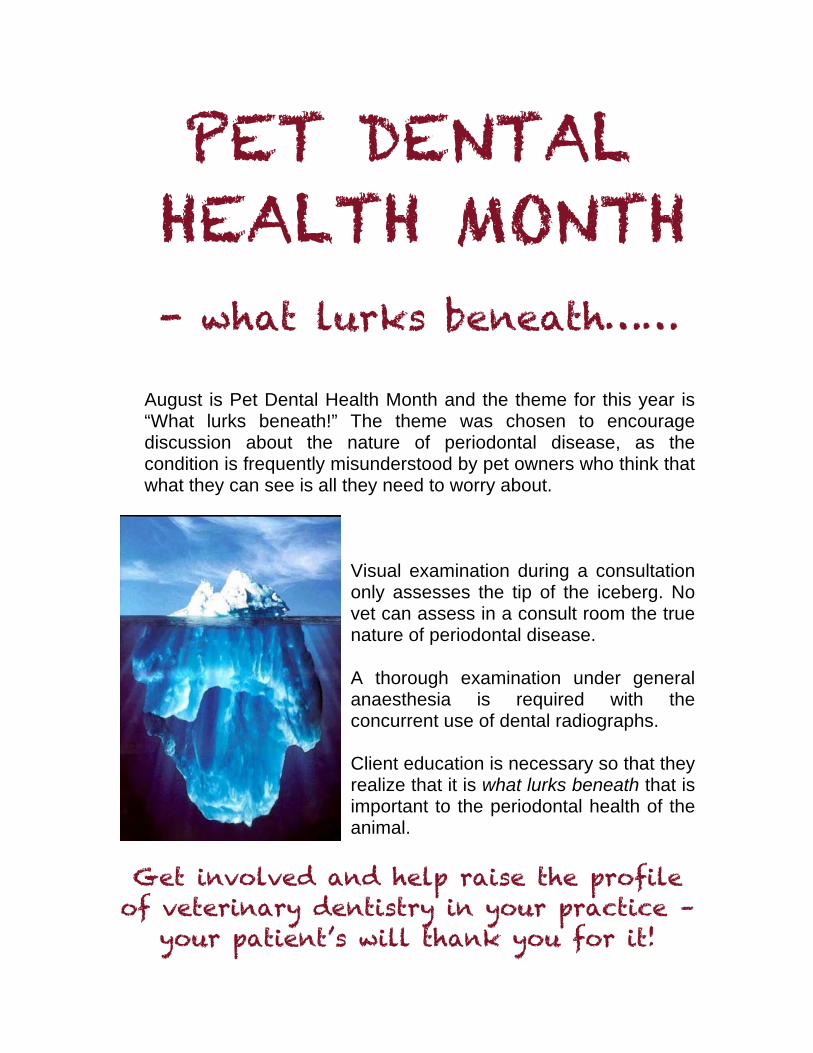
August is Pet Dental Health Month and the theme for this year is “What lurks beneath!” The theme was chosen to encourage discussion about the nature of periodontal disease, as the condition is frequently misunderstood by pet owners who think that what they can see is all they need to worry about.
Visual examination during a consultation only assesses the tip of the iceberg. No vet can assess in a consult room the true nature of periodontal disease. A thorough examination under general anaesthesia is required with the concurrent use of dental radiographs. Client education is necessary so that they realize that it is what lurks beneath that is important to the periodontal health of the animal.
Get involved and help raise the profile
of veterinary dentistry in your practice – your patient’s will thank you for it!
• Check out these great resources available FREE from the AVA website:
For your practice:
☺ Pet Dental Health Month Flyer ☺ media release template – just fill in the blanks!
For your clients:
☺ client handouts on: " Dental health for your cats and dogs " Caring for your horse’s mouth " About your rabbits teeth
For your education:
☺ FREE WEBINAR - Aug 19 @ 6pm (EST) ☺ a Veterinarian’s guide to gum disease in dogs and cats ☺ You can search all past AVDS conference and AVA
proceedings online via the “VETED” tab at the top of the screen. There’s also an international journal search as well!
Also available on the AVA website if you search “dental policy” in the search box you will find details of the following AVA policies: 6.9 Guidelines for dental treatment in dogs and cats 7.2 Equine dentistry 17.9 Dental Guidelines for small mammals
LOOKING FOR IDEAS AND SUPPORT?
www.ava.com.au/dental-
month dental-month
• When: Tues 19 August 2014 • Time: 6-7pm (Sydney time!) • Where: online wherever your computer
goes! • Host: Dr Anthony Caiafa • Cost: FREE to members (non-members
$75) • Topic: What lurks beneath!
What you will learn!
Periodontal disease is no longer considered a local disease affecting only the mouth. Participants will learn about the current human and veterinary research that is looking at the links between periodontal disease and important diseases affecting other parts of the body. This lecture will also look at the similarities between periodontal disease and other chronic diseases such as osteoarthritis. REGISTER ONLINE www.ava.com.au/node/14822
FREE WEBINAR
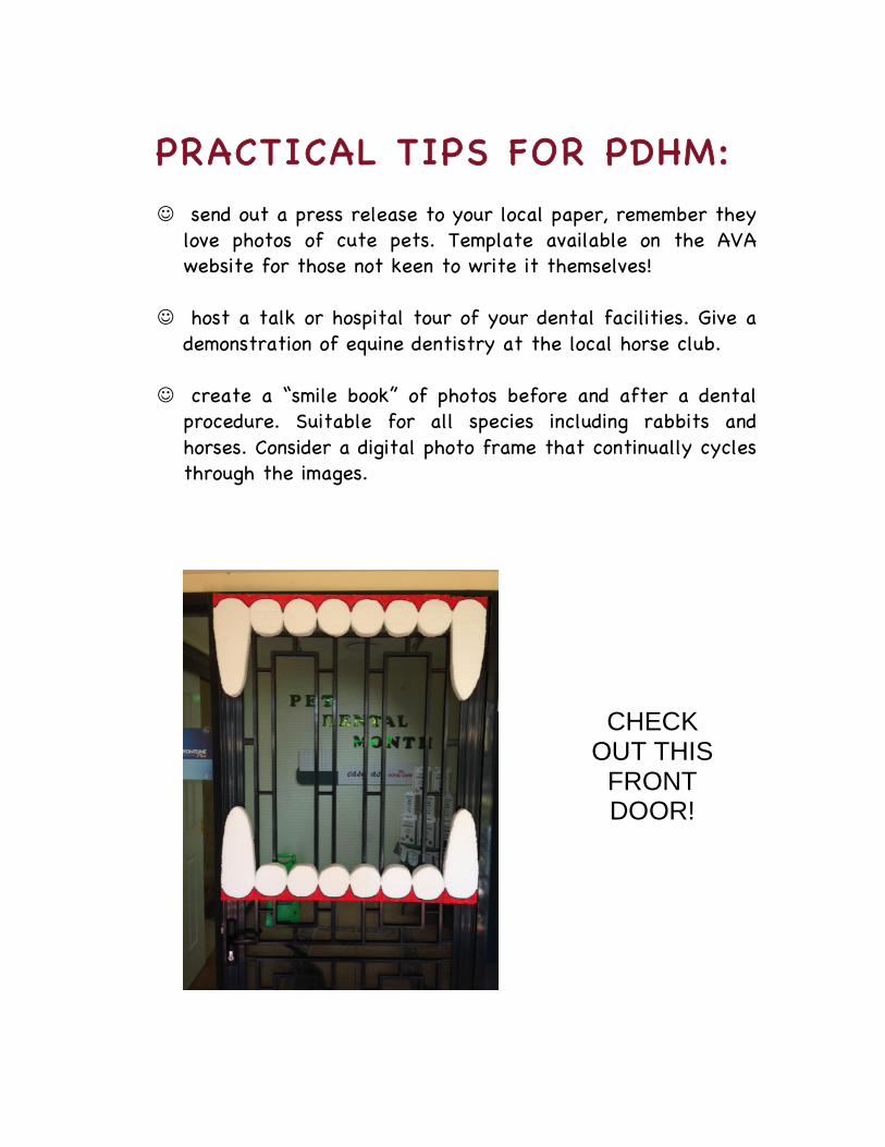
PRACTICAL TIPS FOR PDHM: ☺ send out a press release to your local paper, remember they
love photos of cute pets. Template available on the AVA website for those not keen to write it themselves!
☺ host a talk or hospital tour of your dental facilities. Give a demonstration of equine dentistry at the local horse club.
☺ create a “smile book” of photos before and after a dental
procedure. Suitable for all species including rabbits and horses. Consider a digital photo frame that continually cycles through the images.
CHECK OUT THIS
FRONT DOOR!
☺ get creative and decorate your waiting room with a set of
false teeth or a huge toothbrush – children’s poster paint and recycling materials such as cardboard and foam packing are all you need.
☺ make sure all the nurses ask every client – “what do you do
to keep them smiling?”
☺ local service clubs such as Lions Club are often looking for a speaker for their meetings. Just ask!
☺ share the AVA resource brochures (in print or online) on
dogs, cats, rabbit and horses with your clients – they are bright, colourful and have no company logos!
☺ use social media such as Facebook and Twitter to tell others
what you are doing. Ask for smiling pet photos as a clinic competition. Update your website news section.
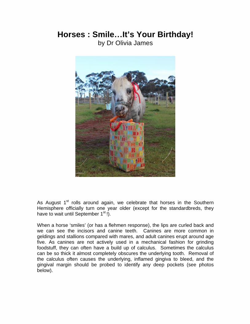
Horses : Smile…It’s Your Birthday! by Dr Olivia James
As August 1st rolls around again, we celebrate that horses in the Southern Hemisphere officially turn one year older (except for the standardbreds, they have to wait until September 1st !). When a horse ‘smiles’ (or has a flehmen response), the lips are curled back and we can see the incisors and canine teeth. Canines are more common in geldings and stallions compared with mares, and adult canines erupt around age five. As canines are not actively used in a mechanical fashion for grinding foodstuff, they can often have a build up of calculus. Sometimes the calculus can be so thick it almost completely obscures the underlying tooth. Removal of the calculus often causes the underlying, inflamed gingiva to bleed, and the gingival margin should be probed to identify any deep pockets (see photos below).
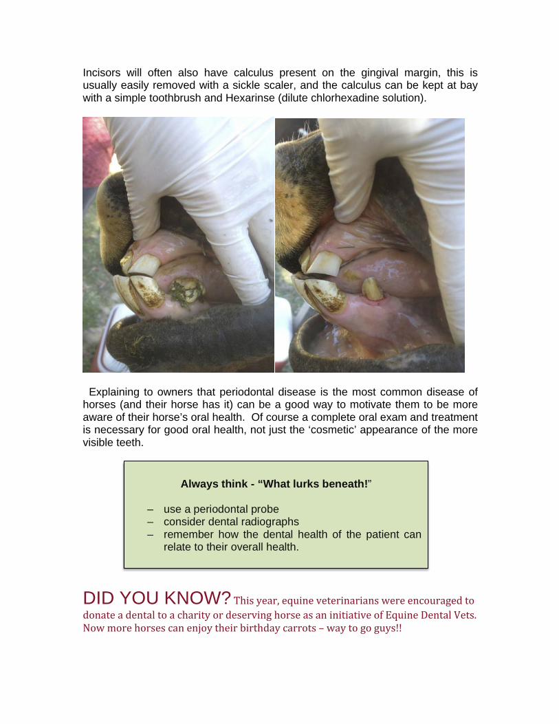
Incisors will often also have calculus present on the gingival margin, this is usually easily removed with a sickle scaler, and the calculus can be kept at bay with a simple toothbrush and Hexarinse (dilute chlorhexadine solution).
Explaining to owners that periodontal disease is the most common disease of horses (and their horse has it) can be a good way to motivate them to be more aware of their horse’s oral health. Of course a complete oral exam and treatment is necessary for good oral health, not just the ‘cosmetic’ appearance of the more visible teeth.
DID YOU KNOW? This year, equine veterinarians were encouraged to donate a dental to a charity or deserving horse as an initiative of Equine Dental Vets. Now more horses can enjoy their birthday carrots – way to go guys!!
Always think - “What lurks beneath!”
– use a periodontal probe – consider dental radiographs – remember how the dental health of the patient can
relate to their overall health.
CASE REPORT : Unilateral facial swelling in an elderly dog.
by Adjunct Assoc Prof Anthony Caiafa
Introduction A 9 year old desexed male Golden Retriever cross Labrador “Foster” was referred to a Veterinary Specialist Referral Practice with a history of a sudden onset of a firm swelling ventral to the left eye (figure 1). The dog had a history of occasionally being offered bones to chew as well as playing games of retrieval of large sticks with the owner. The dog had also recently been on a farm with other dogs. Since the swelling had come up, the dog had become slightly lethargic but was still eating but had no pyrexia on presentation to the referring veterinarian. The referring veterinarian did not have intraoral radiography capabilities and assuming that the swelling was dental in origin and mostly likely a tooth root abscess involving the maxillary 4th premolar (208) or maxillary 1st molar (209), the veterinarian could only offer the client extraction of one or both teeth. The referring veterinarian did however offer the option of referral to a facility that could take intraoral radiographs and thus determine the origin of the swelling. The client accepted this advice. Examination (awake) including whole body, extra-‐ and intra-‐oral examinations On presentation to the referral practice, the dog appeared well with no raised temperature. The swelling just below the left eye was firm and fixed and the dog resented the mouth and muzzle being handled on that side. There was no pain on opening the mouth. The left draining mandibular lymph node was slightly enlarged, non-‐ painful on palpation and mobile. A cursory look at the maxillary caudal teeth on the left side revealed no obvious crown fractures. The medical history revealed a long history of atopic dermatitis which had been controlled by cyclosporine (Atopica® capsules, Novartis Animal Health, Australia). Since unilateral facial swelling (especially being just ventral to the eye) is often associated with an endodontically involved tooth or teeth and because a concussive or subluxation injury to a tooth does not always lead to crown fracture, but can still cause a pulp to become necrotic, at the top of the differential diagnoses was pulp necrosis due to concussion or subluxation injury of the maxillary 4th premolar (208). The most likely cause was bone, rock or stick chewing. The client was also advised that other causes of the swelling could be due to foreign body penetration, dog fight or considering the age of the dog, a facial tumour was possible. The dog’s pre-‐anaesthetic blood analysis was unremarkable and there was no evidence of a white cell elevation or hepatopathy (a known contra-‐indication for long term cyclosporine use) in the biochemistry results.
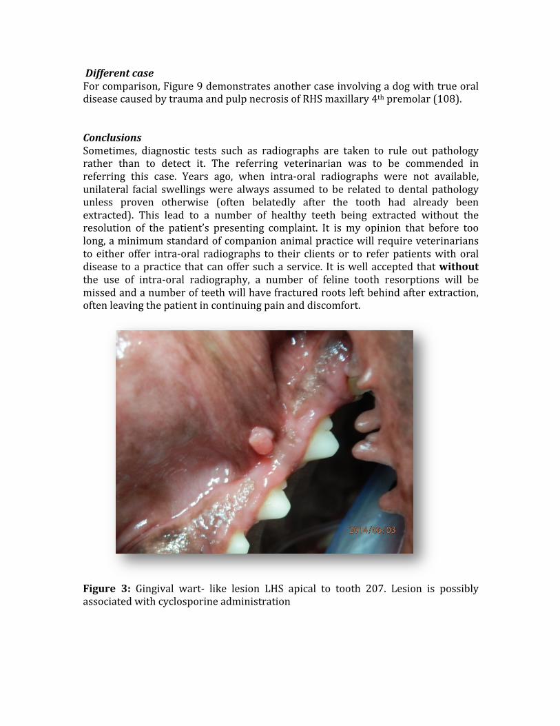
Extra-‐ and intra-‐oral examination under general anaesthesia Under general anaesthesia, an examination of the facial mass revealed it to be firm and slightly mobile. A fine needle aspiration revealed some whole blood. There was no purulent discharge present in the sample. The draining left mandibular lymph node was slightly enlarged. An oral examination revealed evidence of abrasive wear of the teeth (rocks, bones, sticks). Periodontal probing depths were generally 4mm or below (normal is <3mm) and there was some evidence of generalised gingival hypertrophy (a known side effect of long term cyclosporine use) (figure 2). There was also a papilloma like mass just dorsal to the left maxillary 3rd premolar (207) (figure 3). Another known side effect of cyclosporine usage is verruciform lesions or papilloma formation. Because of the dog’s age, the client requested a whole mouth series of intraoral radiographs to rule out any other oral pathology. A whole mouth series of intra-‐oral radiographs were taken using a size 4 phosphor storage plate (5.7 x 7.5cm) (Durr CR7, iM3 Inc., Sydney, Australia). Because the dog was a large breed (32Kg), the size 4 plate was ideal for the task and reduced the number of exposures required to do the whole mouth series of radiographs from the usual 10 or sometimes more exposures (with a size 2 film) down to 6 exposures. This helped reduced anaesthesia time, as well as being more diagnostic by covering more of the oral and nasal cavities in the one exposure. Imaging of the left (figure 4) and right caudal maxillae (figure 5) revealed no obvious dental or nasal problems (if in doubt, when faced with unilateral oral disease, radiograph the other side for comparison). Having ruled out an obvious dental cause for the swelling, it was decided to make a small incision into the mass and with a 5mm skin biopsy punch, and take incisional type biopsies of the facial mass (figure 6). The biopsied tissues were firm and greyish-‐ red in appearance. The oral papilloma type mass apical to the left maxillary 3rd premolar (207) was also excised just in case it was associated with the facial swelling. On biopsy of this wart-‐like lesion, the lesion disintegrated and revealed a mucous blood tinged discharge. Mosquito forceps were used to enlarge the cavity below the intra-‐oral lesion and a size 60 gutta percha point was placed into the facial swelling and found to communicate with the intra-‐oral lesion (figure 7). Exploration of the lesion inside the mouth revealed a small grass seed (figure 8) and this was removed. No other grass seeds were found. A diagnosis of foreign body granuloma was made and the dog was discharged on amoxicillin/ clavulonic acid combination for 7 days. Within 4 days post-‐ surgery, the facial swelling had reduced significantly (>80%) and by 7 days had disappeared and has since not returned. “Foster” is back to his normal self, but is having his teeth brushed regularly to help control plaque accumulation due to the cyclosporine induced gingival hypertrophy.
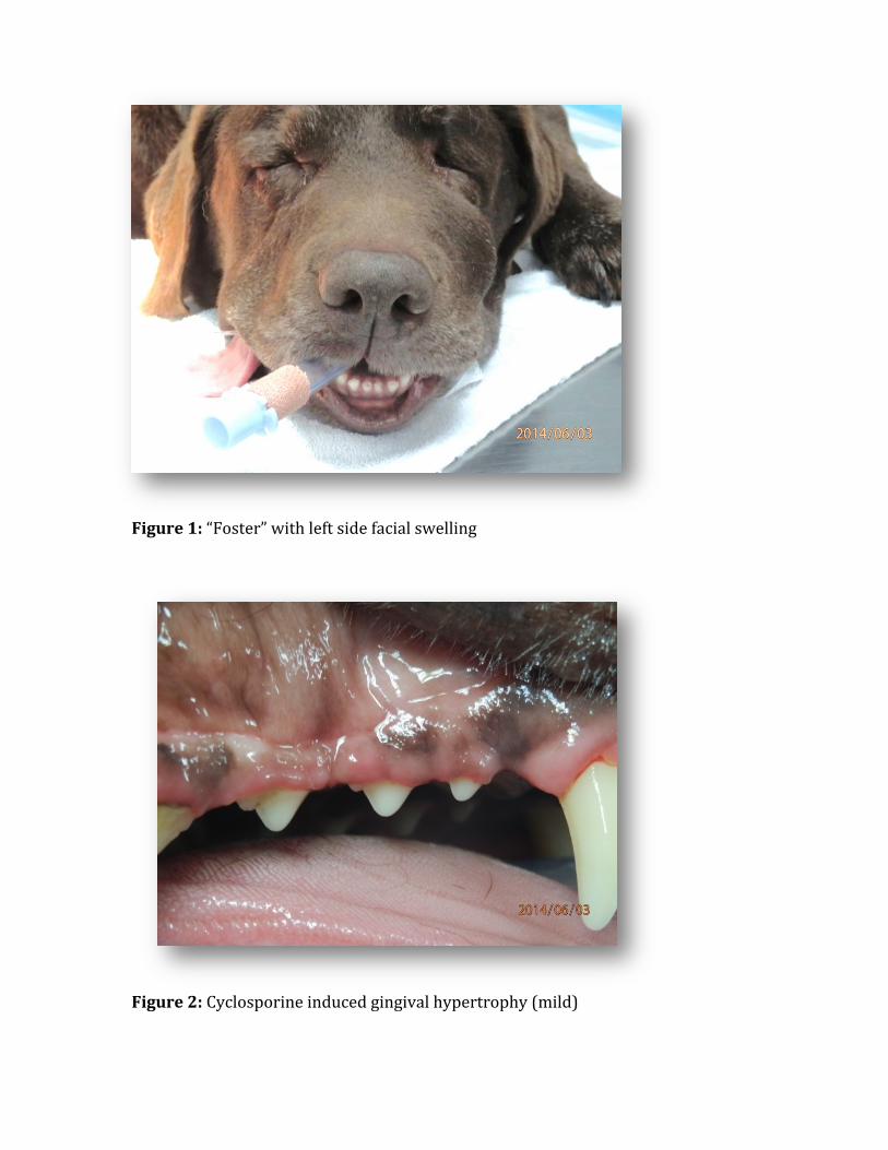
Figure 1: “Foster” with left side facial swelling
Figure 2: Cyclosporine induced gingival hypertrophy (mild)
Different case For comparison, Figure 9 demonstrates another case involving a dog with true oral disease caused by trauma and pulp necrosis of RHS maxillary 4th premolar (108). Conclusions Sometimes, diagnostic tests such as radiographs are taken to rule out pathology rather than to detect it. The referring veterinarian was to be commended in referring this case. Years ago, when intra-‐oral radiographs were not available, unilateral facial swellings were always assumed to be related to dental pathology unless proven otherwise (often belatedly after the tooth had already been extracted). This lead to a number of healthy teeth being extracted without the resolution of the patient’s presenting complaint. It is my opinion that before too long, a minimum standard of companion animal practice will require veterinarians to either offer intra-‐oral radiographs to their clients or to refer patients with oral disease to a practice that can offer such a service. It is well accepted that without the use of intra-‐oral radiography, a number of feline tooth resorptions will be missed and a number of teeth will have fractured roots left behind after extraction, often leaving the patient in continuing pain and discomfort.
Figure 3: Gingival wart-‐ like lesion LHS apical to tooth 207. Lesion is possibly associated with cyclosporine administration
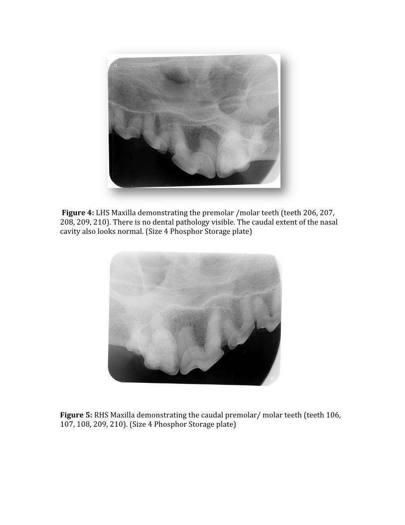
Figure 4: LHS Maxilla demonstrating the premolar /molar teeth (teeth 206, 207, 208, 209, 210). There is no dental pathology visible. The caudal extent of the nasal cavity also looks normal. (Size 4 Phosphor Storage plate) Figure 5: RHS Maxilla demonstrating the caudal premolar/ molar teeth (teeth 106, 107, 108, 209, 210). (Size 4 Phosphor Storage plate)
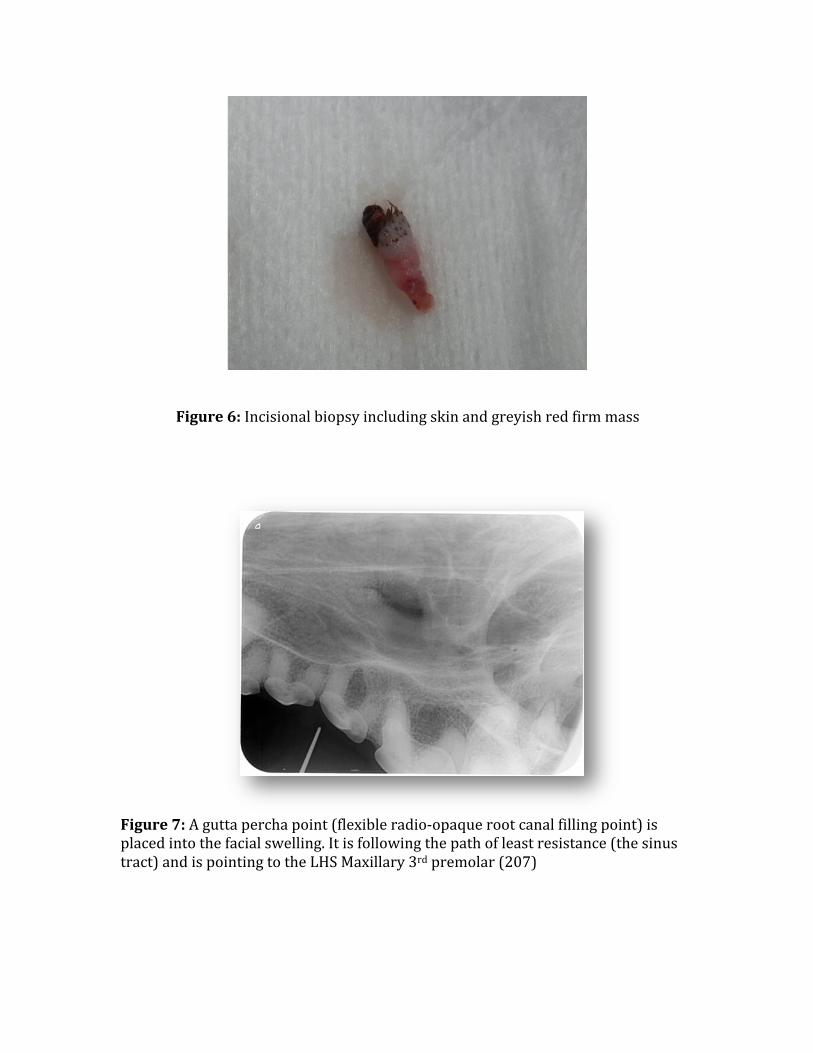
Figure 6: Incisional biopsy including skin and greyish red firm mass
Figure 7: A gutta percha point (flexible radio-‐opaque root canal filling point) is placed into the facial swelling. It is following the path of least resistance (the sinus tract) and is pointing to the LHS Maxillary 3rd premolar (207)
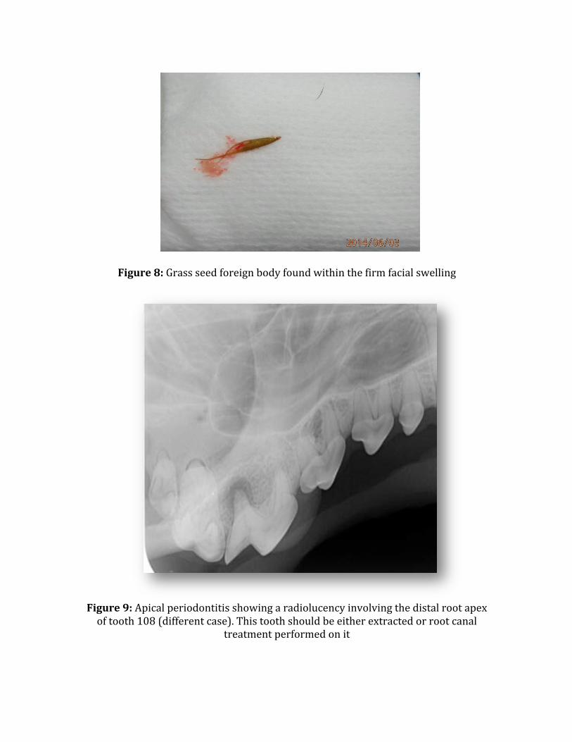
Figure 8: Grass seed foreign body found within the firm facial swelling
Figure 9: Apical periodontitis showing a radiolucency involving the distal root apex of tooth 108 (different case). This tooth should be either extracted or root canal
treatment performed on it
FURTHER AFIELD: ANZCVS REPORT
Current College Veterinary Dentistry Chapter Secretary and fellow AVDS member Dr Kevin Ng sends this report from “Science Week” – 3 days of advanced dental education in small animal and equine topics. Interested vets are more than welcome to attend any of the sessions regardless of whether they have attained Membership to the College. If you are looking for more, this is the place to start in 2015!
ANZCVS Science Week 2014 Report
Science Week 2014 was held on the Gold Coast from 10-12 July. This year marked 21 years since the creation of the Dentistry Chapter of the Australian and New Zealand College of Veterinary Scientists. In honour of this, Dr. Rod Salter BVSc MANZCVS coordinated a fantastic 3-day program covering multiple aspects of both companion animal and equine dentistry. 21st birthday celebrations were also conducted in a more social setting at the Chiangmai Thai restaurant in Surfer’s Paradise with a delicious 5-course banquet. The lecture program this year included a collaborative day-long session with the Emergency, Critical Care and Anaesthesia Chapter, a keynote presentation on rotary endodontics by Dr. Ross Applegarth BDS MDSc FICD MRACDS (Endo), and the inaugural Dentistry Chapter workshop on rotary endodontics conducted by Dr. David Clarke BVSc DAVDC FAVD MANZCVS. We are lining up another exciting program from 9-11 July 2015. Details and registration forms for Science Week 2015 will be available on the College website at www.anzcvs.org.au in due time and registration is open to all veterinarians. The Australian and New Zealand College of Veterinary Scientists provides professional qualifications to veterinarians with superior competence. Entry to the College is by examination. If you are interested in becoming a Member or Fellow of the College, relevant information can be found on the College website at www.anzcvs.org.au.
Anaesthesia free dentistry – Why is it an issue and what is the AVDS going to do about it? by Dr Amanda Huland-Naves Some of you may have come across services that are offered by lay dentists and groomers call “Anaesthesia-free dentistry”. The marketing of these services prey on the public’s unnecessary fear of anaesthesia for their pets and also perpetuates the myth that dentistry is merely a cosmetic procedure. Anaesthesia-free dentistry has been made illegal in California and there has been a recent conviction of a lay dentist there after performing anaesthesia-free dentistry on small animal patients on the grounds that it constituted animal cruelty. So, what is all the fuss about? Well, there are several risks posed by anaesthesia-free dentistry. Risks to the patient :
" Pain from sharp instruments " Inadequate pain relief (maxillary nerve blocks can only be administered
under general anaesthetic and local anaesthetics, general anaesthetics and pain relief drugs are all Schedule 4 drugs unavailable to lay people)
" Stress from excessive physical restraint (if no chemical restraint is used) " Risk of serious injury from movements due to stress and sharp
instruments " Risk of airway compromise due to large amounts of water being used to
cool instruments and flush away tartar. Dogs cannot be trained to spit this water out
" Inadequate diagnosis of periodontal disease as intraoral radiography cannot be performed in a conscious animal and the technician needs to hold a radiation licence to perform radiography (70% of dogs >3 years old have periodontal disease as their primary dental disease and this is a disease of the tissues BELOW the gum line)
" Ongoing pain, periodontal disease and resultant organ disease due to inadequate diagnosis and treatment
Risk to the technician :
" Risk of causing serious injury from trauma inflicted by the patient (due to a combination of stress, fear and pain). This is a considerable Occupational Health and Safety risk to the technician.
Risk to the owner : " A false sense of security that they have adequately facilitated the
treatment of their pet’s dental disease " Paying for a service that has failed to deliver the promised outcome
What protections are in place? In states where there are defined “Acts of Veterinary Science”, it can be argued that use of ultrasonic equipment, especially once it is inserted below the gum is in breach of the act. In states where there are no defined “Acts” within the legislation, there is nothing to prevent this occurring. The Victorian Veterinary Practitioner’s Board has stated that it is powerless to act unless these lay people are misrepresenting themselves as veterinarians. Currently lay people cannot access licences to hold and use radiation equipment or S4 or S8 drugs. They have avoided the need to hold pharmaceuticals by not using them and creating a marketing scheme that makes this a “favourable” thing, even though we know it to be cruel and inadequate. By making the case for intraoral radiography to be the minimum standard amongst the veterinary practitioners performing veterinary dentistry (and there is a lot of science to support this as the right thing to do) we will help to educate the public as this being a point of difference. Again, without “Acts of Veterinary Science” there is no regulation authority to enforce though. So what is the AVDS going to do?
• The committee is currently reviewing the guidelines for small animal dentistry, emphasising the need for general anaesthesia and intraoral radiography to perform adequate small animal dentistry.
• The committee is planning a position statement on anaesthesia free
dentistry in small animals.
• The AVA Victoria Division is using dentistry as a classic example for
lobbying the Victorian State Government and Opposition in the lead up to the state election for the return of “Acts of Veterinary Science”. The change in this legislation should also go some way in benefitting equine dental veterinarians.
• We also intend to lobby each state government in turn as their Veterinary
Surgeons’ Acts come up for review
We hope you have enjoyed this edition of the AVDS Newsletter!
If you have any feedback or
have ideas on member services that you would like to see, feel free to contact one of
the committee.
Dr Rod Salter – [email protected] Dr Tara Cashman – [email protected] Dr Amanda Hulands-Nave – [email protected]