autotransplant tissue selection criteria with or … · autotransplant tissue selection criteria...
TRANSCRIPT
B
O
Aso
MAO
a
Sb
(c
RA
c2
h1r
raz J Otorhinolaryngol. 2014;80(4):318---324
Brazilian Journal of
OTORHINOLARYNGOLOGYwww.bjorl.org
RIGINAL ARTICLE
utotransplant tissue selection criteria with or withouttereomicroscopy in parathyroidectomy for treatmentf renal hyperparathyroidism�
onique Nakayama Ohea,∗, Rodrigo Oliveira Santosb, Murilo Catafesta das Nevesb,luizio Barbosa Carvalhoc, Ilda Sizue Kunii a, Márcio Abrahãob,nivaldo Cervantesb, Marise Lazaretti-Castroa, José Gilberto Henriques Vieiraa
Department of Endocrinology and Metabology, Escola Paulista de Medicina, Universidade Federal de São Paulo (EPM-UNIFESP),ão Paulo, SP, BrazilDepartment of Otorhinolaryngology, Head and Neck, Escola Paulista de Medicina, Universidade Federal de São Paulo
EPM-UNIFESP), São Paulo, SP, BrazilDepartment of Nephrology, Escola Paulista de Medicina, Universidade Federal de São Paulo (EPM-UNIFESP), São Paulo, SP, Brazil
eceived 2 July 2013; accepted 24 January 2014vailable online 11 June 2014
KEYWORDSParathyroid hormone;Hyperparathyroidism,secondary;Parathyroidectomy
AbstractIntroduction: Several methods have been proposed to improve operative success in renal hyper-parathyroidism.Objective: To evaluate stereomicroscopy in parathyroid tissue selection for total parathyroidec-tomy with autotransplantation in secondary (SHPT)/tertiary (THPT) hyperparathyroidism.Methods: 118 renal patients underwent surgery from April of 2000 to October 2009. They weredivided into two groups: G1, 66 patients operated from April of 2000 to May of 2005, with tissueselection based on macroscopic observation; G2, 52 patients operated from March of 2008 toOctober 2009 with stereomicroscopy for tissue selection searching for the presence of adiposecells. All surgeries were performed by the same surgeon. Patients presented SHPT (dialysistreatment) or THPT (renal-grafted). Follow-up was 12---36 months. Intra-operative parathyroidhormone (PTH) was measured in 100/118 (84.7%) patients.Results: Data are presented as means. G1 included 66 patients (38 SHPT, 24 females/14 males;40.0 years of age; 28 THPT, 14 females/14 males; 44 years of age). G2 included 52 patients (29SHPT, 11 females/18 males; 50.7 years of age; 23 THPT, 13 females/10 males, 44.4 years of
age). SHPT patients from G2 presented preoperative serum calcium higher than those of SHPTpatients in G1 (p < 0.05), suggesting a more severe disease. Definitive hypoparathyroidism wasfound in seven of 118 patients (5.9%). Graft-dependent recurrence occurred in four patients,two in each group. All occurred in dialysis patients.� Please cite this article as: Ohe MN, Santos RO, Neves MCd, Carvalho AB, Kunii IS, Abrahão M, et al. Autotransplant tissue selectionriteria with or without stereomicroscopy in parathyroidectomy for treatment of renal hyperparathyroidism. Braz J Otorhinolaryngol.014;80:318---24.∗ Corresponding author.
E-mail: [email protected] (M.N. Ohe).
ttp://dx.doi.org/10.1016/j.bjorl.2014.05.012808-8694/© 2014 Associacão Brasileira de Otorrinolaringologia e Cirurgia Cérvico-Facial. Published by Elsevier Editora Ltda. All rightseserved.
Autotransplant tissue selection criteria with or without stereomicroscopy 319
Conclusion: Stereomicroscopy in SHPT/THPT surgical treatment may be a useful tool to stan-dardize parathyroid tissue selection.© 2014 Associacão Brasileira de Otorrinolaringologia e Cirurgia Cérvico-Facial. Published byElsevier Editora Ltda. All rights reserved.
PALAVRAS-CHAVEHormônioparatireóideo;Hiperparatireoidismosecundário;Paratireoidectomia
Influência do critério de selecão de tecido paratiroideano com ou semestereomicroscopia para autoimplante no resultado do tratamento cirúrgico dohiperparatiroidismo associado a doenca renal crônica
ResumoIntroducão: Diversos métodos têm sido propostos com intuito de melhorar índices de sucessocirúrgico no tratamento do hiperparatiroidismo associado à doenca renal crônica (DRC).Objetivos: Avaliar uso do estereomicroscópio na selecão de tecido paratiroideano naparatiroidectomia total com autoimplante em pacientes com DRC.Métodos: 118 pacientes DRC operados entre 04/2000-10/2009 foram divididos em: G1-66pacientes operados entre 04/2000-05/2005 cuja selecão de tecido foi realizada por métodoconvencional (macroscopia); G2-52 pacientes operados entre 03/2008-10/2009, cuja selecãode tecido foi realizada com uso da estereomicroscopia: Leica-Stereomicroscope (amplificacão:10×-80×). Pacientes foram ainda categorizados em hiperparatiroidismo secundário (HPS) outerciário (HPT) (HPS-diálise/HPT-transplantados renais). Seguimento pós-operatório: 12-36meses. PTH intraoperatório medido 100/118 pacientes (84.7%). Todos pacientes foram operadospelo mesmo cirurgião.Resultados: Dados em média. G1, 66 pacientes (38 HPS, 24f/14m; 40 anos; 28 HPT, 14f/14m;44 anos). G2, 52 pacientes (29 HPS, 11f/18m; 50,7 anos; 23 HPT, 13f/10m; 44,4 anos). Pacientesdialíticos do G2 apresentaram cálcio pré-operatório maior que G1 (p < 0,05), sugerindo doencamais severa. Hipoparatiroidismo definitivo: 7/118 (5,9%) pacientes: G1, 4/66 (6%); G2, 3/52(5,7%). Recorrência do hiperparatiroidismo no autoimplante: 4 pacientes, 2 em cada grupo.Todas foram em pacientes em diálise.Conclusão: Estereomicroscopia no tratamento do hiperparatiroidismo associado à DRC é útil napadronizacão da técnica de selecão de tecido para o autoimplante.© 2014 Associacão Brasileira de Otorrinolaringologia e Cirurgia Cérvico-Facial. Publicado porElsevier Editora Ltda. Todos os direitos reservados.
uimtfa(ct
tsas
P
P
Introduction
The longer survival of chronic renal patients has increasedthe incidence of symptomatic hyperparathyroidism, increas-ing the need of surgical procedure.1 The best surgicalapproach for renal hyperparathyroidism is still debated andcontroversy remains. Relevant questions have been raisedregarding treatment election, since post-surgical recurrenceand the risk of definitive hypoparathyroidism should beavoided. Total parathyroidectomy with parathyroid tissueautotransplantation is a well-accepted technique for themanagement of these patients.2---7 Since recurrence ratesof 8%8 to 20%9 and up to 76%10 have been reported inthe literature, the great majority graft-dependent, tissueselection for autotransplantation has been presented asa considerable challenge. Analysis of parathyroid hyper-plastic tumors, using X-chromosome inactivation, havedemonstrated that monoclonal transformation is a frequentoccurrence in uremic patients.11 In fact, the great major-ity (64%) of parathyroid glands with generalized hyperplasia(non-nodular component) were unequivocally monoclonal,
as demonstrated by Arnold et al.11 This finding imposes adifficult condition in parathyroid tissue selection for auto-transplantation.1io
Intraoperative tissue selection of parathyroid fragmentssing a stereomagnifier was first described by Neyer et al.n 2002.12 The technique can differentiate parathyroid nor-otropic areas by the presence of stromal fat cells from
hose that are disfunctional and hyperplastic tissue withoutat cells.12 Cells from these lipid-containing areas presentn optimal in vitro suppression of parathyroid hormonePTH) secretion by high calcium levels,13 indicating a normalalcium set-point and suggesting the eligibility for auto-ransplantation.
The aim of this study was to compare stereomicroscopyo conventional macroscopic observation in parathyroid tis-ue selection using a historical control, in order to provide
standardized procedure for renal hyperparathyroidismurgery.
atients and methods
atients
18 renal patients underwent total parathyroidectomy withntramuscular presternal autotransplantation7 from Aprilf 2000 to October of 2009. Patients were followed-up

3
awcpoarsmo
cofttotaGSt1
pcsaSwopwplttp
caeftDmpu
l(eg
fOwswasa
M
S
Temg
ess
S
Beii
iframssglpncartag
P
Gaorpp
uMm5mfc
B
20
t the Renal Osteodystrophy Unit in this institution andere referred to surgical treatment for persistent hyper-alcemia not responsive to medical interventions and/orersistent hyperphosphatemia despite the continued usef dietary phosphorus restriction and phosphate-bindinggents, with signs and symptoms such as intractable pru-itus, severe bone pain, fractures or high risk of fracture,keletal deformities, extra skeletal calcifications, develop-ent of calciphylaxis, and radiographic evidences of renal
steodystrophy.Patients were divided into two groups: group 1 (G1)
onsisted of 66 patients who underwent surgery from Aprilf 2000 to May of 2005, and parathyroid tissue selectionor autotransplantation was performed using conventionalechnique based on macroscopic parathyroid tissue observa-ion by a single experienced surgeon; group 2 (G2) consistedf 52 patients who underwent surgery from March of 2008o October of 2009, and parathyroid tissue selection forutotransplantation was performed by the same surgeon as1, based on stereomicroscopy observation using a Leicatereo Zoom S8 APO Stereomicroscope (Leica Microsys-ems GmbH --- Wetzlar, Germany), with magnification of0---80×.
Patients were classified as secondary or tertiary hyper-arathyroidism: secondary hyperparathyroidism (SHPT) washaracterized as an acquired disorder observed in end-tage renal disease, in which the uremic state presents
continuous stimulus to the parathyroid glands. TheHPT group included patients under dialysis treatmentho presented severe hyperparathyroidism with normalr high serum calcium levels. The tertiary hyper-arathyroidism (THPT) group comprised renal patientsith functioning kidney transplant and nonsuppressiblearathyroid hyperplasia, with persistent increased PTHevels and hypercalcemia. Hypercalcemia after kidneyransplantation is usually due to hyperparathyroidismhat persists from the preceding chronic kidney diseaseeriod.14
Surgical cure was defined as restoration of serum cal-ium and PTH levels14 throughout the first six monthsfter surgery. Recurrence was defined when high lev-ls of PTH were observed throughout late post-operativeollow-up (one year after surgical procedure) that failedo respond to medical/pharmacological management.efinitive hypoparathyroidism was defined when PTHeasurements under 10 pg/mL endured one year afterarathyroidectomy, with normal or low serum calcium levelsnder vitamin D and oral calcium supplementation.
Serum ionized calcium (iCa), phosphorus (P), alka-ine phosphatase (AP), and intact parathyroid hormoneiPTH) were measured before parathyroidectomy andvery six months after surgery in all patients from bothroups.
Regarding the follow-up, patients from both groups wereollowed-up after surgery on a regular basis in the Renalsteodystrophy Unit, and patient data from medical reportsere available for this study. Patients from G1 underwent
urgery from 2000 to 2005, while patients from G2 under-
ent surgery from 2008 to 2009. Thus, for surgical outcomenalysis, the first 36 months were the period selected fortudy in both groups, since it was the longest period avail-ble for G2 patients.Ipo
Ohe MN et al.
ethods
tudy design
his was a comparative study using a historical control,valuating stereomicroscopy in comparison to conventionalacroscopic technique in parathyroid tissue selection in sur-
ical treatment of renal hyperparathyroidism.This investigation was approved by the institutional
thics committee (approval No. CEP 0234/06) and patientsigned an informed consent prior to their inclusion in thetudy.
urgical strategy
ilateral cervical exploration with at least four-glandxcision confirmed by frozen section examination and/orntra-operative PTH (IO-PTH) measurement was performedn all 118 patients (IO-PTH available in 100 patients).
Removed parathyroid glands were carefully examinedn order to select a parathyroid area (non-nodular region)or implant and cryopreservation. The selected parathy-oid fragment was gently diced into small pieces measuringpproximately 2.0 mm3. Approximately 30 parathyroid frag-ents were implanted in presternal musculature over a
ingle area of 1.5 cm in length.7 Another 30 fragments wereelected for cryopreservation. The reasons for changing therafted area from the forearm to the presternal muscu-ature are related to some potential advantages. First, itreserves the forearm for arterio-venousus-fistula dialysis ifeeded. Second, the presternal region is close to the cervi-otomy, exposing the same surgical area. Third, it allows forn easier graft-removal in case of graft-dependent recur-ence; the sternal bone presents a posterior boundary forhe grafted tissue, enabling a graft removal under localnestesia.7 All surgeries were performed by the same sur-eon in both groups G1 and G2.
rotocol for intraoperative tissue selection
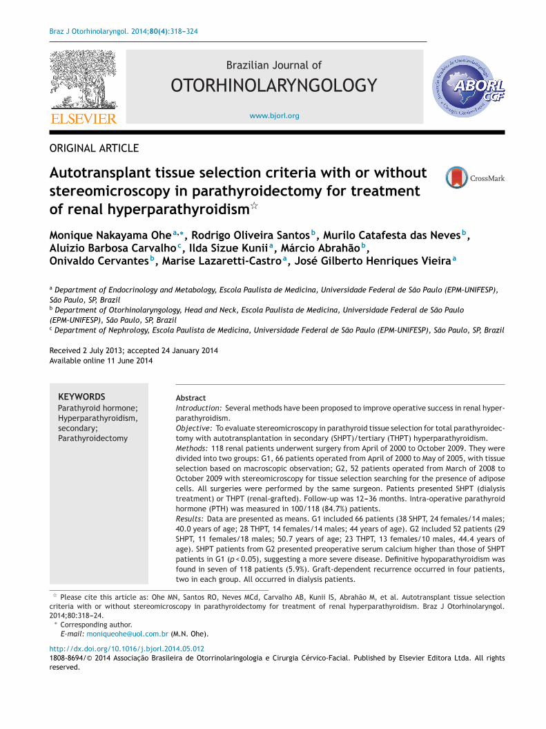
roup 1 --- Conventional technique: tissue selection forutotransplantation based on macroscopic parathyroidbservation was performed in 66 patients. Nodular parathy-oid regions were avoided, while hyperplasic non-nodulararathyroid areas were considered eligible for autotrans-lantation (Fig. 1).
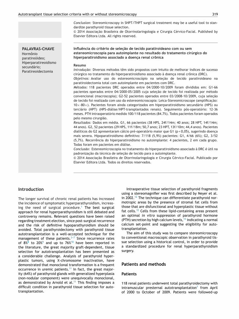
Group 2 --- Stereomicroscopy: parathyroid tissue selectionsing Leica Stereo Zoom S8 APO Stereomicroscope (Leicaicrosystems GmbH --- Wetzlar, Germany) with apochro-atic 8:1 zoom, 10---80× magnification, was performed in
2 patients. Resected parathyroid glands were first observedacroscopically, and stereomicroscopic analysis was per-
ormed thereafter, searching for the presence of stromal fatells in the parathyroid glands (Fig. 2, arrows).
iochemistry
O-PTH was performed to confirm total removal of thearathyroid glands, in order to avoid overlooking remainingr supernumerary hyperfunctioning glands. IO-PTH was

Autotransplant tissue selection criteria with or without stereomicroscopy 321
Figure 1 Macroscopic parathyroid tissue observation: A, nodular parathyroid regions not eligible for autotransplantation; B,hyperplasic non-nodular parathyroid area eligible for autotransplantation.
Figure 2 Stromal fat cells (arrows)
in resected parathyroid glands.
322 Ohe MN et al.
Table 1 Pre-operative data and laboratory findings.
G1 (n = 66) G2 (n = 52)
SHPT THPT SHPT THPT
n 38 28 29 23Age 40.0 (14---58) 44.0 (24---62) 50.7 (26---74) 44.4 (28---63)Female/male 24/14 14/14 11/18 13/10Years on dialysis 7.7 (0.6---16) 6.5 (2---20) 7.9 (3---25) 6.4 (0.8---13)Years of renal graft --- 2.8 (0.5---7) --- 3.3 (0.4---10)Pre-op iCa 1.36a (1.19---1.83) 1.59 (1.42---1.77) 1.41a (1.21---1.59) 1.58 (1.41---1.90)Pre-op iPTH 1618.7 (298---2500) 442.2 (109---1758) 1810.2 (561---3500) 465.8 (181---2237)
Reference values: iCa = 1.20---1.40 mmol/L; iPTH = 10---65 pg/mL.Data expressed as mean and range.SHPT, secondary hyperparathyroidism; THPT, tertiary hyperparathyroidism; iCa, serum ionized calcium; iPTH, intact parathyroid hor-mone.
a t-test: p < 0.05.
Table 2 Post-operative mean iCa and iPTH in cured patients in G1-group (conventional).
SHPT THPT
iCa iPTH iCa iPTH
12 months (n = 34) 1.17 79.8 (n = 25) 1.25 58.424 months (n = 29) 1.22 86.0 (n = 19) 1.22 64.736 months (n = 21) 1.19 83.0 (n = 16) 1.22 78.5
Reference values: iCa = 1.20---1.40 mmol/L; iPTH = 10---65 pg/mL.Data expressed as means.
hyroi
mSfaPir
psieGl
(1
h
R
P
SHPT, secondary hyperparathyroidism; THPT, tertiary hyperparatmone.
easured using the Elecsys PTH Immunoassay (Elecsys 1010ystem; Roche --- Mannheim, Germany) and was availableor 100 patients (84.7%). The time required to perform thessay is nine minutes and reference values are 10---70 pg/mL.eripheral venous blood sample (4.0 mL) was obtainedmmediately after induction of anesthesia and 20 min afteremoval of all parathyroid glands.15---17
Total serum calcium, phosphorus, total alkaline phos-hatase, and creatinine were measured by means oftandard automatic assays (Hitachi 912 --- Roche). Serum
onized calcium was measured by using an ion-specificlectrode (AVL 9180 Electrolyte Analyzer --- Roswell,eorgia, United States). Parathyroid hormone in ambu-atory conditions was measured by immunometric assay
T
ds
Table 3 Post-operative mean iCa and iPTH in cured patients in G
SHPT
iCa iPT
12 months (n = 26) 1.15 624 months (n = 18) 1.09 1136 months (n = 13) 1.13 12
Reference values: iCa = 1.20---1.40 mmol/L; iPTH = 10---65 pg/mL.Data expressed as mean.SHPT, secondary hyperparathyroidism; THPT, tertiary hyperparathyroimone.
dism; iCa, serum ionized calcium; iPTH, intact parathyroid hor-
Immulite; Siemens --- São Paulo, Brazil: reference values0---65 pg/mL).
All removed parathyroid tissue underwent completeistopathology analysis.
esults
re-operative data and laboratory findings are depicted in
able 1.Mean post-operative iPTH and iCa serum measurementsuring the follow-up of cured patients in both groups arehown in Tables 2 and 3.
2-group (stereomicroscopy).
THPT
H iCa iPTH
1.3 (n = 21) 1.20 60.83.2 (n = 14) 1.18 65.05.4 (n = 9) 1.22 80.1
dism; iCa, serum ionized calcium; iPTH, intact parathyroid hor-

Autotransplant tissue selection criteria with or without stereomicroscopy 323
Table 4 Graft-dependent recurrence in patients from G1 (2/66) --- conventional technique, and from G2 (2/52) ---stereomicroscopy.
12 months 24 months 36 months
iCa iPTH iCa iPTH iCa iPTH
Patient 1 (G1) 1.46 13 --- --- 1.29 1165Patient 2 (G1) 0.90 22 1.29 104 1.56 168Patient 3 (G2) 1.49 70 1.40 717 1.49 940Patient 4 (G2) 1.21 224 0.97 1259 --- ---
Reference values: iCa = 1.20---1.40 mmoL/L; iPTH = 10---65 pg/mL.
stsTNtrp
dottri
ot
tiitpnap
iptatlocw
C
St
iCa, serum ionized calcium; iPTH, intact parathyroid hormone.
Persistent hyperparathyroidism due to supernumeraryparathyroid glands not recognized during surgical procedurewas observed in two patients, one from each group; thosepatients were excluded from the study.
Definitive hypoparathyroidism was observed in 7/118(5.9%) patients, 4/66 (6%) in G1, and 3/52 (5.7%) in G2.
Graft-dependent recurrence was observed in 4/118(3.3%) patients, two in each group (G1, 2/66; 3.0% andG2, 2/52; 3.8%) (Table 4). All graft-dependent recurrenceswere observed in dialysis patients. None occurred in kidney-grafted patients.
Discussion
Surgery for renal hyperparathyroidism is performed in Brazilin relatively few medical centers, sometimes leading to longwaiting times and, consequently, to worse clinical condi-tions at the time of surgery. Throughout the years, theauthors have observed a worsening in medical conditionsamong renal patients at the time of surgery, when com-paring patients from G1 (surgeries performed from 04/2000to 05/2005) to G2 (surgeries performed from 03/2008 to10/2009). As observed in SHPT patients from G2, serum cal-cium levels were higher than those seen in SHPT patientsfrom G1 (t-test; p < 0.05). This finding is in agreement withpatients with a more severe disease and, moreover, theseare indicators that patients in G2 underwent surgery ina worse condition. Thus, the apparently similar surgicalresults in graft-dependent recurrence observed between G1and G2 (two recurrences in each group) could be assessed asa positive outcome for stereomicroscopy. The more severedisease observed in G2 may be the explanation for thefinding of a similar recurrence in both groups. Since themacroscopic examination protocol used in G1 was performedby the same surgeon that also supervised the stereomi-croscopy in G2, a fair comparison can be performed.
From the 118 overall patients, graft-dependent recur-rence was observed only in four (3.3%). This can beconsidered a low graft-dependent recurrence rate com-pared to literature data.18---20 Considering the gravity ofthe illness observed in these patients, the experience ofthe surgeon probably contributed to such a positive out-
come.The clearly abnormal macroscopic findings observedin removed parathyroid indicate the severity of the ill-ness and impose an especially difficult challenge in tissue
siAt
election. The effort for finding eligible areas for auto-ransplantation among abnormal parathyroid glands makestereomicroscopy an interesting tool for its standardization.he search for stromal fat cells as previously described byeyer et al.12 may provide a reproducible technique forissue selection in attempts to reach low graft-dependentecurrences rates, even for surgeons not so experienced inarathyroid surgeries.
Another important observation is that all graft-ependent recurrences were in dialysis patients. Noneccurred in kidney-grafted patients. It is probable thathe continuous exposure of the grafted parathyroid tissueo uremic environment may be the main driver for tumorecurrence in patients under long-term dialysis treatment,ndependent of the surgical technique employed.
Post-operative PTH measurements during the follow-upf cured patients were similar in G1 and G2, as were defini-ive hypoparathyroidism rates.
As mentioned above, persistent hyperparathyroidism dueo supernumerary parathyroid glands not recognized dur-ng surgical procedure was observed in two patients, onen each group and those patients were excluded fromhe study. Occurrence of supernumerary and/or ectopicarathyroid glands is more frequent among chronic kid-ey failure patients,21 and this presents an important anddditional challenge in surgical treatment of renal hyper-arathyroidism.
Limitations are related to the heterogeneous character-stics of renal patients enrolled in this study, with theirarticular clinical and laboratory findings, and consideringhem as a group presents a real challenge, especially when
historical control is used. Another limitation is related tohe severity of these patients: the marked illness and long-asting renal condition makes them a special group, whoseutcome would not be expected in ordinary patients withhronic renal disease. In addition, levels of 25-OH vitamin Dere not available in this study.
onclusion
tereomicroscopy in renal hyperparathyroidism surgicalreatment may provide a standardized parathyroid tissue
election for autotransplantation, and is potentially helpfuln obtaining a low incidence of graft-dependent recurrence.dditional controlled and randomized studies are necessaryo confirm these results.
3
F
T
C
T
R
1
1
1
1
1
1
1
1
1
1
2
24
unding
his study was supported by FAPESP: N◦ 07/51056-5.
onflicts of interest
he authors declare no conflicts of interest.
eferences
1. Monchik JM, Bendinelli C, Passero Jr MA, Roggin KK. Sub-cutaneous forearm transplantation of autologous parathyroidtissue in patients with renal hyperparathyroidism. Surgery.1999;126:1152---8.
2. Wells Jr SA, Gunnells JC, Shelburne JD, Schneider AB, SherwoodLM. Transplantation of the parathyroid glands in man: clinicalindications and results. Surgery. 1975;78:34---44.
3. Wells Jr SA, Ross 3rd AJ, Dale JK, Gray RS. Transplantationof the parathyroid glands: current status. Surg Clin North Am.1979;59:167---77.
4. Packman KS, Demeure MJ. Indications for parathyroidectomyand extent of treatment for patients with secondary hyper-parathyroidism. Surg Clin North Am. 1995;75:465---82.
5. Sokoll LJ, Drew H, Udelsman R. Intraoperative parathyroid hor-mone analysis: a study of 200 consecutive cases. Clin Chem.2000;46:1662---8.
6. Weber KJ, Misra S, Lee KJ, Wilhelm SW, DeCresce R, PrinzRA. Intraoperative PTH monitoring in parathyroid hyperplasiarequires stricter criteria for success. Surgery. 2004;136:1154---9.
7. Santos RO, Ohe M, Carvalho ABC, Neves MC, Kunii I, LazarettiCastro M, et al. Total parathyroidectomy with presternal intra-muscular autotransplantation in renal patients: a prospectivestudy of 66 patients. J Osteoporos. 2012;2012 [serial online],Article ID 631243, 6 pp.
8. Tominaga Y, Uchida K, Haba T, Katayama A, Sato T, Hibi Y, et al.More than 1,000 cases of total parathyroidectomy with fore-arm autograft for renal hyperparathyroidism. Am J Kidney Dis.2001;38 4 Suppl. 1:S168---71.
9. Tominaga Y, Matsuoka S, Sato T. Surgical indications and pro-cedures of parathyroidectomy in patients with chronic kidney
disease. Ther Apher Dial. 2005;9:44---7.0. Hampl H, Steinmuller T, Stabell U, Klingenberg HJ, SchnoyN, Neuhaus P. Recurrent hyperparathyroidism after totalparathyroidectomy and autotransplantation in patients with
2
Ohe MN et al.
long-term hemodialysis. Miner Electrolyte Metab. 1991;17:256---60.
1. Arnold A, Brown MF, Urena P, Gaz RD, Sarfati E, Drüek TB.Monoclonality of parathyroid tumors in chronic renal fail-ure and in primary parathyroid hyperplasia. J Clin Invest.1995;95:2047---53.
2. Neyer U, Hoerandner H, Haid A, Zimmermann G, NiederleB. Total parathyroidectomy with autotransplantation inrenal hyperparathyroidism: low recurrence rates after intra-operative tissue selection. Nephrol Dial Transplant. 2002;17:625---9.
3. Niederle B, Horandner H, Roka R, Woloszczuk W. Morphologicand functional studies to prevent graft-dependent recurrencein renal osteodystrophy. Surgery. 1989;106:1043---8.
4. Kidney Disease: Improving Global Outcomes (KDIGO) CKD-MBDWork Group: KDIGO clinical practice guideline for the diagno-sis, evaluation, prevention and treatment of chronic kidneydisease-mineral and bone disorder (CKD-MBD). Kidney Int Suppl.2009;(113):S1---130.
5. Ohe MN, Santos RO, Kunii IS, Carvalho AB, Abrahão M, CervantesO, et al. Usefulness of a rapid immunometric assay for intraop-erative parathyroid hormone measurements. Braz J Med BiolRes. 2003;36:715---21.
6. Ohe MN, Santos RO, Kunii IS, Carvalho AB, Abrahão M, CervantesO, et al. Usefulness of intraoperative PTH measurement in pri-mary and secondary hyperparathyroidism: experience in 109patients. Arq Bras Endocrinol Metabol. 2006;50:869---75.
7. Ohe MN, Santos RO, Kunii IS, Carvalho AB, Abrahão M, Neves MC,et al. Intraoperative PTH cutoff definition to predict successfulparathyroidectomy in secondary and tertiary hyperparathy-roidism. Braz J Otorhinolaryngol. 2013;79:494---9.
8. Gagné ER, Urena P, Leite-Silva S, Zingraff J, Chevalier A, SarfatiE, et al. Short- and long-term efficacy of total parathyroidec-tomy with immediate autografting compared with subtotalparathyroidectomy in hemodialysis patients. J Am Soc Nephrol.1992;3:1008---17.
9. Higgins RM, Richardson AJ, Ratcliffe PJ, Woods CG, Oliver DO,Morris PJ. Total parathyroidectomy alone or with autografty forrenal hyperparathyroidism? Q J Med. 1991;79(288):323---32.
0. Tominaga Y, Nuomano M, Tanaka Y, Uchida K, Takagi H. Surgi-cal treatment of renal hyperparathyroidism. Semin Surg Oncol.1997;13:87---96.
1. Montenegro FL, Tavares MR, Cordeiro AC, Ferraz AR, Ianhez LE,Buchpigel CA. Intrathyroidal supernumerary parathyroid glandin hyperparathyroidism after renal transplantation. NephrolDial Transplant. 2007;22:293---5.