automated perimetry in detecting threats to fixation
TRANSCRIPT
Automated Perimetry in petecting Threats to Fixation
Lili Zhang, MD,I.2 Stephen M. Drance, MD, 1 Gordon R. Douglas, MD 1
Purpose: To study in glaucoma patients the threat to fixation on a Humphrey field analyzer program 10-2 when one of the four innermost paracentral points is defective on program 30-2.
Methods: Forty-five eyes of 45 patients with chronic open-angle glaucoma in whom at least one of the innermost four defective paracentral points was reproducibly defective on program 30-2 of the Humphrey perimeter, with a size 3 target, on two consecutive tests, were studied with program 10-2.
Results: Of the 45 eyes with an abnormal paracentral point of program 30-2, 30 (66%) also showed involvement of a paracentral point on program 10-2, which was considered a threat to fixation. The remaining 15 were considered not to threaten fixation imminently.
Conclusions: In about one third of glaucomatous fields considered to threaten fixation on the standard programs 30-2 and 24-2, the threat was not imminent. The extra evaluation is therefore useful before making radical and precipitous changes in management of the disease. Ophthalmology 1997; 104:1918-1920
Automated threshold perimeters allow quantification and follow-up of patients with glaucoma. 1
-3 Localized visual
field defects is one of the features of glaucomatous damage. These scotomas may develop away from fixation, particularly when the inferior field is involved exclusively. These scotomas, when they enlarge, may move peripherally or toward the center, in which case they may ultimately threaten fixation. Other scotomas may arise close to fixation from the onset, which is more characteristic in localized defects involving the upper hemifield.
The threat to fixation is an important feature of a scotoma because it may constitute a semiemergency in the treatment of patients to avoid a ''snuff out'' of fixation with consequent reduction in acuity and visual handicap. The current use of the Humphrey program 30-2 or 24-
Originally received: January 6, 1997. Revision accepted: June 9, 1997. 1 Department of Ophthalmology, University of British Columbia, Vancouver, Canada. 2 Department of Ophthalmology, Shanxi Eye Hospital, Taiyuan, China.
Supported in part by Orbis International, New York, New York (LZ).
The authors have no proprietary interest in any instruments or materials used in this investigation.
Reprint requests to Stephen M. Drance, MD, Department of Ophthalmology, 2211 Westbrook Mall, Vancouver, British Columbia, Canada V5T 2B5.
1918
2 as the most common field examination in patients with glaucoma means that a threat of fixation is said to exist when, in the presence of a normal central or foveal threshold, one of the four points closest to the center is involved by the scotoma. These four paracentral points are, in fact, 4.2° from the center.
Previous articles have studied paracentral fixation threats.4
•5 The current study was undertaken to learn about
the implication of the involvement of at least one of the four innermost points by a glaucomatous scotoma.
Materials and Methods
A total of 45 eyes of 45 patients with chronic open-angle glaucoma were studied. At least one of the innermost four defective paracentral points was reproducibly defective on program 30-2 of the Humphrey perimeter, with a size 3 target, on two consecutive tests. Paracentral points were considered defective when their threshold on the total deviation plot had a P < 0.01 chance of being normal and was therefore marked with a solid black box on Statpac 1 of the Humphrey perimeter. Patients with extensive visual field damage were excluded as were patients with aphakia and pseudophakia. If the pupils were less than 3 mm in diameter, a drop of 0.5% mydriacyl was used to dilate them. Patients with retinal pathology were excluded.

Zhang et al · Glaucoma, Threats to Fixation, and Paracentral Perimetry
Sllll.l\6 JU,IIt!fE,£0003!.5AS8 a.INlWTOOXSIZE Ill
Sl;,li£Ct' fU.L TK!W FI»H!Illl~l l!Nli:R til( e2:3'i::Jf'll SWU.I.S llt. III IT£ , 0003!.5AS3 ll!OSf'OTOECKSiii" Ill fi:GH()IT~ COOill 10 rll£ tl:~:Sifll
~ IIXD t 5.58 OS tt< w; f\Plllill7f'£t!R ~.5 111 W ~ msiTm' fU1 nmtl.O iXl&D• 5.58!5 OCX tlG f\PILC1A'ETER4.5Itl W: 2e1S8
1100 ra ~
Fl~lCtii.CS:iS 1/{4
FitStrosEm ttiS <8 <e F~~lf:C~M4
lllSllfl600 ~ FMA: ~IllS?.
r{STTUt: !4:l)
-ll·U-ll-al -Z4-25-22-28·Z'·l1
<8 {3 (8 <8
15 26 19 211 10 -17-18 ·16-16
·29·21·11 -!5-..'I·IZ ·ZS-?2 -l1 -U -5 ·II-' -3
16 ,jJj, I!
l!<llliUTY I\!l\lS :: p( $':
I?. P< ~
• P< n • , , es:
"'-3 -1 -' e 1 -?sa-? ·3 -5 "' -5 ~ -? -' I
-5 ·I -3 8 ·2 -5 r.!TTU!I -2 -~ ·U ·I
tEi'llllllW 0.17. ::.
... .. " . . .
. " ..
~)£l!Jf!El.DT£Sl , " J ,
OJi;i(((f!tll!mS
1(1 -tz.nct , ~ ~s . PS& !Ul£6 P<U : (j I.SJ CE: CPStl Je.3BtE; P ( U.
lljj,
i:IO.l I dJ, dl> 14 IY> N ra ~
FJ):AJII)t~ 9124
F<llfllliEJ!illiSi'li FIUI <EtEJ!illiS I'll
Q.ISTJ!IISOO ·~ ro.u;: )Jill : :
II II <111 <I> 11 Jj, Jj, IS
11 11 ,!J, ,f.,!, ,~, n ,!1, <t <I <8 t <I Z2 .~ 2S 24 zz v n u 2121 Hw~
rrsr 111£ 13=!3
'I l .li~
WOS<I 6ll·~n
z•v ,~, ~I1J11 ~;s
~ .~, '*' v 11 <!l, <!> ~
·14-1
-..'1·15 ·14-15~ .. -ll -31 -31-ll·12.$ ·5 ·12
·~ ·~ '32·19-18 ·II~ ·N
·~'ll -~.:ll'l24 • ·I • ~
~ ·l -1 ·l ·I ·1 ·l ·l ·l ~ -6 .J -l-5 .J -3-5 .J -6 ... -l-3 .J -l-4 -5
.$ .$ ... -4-4 -3 TGTil -4 .J
IIVIAIIIJI
8 . " . ••
. ~ .
. " ~
fl(8:11!liTYS'rmS :: p ( ~
~p ( ~
S P< It . PIES<
-24 -12-11-12-3 -5
.ZS.ZS.ZS-1! ·l ~ ~ ~
-29-29-25 -16-lS-r -s -17
-1! -1! .ZS <1 -1! ·I ·I I ·I ·I
~ i • I I I i I I ~ -J e e -2 a 1-2 a -l-1 1 e • • -1 -2
-5..$-1-1-1 e fll1lWI -1'
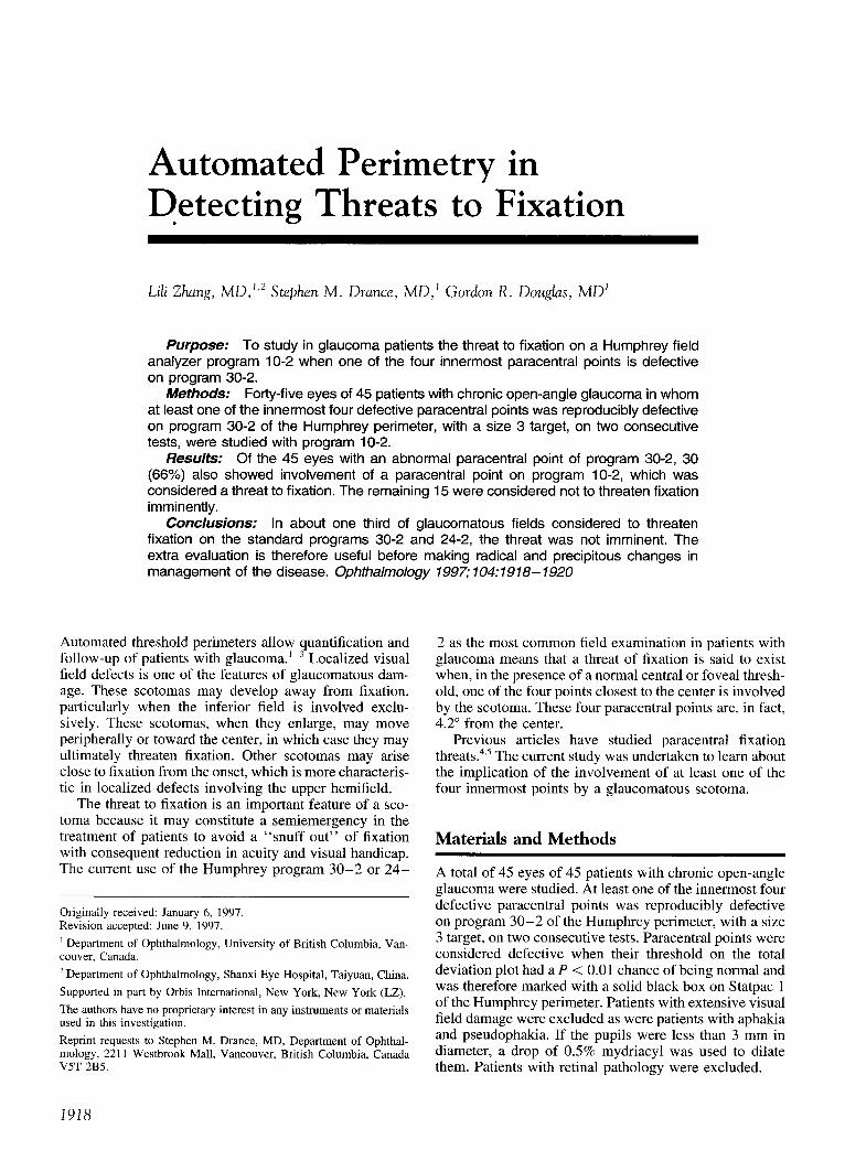
Figure 1. A , a standard Humphrey program 30-2 visual field of a patient with glaucoma indicated one paracentral scotoma point on its total deviation
with P value < 1 o/o and within 2.4° from fixation. B, the Humphrey program 10- 2 visual field of the same patient showed two paracentral scotoma
points within 1.4° from fixation on tom! deviation with the same range of the probability value.
The patients were not selected by their visual acuity nor
foveal sensitivity, but 80% had a visual acuity better than 6/
9 and 91 % had a foveal sensitivity equal to or greater than
32 dB. All patients had a visual field done with program 10-
2, target size 3, on the Humphrey perimeter. All patients had
less than 5% fixation losses as well as false-positive and false
negative responses less than 20% of the time.
Classification and Analysis
The four innermost paracentral points of the total devia
tion plot on program 30-2 are 4.2° from the center. In
program 10-2, they are 1.4° from the center, whereas the
next innermost point is 2.24° from the center.
Results
Of the 45 fields that showed an involvement of at least 1
of the 4 most central paracentral points situated 4.2° from
fixation on program 30-2 of the Humphrey perimeter, 30
(66%) showed involvement of a paracentral point 1.4° from
fixation when tested on program 10-2 and were considered
to have a confirmed threat to fixation (Fig 1). The remaining
15 did not show such a threat. Of the 45 fields, 42 (93%)
had a paracentral scotoma involving 1 of the points 2.24°
from fixation on program 10-2 (Fig 2).
Discussion
The current study shows that when at least one of the
innermost paracentral points of program 30-2 is dis-
turbed and suggests a threat to fixation, almost a third of
patients do not have a disturbance within 1.4° of fixation.
However, in the other two thirds, the scotomas involve
the innermost paracentral points on program 10-2. In the
presence of a 617.5 visual acuity or a foveal threshold
greater than 32 dB, one can presume that fixation, al
though imminently threatened, is not yet affected. Para
central scotomas are well documented as one of the typi
cal early visual field defects of open-angle glaucoma.6•7
The ability to detect threshold changes generally is more
precise in the paracentral area than at the periphery. 8 In
the presence of such a disturbance of the paracentral
points on the commonly used program 30-2 or 24-2, a
program 10-2 or other macular program should be car
ried out to see whether the field defect in fact imminently
threatens fixation. Such a threat may constitute a clinical
urgency that sometimes requires intervention, which may
not be judged as quite as urgent when fixation is less
immediately or not threatened. The current study was not designed to evaluate the
effects of long-term fluctuation on the paracentral visual
field, which might be one explanation for the results ob
tained. However, all patients had many previous visual
field examinations, and the defective innermost paracen
tral involvement was reproducible. Our experience with
the program 10-2 in observing patients with glaucoma,
particularly when advanced disease is excluded, suggests
that when an innermost paracentral point is defective and
indicates a fixation threat, then a detailed examination of
the parafoveal area is indicated. In a third of such patients,
the threat is not as imminent and allows for a nonemergent
approach to their treatment. There are many ways of testing the central 5° to I 0°
1919

Ophthalmology Volume 104, Number 11, November 1997
SIIJUIIi ttt. UUtt. !miG b .S ISS l !Nl 901 OOJ St~ ttl Stll>t!Ct Rll tlfi!90.D
RIOO II;[ ~
f!IOTIIIIU6SIS 4125 fllS!Al>IIIIIISi'IS fllS!IECIIIIIIS l'll IWliii<SASl:DI lOI;
ftMJI' "18 t£S11!1( 16:82
iftl<l 141·1113
·1 4 -1 I ·11-l -3 -1 I
·It ol -1 4 I ·I I l ·IH -7 ol 4 I I ·I~ I
(I <I l 17 ~ <~>
'1 '
1 1L t! ·11-l .J '" l ·II~ -1 -7 I I I 3
-29 -31 -32 .(8 ·II ·11 -4 -$
ol!·J-lJ.IH" TOTIL -21·2:9 ·14-8 ll'I!Rtllll
A
~
ll f'iliiiitiT'f m&l.S :: p ( st
I?.P< ll • ,( 1\ . , ( .~
.a~-31-l1·3 ·16-l -4
-21-l!~HI• ol
ll'IIMIIII
IZ ' .... •••• •••• •••
10 TJI{83:l1:Slf71 £G PIIIl o:A(l[R 4.$ m w 2Va
o..R.Iln11£111fi8.DTES1 1Q!t) OJISIOC!Glll. llKIIS
10 -ll.UCE! P<Ul PSD IU1C8 P<t.St ~ 1.5118 " ~ <P!U tl.~l8 P<U
: .'-l .:: ill · .J.r:. :-o·x ::.~ ~ :..:o,c :K' J£' :::! ::: ;-:~rm =..u ·.;:~:;Q.:
: !·~D•'~l: m·:.il :, .~t: .;.:s :·: X.·
::1);: :.:< ~
~~ ~
!l i!• !j - ~ ' :-; .r: \• ~ ~
?:. =:·~T!')I ~s t<1 r-<::t~~tf'i ~ ~ ·
=:t;r ~; S:KfS i- :~
).t;•:.:JiS~W :!'
=~·9= i£(t ·:::· a~ :::.::
11 x r~ · 2: [t ,;!1 J: :t
Y t .r~:t •: :.: •. !. /i ' ~ ~
1 i 1
~ :: :~ :l i:i :: :; :: ..; .; ·) : ~ ? : : : ·!
.;_; -~ .;: ·! ~ I i .; I ? .; -::-!.: -: ' :! -: .; .;:.: .· .· i.: -:-: .;
.;; ·!= -~: ·~ -~: .;.: ·:·:.. .;~: .:; :r.:~m~~
11. · : r: .. : ~!: : : :
• .z • ·
8
• 11. " ·!. •••• •
I a •! I a a • •
\ :~ ~?, . :~ -~;
:: :. !'. SP 7. • I :· . a; ?.~ .
111 .: ·l ,, I .. . :.·
-. ~ :: :; :; ::: :: ·: :; .:
-: .? -! .;l.; l l -:: l-: -:: ·! ..: .: ! : ~ ·! ·1 -:~ ·= .· . . .: .: .;
(l · . ' . . :: .. , • •• •i .. •••••• ••
Figure 2. A, a standard Humphrey program 30- 2 visual field of a patient with glaucoma indicated one paracentral scotoma point on its total deviation with P value < 1% and within 2.4° from fixation. B, the Humphrey program 10-2 visual field of the same patient showed clear normal four points within 1.4° from fixation on its total deviation plot.
of the visual field. The Humphrey program 10-2 takes the same length of time and effort as does the current program 30-2. It is possible to obtain the same information by using some of the other options available on the perimeter or some programs on other perimeters that would almost certainly provide the same information more expeditiously. It also would be interesting in future studies to find out how long it takes and what the pattern of field change is that finally involves fixation itself.
References
1 . Heijl A, Drance SM. A clinical comparison of three computerized automatic perimeters in the detection of glaucoma defects. Arch Ophthalmol 1981;99:832-6.
2. Trope GE, Britton R. A comparison of Goldmann and Humphrey automated perimetry in patients with glaucoma. Br J Ophthalmol1987;71:489-93.
1920
3. Beck RW, Bergstrom TJ, Lichter PR. A clinical comparison of visual field testing with a new automated perimeter, the Humphrey Field Analyzer and the Goldmann perimeter. Ophthalmology 1985; 92:77-82.
4. Kolker AE. Visual prognosis in advanced glaucoma: a comparison of medical and surgical therapy for 101 retention of vision in eyes with advanced glaucoma. Trans Am Ophthalmol Soc 1977;75:539-55.
5. Lieberman MC, Ewing RH. Reassessing split fixation in advanced glaucoma: preliminary studies with computerized perimetry. In: Mills RP, Heijl A, eds. Perimetry Update 1990/1991. New York: Kugler & Ghedini, 1991;473.
6. Armaly MF. Selective perimetry for glaucomatous defects in ocular hypertension. Arch Ophthalmol1972;87:518-24.
7. Hart WM Jr, Becker B. The onset and evolution of glaucomatous visual field defects. Ophthalmology 1982; 89:268-79.
8. McKee SP, Taylor DG. Discrimination of time: comparison of fovea and peripheral sensitivity. J Opt Soc Am [A] 1984; 1:620-7.