author manuscript nih public access isabelle audo , and ... · crb1; lca; retinitis pigmentosa;...
TRANSCRIPT

CRB1 mutations in inherited retinal dystrophies
Kinga Bujakowska1,2,3, Isabelle Audo1,2,3,4,5, Saddek Mohand-Saïd1,2,3,4, Marie-EliseLancelot1,2,3, Aline Antonio1,2,3,4, Aurore Germain1,2,3, Thierry Léveillard1,2,3, MélanieLetexier6, Jean-Paul Saraiva6, Christine Lonjou7, Wassila Carpentier7, José-AlainSahel1,2,3,4,5,8, Shomi S. Bhattacharya1,2,3,5,9, and Christina Zeitz1,2,3
1INSERM, U968, Paris, F-75012, France2CNRS, UMR_7210. Paris, F-75012, France3UPMC Univ Paris 06, UMR_S 968, Department of Genetics, Institut de la Vision, Paris, F-75012,France4Centre Hospitalier National d’Ophtalmologie des Quinze-Vingts, INSERM-DHOS CIC 503, Paris,F-75012, France5UCL-Institute of Ophthalmology, London, UK6IntegraGen SA, Genopole CAMPUS 1 bat G8 FR-91030 Evry, France7Plateforme Post-génomique P3S, Hôpital Pitié Salpêtrière, Paris, France8Fondation Ophtalmologique Adolphe de Rothschild, Paris, France9Department of Cellular Therapy and Regenerative Medicine, Andalusian Centre for MolecularBiology and Regenerative Medicine (CABIMER), Isla Cartuja, Seville, Spain
AbstractMutations in the CRB1 gene are associated with variable phenotypes of severe retinal dystrophies,ranging from Leber Congenital Amaurosis (LCA) to rod-cone dystrophy (also called retinitispigmentosa (RP)). Moreover, retinal dystrophies resulting from CRB1 mutations may beaccompanied by specific fundus features: preservation of the para-arteriolar retinal pigmentepithelium (PPRPE) and retinal telangiectasia with exudation (also referred to as Coats-likevasculopathy). In this publication we report seven novel mutations and classify over 150 reportedCRB1 sequence variants that were found in more that 240 patients. The data from previous reportswas used to analyse a potential correlation between CRB1 variants and the clinical features ofrespective patients. This meta-analysis suggests that the differential phenotype of patients withCRB1 mutations is due to additional modifying factors rather than particular mutant allelecombination.
KeywordsCRB1; LCA; Retinitis Pigmentosa; rod-cone dystrophy
BackgroundMutations in the CRB1 gene (MIM# 604210) are associated with variable phenotypes ofsevere retinal dystrophies, ranging from Leber Congenital Amaurosis (LCA) to rod-cone
Corresponding authors: Isabelle Audo and Christina Zeitz Institut de la Vision Department of Genetics 17, Rue Moreau, 75012 ParisFrance [email protected], [email protected].
NIH Public AccessAuthor ManuscriptHum Mutat. Author manuscript; available in PMC 2013 February 1.
Published in final edited form as:Hum Mutat. 2012 February ; 33(2): 306–315. doi:10.1002/humu.21653.
NIH
-PA Author Manuscript
NIH
-PA Author Manuscript
NIH
-PA Author Manuscript

dystrophy (also called retinitis pigmentosa (RP)) (Azam, et al., 2011; Benayoun, et al., 2009;Bernal, et al., 2003; Booij, et al., 2005; Clark, et al., 2010; Coppieters, et al., 2010; denHollander, et al., 2004; den Hollander, et al., 2001a; den Hollander, et al., 2007; denHollander, et al., 1999; Galvin, et al., 2005; Gerber, et al., 2002; Hanein, et al., 2004;Henderson, et al., 2010; Henderson, et al., 2007; Jacobson, et al., 2003; Khaliq, et al., 2003;Li, et al., 2011; Lotery, et al., 2001a; Lotery, et al., 2001b; Riveiro-Alvarez, et al., 2008;Seong, et al., 2008; Siemiatkowska, et al., 2011; Simonelli, et al., 2007; Tosi, et al., 2009;Vallespin, et al., 2007; Walia, et al., 2010; Yzer, et al., 2006a; Yzer, et al., 2006b; Zernant,et al., 2005). LCA is a group of the most severe and the earliest occurring retinal dystrophiesresulting in congenital blindness (den Hollander, et al., 2008). The onset of the diseaseoccurs at birth and the characteristic features include non-recordable electroretinogram(ERG), nystagmus, sluggish or absent pupillary responses and oculo-digital reflexes, adistinctive eye-rubbing also called the Franschetti sign (den Hollander, et al., 2008;Franceschetti and Dieterle, 1954; Leber, 1869). RP is a clinically heterogeneous disordercharacterised by a progressive degeneration of the photoreceptors and leading to a visualimpairment of variable severity that can end in complete blindness. The disease onset ishighly variable: it may commence in the first decade of life or much later. There is aconsiderable clinical overlap between LCA and early-onset RP and in some cases/reports thediagnosis is ambiguous. Early-onset RP, however, is considered as a relatively milder form,where patients do not have a congenital onset of visual impairment.
LCA and RP resulting from CRB1 mutations may be accompanied by specific fundusfeatures: preservation of the para-arteriolar retinal pigment epithelium (PPRPE) (Bernal, etal., 2003; den Hollander, et al., 2004; den Hollander, et al., 1999; Heckenlively, 1982;Henderson, et al., 2010; Khaliq, et al., 2003; Simonelli, et al., 2007; Yzer, et al., 2006b) andretinal telangiectasia with exudation (also referred to as Coats-like vasculopathy)(Coppieters, et al., 2010; den Hollander, et al., 2004; den Hollander, et al., 2001a;Henderson, et al., 2010; Yzer, et al., 2006b). PPRPE is characterized by a relativepreservation of retinal pigment epithelium (RPE) adjacent to retinal arterioles despite apanretinal RPE degeneration (Heckenlively, 1982). This is, however, not consistent inCRB1-associated RP and the absence of PPRPE in a severe RP should not exclude CRB1 asa potential causal gene (Lotery, et al., 2001b). Retinal telangiectasia is a condition ofabnormally permeable blood vessels, leading to exudation and retinal detachment (Cahill, etal., 2001). Some patients with CRB1 mutations show macular atrophy (Henderson, et al.,2010), similar features were found for other LCA causing genes (GUCY2D MIM# 600179,AIPL1 MIM# 604392 and RPGRIP1 MIM# 605446), which lead to classification of LCAinto cone-rod LCA and rod-cone LCA (Hanein, et al., 2004). Patients with CRB1 mutationsbelong to both categories. Predisposition of the CRB1 patients to keratoconus (McKibbin, etal., 2010; McMahon, et al., 2009) and implication for pigmented paravenous chorioretinalatrophy (McKay, et al., 2005) and nanophthalmos (Zenteno, et al., 2011) have also beenreported.
CRB1 is a human homologue of the Drosophila melanogaster gene coding for proteincrumbs (crb) and it is expressed in the retina and the brain (den Hollander, et al., 1999).CRB1 consists of 12 exons and exhibits alternative splicing at the 3′ end, yielding twoproteins of 1376 and 1406 amino acids (den Hollander, et al., 2001b). Both proteins contain19 epidermal growth factor (EGF)-like domains, three laminin A globular (AG)-likedomains and a signal peptide sequence. In addition, the longer isoform containstransmembrane and cytoplasmic domains (den Hollander, et al., 2001b; Gosens, et al.,2008). The cytoplasmic domain includes conserved FERM and PDZ binding motifs, throughwhich CRB1 participates in the formation of adherens junction and links to the actincytoskeleton (Gosens, et al., 2008).
Bujakowska et al. Page 2
Hum Mutat. Author manuscript; available in PMC 2013 February 1.
NIH
-PA Author Manuscript
NIH
-PA Author Manuscript
NIH
-PA Author Manuscript
In Drosophila, crb determines the polarity of the embryonic epithelium and peripheralneurons; it is important for the maintenance of zonula adherens (ZA) and it is localized inthe apical membrane (Tepass, et al., 1990). In the mouse retina, Crb1 is present in the apicalmembranes of the epithelial cells, in Muller cells and in photoreceptor inner segments,where it concentrates in the vicinity of the outer limiting membrane (den Hollander, et al.,2002; Mehalow, et al., 2003; Pellikka, et al., 2002; van de Pavert, et al., 2004). A similardistribution was found in the human retina (van de Pavert, et al., 2004). Crumbs and itsmouse homolog Crb1 is involved in the photoreceptor morphogenesis (Pellikka, et al., 2002;Tepass, et al., 1990). Analysis of the naturally occurring Crb1rd8 mouse mutant, suggests adevelopmental defect of the retina, where disruption of the outer limiting membrane andformation of retinal folds (pseudorosettes) are observed (Mehalow, et al., 2003).Disorganization of the retinal layers was also noted in other Crb1 mouse models (van dePavert, et al., 2004; van de Pavert, et al., 2007). These findings are in accordance withclinical features of the patients carrying CRB1 mutations, whose retinas are thickened andshow an altered laminar organization, resembling an immature normal retina (Jacobson, etal., 2003). The latter further supports the importance of CRB1 in the development of theretina.
This study presents an overview of the previously published CRB1 variants and novelmutations identified in a French cohort of simplex and autosomal recessive RP (arRP)patients. Based on the available genetic and phenotypic data from the literature and on ouroriginal findings, we classify all variants into one of the three groups (likely pathogenic,unclassified variants and unlikely pathogenic, Supp. Tables S1-S3). We discuss the clinicalvariability of patients harboring CRB1 mutations and analyse the phenotype-genotypecorrelation of likely pathogenic changes. Identification of novel mutations in the Frenchcohort is described (Supp. Methods and Results) and precise clinical characterisation isgiven.
Novel CRB1 VariantsEleven unrelated patients with ar or isolated RP in the French cohort carried likelypathogenic variants of CRB1 (Table 1). Seven mutations were novel: three missense changes(p.Ser740Phe, p.Tyr1198Cys and p.Cys1223Ser), one nonsense mutation (p.Cys423*), onein-frame deletion (p.Asn789del) and two frameshift deletions (p.Leu655Trpfs*10,p.Ser1220Asnfs*62) (Table 1). Mutations identified in this study were not present in theSNP databases nor listed as non-pathogenic variants in the literature. None of the novelmutations was present in at least 362 control alleles and the mutations co-segregated inavailable family members (Supp. Figure S1). In all but one patient (547) two mutated CRB1alleles were found.
The three novel missense mutations are in the conserved domains of the CRB1 protein. Thep.Ser740Phe exchange replaces a highly conserved serine in the second laminin AG-likedomain, the p.Tyr1198Cys mutation replaces a conserved tyrosine with a cysteine in the 16th
calcium binding EGF-like domain and the p.Cys1223Ser is a replacement of a conservedcysteine with a serine in the 17th calcium binding EGF-like domain (Figure 1). The in-framedeletion p.Asn789del is also located in the second laminin AG-like domain. Other novelmutations (p.Cys423*, p.Leu655Trpfs*10, p.Ser1220Asnfs*62) result in premature stopcodons, which most likely lead to nonsense mediated decay (Chang, et al., 2007) andtherefore these alleles are considered as null alleles. Five novel mutations are within exons 7and 9, which are the most frequently mutated (Figure 1).
Bujakowska et al. Page 3
Hum Mutat. Author manuscript; available in PMC 2013 February 1.
NIH
-PA Author Manuscript
NIH
-PA Author Manuscript
NIH
-PA Author Manuscript
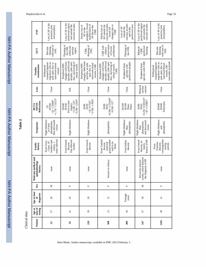
Clinical Characterisation of Patients with CRB1 MutationsClinical findings of French patients with CRB1 mutations are summarized in Tables 2 and 3.The average age at time of diagnosis was 17. Visual acuity was decreased in all patientsranging from 20/50 to light perception with no clear correlation with age or duration of thedisease. Hyperopia was noted for 6/11 patients including three for whom sphericalequivalent was equal or above +5 diopters. Night blindness was present in all patients butthree, for whom a decrease of central vision and photophobia dominated. None of thepatients had nystagmus. Most patients (9/11) had a clear lens; in the remaining two, one hadundergone cataract surgery and one had significant lens opacities. These two patients wereover 40 years of age. Two patterns of fundus pigmentary changes were present in thiscohort: 7/11 had typical bone spicule-shaped pigment migration within the peripheral retinawhereas 4/11 had widespread clumped pigmentary changes of nummular appearance at thelevel of the retinal pigment epithelium (Figure 2). Clumped pigmentation is therefore highlysuggestive of CRB1 mutations but it is not specific since it has also been associated withmutations in NR2E3 (Schorderet and Escher, 2009; Sharon, et al., 2003), NRL (Nishiguchi,et al., 2004) or TULP1 (Mataftsi, et al., 2007). None of the patients displayed preservation ofthe para-arteriolar retinal pigment epithelium as previously described in association withCRB1 mutations (Bernal, et al., 2003; den Hollander, et al., 2004; den Hollander, et al.,1999; Heckenlively, 1982; Henderson, et al., 2010; Khaliq, et al., 2003; Simonelli, et al.,2007; Yzer, et al., 2006b). In addition, none of the patients displayed Coats-like changes inthe periphery. All patients had macular involvement. Six of the patients displayed cystoidmacular edema whereas the other five had macular thinning with loss of the outer retinallayers and corresponding loss of autofluorescence (Figure 2). Color vision was normal infour patients or showed either tritan deficit or a dyschromatopsia with no clear axis whenvisual acuity allowed color vision testing. Full field electroretinogram showed severegeneralized retinal dysfunction with no detectable responses in all patients except three forwhom some residual rod and cone function was detectable. Among those three, the bestresponses on ERG were obtained in the youngest patients. Residual responses on ERG werecorrelated with better preservation of the visual field.
All patients displayed severe retinal involvement with early macular changes, half of themhad cystoid macular edema, a higher percentage than the usually reported prevalence ofabout 30% in overall RP (Hajali, et al., 2008). This higher prevalence could at least be inpart related to vascular abnormalities with Coats-like changes encountered in patients withCRB1 mutations (Coppieters, et al., 2010; den Hollander, et al., 2004; den Hollander, et al.,2001a; Henderson, et al., 2010; Yzer, et al., 2006b). Alternatively, these changes could berelated to abnormal laminar structure associated with CRB1-mutations (Jacobson, et al.,2003). None of our patients developed Coats-like changes or para-arteriolar retinal pigmentepithelium suggesting that these changes are not consistant in CRB1-related RP (Lotery, etal., 2001b). Four subjects displayed clumped retinopathies reinforcing that CRB1 should beconsidered as a potential causal gene for this specific phenotype along with NR2E3 (Sharon,et al., 2003) or NRL (Nishiguchi, et al., 2004).
CRB1 Variants and Their ClassificationOver 240 patients with CRB1 mutations and more than 150 gene variants have beendescribed in the literature (Azam, et al., 2011; Benayoun, et al., 2009; Bernal, et al., 2003;Booij, et al., 2005; Clark, et al., 2010; Coppieters, et al., 2010; den Hollander, et al., 2004;den Hollander, et al., 2001a; den Hollander, et al., 2007; den Hollander, et al., 1999; Galvin,et al., 2005; Gerber, et al., 2002; Hanein, et al., 2004; Henderson, et al., 2010; Henderson, etal., 2007; Jacobson, et al., 2003; Khaliq, et al., 2003; Li, et al., 2011; Lotery, et al., 2001a;Lotery, et al., 2001b; Riveiro-Alvarez, et al., 2008; Seong, et al., 2008; Siemiatkowska, et
Bujakowska et al. Page 4
Hum Mutat. Author manuscript; available in PMC 2013 February 1.
NIH
-PA Author Manuscript
NIH
-PA Author Manuscript
NIH
-PA Author Manuscript

al., 2011; Simonelli, et al., 2007; Tosi, et al., 2009; Vallespin, et al., 2007; Yzer, et al.,2006a; Yzer, et al., 2006b; Zenteno, et al., 2011; Zernant, et al., 2005). The most frequentlyoccurring of the known mutations is the p.Cys948Tyr in exon 9 (96 alleles reported, 24% ofknown CRB1 mutations) (Bernal, et al., 2003; Booij, et al., 2005; Clark, et al., 2010;Coppieters, et al., 2010; den Hollander, et al., 2004; den Hollander, et al., 2001a; denHollander, et al., 2007; den Hollander, et al., 1999; Galvin, et al., 2005; Hanein, et al., 2004;Henderson, et al., 2010; Henderson, et al., 2007; Jacobson, et al., 2003; Lotery, et al., 2001a;Riveiro-Alvarez, et al., 2008; Tosi, et al., 2009; Vallespin, et al., 2007; Yzer, et al., 2006a;Zernant, et al., 2005). In general most of the mutations are in exons 9 (41%) and 7 (27%),therefore as a screening strategy these exons can be tested in the first instance (Figure 1,Supp. Table S1). Exons 7 and 9 encode second and third laminin AG-like domainsrespectively, implying that these domains are particularly important for CRB1 function.Missense mutations constitute 66% of all known mutations, the remaining being frameshift,truncation and splice site mutations.
We have attempted to classify all the reported mutations in three groups: 1) likelypathogenic, 2) unclassified variants, 3) unlikely pathogenic. This classification was based onthe genetic data available from the literature, amino acid conservation and bioinformaticpathogenicity prediction tools (Supp. Tables S1-S3). An important criterion was thepresence of two mutant alleles and co-segregation in the family. Approximately 30% ofcases were reported with only one mutant allele, assuming that the second mutation is withinthe intronic region. For these patients however, one cannot exclude the possibility that thereis another molecular cause of the pathology. The lack of the second mutant CRB1 allele issometimes explained by a digenic inheritance, however so far it has not been proven by co-segregation analysis (Li, et al., 2011; Vallespin, et al., 2007).
Pathogenicity is easier to asses in deletions and frameshift variants than in the case ofmissense changes, hence the importance of the bioinformatic analysis of the pathogenicity,amino acid conservation and functional analysis of the variants. On this basis we have notconsidered two changes identified in our cohort as pathogenic (p.Gly959Ser andp.Ala1354Thr) (den Hollander, et al., 2004; den Hollander, et al., 2001a)). The respectivepatients did not carry a second CRB1 mutation and we did not consider the p.Gly959Ser andp.Ala1354Thr substitutions as likely pathogenic, based on poor conservation of the residuesand low pathogenicity predictions using online bioinformatic tools: PolyPhen-2 and SIFT(Supp. Tables S2 and S3). One report suggests involvement of CRB1 in autosomal dominantpigmented paravenous chorioretinal atrophy (McKay, et al., 2005), though the reportedmutation p.Val162Met has a questionable pathogenicity, since valine is not conserved andmethionine is present in this position in other mammals (Supp. Table S2).
PrevalenceIn the investigated cohort, at least 2.5% of arRP patients carry CRB1 gene defects, whichlies within the previously published range of 0-6.5% (Bernal, et al., 2003; den Hollander, etal., 2004; Vallespin, et al., 2007), or 2.7% after cohort averaging (Table 4). The highpreponderance of novel CRB1 mutations in our cohort suggests, however, that probablymore arRP patients carry CRB1 pathogenic defects, which are novel and thereforeundetectable by arRP microarray. Much higher prevalence is observed in LCA/EORDcohorts and RP with additional features like PPRPE and retinal telangiectasia, representing10.1%, 74.1%, 53.3% respectively in averaged cohorts (Table 4) (Bernal, et al., 2003;Coppieters, et al., 2010; den Hollander, et al., 2004; den Hollander, et al., 2001a; denHollander, et al., 2007; den Hollander, et al., 1999; Hanein, et al., 2004; Henderson, et al.,2010; Henderson, et al., 2007; Lotery, et al., 2001a; Seong, et al., 2008; Simonelli, et al.,2007; Vallespin, et al., 2007; Walia, et al., 2010).
Bujakowska et al. Page 5
Hum Mutat. Author manuscript; available in PMC 2013 February 1.
NIH
-PA Author Manuscript
NIH
-PA Author Manuscript
NIH
-PA Author Manuscript

Genotype-Phenotype CorrelationWe were not able to establish a clear genotype/phenotype correlation for our cohort, whichmight be due to the small number of patients with CRB1 mutations and their variablephenotype. In addition, the nature of existing published data makes it difficult to correlatethe recurring CRB1 mutations with different phenotypes for a number of reasons. First, thephenotyping of patients is complex and distinguishing between early-onset RP and LCA isoften arbitrary and depends on the guidelines of a particular clinical center. Second, preciseclinical data is often omitted in the publications and therefore it is difficult to adjust for thesediagnostic differences in a cross-paper analysis. Despite these inconsistencies, we attemptedto analyse data from previous reports in order to find the relationship between the CRB1variants and the clinical features of respective patients. In this meta-analysis we used 171patients, who carried two likely pathogenic mutations in trans (Benayoun, et al., 2009;Bernal, et al., 2003; Booij, et al., 2005; Clark, et al., 2010; Coppieters, et al., 2010; denHollander, et al., 2004; den Hollander, et al., 2001a; den Hollander, et al., 2007; denHollander, et al., 1999; Galvin, et al., 2005; Hanein, et al., 2004; Henderson, et al., 2010;Henderson, et al., 2007; Jacobson, et al., 2003; Khaliq, et al., 2003; Li, et al., 2011; Lotery,et al., 2001a; Lotery, et al., 2001b; McKibbin, et al., 2010; Riveiro-Alvarez, et al., 2008;Seong, et al., 2008; Simonelli, et al., 2007; Tosi, et al., 2009; Vallespin, et al., 2007; Yzer, etal., 2006a). Combination of two mutant alleles was analysed in relation to clinicalcharacteristics of the published cases. Based on the reports we distinguished the followingphenotypes: LCA, early onset retinal degeneration (EORD), RP, presence of PPRPE andCoats-like vasculopathy. The mutations were classed as null mutations (all mutationsleading to a premature stop codon) or as variants leading to an altered protein (missense andin frame deletions). The likely pathogenic mutations were plotted on a graph, where affectedcodons on allele 1 and allele 2 served as coordinates (codon 0 was assigned to nullmutations). The results show that we cannot assign a specific allele combination to aparticular phenotype, e.g. homozygous null alleles or homozygous p.Cys948Tyr alleles arefound in LCA, EORD and RP patients (Figure 3 A). Null alleles are however more frequentin LCA cohorts (Figure 3 B) as previously suggested (den Hollander, et al., 2004). Thepresence/absence of PPRPE or Coats-like vasculopathy did not reveal a particular mutationpattern (Figure 3 C). These findings suggest the involvement of additional modifying factors(genetic and/or environmental), which are responsible for the modulation of the phenotypein patients harboring CRB1 mutations.
Future DirectionsThe above analysis of the phenotype-genotype correlation suggests that the diseaseseverities associated with CRB1 mutations are in fact a continuum of the same clinical entitywith possible additional modifying factors influencing disease onset and progression. Thereis increasing evidence of the involvement of multiple alleles in the patient’s phenotype, ashas been shown for the Bardet-Biedl patients (Katsanis, et al., 2001) and more recently for aPRPH2-associated macular dystrophy family, where the phenotype has been modulated byadditional heterozygous mutations in ABCA4 (MIM# 601691) and ROM1 (MIM# 180721)(Poloschek, et al., 2010). It is likely that the new next generation sequencing (NGS)technology will help to shed light on the potential genetic modifiers that influence diseasephenotype. One has, however, to analyse the data with caution since NGS will reveal largenumbers of polymorphic changes, which do not modulate the disease. The potential newmodifying changes will have to be confirmed by appropriate genetic and functional analysis.The certainty of the molecular cause of a disease is particularly important in the era of genetherapy trials. Genetic treatment of recessive disorders should not be undertaken beforeobtaining proof that both alleles of a given gene are dysfunctional. In-depth genetic analysis,as presented here, is necessary to provide a basis for conducting such therapies.
Bujakowska et al. Page 6
Hum Mutat. Author manuscript; available in PMC 2013 February 1.
NIH
-PA Author Manuscript
NIH
-PA Author Manuscript
NIH
-PA Author Manuscript
Supplementary MaterialRefer to Web version on PubMed Central for supplementary material.
AcknowledgmentsThe authors would like to thank patients and families for participation in this study, Dominique Santiard-Baron,Christine Chaumeil and clinical staff for their help in clinical data and DNA collection, Sandro Banfi, RobertHenderson and Qingjiong Zhang for additional information on genotype-phenotype correlations of previouslypublished mutations and Robert Gillan for help with the manuscript. The project was financially supported by theFoundation Fighting Blindness (I.A. FFB Grant No: CD-CL-0808-0466-CHNO and the CIC503 recognized as anFFB center, FFB Grant No: C-CMM-0907-0428-INSERM04), Agence Nationale de la Recherche (SSB), FondationVoir et Entendre (CZ), GIS-maladies rares (CZ), Ville de Paris and Région Ile de France, National Institutes ofHealth (USA) (KB NIH, Grant No: 1R01EY020902 - 01A1).
Financial Support: Foundation Fighting Blindness (I.A. FFB Grant No: CD-CL-0808-0466-CHNO and the CIC503recognized as an FFB center, FFB Grant No: C-CMM-0907-0428-INSERM04), Agence Nationale de la Recherche(SSB), Fondation Voir et Entendre (CZ), GIS-maladies rares (CZ), Ville de Paris and Région Ile de France,National Institutes of Health (USA) (KB NIH, Grant No: 1R01EY020902 - 01A1). European Reintegration GrantPERG04-GA-2008-231125 (to K.B.).
ReferencesAdzhubei IA, Schmidt S, Peshkin L, Ramensky VE, Gerasimova A, Bork P, Kondrashov AS, Sunyaev
SR. A method and server for predicting damaging missense mutations. Nat Methods. 2010; 7:248–9. [PubMed: 20354512]
Audo I, Sahel JA, Mohand-Said S, Lancelot ME, Antonio A, Moskova-Doumanova V, Nandrot EF,Doumanov J, Barragan I, Antinolo G, Bhattacharya SS, Zeitz C. EYS is a major gene for rod-conedystrophies in France. Hum Mutat. 2010; 31:E1406–35. [PubMed: 20333770]
Azam M, Collin RW, Malik A, Khan MI, Shah ST, Shah AA, Hussain A, Sadeque A, Arimadyo K,Ajmal M, Azam A, Qureshi N, Bokhari H, Strom TM, Cremers FP, Qamar R, den Hollander AI.Identification of novel mutations in pakistani families with autosomal recessive retinitis pigmentosa.Arch Ophthalmol. 2011; 129:1377–8. [PubMed: 21987686]
Benayoun L, Spiegel R, Auslender N, Abbasi AH, Rizel L, Hujeirat Y, Salama I, Garzozi HJ, Allon-Shalev S, Ben-Yosef T. Genetic heterogeneity in two consanguineous families segregating earlyonset retinal degeneration: the pitfalls of homozygosity mapping. Am J Med Genet A. 2009; 149A:650–6. [PubMed: 19140180]
Bernal S, Calaf M, Garcia-Hoyos M, Garcia-Sandoval B, Rosell J, Adan A, Ayuso C, Baiget M. Studyof the involvement of the RGR, CRPB1, and CRB1 genes in the pathogenesis of autosomalrecessive retinitis pigmentosa. J Med Genet. 2003; 40:e89. [PubMed: 12843338]
Booij JC, Florijn RJ, ten Brink JB, Loves W, Meire F, van Schooneveld MJ, de Jong PT, Bergen AA.Identification of mutations in the AIPL1, CRB1, GUCY2D, RPE65, and RPGRIP1 genes in patientswith juvenile retinitis pigmentosa. J Med Genet. 2005; 42:e67. [PubMed: 16272259]
Cahill M, O’Keefe M, Acheson R, Mulvihill A, Wallace D, Mooney D. Classification of the spectrumof Coats’ disease as subtypes of idiopathic retinal telangiectasis with exudation. Acta OphthalmolScand. 2001; 79:596–602. [PubMed: 11782226]
Chang YF, Imam JS, Wilkinson MF. The nonsense-mediated decay RNA surveillance pathway. AnnuRev Biochem. 2007; 76:51–74. [PubMed: 17352659]
Clark GR, Crowe P, Muszynska D, O’Prey D, O’Neill J, Alexander S, Willoughby CE, McKay GJ,Silvestri G, Simpson DA. Development of a diagnostic genetic test for simplex and autosomalrecessive retinitis pigmentosa. Ophthalmology. 2010; 117:2169–77. e3. [PubMed: 20591486]
Coppieters F, Casteels I, Meire F, De Jaegere S, Hooghe S, van Regemorter N, Van Esch H,Matuleviciene A, Nunes L, Meersschaut V, Walraedt S, Standaert L, Coucke P, Hoeben H, KroesHY, Vande Walle J, de Ravel T, Leroy BP, De Baere E. Genetic screening of LCA in Belgium:predominance of CEP290 and identification of potential modifier alleles in AHI1 of CEP290-related phenotypes. Hum Mutat. 2010; 31:E1709–66. [PubMed: 20683928]
Bujakowska et al. Page 7
Hum Mutat. Author manuscript; available in PMC 2013 February 1.
NIH
-PA Author Manuscript
NIH
-PA Author Manuscript
NIH
-PA Author Manuscript

den Hollander AI, Davis J, van der Velde-Visser SD, Zonneveld MN, Pierrottet CO, Koenekoop RK,Kellner U, van den Born LI, Heckenlively JR, Hoyng CB, Handford PA, Roepman R, Cremers FP.CRB1 mutation spectrum in inherited retinal dystrophies. Hum Mutat. 2004; 24:355–69. [PubMed:15459956]
den Hollander AI, Ghiani M, de Kok YJ, Wijnholds J, Ballabio A, Cremers FP, Broccoli V. Isolationof Crb1, a mouse homologue of Drosophila crumbs, and analysis of its expression pattern in eyeand brain. Mech Dev. 2002; 110:203–7. [PubMed: 11744384]
den Hollander AI, Heckenlively JR, van den Born LI, de Kok YJ, van der Velde-Visser SD, Kellner U,Jurklies B, van Schooneveld MJ, Blankenagel A, Rohrschneider K, Wissinger B, Cruysberg JR,Deutman AF, Brunner HG, Apfelstedt-Sylla E, Hoyng CB, Cremers FP. Leber congenitalamaurosis and retinitis pigmentosa with Coats-like exudative vasculopathy are associated withmutations in the crumbs homologue 1 (CRB1) gene. Am J Hum Genet. 2001a; 69:198–203.[PubMed: 11389483]
den Hollander AI, Johnson K, de Kok YJ, Klebes A, Brunner HG, Knust E, Cremers FP. CRB1 has acytoplasmic domain that is functionally conserved between human and Drosophila. Hum MolGenet. 2001b; 10:2767–73. [PubMed: 11734541]
den Hollander AI, Lopez I, Yzer S, Zonneveld MN, Janssen IM, Strom TM, Hehir-Kwa JY, VeltmanJA, Arends ML, Meitinger T, Musarella MA, van den Born LI, Fishman GA, Maumenee IH,Rohrschneider K, Cremers FP, Koenekoop RK. Identification of novel mutations in patients withLeber congenital amaurosis and juvenile RP by genome-wide homozygosity mapping with SNPmicroarrays. Invest Ophthalmol Vis Sci. 2007; 48:5690–8. [PubMed: 18055821]
den Hollander AI, Roepman R, Koenekoop RK, Cremers FP. Leber congenital amaurosis: genes,proteins and disease mechanisms. Prog Retin Eye Res. 2008; 27:391–419. [PubMed: 18632300]
den Hollander AI, ten Brink JB, de Kok YJ, van Soest S, van den Born LI, van Driel MA, van de PolDJ, Payne AM, Bhattacharya SS, Kellner U, Hoyng CB, Westerveld A, Brunner HG, Bleeker-Wagemakers EM, Deutman AF, Heckenlively JR, Cremers FP, Bergen AA. Mutations in a humanhomologue of Drosophila crumbs cause retinitis pigmentosa (RP12). Nat Genet. 1999; 23:217–21.[PubMed: 10508521]
Franceschetti A, Dieterle P. [Diagnostic and prognostic importance of the electroretinogram intapetoretinal degeneration with reduction of the visual field and hemeralopia]. Confin Neurol.1954; 14:184–6. [PubMed: 13190865]
Galvin JA, Fishman GA, Stone EM, Koenekoop RK. Evaluation of genotype-phenotype associationsin leber congenital amaurosis. Retina. 2005; 25:919–29. [PubMed: 16205573]
Gerber S, Perrault I, Hanein S, Shalev S, Zlotogora J, Barbet F, Ducroq D, Dufier J, Munnich A, RozetJ, Kaplan J. A novel mutation disrupting the cytoplasmic domain of CRB1 in a largeconsanguineous family of Palestinian origin affected with Leber congenital amaurosis. OphthalmicGenet. 2002; 23:225–35. [PubMed: 12567265]
Gosens I, den Hollander AI, Cremers FP, Roepman R. Composition and function of the Crumbsprotein complex in the mammalian retina. Exp Eye Res. 2008; 86:713–26. [PubMed: 18407265]
Hajali M, Fishman GA, Anderson RJ. The prevalence of cystoid macular oedema in retinitispigmentosa patients determined by optical coherence tomography. Br J Ophthalmol. 2008;92:1065–8. [PubMed: 18653601]
Hanein S, Perrault I, Gerber S, Tanguy G, Barbet F, Ducroq D, Calvas P, Dollfus H, Hamel C,Lopponen T, Munier F, Santos L, Shalev S, Zafeiriou D, Dufier JL, Munnich A, Rozet JM, KaplanJ. Leber congenital amaurosis: comprehensive survey of the genetic heterogeneity, refinement ofthe clinical definition, and genotype-phenotype correlations as a strategy for molecular diagnosis.Hum Mutat. 2004; 23:306–17. [PubMed: 15024725]
Heckenlively JR. Preserved para-arteriole retinal pigment epithelium (PPRPE) in retinitis pigmentosa.Br J Ophthalmol. 1982; 66:26–30. [PubMed: 7055539]
Henderson RH, Mackay DS, Li Z, Moradi P, Sergouniotis P, Russell-Eggitt I, Thompson DA, RobsonAG, Holder GE, Webster AR, Moore AT. Phenotypic variability in patients with retinaldystrophies due to mutations in CRB1. Br J Ophthalmol. 2010
Henderson RH, Waseem N, Searle R, van der Spuy J, Russell-Eggitt I, Bhattacharya SS, ThompsonDA, Holder GE, Cheetham ME, Webster AR, Moore AT. An assessment of the apex microarray
Bujakowska et al. Page 8
Hum Mutat. Author manuscript; available in PMC 2013 February 1.
NIH
-PA Author Manuscript
NIH
-PA Author Manuscript
NIH
-PA Author Manuscript

technology in genotyping patients with Leber congenital amaurosis and early-onset severe retinaldystrophy. Invest Ophthalmol Vis Sci. 2007; 48:5684–9. [PubMed: 18055820]
Jacobson SG, Cideciyan AV, Aleman TS, Pianta MJ, Sumaroka A, Schwartz SB, Smilko EE, MilamAH, Sheffield VC, Stone EM. Crumbs homolog 1 (CRB1) mutations result in a thick human retinawith abnormal lamination. Hum Mol Genet. 2003; 12:1073–8. [PubMed: 12700176]
Katsanis N, Ansley SJ, Badano JL, Eichers ER, Lewis RA, Hoskins BE, Scambler PJ, Davidson WS,Beales PL, Lupski JR. Triallelic inheritance in Bardet-Biedl syndrome, a Mendelian recessivedisorder. Science. 2001; 293:2256–9. [PubMed: 11567139]
Khaliq S, Abid A, Hameed A, Anwar K, Mohyuddin A, Azmat Z, Shami SA, Ismail M, Mehdi SQ.Mutation screening of Pakistani families with congenital eye disorders. Exp Eye Res. 2003;76:343–8. [PubMed: 12573663]
Leber T. Ueber Retinitis pigmentosa und angeborene Amaurose. Graefe’s Archive For Clinical AndExperimental Ophthalmology. 1869; 15:1–25.
Li L, Xiao X, Li S, Jia X, Wang P, Guo X, Jiao X, Zhang Q, Hejtmancik JF. Detection of variants in15 genes in 87 unrelated chinese patients with leber congenital amaurosis. PLoS One. 2011;6:e19458. [PubMed: 21602930]
Lotery AJ, Jacobson SG, Fishman GA, Weleber RG, Fulton AB, Namperumalsamy P, Heon E, LevinAV, Grover S, Rosenow JR, Kopp KK, Sheffield VC, Stone EM. Mutations in the CRB1 genecause Leber congenital amaurosis. Arch Ophthalmol. 2001a; 119:415–20. [PubMed: 11231775]
Lotery AJ, Malik A, Shami SA, Sindhi M, Chohan B, Maqbool C, Moore PA, Denton MJ, Stone EM.CRB1 mutations may result in retinitis pigmentosa without para-arteriolar RPE preservation.Ophthalmic Genet. 2001b; 22:163–9. [PubMed: 11559858]
Mataftsi A, Schorderet DF, Chachoua L, Boussalah M, Nouri MT, Barthelmes D, Borruat FX, MunierFL. Novel TULP1 mutation causing leber congenital amaurosis or early onset retinal degeneration.Invest Ophthalmol Vis Sci. 2007; 48:5160–7. [PubMed: 17962469]
McKay GJ, Clarke S, Davis JA, Simpson DA, Silvestri G. Pigmented paravenous chorioretinal atrophyis associated with a mutation within the crumbs homolog 1 (CRB1) gene. Invest Ophthalmol VisSci. 2005; 46:322–8. [PubMed: 15623792]
McKibbin M, Ali M, Mohamed MD, Booth AP, Bishop F, Pal B, Springell K, Raashid Y, Jafri H,Inglehearn CF. Genotype-phenotype correlation for leber congenital amaurosis in NorthernPakistan. Arch Ophthalmol. 2010; 128:107–13. [PubMed: 20065226]
McMahon TT, Kim LS, Fishman GA, Stone EM, Zhao XC, Yee RW, Malicki J. CRB1 gene mutationsare associated with keratoconus in patients with leber congenital amaurosis. Invest OphthalmolVis Sci. 2009; 50:3185–7. [PubMed: 19407021]
Mehalow AK, Kameya S, Smith RS, Hawes NL, Denegre JM, Young JA, Bechtold L, Haider NB,Tepass U, Heckenlively JR, Chang B, Naggert JK, Nishina PM. CRB1 is essential for externallimiting membrane integrity and photoreceptor morphogenesis in the mammalian retina. Hum MolGenet. 2003; 12:2179–89. [PubMed: 12915475]
Ng PC, Henikoff S. SIFT: Predicting amino acid changes that affect protein function. Nucleic AcidsRes. 2003; 31:3812–4. [PubMed: 12824425]
Nishiguchi KM, Friedman JS, Sandberg MA, Swaroop A, Berson EL, Dryja TP. Recessive NRLmutations in patients with clumped pigmentary retinal degeneration and relative preservation ofblue cone function. Proc Natl Acad Sci U S A. 2004; 101:17819–24. [PubMed: 15591106]
Pellikka M, Tanentzapf G, Pinto M, Smith C, McGlade CJ, Ready DF, Tepass U. Crumbs, theDrosophila homologue of human CRB1/RP12, is essential for photoreceptor morphogenesis.Nature. 2002; 416:143–9. [PubMed: 11850625]
Poloschek CM, Bach M, Lagreze WA, Glaus E, Lemke JR, Berger W, Neidhardt J. ABCA4 andROM1: implications for modification of the PRPH2-associated macular dystrophy phenotype.Invest Ophthalmol Vis Sci. 2010; 51:4253–65. [PubMed: 20335603]
Riveiro-Alvarez R, Vallespin E, Wilke R, Garcia-Sandoval B, Cantalapiedra D, Aguirre-Lamban J,Avila-Fernandez A, Gimenez A, Trujillo-Tiebas MJ, Ayuso C. Molecular analysis of ABCA4 andCRB1 genes in a Spanish family segregating both Stargardt disease and autosomal recessiveretinitis pigmentosa. Mol Vis. 2008; 14:262–7. [PubMed: 18334942]
Bujakowska et al. Page 9
Hum Mutat. Author manuscript; available in PMC 2013 February 1.
NIH
-PA Author Manuscript
NIH
-PA Author Manuscript
NIH
-PA Author Manuscript
Schorderet DF, Escher P. NR2E3 mutations in enhanced S-cone sensitivity syndrome (ESCS),Goldmann-Favre syndrome (GFS), clumped pigmentary retinal degeneration (CPRD), and retinitispigmentosa (RP). Hum Mutat. 2009; 30:1475–85. [PubMed: 19718767]
Seelow D, Schuelke M, Hildebrandt F, Nurnberg P. HomozygosityMapper--an interactive approach tohomozygosity mapping. Nucleic Acids Res. 2009; 37:W593–9. [PubMed: 19465395]
Seong MW, Kim SY, Yu YS, Hwang JM, Kim JY, Park SS. Molecular characterization of Lebercongenital amaurosis in Koreans. Mol Vis. 2008; 14:1429–36. [PubMed: 18682808]
Sharon D, Sandberg MA, Caruso RC, Berson EL, Dryja TP. Shared mutations in NR2E3 in enhancedS-cone syndrome, Goldmann-Favre syndrome, and many cases of clumped pigmentary retinaldegeneration. Arch Ophthalmol. 2003; 121:1316–23. [PubMed: 12963616]
Siemiatkowska AM, Arimadyo K, Moruz LM, Astuti GDN, Castro-Miro Md, Zonneveld MN, StromTM, Wijs IJd, Hoefsloot LH, Faradz SMH, Cremers FPM, Hollander AId, Collin RWJ. Moleculargenetic analysis of retinitis pigmentosa in Indonesia using genome-wide homozygosity mapping.Molecular Vision. 2011 (in press).
Simonelli F, Ziviello C, Testa F, Rossi S, Fazzi E, Bianchi PE, Fossarello M, Signorini S, Bertone C,Galantuomo S, Brancati F, Valente EM, Ciccodicola A, Rinaldi E, Auricchio A, Banfi S. Clinicaland molecular genetics of Leber’s congenital amaurosis: a multicenter study of Italian patients.Invest Ophthalmol Vis Sci. 2007; 48:4284–90. [PubMed: 17724218]
Tepass U, Theres C, Knust E. crumbs encodes an EGF-like protein expressed on apical membranes ofDrosophila epithelial cells and required for organization of epithelia. Cell. 1990; 61:787–99.[PubMed: 2344615]
Tosi J, Tsui I, Lima LH, Wang NK, Tsang SH. Case report: autofluorescence imaging and phenotypicvariance in a sibling pair with early-onset retinal dystrophy due to defective CRB1 function. CurrEye Res. 2009; 34:395–400. [PubMed: 19401883]
Vallespin E, Avila-Fernandez A, Velez-Monsalve C, Almoguera B, Martinez-Garcia M, Gomez-Dominguez B, Gonzalez-Roubaud C, Cantalapiedra D, Trujillo-Tiebas MJ, Ayuso C. Novelhuman pathological mutations. Gene symbol: CRB1. Disease: Leber congenital amaurosis. HumGenet. 2010; 127:119. [PubMed: 20108431]
Vallespin E, Cantalapiedra D, Riveiro-Alvarez R, Aguirre-Lamban J, Avila-Fernandez A, MartinezMA, Gimenez A, Trujillo-Tiebas MJ, Ayuso C. Human gene mutations. Gene symbol: CRB1.Disease: late onset retinitis pigmentosa. Hum Genet. 2007a; 122:212. [PubMed: 18386368]
Vallespin E, Cantalapiedra D, Riveiro-Alvarez R, Wilke R, Aguirre-Lamban J, Avila-Fernandez A,Lopez-Martinez MA, Gimenez A, Trujillo-Tiebas MJ, Ramos C, Ayuso C. Mutation screening of299 Spanish families with retinal dystrophies by Leber congenital amaurosis genotypingmicroarray. Invest Ophthalmol Vis Sci. 2007b; 48:5653–61. [PubMed: 18055816]
van de Pavert SA, Kantardzhieva A, Malysheva A, Meuleman J, Versteeg I, Levelt C, Klooster J,Geiger S, Seeliger MW, Rashbass P, Le Bivic A, Wijnholds J. Crumbs homologue 1 is requiredfor maintenance of photoreceptor cell polarization and adhesion during light exposure. J Cell Sci.2004; 117:4169–77. [PubMed: 15316081]
van de Pavert SA, Meuleman J, Malysheva A, Aartsen WM, Versteeg I, Tonagel F, Kamphuis W,McCabe CJ, Seeliger MW, Wijnholds J. A single amino acid substitution (Cys249Trp) in Crb1causes retinal degeneration and deregulates expression of pituitary tumor transforming gene Pttg1.J Neurosci. 2007; 27:564–73. [PubMed: 17234588]
Walia S, Fishman GA, Jacobson SG, Aleman TS, Koenekoop RK, Traboulsi EI, Weleber RG, PennesiME, Heon E, Drack A, Lam BL, Allikmets R, Stone EM. Visual acuity in patients with Leber’scongenital amaurosis and early childhood-onset retinitis pigmentosa. Ophthalmology. 2010;117:1190–8. [PubMed: 20079931]
Yzer S, Fishman GA, Racine J, Al-Zuhaibi S, Chakor H, Dorfman A, Szlyk J, Lachapelle P, van denBorn LI, Allikmets R, Lopez I, Cremers FP, Koenekoop RK. CRB1 heterozygotes with regionalretinal dysfunction: implications for genetic testing of leber congenital amaurosis. InvestOphthalmol Vis Sci. 2006a; 47:3736–44. [PubMed: 16936081]
Yzer S, Leroy BP, De Baere E, de Ravel TJ, Zonneveld MN, Voesenek K, Kellner U, Ciriano JP, deFaber JT, Rohrschneider K, Roepman R, den Hollander AI, Cruysberg JR, Meire F, Casteels I, vanMoll-Ramirez NG, Allikmets R, van den Born LI, Cremers FP. Microarray-based mutation
Bujakowska et al. Page 10
Hum Mutat. Author manuscript; available in PMC 2013 February 1.
NIH
-PA Author Manuscript
NIH
-PA Author Manuscript
NIH
-PA Author Manuscript
detection and phenotypic characterization of patients with Leber congenital amaurosis. InvestOphthalmol Vis Sci. 2006b; 47:1167–76. [PubMed: 16505055]
Zenteno JC, Buentello-Volante B, Ayala-Ramirez R, Villanueva-Mendoza C. Homozygosity mappingidentifies the Crumbs homologue 1 (Crb1) gene as responsible for a recessive syndrome of retinitispigmentosa and nanophthalmos. Am J Med Genet A. 2011; 155A:1001–6. [PubMed: 21484995]
Zernant J, Kulm M, Dharmaraj S, den Hollander AI, Perrault I, Preising MN, Lorenz B, Kaplan J,Cremers FP, Maumenee I, Koenekoop RK, Allikmets R. Genotyping microarray (disease chip) forLeber congenital amaurosis: detection of modifier alleles. Invest Ophthalmol Vis Sci. 2005;46:3052–9. [PubMed: 16123401]
Bujakowska et al. Page 11
Hum Mutat. Author manuscript; available in PMC 2013 February 1.
NIH
-PA Author Manuscript
NIH
-PA Author Manuscript
NIH
-PA Author Manuscript

Figure 1.Distribution of CRB1 mutations in the gene and protein. A) Nucleotide numbering is basedon cDNA sequence of CRB1 (Ref. NM_201253.2) where A of the ATG initiation codon is1. The stop and frameshift mutations are indicated above the structure of the gene and theposition of the missense mutations are drawn in relation to protein domains. The novelmutations are indicated in red. B) The structures of EGF-like and Ca++ binding EGF-likedomains with indications of conserved residues and recurrent mutations. The highlyconserved cysteine residues are in black, the conserved residues between both domains arein grey and the conserved amino acids specific to the Ca2+ binding domain are in blue. C)Evolutionary conservation of the likely pathogenic CRB1 residue changes identified in thiswork.
Bujakowska et al. Page 12
Hum Mutat. Author manuscript; available in PMC 2013 February 1.
NIH
-PA Author Manuscript
NIH
-PA Author Manuscript
NIH
-PA Author Manuscript

Figure 2.Fundus color photographs and Optical Coherence Tomography (OCT). A) Color fundusphotograph of the left eye of 3969 showing nummular pigmentary migration in the midperiphery in addition to pigmentary changes within the macula. B) Vertical scan OCT of theleft eye of 3969 showing cystic changes in the macular region. C) Color fundus photographof the right eye of 547 showing bone spicules pigmentary migration in the periphery inaddition to atrophic changes within the macula. D) Vertical scan OCT of the right eye of 547showing atrophic changes in the macular region after resolution of episodes of cystoidchanges.
Bujakowska et al. Page 13
Hum Mutat. Author manuscript; available in PMC 2013 February 1.
NIH
-PA Author Manuscript
NIH
-PA Author Manuscript
NIH
-PA Author Manuscript

Figure 3.Genotype-phenotype correlation of patients with CRB1 mutations. A) Distribution of CRB1mutations in LCA, EORD and RP. XY axes represent allele 1 and 2 of the patients, theaffected codons serve as xy coordinates, null allele coordinate is designated as 0. The size ofthe circles is proportional to the number of the CRB1 patients with a given genotype. B)Frequency of null and missense allele combinations in LCA, EORD and RP patients. C)Distribution of CRB1 mutations in patients with/without additional features: PPRPE andCoats-like vasculopathy.
Bujakowska et al. Page 14
Hum Mutat. Author manuscript; available in PMC 2013 February 1.
NIH
-PA Author Manuscript
NIH
-PA Author Manuscript
NIH
-PA Author Manuscript

NIH
-PA Author Manuscript
NIH
-PA Author Manuscript
NIH
-PA Author Manuscript
Bujakowska et al. Page 15
Tabl
e 1
Patie
nts w
ith C
RB1
mut
atio
ns id
entif
ied
in th
is st
udy
Patie
ntnu
mbe
rFa
mily
Alle
le 1
Alle
le 2
Exo
nN
ucle
otid
e ch
ange
Prot
ein
chan
geE
xon
Nuc
leot
ide
chan
gePr
otei
n ch
ange
229
159
2c.
613_
619d
elp.
Ile20
5Asp
fs*1
37
c.23
65_2
367d
elA
AT
p.A
sn78
9del
53N
o fa
mily
mem
bers
6c.
1269
C>A
p.C
ys42
3*7
c.25
06C
>Ap.
Pro8
36Th
r
368
249
6c.
1750
G>T
p.A
sp58
4Tyr
7c.
2506
C>A
p.Pr
o836
Thr
547
372
6c.
1963
delC
p.L
eu65
5Trp
fs*1
0?
4240
a20
257
c.22
19C
>Tp.
Ser7
40Ph
e7
c.22
19C
>Tp.
Ser7
40Ph
e
5439
7c.
2222
T>C
p.M
et74
1Thr
9c.
3593
A>G
p.Ty
r119
8Cys
3969
No
fam
ilym
embe
rs7
c.25
06C
>Ap.
Pro8
36Th
r7
c.25
06C
>Ap.
Pro8
36Th
r
409
281
9c.
2843
G>A
p.C
ys94
8Tyr
9c.
3668
G>C
p.C
ys12
23Se
r
1183
b70
99
c.36
59_3
660d
elin
sAp.
Ser1
220A
snfs
*62
9c.
3659
_366
0del
insA
p.Se
r122
0Asn
fs*6
2
1731
1008
9c.
2843
G>A
p.C
ys94
8Tyr
9c.
2843
G>A
p.C
ys94
8Tyr
3144
1302
9c.
2843
G>A
p.C
ys94
8Tyr
7c.
3307
G>A
p.G
ly11
03A
rg
a mut
atio
n in
this
pat
ient
was
iden
tifie
d by
NG
S
b mut
atio
n in
this
pat
ient
was
foun
d th
roug
h ho
moz
ygos
ity m
appi
ng n
ovel
mut
atio
ns a
re in
bol
d
Hum Mutat. Author manuscript; available in PMC 2013 February 1.
NIH
-PA Author Manuscript
NIH
-PA Author Manuscript
NIH
-PA Author Manuscript
Bujakowska et al. Page 16
Tabl
e 2
Clin
ical
dat
a
Patie
ntA
ge a
ttim
e of
test
ing
Age
at t
ime
ofdi
agno
sis
Sex
Rel
evan
t med
ical
and
opht
halm
olog
yhi
stor
yFa
mily
hist
ory
Sym
ptom
sB
CV
AO
D/O
SR
efra
ctio
nL
ens
Fund
usex
amin
atio
nO
CT
FAF
5327
20M
none
From
Ivor
yC
oast
, 10
brot
hers
and
sist
ers,
1si
ster
aff
ecte
d
Nig
ht b
lindn
ess
at 6
then
phot
opho
bia
then
dec
reas
edvi
sion
LP20
/500
+2(−
1.50
)60°
+1.7
5(−
1.5)
125°
Cle
ar
Wid
espr
ead
clum
ped
pigm
ent
mig
ratio
n w
ith n
opa
le o
ptic
dis
c or
narr
owed
retin
alve
ssel
s
Mac
ular
thin
ning
with
loss
of
ON
L
Loss
of A
F at
the
post
erio
r pol
ean
d pe
riphe
ry
5441
25F
none
From
Fre
nch
desc
ent
One
aff
ecte
dbr
othe
rN
ight
blin
dnes
s
20/6
4020
/100
Prio
r to
lens
surg
ery:
+5.5
0(−
1)5°
+5.5
0(−
1)16
5°
IOL
Perip
hera
l RPE
chan
ges w
ith b
one
spic
ules
, per
ifove
alat
roph
y, p
ale
optic
disc
, nar
row
ing
ofre
tinal
ves
sels
Thin
ning
of
the
ON
Lw
ithin
the
mac
ular
regi
on
Loss
of A
F in
the
perif
ovea
l reg
ion
and
outs
ide
the
vasc
ular
arc
ades
229
2920
Fno
neFr
om F
renc
hde
scen
tN
ight
blin
dnes
s20
/80
20/5
0+2
(−0.
75)5
°+2
.50(−
1.50
)5°
Cle
ar
Perip
hera
l RPE
chan
ges,
little
bon
esp
icul
es, n
o pa
leop
tic d
isc
orna
rrow
ed re
tinal
vess
els,
CM
E
CM
E,th
inni
ng o
fO
NL
Patc
hy lo
ss o
fA
F in
the
perip
hery
; fov
eal
mod
ifica
tion
ofA
F du
e to
the
CM
E
368
1312
FSe
izur
e in
infa
ncy
From
Tur
kish
desc
ent
mat
erna
lgr
and-
mot
her
said
to b
ebl
ind
phot
opho
bia
20/8
020
/63
+6.5
0(−
1.25
)160
°+6
.50(−
1)7°
Cle
ar
Perip
hera
l RPE
chan
ges w
ith b
one
spic
ules
, per
ifove
alat
roph
y, p
ale
optic
disc
, nar
row
ing
ofre
tinal
ves
sels
,C
ME
CM
E w
ithre
lativ
epr
eser
vatio
nof
fove
alar
chite
ctur
e
Patc
hy lo
ss o
fA
F ou
tsid
e th
eva
scul
ar a
rcad
es,
fove
al A
Fch
ange
s due
toC
ME
409
43Te
enag
eye
ars
Fno
neFr
om It
alia
nde
scen
tN
ight
blin
dnes
sth
enph
otop
hobi
a
20/1
6020
/100
Plan
oPl
ano
Cle
arPe
riphe
ral b
one
spic
ules
with
perif
ovea
l atro
phy
Thin
ning
of
the
ON
L
Loss
of A
Fou
tsid
e th
eva
scul
ar a
rcad
esan
d in
the
perif
ovea
l are
a
547
5739
MR
ecur
rent
ant
erio
ruv
eitis
, whi
ch d
elay
edth
e di
agno
sis o
f RP
From
Fre
nch
desc
ent,
nofa
mily
hist
ory
of R
P
Nig
ht b
lindn
ess
then
phot
opho
bia
and
decr
ease
dvi
sion
20/8
020
/63
+0.2
5(−
0.50
)110
°−2(−1.25)65°
Bila
tera
lnu
clea
rca
tara
ctPe
riphe
ral b
one
spic
ules
with
CM
E
Bila
tera
lC
ME,
perif
ovea
lth
inni
ng
Loss
of A
F in
the
perif
ovea
l reg
ion
and
outs
ide
the
vasc
ular
arc
ades
1183
3815
Fno
ne
From
Tuni
sian
desc
ent;
cons
angu
inity
amon
gpa
rent
s
Nig
ht b
lindn
ess
and
phot
opho
bia
20/6
4020
/640
Emet
ropi
aC
lear
Wid
espr
ead
clum
ped
pigm
ent
mig
ratio
n w
ith n
opa
le o
ptic
dis
c or
narr
owed
blo
odve
ssel
s; O
Das
tero
ides
hya
loid
s
Mac
ular
thin
ning
with
loss
of
ON
L
Loss
of A
F at
the
post
erio
r pol
ean
d pe
riphe
ry
Hum Mutat. Author manuscript; available in PMC 2013 February 1.

NIH
-PA Author Manuscript
NIH
-PA Author Manuscript
NIH
-PA Author Manuscript
Bujakowska et al. Page 17
Patie
ntA
ge a
ttim
e of
test
ing
Age
at t
ime
ofdi
agno
sis
Sex
Rel
evan
t med
ical
and
opht
halm
olog
yhi
stor
yFa
mily
hist
ory
Sym
ptom
sB
CV
AO
D/O
SR
efra
ctio
nL
ens
Fund
usex
amin
atio
nO
CT
FAF
1731
2317
MD
eafn
ess s
ince
age
9
From
Spa
nish
desc
ent;
pare
nts f
irst
cous
ins;
one
brot
her
affe
cted
Low
vis
ion
sinc
e ea
rlych
ildho
od
HM
20/8
0Em
etro
pia
Cle
ar
Wid
espr
ead
clum
ped
pigm
ent
mig
ratio
n w
ithre
lativ
e sp
arin
g of
the
mac
ula,
with
no
pale
opt
ic d
isc
orna
rrow
ed b
lood
vess
els
Mac
ular
thin
ning
with
loss
of
ON
L
Loss
of A
F at
the
post
erio
r pol
ean
d pe
riphe
ry
3144
209
Fno
neFr
om F
renc
hde
scen
tN
ight
blin
dnes
ssi
nce
early
child
hood
20/8
020
/80
+9(−
1.50
)170
°+7
.50
Cle
ar
Som
e R
PE c
hang
esin
the
perip
hery
,no
rmal
dis
c co
lor
and
no n
arro
win
g of
bloo
d ve
ssel
s; C
ME
CM
E w
ithre
lativ
ely
spar
edfo
veal
stru
ctur
e
Patc
hy lo
ss o
fA
F ou
tsid
e th
eva
scul
ar a
rcad
es,
fove
al A
Fch
ange
s due
toC
ME
3969
2812
Fno
neFr
om M
ali
Nig
ht b
lindn
ess
then
phot
opho
bia
20/1
2520
/320
+0.5
0(−
1.50
)90°
+1.7
5(−
1.25
)95°
Cle
ar
Wid
espr
ead
clum
ped
pigm
ent
mig
ratio
n in
the
post
erio
r pol
e an
dpe
riphe
ryC
ME
CM
ETh
inni
ng o
fO
NL
Diff
use
patc
hylo
ss o
f AF
with
inth
e po
ster
ior p
ole
and
perip
hery
4240
76
Mno
neO
ne si
ster
affe
cted
,fr
om T
urki
shde
scen
t
Dec
reas
edvi
sion
20/6
320
/80
−1.50(−1.50)10°
−2(−0.75)180°
Cle
arM
oder
ate
RPE
chan
ges i
n th
epe
riphe
ryC
ME
CM
E w
ithre
lativ
ely
spar
edpa
rafo
veal
stru
ctur
e
Patc
hy lo
ss o
fA
F ou
tsid
e th
eva
scul
ar a
rcad
e,no
rmal
AF
with
in p
oste
rior
pole
exc
ept A
Fm
odifi
catio
n du
eto
CM
E in
the
fove
a
BC
VA
: bes
t cor
rect
ed v
isua
l acu
ity; C
ME:
cys
toid
mac
ular
ede
ma;
ND
: not
det
ecta
ble;
FA
F: F
undu
s Aut
oflu
ores
cenc
e; O
D: O
culis
dex
tra (r
ight
eye
); O
S: O
culis
Sin
istra
(lef
t eye
); IO
L: in
tra o
cula
r len
s;C
F: c
ount
ing
finge
rs; H
M: h
and
mot
ion;
LP:
ligh
t per
cept
ion;
RPE
: ret
inal
pig
men
t epi
thel
ium
; RP:
retin
itis p
igm
ento
sa; O
HT:
ocu
lar h
yper
tens
ion;
ON
L: O
uter
Nuc
lear
Lay
er
Hum Mutat. Author manuscript; available in PMC 2013 February 1.

NIH
-PA Author Manuscript
NIH
-PA Author Manuscript
NIH
-PA Author Manuscript
Bujakowska et al. Page 18
Table 3
Function data
Patient Colour vision(15 saturated Hue)
Binocular Goldman visual field, III4isopter Full field ERG Multifocal ERG
53 NP Inf to 5° ND ND
54 Dyschromatopsiawithout axis Inf to 5° ND ND
229 Normal40 central degree with 2 peripheral
islandof perception
ND ND
368 Normal 120° horizontally, 60° vertically withrelative central annular scotoma
Residual responses consistent withsevere rod-
cone dysfunctionResidual responses to
central hexagones
409 Dyschromatopsiawithout axis
100° horizontally, 60° vertically withannular scotoma Residual cone responses ND
547 Bilateral tritaonopia20 central degrees both horizontally
andvertically
ND ND
1183 NP Inf to 5° ND ND
1731 OD NP, OS tritaonopia 5 central degrees ND ND
03144 Normal20 central degrees both horizontally
andvertically
ND ND
3969 Dyschromatopsiawithout axis
20 central degree with 2 peripheralisland
of perceptionND ND
4240 Normal 130° vertically and 110° horizontally30% decreased scotopic responses
with photopicresponses at the lower limit of
normal
Decreased responses tocentral hexagones
NP: not performed; ND: not detectable
Hum Mutat. Author manuscript; available in PMC 2013 February 1.

NIH
-PA Author Manuscript
NIH
-PA Author Manuscript
NIH
-PA Author Manuscript
Bujakowska et al. Page 19
Tabl
e 4
Ave
rage
pre
vale
nce
of C
RB1
mut
atio
ns in
retin
al d
ystro
phy
patie
nts i
n pu
blis
hed
repo
rts
Dys
trop
hyPr
eval
ence
*Pa
tient
s with
two
CR
B1
alle
les
Patie
nts w
ithon
eC
RB
1 al
lele
Add
edco
hort
size
Ref
eren
ces
LCA
/EO
RD
10.1
%10
957
1645
(Ber
nal,
et a
l., 2
003;
Cop
piet
ers,
et a
l., 2
010;
den
Hol
land
er, e
t al.,
200
4; d
en H
olla
nder
, et a
l., 2
001;
den
Hol
land
er, e
t al.,
200
7; d
en H
olla
nder
, et a
l., 1
999;
Han
ein,
et a
l., 2
004;
Hen
ders
on, e
t al.,
201
0; H
ende
rson
, et a
l., 2
007;
Li,
et a
l., 2
011;
Lot
ery,
et a
l., 2
001;
Seo
ng, e
t al.,
200
8;Si
mon
elli,
et a
l., 2
007;
Val
lesp
in, e
t al.,
200
7; W
alia
, et a
l.)
RP
2.7%
45
335
(Ber
nal,
et a
l., 2
003;
den
Hol
land
er, e
t al.,
200
4; V
alle
spin
, et a
l., 2
007)
RP+
PPR
PE74
.1%
182
27(d
en H
olla
nder
, et a
l., 2
004;
den
Hol
land
er, e
t al.,
199
9)
RP+
ret
tela
ngie
ctas
ia53
.3%
88
30(d
en H
olla
nder
, et a
l., 2
004;
den
Hol
land
er, e
t al.,
200
1;H
ende
rson
, et a
l., 2
010)
Cla
ssic
Coa
tsdi
seas
e0.
0%0
018
(den
Hol
land
er, e
t al.,
200
4)
* The
aver
age
prev
alen
ce w
as c
alcu
late
d on
the
basi
s of a
ll th
e pu
blis
hed
repo
rts in
dica
ting
phen
otyp
es o
f pat
ient
s with
CRB
1 m
utat
ions
and
the
size
of s
cree
ned
coho
rts.
Hum Mutat. Author manuscript; available in PMC 2013 February 1.