author manuscript nih public access ashley r. morgan
TRANSCRIPT

Acute effect of a high nitrate diet on brain perfusion in olderadults
Tennille D. Presley1,2, Ashley R. Morgan3, Erika Bechtold4, William Clodfelter4, Robin W.Dove2,5, Janine M. Jennings2,5, Robert A. Kraft6, S. Bruce King2,4, Paul J. Laurienti2,3, W.Jack Rejeski2,7, Jonathan H. Burdette2,3,*, Daniel B. Kim-Shapiro1,2,*, and Gary D.Miller2,7,*1Department of Physics, Wake Forest University, Winston-Salem, NC, 271092Translational Science Center, Reynolda Campus, Wake Forest University, Winston-Salem, NC271093Department of Radiology, Wake Forest University School of Medicine, Winston-Salem, NorthCarolina, 271574Department of Chemistry, Wake Forest University, Winston-Salem, NC, 271095Department of Psychology, Wake Forest University, Winston-Salem, NC, 271096Department of Biomedical Engineering, Wake Forest University School of Medicine, Winston-Salem, North Carolina, 271577Department of Health and Exercise Science, Wake Forest University, Winston-Salem, NC,27109
AbstractAims—Poor blood flow and hypoxia/ischemia contribute to many disease states and may also bea factor in the decline of physical and cognitive function in aging. Nitrite has been discovered tobe a vasodilator that is preferentially harnessed in hypoxia. Thus, both infused and inhaled nitriteare being studied as therapeutic agents for a variety of diseases. In addition, nitrite derived fromnitrate in the diet has been shown to decrease blood pressure and improve exercise performance.Thus, dietary nitrate may also be important when increased blood flow in hypoxic or ischemicareas is indicated. These conditions could include age-associated dementia and cognitive decline.The goal of this study was to determine if dietary nitrate would increase cerebral blood flow inolder adults.
© 2010 Elsevier Inc. All rights reserved.Address correspondence to, Gary D. Miller, Department of Health and Exercise Science, Wake Forest University, Winston-Salem,NC, 27109, [email protected] or Daniel B. Kim-Shapiro, Department of Physics, Wake Forest University, Winston-Salem, NC,27109, [email protected] or Jonathan H. Burdette, Department of Radiology, Wake Forest University School of Medicine, Winston-Salem, North Carolina, 27157, [email protected].*These authors share senior authorshipPublisher's Disclaimer: This is a PDF file of an unedited manuscript that has been accepted for publication. As a service to ourcustomers we are providing this early version of the manuscript. The manuscript will undergo copyediting, typesetting, and review ofthe resulting proof before it is published in its final citable form. Please note that during the production process errors may bediscovered which could affect the content, and all legal disclaimers that apply to the journal pertain.Conflicts of InterestDr. Kim-Shapiro is listed as a co-author on a patent application entitled "USE OF NITRITE SALTS FOR THE TREATMENT OFCARDIOVASCULAR CONDITIONS".
NIH Public AccessAuthor ManuscriptNitric Oxide. Author manuscript; available in PMC 2012 January 1.
Published in final edited form as:Nitric Oxide. 2011 January 1; 24(1): 34–42. doi:10.1016/j.niox.2010.10.002.
NIH
-PA Author Manuscript
NIH
-PA Author Manuscript
NIH
-PA Author Manuscript

Methods and Results—In this investigation we administered a high vs. low nitrate diet to olderadults (74.7 ± 6.9 years) and measured cerebral perfusion using arterial spin labeling magneticresonance imaging. We found that the high nitrate diet did not alter global cerebral perfusion, butdid lead to increased regional cerebral perfusion in frontal lobe white matter, especially betweenthe dorsolateral prefrontal cortex and anterior cingulate cortex.
Conclusion—These results suggest that dietary nitrate may be useful in improving regionalbrain perfusion in older adults in critical brain areas known to be involved in executivefunctioning.
KeywordsNitric Oxide; Nitrite; Nitrate; Cerebral Blood Flow; Aging; Magnetic Resonance Imaging
1. IntroductionUntil recently, nitrite was thought to be relatively inert biologically [1]. However, nitriteinfusions leading to slightly supraphysiologic levels of plasma nitrite, from 180 nM atbaseline to 2,600 nM, elevated forearm blood flow and was associated with nitric oxide(NO) formation [2]. Further research showed that a smaller rise in plasma nitrite to only 350nM also increased forearm blood flow [3]. Levels of nitrite in plasma that are shown toimprove blood flow can be achieved by consuming foods high in nitrate [4]. Once ingested,nitrate is absorbed from the upper part of the intestine, and transported via plasma intosalivary glands where it is concentrated and released into saliva. Nitrate is subsequentlyreduced to nitrite by symbiotic, oral bacteria. The nitrite is swallowed and ultimatelyabsorbed from the intestine into the circulatory system [4,5]. Several groups have shown thatingesting diet sources high in nitrate leads to substantial increases in plasma nitrite [6–9].This conversion of nitrate to nitrite can be eliminated when volunteers either expectorate oruse mouthwash which kills oral bacteria that are key to the conversion process [6,8].Physiologic effects from increasing dietary nitrate and plasma nitrite include reduction inblood pressure [6,8], improvement in intestinal health [10], and increases in exerciseperformance [9] which are all attributed to the further reduction of nitrite to NO.Interestingly, nitrite is being investigated for use in a variety of conditions, includingischemic-reperfusion injury, pulmonary hypertension, stroke, sickle cell disease, and gastricdiseases [5,11]. A major feature of nitrite’s ability to increase blood flow is that it actspreferentially in hypoxic conditions, allowing nitrite to increase blood flow precisely in theareas where it is needed most [2,12,13]. We hypothesized that we could use this feature toincrease cerebral blood flow in older adults.
As the average age in the United States continues to rise, there has been increasing interestin gaining a better understanding of the aging brain and the neurological morbidities thataccompany aging. Cognitive decline in general and dementia in particular, are sources ofmorbidity and dependency for older adults. These conditions also place an enormous burdenon the family and society. It has been shown that diminished blood flow to the braincontributes to cognitive impairment [14]. In addition, from the Rotterdam Study, it has beenshown that cerebral hypoperfusion precedes and probably contributes to the onset of clinicaldementia [15]. Moreover, diminished cerebral blood flow has been linked to poorercognition, such as a reduction in information processing speed, and to dementia [16,17]. Aubiquitous finding in neurocognitive disorders associated with aging is the so-called “whitematter hyperintensity” (WMH). Chronic ischemia appears to be the fundamental processthat leads to WMHs, often referred to as leukoaraiosis. Several underlying mechanisms havebeen shown to contribute to this chronic ischemic state, including abnormal arterioles,capillaries and venules, and increased tortuosity of arterioles as we age [18–20]. Chronic
Presley et al. Page 2
Nitric Oxide. Author manuscript; available in PMC 2012 January 1.
NIH
-PA Author Manuscript
NIH
-PA Author Manuscript
NIH
-PA Author Manuscript

ischemia in the white matter appears to be the complex endgame leading to the WMHs andis associated with cognitive decline [21]. In particular, age-related white matter degenerationhas been linked to poor executive functioning as assessed by measures of working memory[22,23] dichotic listening [24], task switching [25] and episodic retrieval [26].
The current analyses are on 2 separate studies. The first was a preliminary study that testedthe time course of plasma nitrate and nitrite levels over a 3-hour period followingconsumption of a high nitrate breakfast, which included beetroot juice. This provided uswith a target time for measuring cerebral blood flow in the subsequent study based on thepeak level achieved and kinetics of the plasma nitrate and nitrite concentrations. Thesubsequent and primary study examined in a within subject design the effect of two levels ofdietary nitrate (high vs. low) over a 24-hour feeding period on acute changes in cerebralblood flow in older adults as measured by magnetic resonance (MR) imaging. Each personwas provided both dietary conditions in a randomized order on separate days and blood flowwas assessed with each diet. The hypothesis for the primary study was that a diet high innitrate containing foods and beverages would produce changes in cerebral blood flow in keyand critical areas of the brain, principally those associated with executive function,compared to a diet of foods that are low in nitrate. The fact that nitrite-based increases inblood flow targets areas of hypoxia [2,12,13,27–29] supports the hypothesis that increasingplasma nitrite will increase cerebral brain perfusion in these areas where it is needed most
2. Methods2.1 Subject Description
Subject selection for both studies followed the same criteria with an age cutoff of ≥ 70 yearsold. In the preliminary study, a total of 5 individuals were recruited, and the primary aim forthe larger trial was addressed by recruiting a total of 16 older adults to undergo both a highnitrate and a low nitrate dietary intervention as approved by the Institutional Review Boardat Wake Forest University. The investigations conformed to the principles outlined in theDeclaration of Helsinki. A phone screen was administered to interested individuals foreligibility requirements. Exclusion criteria included those with systemic uncontrolleddiseases, use of medications that may be contraindicated or interact with the high nitratediet, such as nitroglycerin or nitrate preparations used for angina, or phosphodiesterase type5 (PDE5) inhibitors, including sildenafil (Viagra®). Individuals with clinical hypotension orthose with a resting blood pressure less than 100/60 were also excluded. In addition,individuals had to agree to report to the testing facilities, had no avoidance to the foodsprovided, had a Mini-Mental State Exam (MMSE, [30]) score of at least 24 (older adultswith a score below 24 would not be able to complete the assessments reliably), and had nohistory of medical conditions that could impair cognition, such as head injury, stroke ordementia. They were also appropriately screened to be eligible for MR imaging.
2.2 Study DesignFor the preliminary time course study, participants reported to the laboratory after anovernight fast. Blood was drawn and then they were fed a high nitrate breakfast thatcontained 500 ml of beetroot juice (see high nitrate breakfast in Table 1 for specific foods).At 30-minutes, and 1, 2, and 3 hours after finishing the meal, blood was drawn to determineplasma levels of nitrate and nitrite.
In the cerebral perfusion trial, all participants received both treatments in a within-subjectresearch design with diet conditions being delivered in a Latin-Square arrangement, that is,on days 1 and 2, one-half of the participants received the low nitrate diet first and the otherhalf received the high nitrate diet first, and then for days 3 and 4 they switched treatments
Presley et al. Page 3
Nitric Oxide. Author manuscript; available in PMC 2012 January 1.
NIH
-PA Author Manuscript
NIH
-PA Author Manuscript
NIH
-PA Author Manuscript

(see section below for description of the diets). Comparisons were made between the lowand high nitrate diets with the low nitrate diet serving as the control treatment. The timelinefor the study design is shown in Figure 1. All participants reported to the laboratory on 4consecutive days; each diet treatment lasted two days. On day 1, participants arrived at thelaboratory in the morning following a 10-hour overnight fast and completed an informedconsent and provided a medical history and health status report. Participants were then fedbreakfast comprised of foods from either the low nitrate or the high nitrate diet for theirrespective treatment. Foods for lunch and dinner were given to the individuals to take hometo eat for the rest of day 1 following the same diet treatment given at breakfast (low or highnitrate). They reported back to the laboratory after a 10-hour overnight fast for breakfast onday 2. The same diet as for day 1 (low or high nitrate) was consumed for breakfast on day 2.Blood was drawn prior to breakfast on day 2 and at 1-hour after finishing breakfast. Brainimaging occurred immediately after the 1-hour post-breakfast blood draw. These times werebased on the results from the preliminary time course study. They reported back to thelaboratory on day 3 and performed the second experimental condition: either low or highnitrate diet. This followed the same pattern as day 1 with breakfast consumed in thelaboratory, and food for lunch and dinner, which was provided to them, on day 3 consumedat home. On day 4, they reported back to the laboratory after a 10-hour fast, had blooddrawn prior to eating, then ate breakfast of their second experimental condition. One hourafter eating, participants had another blood draw, proceeded by brain imaging following thesame procedure as in the first experimental condition. The time from after the imaging onday 2 to breakfast on day 3 was considered the washout period and participants resumedtheir normal diet during this period. This provided a 24 hour washout period between diettreatments (breakfast of day 2 until breakfast of day 3), as well as 48 hours betweenassessments (1-hour after breakfast on day 2 until fasting pre-breakfast assessment on day4). No blood assessments were performed on day 1 or 3 prior to starting the respectivetreatments since individuals were on varied diets and nitrate and nitrite levels would havereflected their typical intake; thus not providing a control level for comparison purposeswithin treatment groups of the outcome measures.
Compliance to the diet treatments was determined based on direct observation by researchstaff of food and drink consumption at breakfast on all days, as well as obtaining a dietrecord for lunch, dinner, and snacks on days 1 and 3. Participants were asked to record anyfoods they did not consume that were part of the diet as well as record any foods they ate inaddition to provided foods.
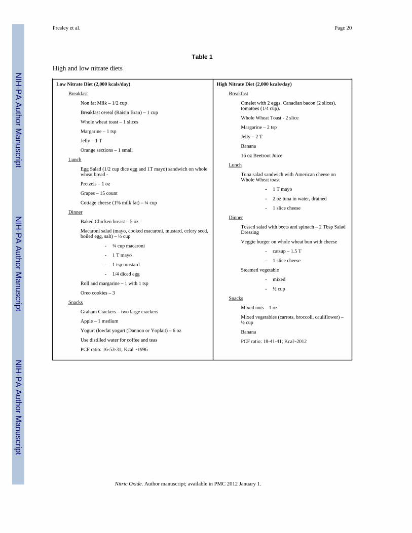
2.3 Diets (See Table 1 for complete listing of foods)The low nitrate diet was low in fruits and vegetables and contained primarily grains, meats,and dairy products. Processed meats high in nitrates and nitrites, such as hotdogs, bacon, andsausage were avoided. The high nitrate diet contained foods high in vegetables, particularlyleafy green vegetables like spinach, lettuce, and broccoli. For the high nitrate breakfast,individuals also consumed 500 ml of beet juice (Biotta, Inc., Carmel, IN). Food andbeverages were liquefied and assayed for their nitrate and nitrite levels using a Sievers NitricOxide Analyzer. For the beet juice, nitrate and nitrite concentrations were 17 mM and 31nM, respectively. The beet juice thus provided 8.5 mmol of nitrate (530 mg). The valuesmeasured for the high vs. low nitrate foods, respectively, were 3.9 mmol vs. 0.089 mmol fornitrate, and 20 µmol vs. 0.4 µmol for nitrite. Thus, the two diets were similarly low in nitritecontent, but the high nitrate diet had a total of 12.4 mmol compared to 0.089 mmol for thelow nitrate diet, nearly a 150 fold difference. For both diets, distilled water was provided todrink.
Presley et al. Page 4
Nitric Oxide. Author manuscript; available in PMC 2012 January 1.
NIH
-PA Author Manuscript
NIH
-PA Author Manuscript
NIH
-PA Author Manuscript

2.4 MeasuresFor both studies, blood was collected using similar techniques. Blood was taken from anantecubital vein and collected in two 4 mL Lithium heparin vials. The tubes wereimmediately centrifuged at 5,000 rpm for 2 min, plasma removed, immediately frozen ondry ice in aliquots with ~0.4 mL of plasma, and stored in a −80° C freezer. For the timecourse study, plasma nitrate and nitrite were determined from blood obtained before andthen at 30-minutes and hourly for 3-hours after eating the high nitrate meal. In the perfusionstudy, age, gender, medical history, and MMSE were obtained on day 1 at the firstlaboratory visit. Blood was obtained on days 2 and 4 prior to breakfast and at 1-hour aftereating.
Plasma and diet nitrite and nitrate levels were measured using chemiluminescence-basedNitric Oxide Analyzers (Sievers, Inc) according to instructions of the manufacturer. For allmeasurements, standard curves were obtained and used for quantitative measurements.
Cerebral blood flow (CBF) was determined from MR images collected 1 hour followingbreakfast on days 2 and 4. All scans were performed on a 1.5T GE scanner using an 8-channel head coil (GE Medical Systems, Milwaukee, WI) and included anatomic imaging(3D BRAVO) and perfusion imaging (PASL Q2TIPS, TR3000, voxel size 3.75mm ×3.75mm × 8mm). Non-invasive multi-slice quantitative CBF was measured withQuantitative Imaging of Perfusion using a Single Subtraction with Thin Slice TI1 PeriodicSaturation (QUIPSS II TIPS a.k.a. Q2TIPS) [31] with a Flow-sensitive AlternatingInversion Recovery (FAIR) [32]. In our implementation of Q2TIPS, saturation pulses wereVery Selective Suppression (VSS) radio frequency pulses [33], which were applied every 25ms between 800 ms (TI1) and 1200ms (TI1s) and which saturated a 2 cm slab of tissue witha 1 cm gap between the saturation slab and the first imaging slice. Other imaging parameterswere as follows: TE 28 ms, TI1 800ms, TI1s 1200 ms, TI 2000 ms, TR 3000 ms, receiverbandwidth 62.5 kHz, flip angle 90 degrees, FOV 24 cm (frequency) × 18 cm (phase), anacquisition matrix 64 × 48 (11 slices, 8 mm thickness, 0 mm slice gap), and frequencyencoding direction anterior/posterior. A diffusion gradient with an equivalent b value of 5.25sec/mm2 was added to suppress intra-arterial spins [34]. An inversion time of 2000 ms wasused for both groups.
Q2TIPS-FAIR acquired data in label/control (slice selective inversion/global inversion)pairs. We acquired 60 label/control pairs to obtain perfusion weighted images with goodsignal-to-noise ratio in a time of 6 minutes 30 seconds. The first 30 seconds (10 volumes)was used to establish steady state and to acquiring a proton density (M0) image. The M0image served as an internal reference to scale the perfusion weighted images appropriatelyto obtain quantitative CBF maps.
Quantitative CBF maps require that the T1 of the tissue be measured at each voxel. T1 mapswere calculated from data acquired from a separate inversion recovery EPI imagingexperiment. Twelve inversion times were acquired logarithmically from 10 milliseconds to 6seconds with a TR of 10 seconds. A total of 13 imaging volumes were acquired in a totalscan time of 2 minutes and 10 seconds. All other imaging acquisition parameters (FOV,matrix size, TE, flip angle, slice thickness, and slice location, etc) were identical to theQ2TIPS-FAIR protocol described previously.
Perfusion imaging preprocessing—Prior to statistical analyses, perfusion data wereprocessed using the following algorithms [35]. Motion correction was applied to theperfusion weighted volumes with a six-parameter rigid body transformation using SPM5.After motion correction the difference images were averaged together, and quantitativeperfusion maps were calculated from the equation:
Presley et al. Page 5
Nitric Oxide. Author manuscript; available in PMC 2012 January 1.
NIH
-PA Author Manuscript
NIH
-PA Author Manuscript
NIH
-PA Author Manuscript

where CBF is the cerebral blood flow, ΔM(TI2) is the mean difference in the signal intensitybetween the label and control images, M0,blood is the equilibrium magnetization of blood, αis the tagging efficiency, TI1 is the time duration of the tagging bolus, TI2 is the inversiontime of each slice, and T1,blood is the longitudinal relaxation time of blood, qp is a correctionfactor that accounts for the difference between the T1 of blood and the T1 of brain tissue.The M0,blood was approximated from the M0, white matter, which was measured directly fromthe M0 image acquired with the perfusion weighted images. The correction factor, qp,required that the T1 of the brain tissue be measured at each voxel, which was measured witha separate inversion recovery (IR) EPI imaging experiment. All other parameters are knownor assumed to be a constant (TI1=800ms, TI1S=1200ms, T1,blood=1200ms). These mapsmeasured perfusion in standard units of milliliters per 100 grams of tissue per minute.
2.5 Statistical AnalysisNitrite and nitrate values are reported as the mean ± one standard deviation. Repeatedmeasures analysis of variance was used to compare the 5 time points for plasma nitrate andnitrite in the time course study. Paired t-tests were used to compare differences between thelow and high diet conditions for the plasma nitrite and nitrate in the perfusion study.Cerebral perfusion analyses were performed using SPM 5 within Matlab. Imaging data werenormalized to standard space within SPM to allow for perfusion comparisons in each voxel.Voxel-wise paired t-tests were performed to evaluate the effects of nitrate in an unbiasedmanner due to a lack of a priori knowledge of where the effects of nitrate would beobserved. The voxel wise statistics can result in large numbers of false positives due tomultiple comparisons. Therefore, data were corrected for multiple comparisons by firstapplying a relatively stringent threshold (p<0.005), and then applying an extent correctionsuch that significant areas had to contain a minimum of 180 contiguous voxels.
3. ResultsFor both studies presented, the beetroot beverage and diets were generally well-tolerated byparticipants. The common expected side effects of red stools and urine associated withdrinking beetroot juice (beeturia) were experienced and reported. One individual in theperfusion study was excluded from the analysis as they refused to drink the beetroot juice onday 2 of the high nitrate diet. No other symptoms, such as dizziness, orthostatic hypotension,or headaches were reported by participants. Compliance to the diet was high as the foodswere well tolerated and no participant reported consuming additional food outside of the dietprovided during testing days.
Five individuals completed the time course study. Figure 2 demonstrates the mean values forboth plasma nitrate and nitrite at each data collection time. Following the overnight fast,plasma nitrate was 51.1 ± 22.3 µM and it increased at 30-minutes and remained elevatedabove the initial fasting draw throughout the 3-hours, F(4,16)=21.31, p<0.001. There wereno differences between 1, 2, and 3 hours for nitrate, and all were higher than at 30-minutes.A similar increase in plasma nitrite was seen over time, F(4,16)=9.90, p<0.001. Post hoccomparisons showed a marginally significant trend for the 30-minute plasma to be higherthan the pre-breakfast blood draw (p=0.058). By 1-hour, nitrite was elevated above both pre-breakfast and 30-minutes, and it remained higher than the fasting draw throughout the 3hours.
Presley et al. Page 6
Nitric Oxide. Author manuscript; available in PMC 2012 January 1.
NIH
-PA Author Manuscript
NIH
-PA Author Manuscript
NIH
-PA Author Manuscript

A total of 16 individuals were recruited for the perfusion study with 14 completing allcomponents (mean age = 74.7 ± 6.9 years). One subject was excluded as they did not drinkthe beetroot juice on the second day for the high nitrate diet. A second subject was excludeddue to a large susceptibility artifact in the MR images. Plasma nitrite and nitrate levels weremeasured both prior to the last breakfast (Pre) and one hour after (Post) the breakfast ondays 2 and 4 of the study. As shown in Figure 3, consumption of the high nitrate dietresulted in substantially greater plasma nitrate (Figure 3A) and nitrite (Figure 3B) levels atboth time points (Pre and Post) compared to low nitrate diet. The average nitrate levels onehour after consuming the high nitrate breakfast on the test day was 750 ± 230 µM comparedto 80 ± 30 µM measured one hour after consumption of the low nitrate breakfast, over a 9fold change (t(14)=-11.1, p<0.001). Average plasma nitrite level one hour after consumingthe high nitrate breakfast on the test day was 950 ± 470 nM compared to 120 ± 70 nMmeasured one hour after consumption of the low nitrate breakfast, nearly an 8 fold change(t(14)=−7.1, p<0.001). Interestingly, plasma nitrite and nitrate levels were also higher afterthe overnight fast on the high nitrate diet compared to the low nitrate diet (compare Pre forhigh vs. low nitrate diets). The average ± standard deviation plasma nitrate values measuredin these post-overnight fast, pre-breakfast time points were 300 ± 140 µM for the highnitrate breakfast compared to 120 ± 60 µM for the low nitrate breakfast (t(14)=−5.0,p<0.001). The average plasma nitrite values measured in this post-overnight fast, pre-breakfast draws were 320 ± 250 nM for the high nitrate breakfast compared to 84 ± 40 nMfor the low nitrate breakfast (t(14)=−3.8, p=0.002). Interestingly, eating the low nitratebreakfast increased plasma nitrite but decreased plasma nitrate (nitrite goes from 84 ± 40nM to 120 ± 70 nM (p = 0.005) and nitrate goes from 120 ± 46 µM to 80 ± 30 µM (p =0.01)). These data for plasma nitrate and nitrite are pooled and are not based on treatmentorder, but on treatment assignment. Further analysis to examine for first order effect showedno differences in outcome variables based on treatment order. That is, participants exposedto the high nitrate diet first did not show a significant increase in plasma nitrate and nitriteduring their low nitrate diet treatment for either pre or post measures as compared to thosethat received the low nitrate diet first. Most importantly, the data in Figure 3 demonstratethat dietary nitrate in our study of older adults led to dramatic increases in plasma nitrite justprior to imaging; levels one hour after eating the high nitrate breakfast were several timeshigher than one hour after eating the low nitrate breakfast (see Post data in Figure 3B).
There were no global perfusion differences when the 14 subjects were on their low nitrateand high nitrate diets. Specifically, average global CBF was 43 ± 10 ml/100g/min on the lownitrate diet and 44 ± 10 on the high nitrate diet. However, when a voxel wise analysis wasperformed (comparison of each brain voxel across subjects), the subjects demonstratedincreased CBF within the subcortical and deep white matter of the frontal lobe (Figures 4and 5). These differences were located between the dorsolateral prefrontal cortex region andanterior cingulate gyrus. Figure 4 shows CBF on a coronal slice through the frontal lobes forthe 14 subjects on the high nitrate and low nitrate diets. Note the subtle increased perfusionwithin the frontal lobe gray matter and increased perfusion at the gray-white junction andwithin the white matter itself (smaller blue and purple regions). Figure 5 shows the statisticalcomparison of the 14 subjects on the high versus low diets. While there were regions in thefrontal lobe gray matter that showed increased flow in the high nitrate state, these areas didnot reach statistical significance. However, there were four statistically significant areas ofincreased CBF within the bilateral white matter of the frontal lobes, areas known to be atrisk for chronic ischemia in the elderly. The peak location, mean perfusion, and standarddeviation for these areas are listed in Table 2. The cross-hairs on the native CBF maps inFigure 4 were placed to illustrate those regions in Figure 5 where significant increases inCBF were present on the high nitrate diet. A post-hoc analysis evaluating the effects of dietorder was performed on the mean measures from the right anterior region-of-interest. This
Presley et al. Page 7
Nitric Oxide. Author manuscript; available in PMC 2012 January 1.
NIH
-PA Author Manuscript
NIH
-PA Author Manuscript
NIH
-PA Author Manuscript
analysis showed that perfusion was greater in the high nitrate diet compared to the lownitrate diet independent of order.
4. DiscussionIn a study led by Gladwin in 2003, it was shown that slightly supraphysiological amounts ofinfused nitrite (going from 180 nM at baseline to 2,600 nM after infusion) led to increasedforearm blood flow [2]. More recently, the Gladwin lab showed that infusion of nitrite toachieve a level of only 350 nM results in increased forearm blood flow [3]. Others havesince confirmed that nitrite acts as a vasodilator in various tissues and that its activity isheightened in hypoxia [36–40]. Rifkind and coworkers showed that nitrite infusions led toincreases in cerebral blood flow in rats as measured by laser Doppler flowmetry [38]. Thus,there is growing evidence that nitrite, rather than being relatively biologically inert, can actas a potent vasodilator harnessed primarily under hypoxic conditions. In this paper, wedemonstrated the ability of oral nitrate to increase plasma nitrite and to increase cerebralblood flow within white matter in older adult humans using perfusion MRI.
Others have previously shown that substantial elevations in plasma nitrite occur throughincreasing dietary nitrate intake [4]. Nitrate from the diet, once absorbed from the intestine,is taken up from the plasma by salivary glands and concentrated in saliva; nitrate issubsequently reduced to nitrite by symbiotic, oral bacteria and ultimately absorbed into thecirculatory system [4,5]. Documented physiological effects from increasing dietary nitrateinclude reduction in blood pressure [6,8], improvement in intestinal health [10], andincreases in exercise performance [9]. These effects are eliminated when volunteers eitherspit or use mouthwash, thereby implicating the importance of nitrate reduction to nitrite byoral bacteria. The level of nitrate utilized in the current study (12.4 mmol) from the dietarymanipulations falls within the range shown by others to reduce blood pressure and improveexercise performance (22.5 mmol and 5.5 mmol, respectively).
To our knowledge, we are the first to demonstrate that these pathways also function in olderadults. One might think that due to xerostomia, altered oral bacterial colonization, andincreased gastric pH, the effect of dietary nitrate on plasma nitrite levels of older adultswould not be as great as for younger adults. However, the increases in plasma nitrite that weobserved in these older adults (an average of 630 nM increase one hour after the high nitratebreakfast compared to before the breakfast, and an average of 830 nM increase in post highnitrate breakfast compared to post low nitrate breakfast, Figure 3) are comparable to whathas been observed in younger adults [6,8]. Moreover, the plasma levels of nitrite achieved inour study using a dietary nitrate intervention (950 ± 470 nM, with a range of 280 to 1890nM measured 1 hour after the high nitrate breakfast) are similar in magnitude to thoseobtained in nitrite infusions which led to increased forearm blood flow [2,3]. Thus, asdietary nitrate is a natural variant, our data support the idea that nitrite is a physiological(rather than simply therapeutic) modulator of blood flow.
Results from the initial kinetics study presented here demonstrate the time course of changesin nitrate and nitrite after consuming the high nitrate breakfast containing 500 ml of beetrootjuice. There was an increase in plasma nitrite levels by over 5 fold above fasting values inone hour, and this level was maintained for at least an additional two hours. These findingssuggest that the plasma nitrite concentrations are maintained at a high level for the timeparticipants in the perfusion study underwent imaging for perfusion measures.
In fact, the effects of dietary nitrate on plasma nitrite and nitrate levels were observed to besustained after the overnight fast (compare levels of different diets pre-breakfast in Figure2), suggesting that the effects of dietary nitrate are prolonged. Consumption of the low
Presley et al. Page 8
Nitric Oxide. Author manuscript; available in PMC 2012 January 1.
NIH
-PA Author Manuscript
NIH
-PA Author Manuscript
NIH
-PA Author Manuscript

nitrate breakfast led to increases in plasma nitrite, but nitrate levels decreased. This couldpossibly be due to increased swallowing and gastric activity leading to more nitrateconversion to nitrite without consumption of substantial amounts of additional nitrate.
Although the effects of the nitrate in the diet on plasma nitrite and nitrate levels was highlysignificant, there was a substantial amount of variability, particularly in the levels of nitrite.The increase in individual subjects in plasma nitrite after the high nitrate breakfast comparedto the low nitrate breakfast ranged from none in one case and a few fold in a couple of casesto up to 20 fold. This variance is not grossly dissimilar to that previously reported in a studyusing beet juice in younger adults [8]. The cause for this large variance could be related tovariability in the nitrate reductase activity of oral bacteria in different individuals, rates ofnitrate uptake, or other causes. Additionally, gastric pH may lead to alterations in nitrate andnitrite metabolism. The presence of achlorhydria and use of pharmacological agents that canaffect gastric pH, such as proton pump inhibitors were not controlled in the present studyand this may have contributed to the variability observed. Clearly, this individual differencein response deserves further study.
There have been several mechanisms proposed for nitrite’s vasodilatory action, all involvingconversion of nitrite to nitric oxide, although sometimes through the intermediacy of othernitrogen oxides [13]. Most (but not all [41]) of these involve the action of a particularprotein and those proposed include hemoglobin [2,42], myoglobin [43,44], xanthineoxidoreductase [45,46], nitric oxide synthase [47], cytochrome c oxidase [48,49], aldehydeoxidase [50,51] and cytochrome c [52,53]. It is likely that several of these act to reducenitrite in different tissues and different conditions. Importantly, all proposed mechanisms ofnitrite reduction to nitric oxide include a potentiation under hypoxic conditions, consistentwith observed physiological nitrite action.
Earlier work has shown that aging is associated with progressive impairment of endothelialfunction [54]. This may lead to a reduction in NO produced from arginine via the nitricoxide synthase (NOS) reaction in the endothelium and contribute to the reduced perfusion intissues with aging. Supplementing L-arginine in older adults has produced equivocal resultswith some groups indicating improvements in flow-mediated dilation and other showing noeffect with L-arginine supplementation [55,56]. The current work indicates improvement intissue perfusion via an endothelium-independent treatment. It would be informative todetermine if L-arginine supplementation would increase cerebral blood flow independent ofthe effect from dietary nitrate, or if there is a potentiating effect from these two agents.
Historically, very high concentrations of nitrate in drinking water were thought to causemethemoglobinemia in infants, but more recent research has questioned whether nitrate ornitrite alone is the cause or if bacterial infection is also required [57,58]. Furthermore, in the1970s, concern over the possibility of nitrite causing cancer arose from the theoreticalassociation between the ability of nitrite to form nitrosamines in the gut and studies showingthat nitrosoamines are carcinogenic [58–61]. However, the evidence that high levels ofnitrate in diet causes any type of cancer is weak and most studies have found no linkbetween dietary nitrate and cancer at all [58,59]. The highest sources of nitrate in our dietare found in certain vegetables like spinach, celery, and beetroot, and epidemiologicevidence does not support these foods as causing diseases from nitrate. It may be that inaddition to nitrate, vegetables also have antioxidants which may protect against nitrosamineformation. This being said, it is possible that some human subpopulations may be especiallysensitive to cancer causing effects of nitrate and continued monitoring of this potentialuntoward health effect is advisable.
Presley et al. Page 9
Nitric Oxide. Author manuscript; available in PMC 2012 January 1.
NIH
-PA Author Manuscript
NIH
-PA Author Manuscript
NIH
-PA Author Manuscript

Chronic ischemia in the white matter is associated with aging. Chronic ischemia appears tobe the fundamental process that leads to so-called white matter hyperintensities (WMHs).Several underlying mechanisms have been shown to contribute to this chronic ischemicstate, including abnormal arterioles, capillaries and venules, and increased tortuosity ofarterioles as we age [18–20]. However, whatever the cause and regardless of the clinicaldiagnosis, chronic ischemia in the white matter appears to be the complex endgame leadingto cognitive decline [21]. As poor cerebral perfusion and ischemia have been associated withcognitive decline and dementia [14–21], our results support the proposal that oral nitratetherapy may be beneficial in treating cognitive decline that is often observed with aging.Towards that end, we show a direct effect of dietary nitrate on cerebral blood flow withinthe subcortical and deep white matter of the frontal lobes. This finding is intriguing as thereis evidence for an anterior-posterior gradient in age-related degeneration of white matter[62,63] suggesting frontal regions are particularly compromised by aging. Moreover, there isa strong relationship between losses in white matter integrity and declines in aspects ofexecutive function, including working memory, task-switching and episodic memoryretrieval [25,26,62], which are important for older adults’ performance of instrumentalactivities of daily living, such as writing checks, using appliances, and shopping [64].
As already discussed, nitrite has been shown to not only increase blood flow to certain areasof the body, but also acts preferentially in hypoxic conditions, allowing nitrite to increaseblood flow precisely in the areas where it is needed [2,12,13]. Based on this notion, our datasuggest that a diet high in nitrate might allow increased perfusion to those areas of the brainknown to be at risk in the elderly and important for cognitive function —the deep whitematter in the frontal lobe.
The diets used in our studies were designed to be isocaloric with similar protein levels, butdiffering levels of nitrate. The measured levels of nitrate were more than 1,000 fold differentbut the nitrite levels were similar between the low and high nitrate treatments. Based on thedifferent food groups that compose each diet, it is expected that the level of antioxidants andother phytochemicals in the diets differ. We recognize that other dietary components besidesnitrate may be partially responsible for the differential response in cerebral perfusion weobserved. However, previous studies using a treatment of beetroot juice only or nitrate byitself have found that minimizing the conversion of nitrate to nitrite, by using mouthwash tokill bacteria or spitting to lower the absorption of nitrite, eliminated the hypotensive effect ofthe nitrate or juice [6,8]. Thus, we suggest that the differences in nitrate in the diet andnitrite in the plasma are primarily responsible for the physiological effect we observed.Nevertheless, using a bactericidal mouthwash or spitting would need to be performed tosubstantiate the effect observed with the current diet is from the nitrate content in the diet.
5. ConclusionsWe have shown, as previously demonstrated in younger adults, that consumption of a highnitrate diet results in substantial increases in plasma nitrite in older adults. The increase inplasma nitrite and nitrate is sustained, even after overnight fast. Generally, we hypothesizethat dietary nitrate may be beneficial in compensating for age-related endothelialdysfunction and associated pathology. In this study we focused on cerebral blood flow andfound that perfusion increased in frontal lobe white matter after consumption of the highnitrate diet compared to the low nitrate diet. Further studies are required to confirm that theeffect we observed is due to nitrate and to refine the nitrate diet. Such work has potential tolead to interventions that could improve cognitive and physical functional health in olderadults.
Presley et al. Page 10
Nitric Oxide. Author manuscript; available in PMC 2012 January 1.
NIH
-PA Author Manuscript
NIH
-PA Author Manuscript
NIH
-PA Author Manuscript

Abbreviations
NO Nitric Oxide
CBF Cerebral Blood Flow
AcknowledgmentsWe thank Erin Reddan, Alesia Goodman, Rebecca Patten, Rudayna Chanouha, Kati Gigler, Andrea Worsham, SaraTurner, Aasha Anderson, and Michael Font for technical assistance.
Funding
This work was supported by the Translational Science Center on the Reynolda Campus at Wake Forest University,The Science Research fund of Wake Forest University, and National Institutes of Health [grant numbersHL058091; HL62198].
References1. Lauer T, Preik M, Rassaf T, Strauer BE, Deussen A, Feelisch M, Kelm M. Plasma nitrite rather than
nitrate reflects regional endothelial nitric oxide synthase activity but lacks intrinsic vasodilatoraction. Proc. Natl. Acad. Sci. USA 2001;98:12814–12819. [PubMed: 11606734]
2. Cosby K, Partovi KS, Crawford JH, Patel RP, Reiter CD, Martyr S, Yang BK, Waclawiw MA,Zalos G, Xu XL, Huang KT, Shields H, Kim-Shapiro DB, Schechter AN, Cannon RO, GladwinMT. Nitrite reduction to nitric oxide by deoxyhemoglobin vasodilates the human circulation. Nat.Med 2003;9:1498–1505. [PubMed: 14595407]
3. Dejam A, Hunter CJ, Tremonti C, Pluta RM, Hon YY, Grimes G, Partovi K, Pelletier MM, OldfieldEH, Cannon RO III, Schechter AN, Gladwin MT. Nitrite Infusion in Humans and NonhumanPrimates. Endocrine Effects, Pharmacokinetics, and Tolerance Formation, Circulation2007;116:1821–1831.
4. Lundberg JO, Weitzberg E, Gladwin MT. The nitrate-nitrite-nitric oxide pathway in physiology andtherapeutics. Nat Rev Drug Discov 2008;7:156–167. [PubMed: 18167491]
5. Lundberg JO, Gladwin MT, Ahluwalia A, Benjamin N, Bryan NS, Butler A, Cabrales P, Fago A,Feelisch M, Ford PC, Freeman BA, Frenneaux M, Friedman J, Kelm M, Kevil CG, Kim-ShapiroDB, Kozlov AV, Lancaster JR, Lefer DJ, McColl K, McCurry K, Patel RP, Petersson J, Rassaf T,Reutov VP, Richter-Addo GB, Schechter A, Shiva S, Tsuchiya K, van Faassen EE, Webb AJ,Zuckerbraun BS, Zweier JL, Weitzberg E. Nitrate and nitrite in biology, nutrition and therapeutics.Nature Chemical Biology 2009;5:865–869.
6. Govoni M, Jansson EA, Weitzberg E, Lundberg JO. The increase in plasma nitrite after a dietarynitrate load is markedly attenuated by an antibacterial mouthwash. Nitric Oxide-Biol Ch2008;19:333–337.
7. Larsen FJ, Ekblom B, Sahlin K, Lundberg JO, Weitzberg E. Effects of dietary nitrate on bloodpressure in healthy volunteers. New Engl J Med 2006;355:2792–2793. [PubMed: 17192551]
8. Webb AJ, Patel N, Loukogeorgakis S, Okorie M, Aboud Z, Misra S, Rashid R, Miall P, Deanfield J,Benjamin N, MacAllister R, Hobbs AJ, Ahluwalia A. Acute blood pressure lowering,vasoprotective, and antiplatelet properties of dietary nitrate via bioconversion to nitrite.Hypertension 2008;51:784–790. [PubMed: 18250365]
9. Bailey SJ, Winyard P, Vanhatalo A, Blackwell JR, DiMenna FJ, Wilkerson DP, Tarr J, Benjamin N,Jones AM. Dietary nitrate supplementation reduces the O-2 cost of low-intensity exercise andenhances tolerance to high-intensity exercise in humans. J Appl Physiol 2009;107:1144–1155.[PubMed: 19661447]
10. Bjorne H, Petersson J, Phillipson M, Weltzberg E, Holm L, Lundberg JO. Nitrite in salivaincreases gastric mucosal blood flow and mucus thickness. J. Clin. Invest 2004;113:106–114.[PubMed: 14702114]
11. Gladwin MT, Schechter AN, Kim-Shapiro DB, Patel RP, Hogg N, Shiva S, Cannon I. Richard O,Kelm M, Wink DA, Espey MG, Oldfield EH, Pluta RM, Freeman BA, Lancaster J. Jack R,
Presley et al. Page 11
Nitric Oxide. Author manuscript; available in PMC 2012 January 1.
NIH
-PA Author Manuscript
NIH
-PA Author Manuscript
NIH
-PA Author Manuscript

Feelisch M, Lundberg JO. The emerging biology of the nitrite anion. Nature Chemical Biology2005;1:308–314.
12. Huang Z, Shiva S, Kim-Shapiro DB, Patel RP, Ringwood LA, Irby CE, Huang KT, Ho C, Hogg N,Schechter AN, Gladwin MT. Enzymatic function of hemoglobin as a nitrite reductase thatproduces Nitric oxide under allosteric control. J. Clin. Invest 2005;115:2099–2107. [PubMed:16041407]
13. van Faassen EE, Babrami S, Feelisch M, Hogg N, Kelm M, Kim-Shapiro DB, Kozlov AV, Li HT,Lundberg JO, Mason R, Nohl H, Rassaf T, Samouilov A, Slama-Schwok A, Shiva S, Vanin AF,Weitzberg E, Zweier J, Gladwin MT. Nitrite as Regulator of Hypoxic Signaling in MammalianPhysiology. Medicinal Research Reviews 2009;29:683–741. [PubMed: 19219851]
14. Meyer JS, Rogers RL, Judd BW, Mortel KF, Sims P. Cognition and cerebral blood flow fluctuatetogether in multi-infarct dementia. Stroke 1988;19:163–169. [PubMed: 3344529]
15. Ruitenberg A, den Heijer T, Bakker SL, van Swieten JC, Koudstaal PJ, Hofman A, Breteler MM.Cerebral hypoperfusion and clinical onset of dementia: the Rotterdam Study. Ann Neurol2005;57:789–794. [PubMed: 15929050]
16. Rabbitt P, Scott M, Thacker N, Lowe C, Jackson A, Horan M, Pendleton N. Losses in gross brainvolume and cerebral blood flow account for age-related differences in speed but not in fluidintelligence. Neuropsychology 2006;20:549–557. [PubMed: 16938017]
17. Spilt A, Weverling-Rijnsburger AW, Middelkoop HA, van Der Flier WM, Gussekloo J, de CraenAJ, Bollen EL, Blauw GJ, van Buchem MA, Westendorp RG. Late-onset dementia: structuralbrain damage and total cerebral blood flow. Radiology 2005;236:990–995. [PubMed: 16014443]
18. Moody DM, Brown WR, Challa VR, Anderson RL. Periventricular venous collagenosis:association with leukoaraiosis. Radiology 1995;194:469–476. [PubMed: 7824728]
19. Moody DM, Brown WR, Challa VR, Ghazi-Birry HS, Reboussin DM. Cerebral microvascularalterations in aging, leukoaraiosis, and Alzheimer's disease. Ann N Y Acad Sci 1997;826:103–116. [PubMed: 9329684]
20. Morris JS, Friston KJ, Buchel C, Frith CD, Young AW, Calder AJ, Dolan RJ. A neuromodulatoryrole for the human amygdala in processing emotional facial expressions. Brain 1998;121(Pt 1):47–57. [PubMed: 9549487]
21. Holland CM, Smith EE, Csapo I, Gurol ME, Brylka DA, Killiany RJ, Blacker D, Albert MS,Guttmann CR, Greenberg SM. Spatial distribution of white-matter hyperintensities in Alzheimerdisease, cerebral amyloid angiopathy, and healthy aging. Stroke 2008;39:1127–1133. [PubMed:18292383]
22. Charlton RA, Schiavone F, Barrick TR, Morris RG, Markus HS. Diffusion tensor imaging detectsage related white matter change over a 2 year follow-up which is associated with working memorydecline. Journal of Neurology Neurosurgery and Psychiatry 2010;81:13–19.
23. Oosterman JM, van Harten B, Weinstein HC, Scheltens P, Sergeant JA, Scherder EJA. Whitematter hyperintensities and working memory: An explorative study. Aging Neuropsychology andCognition 2008;15:384–399.
24. Gootjes L, Scheltens P, Van Strien JW, Bouma A. Subcortical white matter pathology as amediating factor for age-related decreased performance in dichotic listening. Neuropsychologia2007;45:2322–2332. [PubMed: 17382359]
25. O'Sullivan M, Jones DK, Summers PE, Morris RG, Williams SCR, Markus HS. Evidence forcortical "disconnection" as a mechanism of age-related cognitive decline. Neurology 2001;57:632–638. [PubMed: 11524471]
26. Nordahl CW, Ranganath C, Yonelinas AP, DeCarlil C, Fletcher E, Jagust WJ. White matterchanges compromise prefrontal cortex function in healthy elderly individuals. Journal of CognitiveNeuroscience 2006;18:418–429. [PubMed: 16513006]
27. Gladwin MT. Haldane, hot dogs, halitosis, and hypoxic vasodilation: the emerging biology of thenitrite anion. J. Clin. Invest 2004;113:19–21. [PubMed: 14702102]
28. Hunter CJ, Dejam A, Blood AB, Shields H, Kim-Shapiro D, Machado RF, Tarekegn S, Mulla N,Hopper AO, Schechter AN, Power GG, Gladwin MT. Inhaled nebulized nitrite is a hypoxia-sensitive NO-dependent selective pulmonary vasodilator. Nat. Med 2004;10:1122–1127.[PubMed: 15361865]
Presley et al. Page 12
Nitric Oxide. Author manuscript; available in PMC 2012 January 1.
NIH
-PA Author Manuscript
NIH
-PA Author Manuscript
NIH
-PA Author Manuscript

29. Kim-Shapiro DB, Gladwin MT, Patel RP, Hogg N. The reaction between nitrite and hemoglobin:The role of nitrite in hemoglobin-mediated hypoxic vasodilation. J Inorg Biochem 2005;99:237–246. [PubMed: 15598504]
30. Folstein MF, Folstein SE, McHugh PR. MINI-MENTAL STATE - PRACTICAL METHOD FORGRADING COGNITIVE STATE OF PATIENTS FOR CLINICIAN. Journal of PsychiatricResearch 1975;12:189–198. [PubMed: 1202204]
31. Luh WM, Wong EC, Bandettini PA, Hyde JS. QUIPSS II with thin-slice TI1 periodic saturation: amethod for improving accuracy of quantitative perfusion imaging using pulsed arterial spinlabeling. Magn Reson Med 1999;41:1246–1254. [PubMed: 10371458]
32. Kim SG, Tsekos NV. Perfusion imaging by a flow-sensitive alternating inversion recovery (FAIR)technique: application to functional brain imaging. Magn Reson Med 1997;37:425–435. [PubMed:9055234]
33. Tran TK, Vigneron DB, Sailasuta N, Tropp J, Le Roux P, Kurhanewicz J, Nelson S, Hurd R. Veryselective suppression pulses for clinical MRSI studies of brain and prostate cancer. Magn ResonMed 2000;43:23–33. [PubMed: 10642728]
34. Yang Y, Frank JA, Hou L, Ye FQ, McLaughlin AC, Duyn JH. Multislice imaging of quantitativecerebral perfusion with pulsed arterial spin labeling. Magn Reson Med 1998;39:825–832.[PubMed: 9581614]
35. Wong EC, Buxton RB, Frank LR. Quantitative imaging of perfusion using a single subtraction(QUIPSS and QUIPSS II). Magn Reson Med 1998;39:702–708. [PubMed: 9581600]
36. Maher AR, Milsom AB, Gunaruwan P, Abozguia K, Ahmed I, Weaver RA, Thomas P, AshrafianH, Born GVR, James PE, Frenneaux MP. Hypoxic modulation of exogenous nitrite-inducedvasodilation in humans. Circulation 2008;117:670–677. [PubMed: 18212289]
37. Rogers SC, Khalatbari A, Datta BN, Ellery S, Paul V, Frenneaux MP, James PE. NO metaboliteflux across the human coronary circulation. Cardiovasc. Res 2007;75:434–441. [PubMed:17512506]
38. Rifkind JM, Nagababu E, Barbiro-Michaely E, Ramasamy S, Pluta RM, Mayevsky A. Nitriteinfusion increases cerebral blood flow and decreases mean arterial blood pressure in rats: A rolefor red cell NO. Nitric Oxide-Biol Ch 2007;16:448–456.
39. Kumar D, Branch BG, Pattillo CB, Hood J, Thoma S, Simpson S, Illum S, Arora N, Chidlow JH,Langston W, Teng XJ, Lefer DJ, Patel RP, Kevil CG. Chronic sodium nitrite therapy augmentsischemia-induced angiogenesis and arteriogenesis. Proc. Natl. Acad. Sci. USA 2008;105:7540–7545. [PubMed: 18508974]
40. Kozlov AV, Costantino G, Sobhian B, Szalay L, Umar F, Nohl H, Bahrami S, Redl H.Mechanisms of vasodilatation induced by nitrite instillation in intestinal lumen: Possible role ofhemoglobin. Antioxid Redox Sign 2005;7:515–521.
41. Zweier JL, Wang PH, Samouilov A, Kuppusamy P. Enzyme-Independent Formation of Nitric-Oxide in Biological Tissues. Nat. Med 1995;1:804–809. [PubMed: 7585184]
42. Nagababu E, Ramasamy S, Abernethy DR, Rifkind JM. Active nitric oxide produced in the red cellunder hypoxic conditions by deoxyhemoglobin-mediated nitrite reduction. J. Biol. Chem2003;278:46349–46356. [PubMed: 12952953]
43. Shiva S, Huang Z, Grubina R, Sun JH, Ringwood LA, MacArthur PH, Xu XL, Murphy E, Darley-Usmar VM, Gladwin MT. Deoxymyoglobin is a nitrite reductase that generates nitric oxide andregulates mitochondrial respiration. Circ. Res 2007;100:654–661. [PubMed: 17293481]
44. Rassaf T, Flogel U, Drexhage C, Hendgen-Cotta U, Kelm M, Schrader J. Nitrite reductase functionof deoxymyoglobin - Oxygen sensor and regulator of cardiac energetics and function. Circ. Res2007;100:1749–1754. [PubMed: 17495223]
45. Li HT, Samouilov A, Liu XP, Zweier JL. Characterization of the effects of oxygen on xanthineoxidase-mediated nitric oxide formation. J. Biol. Chem 2004;279:16939–16946. [PubMed:14766900]
46. Millar TM, Stevens CR, Benjamin N, Eisenthal R, Harrison R, Blake DR. Xanthine oxidoreductasecatalyses the reduction of nitrates and nitrite to nitric oxide under hypoxic conditions. FEBS Lett1998;427:225–228. [PubMed: 9607316]
Presley et al. Page 13
Nitric Oxide. Author manuscript; available in PMC 2012 January 1.
NIH
-PA Author Manuscript
NIH
-PA Author Manuscript
NIH
-PA Author Manuscript

47. Gautier C, van Faassen E, Mikula I, Martasek P, Slama-Schwok A. Endothelial nitric oxidesynthase reduces nitrite anions to NO under anoxia. Biochem. Biophys. Res. Commun2006;341:816–821. [PubMed: 16442076]
48. Castello PR, David PS, McClure T, Crook Z, Poyton RO. Mitochondrial cytochrome oxidaseproduces nitric oxide under hypoxic conditions: Implications for oxygen sensing and hypoxicsignaling in eukaryotes. Cell Metabolism 2006;3:277–287. [PubMed: 16581005]
49. Castello PR, Woo DK, Ball K, Wojcik J, Liu L, Poyton RO. Oxygen-regulated isoforms ofcytochrome c oxidase have differential effects on its nitric oxide production and on hypoxicsignaling. Proceedings of the National Academy of Sciences 2008;105:8203–8208.
50. Li HT, Kundu TK, Zweier JL. Characterization of the Magnitude and Mechanism of AldehydeOxidase-mediated Nitric Oxide Production from Nitrite. J. Biol. Chem 2009;284:33850–33858.[PubMed: 19801639]
51. Li H, Cui H, Kundu TK, Alzawahra W, Zweier JL. Nitric Oxide Production from Nitrite OccursPrimarily in Tissues Not in the Blood: CRITICAL ROLE OF XANTHINE OXIDASE ANDALDEHYDE OXIDASE. J. Biol. Chem 2008;283:17855–17863. [PubMed: 18424432]
52. Chen YR, Chen LC, Liu X, Li HT, Zweier JL, Mason MP. Involvement of protein radical, proteinaggregation, and effects on NO metabolism in the Hypochlorite-mediated oxidation ofmitochondrial cytochrome c. Free Radical Biol. Med 2004;37:1591–1603. [PubMed: 15477010]
53. Basu S, Azarova NA, Font MD, King SB, Hogg N, Gladwin MT, Shiva S, Kim-Shapiro DB.Nitrite Reductase Activity of Cytochrome c. J. Biol. Chem 2008;283:32590–32597. [PubMed:18820338]
54. Lyons D, Roy S, Patel M, Benjamin N, Swift CG. Impaired nitric oxide-mediated vasodilatationand total body nitric oxide production in healthy old age. Clinical Science 1997;93:519–525.[PubMed: 9497788]
55. Bode-Boger SM, Muke J, Surdacki A, Brabant G, Boger RH, Frolich JC. Oral L-arginine improvesendothelial function in healthy individuals older than 70 years. Vascular Medicine 2003;8:77–81.[PubMed: 14518608]
56. Gates PE, Boucher ML, Silver AE, Monahan KD, Seals DR. Impaired flow-mediated dilation withage is not explained by L-arginine bioavailability or endothelial asymmetric dimethylarginineprotein expression. J Appl Physiol 2007;102:63–71. [PubMed: 16946027]
57. Avery AA. Infantile methemoglobinemia: Reexamining the role of drinking water nitrates. EnvironHealth Persp 1999;107:583–586.
58. Gilchrist M, Winyard PG, Benjamin N. Dietary nitrate – Good or bad? Nitric Oxide. 2009 In Press.59. Milkowski A, Garg HK, Coughlin JR, Bryan NS. Nutritional Epidemiology in the Context of
Nitric Oxide Biology: A Risk-Benefit Evaluation for Dietary Nitrite and Nitrate. Nitric Oxide.2009 In press.
60. Tannenbaum SR, Weisman M, Fett D. EFFECT OF NITRATE INTAKE ON NITRITEFORMATION IN HUMAN SALIVA. Food and Cosmetics Toxicology 1976;14:549–552.[PubMed: 1017770]
61. Spiegelhalder B, Eisenbrand G, Preussmann R. INFLUENCE OF DIETARY NITRATE ONNITRITE CONTENT OF HUMAN SALIVA - POSSIBLE RELEVANCE TO INVIVOFORMATION OF N-NITROSO COMPOUNDS. Food and Cosmetics Toxicology 1976;14:545–548. [PubMed: 1017769]
62. Madden DJ, Bennett IJ, Song AW. Cerebral White Matter Integrity and Cognitive Aging:Contributions from Diffusion Tensor Imaging. Neuropsychology Review 2009;19:415–435.[PubMed: 19705281]
63. Tullberg M, Fletcher E, DeCarli C, Mungas D, Reed BR, Harvey DJ, Weiner MW, Chui HC,Jagust WJ. White matter lesions impair frontal lobe function regardless of their location.Neurology 2004;63:246–253. [PubMed: 15277616]
64. Cahn-Weiner D, Boyle P, Malloy P. Tests of executive function predict instrumental activities ofdaily living in community-dwelling older individuals. Applied Neuropsychology 2002;9:187–191.[PubMed: 12584085]
Presley et al. Page 14
Nitric Oxide. Author manuscript; available in PMC 2012 January 1.
NIH
-PA Author Manuscript
NIH
-PA Author Manuscript
NIH
-PA Author Manuscript

Figure 1.Timeline of study design for the larger study that included MRI scans.
Presley et al. Page 15
Nitric Oxide. Author manuscript; available in PMC 2012 January 1.
NIH
-PA Author Manuscript
NIH
-PA Author Manuscript
NIH
-PA Author Manuscript

Figure 2.Plasma nitrate (A) and nitrite (B) levels prior to and at 30-minutes, 1-hour, 2-hours, and 3-hours following a high nitrate breakfast in the time course study. Plasma nitrite and nitratewere measured after consumption of a high nitrate breakfast following overnight fast.Averages and standard deviations are shown (n=5).* indicates significantly different than fasting values# indicates significantly different than at 30-minutes
Presley et al. Page 16
Nitric Oxide. Author manuscript; available in PMC 2012 January 1.
NIH
-PA Author Manuscript
NIH
-PA Author Manuscript
NIH
-PA Author Manuscript

Figure 3.Plasma nitrate (A) and nitrite (B) levels in the perfusion study. The levels reflect measureson days and 2 and 4 of the study, either before (Pre) or one hour after (Post) consumingbreakfast on the high or low nitrate diet. Subjects consumed either a high or low nitrate dietstarting on days 1 and 3 that continued through the breakfast before and after which bloodwas drawn. Values plotted are averages ± one standard deviation.* indicates significantly different than fasting values
Presley et al. Page 17
Nitric Oxide. Author manuscript; available in PMC 2012 January 1.
NIH
-PA Author Manuscript
NIH
-PA Author Manuscript
NIH
-PA Author Manuscript

Figure 4.Group cerebral blood flow maps (in ml/100g tissue/minute) for the 14 subjects on the highnitrate diet (left) and low nitrate diet (right). Both images are from a coronal slice throughthe frontal lobes. There is subtle increased perfusion within the frontal lobe gray matter andat the gray-white junction and within the white matter itself (smaller blue and purpleregions). The cross-hairs were placed to illustrate those regions in Figure 5 wherestatistically significant increases in CBF were present on the high nitrate diet.
Presley et al. Page 18
Nitric Oxide. Author manuscript; available in PMC 2012 January 1.
NIH
-PA Author Manuscript
NIH
-PA Author Manuscript
NIH
-PA Author Manuscript

Figure 5.Cerebral blood flow (CBF) differences between the high nitrate diet and low nitrate dietstates. Statistical maps show significant differences in regional blood flow for the n=14subjects on the high nitrate diet versus on the low nitrate diet. Note the increased CBF (ml/100g/min) within the bilateral white matter of the frontal lobes, areas known to be at risk forchronic ischemia in the elderly. The bottom left image is a coronal slice at the level of genuof the corpus callosum. The diagonal stacked images are axial slices extending from theuppermost portions of the lateral ventricles superiorly to the basal ganglia/mid-body of thelateral ventricles inferiorly. Although there are some asymmetries to the findings, the effectson CBF from the high nitrate diet clearly manifest bilaterally within the white matter.Statistical analyses were performed at p<0.005, extent corrected at 180 voxels. Color scalerepresents the t-score from a voxel-wise paired-samples t-test.
Presley et al. Page 19
Nitric Oxide. Author manuscript; available in PMC 2012 January 1.
NIH
-PA Author Manuscript
NIH
-PA Author Manuscript
NIH
-PA Author Manuscript
NIH
-PA Author Manuscript
NIH
-PA Author Manuscript
NIH
-PA Author Manuscript
Presley et al. Page 20
Table 1
High and low nitrate diets
Low Nitrate Diet (2,000 kcals/day) High Nitrate Diet (2,000 kcals/day)
Breakfast
Non fat Milk – 1/2 cup
Breakfast cereal (Raisin Bran) – 1 cup
Whole wheat toast – 1 slices
Margarine – 1 tsp
Jelly – 1 T
Orange sections – 1 small
Lunch
Egg Salad (1/2 cup dice egg and 1T mayo) sandwich on wholewheat bread -
Pretzels – 1 oz
Grapes – 15 count
Cottage cheese (1% milk fat) – ¼ cup
Dinner
Baked Chicken breast – 5 oz
Macaroni salad (mayo, cooked macaroni, mustard, celery seed,boiled egg, salt) – ½ cup
- ¼ cup macaroni
- 1 T mayo
- 1 tsp mustard
- 1/4 diced egg
Roll and margarine – 1 with 1 tsp
Oreo cookies – 3
Snacks
Graham Crackers – two large crackers
Apple – 1 medium
Yogurt (lowfat yogurt (Dannon or Yoplait) – 6 oz
Use distilled water for coffee and teas
PCF ratio: 16-53-31; Kcal ~1996
Breakfast
Omelet with 2 eggs, Canadian bacon (2 slices),tomatoes (1/4 cup).
Whole Wheat Toast - 2 slice
Margarine – 2 tsp
Jelly – 2 T
Banana
16 oz Beetroot Juice
Lunch
Tuna salad sandwich with American cheese onWhole Wheat toast
- 1 T mayo
- 2 oz tuna in water, drained
- 1 slice cheese
Dinner
Tossed salad with beets and spinach – 2 Tbsp SaladDressing
Veggie burger on whole wheat bun with cheese
- catsup – 1.5 T
- 1 slice cheese
Steamed vegetable
- mixed
- ½ cup
Snacks
Mixed nuts – 1 oz
Mixed vegetables (carrots, broccoli, cauliflower) –½ cup
Banana
PCF ratio: 18-41-41; Kcal~2012
Nitric Oxide. Author manuscript; available in PMC 2012 January 1.

NIH
-PA Author Manuscript
NIH
-PA Author Manuscript
NIH
-PA Author Manuscript
Presley et al. Page 21
Tabl
e 2
The
mea
n an
d st
anda
rd d
evia
tion
of th
e pe
rfus
ion
(ml/1
00g
tissu
e/m
in) w
as c
alcu
late
d fo
r a 1
0mm
dia
met
er re
gion
-of-
inte
rest
cen
tere
d at
eac
h th
e pe
akof
sign
ifica
nce.
Val
ues a
re p
rese
nted
for l
ow a
nd h
igh
nitra
te d
iets
in e
ach
of th
e fo
ur re
gion
s.
peak
loca
tion
mea
n pe
rfus
ion
stde
v
xy
zhi
gh diet
low
die
thi
gh diet
low
die
t
righ
tan
terio
r30
1428
31.3
2731
22.3
5293
11.9
3273
9.84
7101
post
erio
r-su
perio
r12
−6
5845
.340
1637
.552
139.
4505
3610
.783
69
left
ante
rior
−32
2614
40.2
891
32.5
0811
8.96
4576
13.9
4549
post
erio
r-su
perio
r−16
−8
5637
.526
129
.782
619.
4043
9310
.360
62
Nitric Oxide. Author manuscript; available in PMC 2012 January 1.