author : bogdan dumitrachi coauthor : bogdan lucian bucur coordinator : asist.univ.dr. anca...
TRANSCRIPT

Author : BOGDAN DUMITRACHI Coauthor : BOGDAN LUCIAN BUCUR
Coordinator : Asist.univ.Dr. ANCA TORCATORU
Effect of Magnification on Locating the Canals in Maxillary Molars

Introduction It is well known that one of the causes of the failure of endodontic therapy is the inability to localize
and treat all the canals of the endodontic system. All categories of teeth may have extra roots and/or canals, but the
likelihood of finding aberrant canal
configurations is higher in premolars and molars.
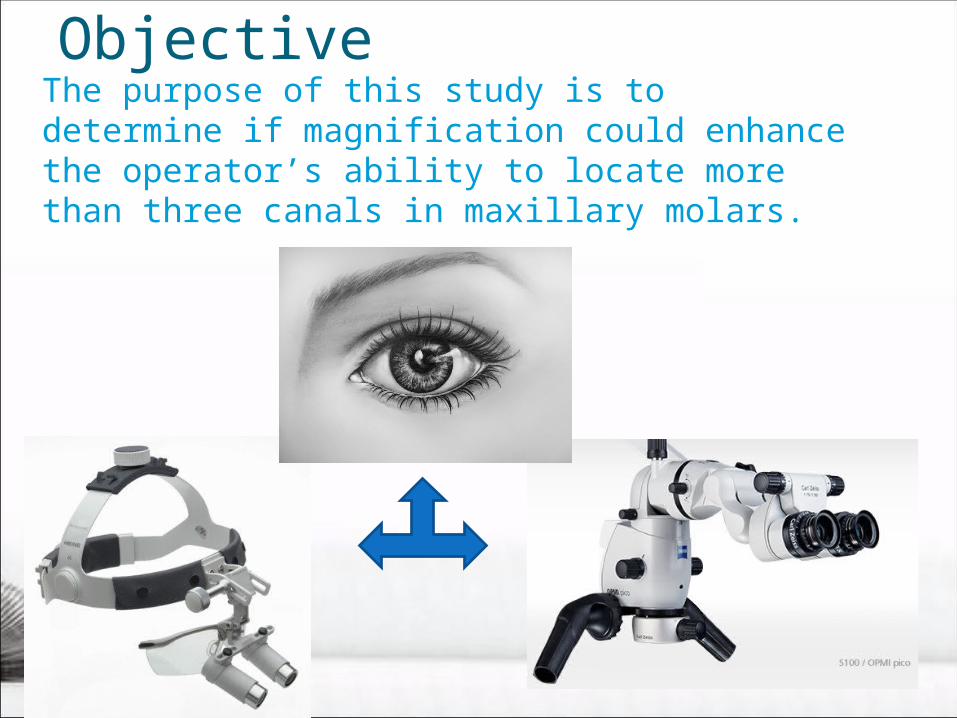
ObjectiveThe purpose of this study is to determine if
magnification could enhance the operator’s ability to locate more than three canals in maxillary molars.

Material and Method
• In this study 18 extracted first and second permanent maxillary molars were used.
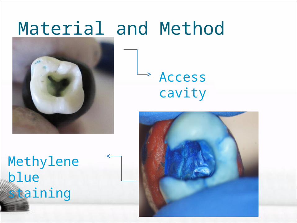
•After creating the access cavity a sharp endodontic explorer was used to identify the canals
•In order to increase the canal entrance detection, the pulp floor was stained using methylene blue

Material and Method
Extracted teeth
Material and Method
Access cavity
Methylene blue staining

Material and Method
• Each tooth was examined by single blinded operators with different experience ( two students and one endodontic specialist):
1.without magnification
2.using dental loupes at 4x magnification
3.using surgical operative microscope (SOM) at 29.5x magnification.

Material and Method

ResultsUsing no magnification ,student #1 and student # 2 found 4 canals in 2 teeth (11.11%), meanwhile the endodontic specialist found 4 canals in 4 teeth (22.22%).

ResultsUsing the dental loupes ,student #1 and student#2 found 4 canals in 2 teeth(11.11%) meanwhile the endodontic specialist found 4 canals in 8 teeth(44.44%) and 5 canals in 1 tooth(5.56%).

ResultsUsing the microscope (SOM),student #1 and student #2 found 4 canals in 4 teeth(22.22%) meanwhile the endodontic specialist found 4 canals in 8 teeth(44.44%) and 5 canals in 1 tooth(5.56%)

Conclusions
1. For a successful endodontic treatment each operator must know very precisely the internal anatomy of the maxillary molars.
2. It is important to remember that each maxillary molar may have at least one extra root/canal.

Conclusions
3.Staining the pulp chamber floor with blue methylene helps the operator to detect canals entrances but no difference was observed between inspection with or without staining in finding extra canals in maxillary molars.
Conclusions
4.The use of the loupes and the operative microscope (SOM) increases the ability of the operator to find more than 3 canals in the first and second permanent maxillary molars.

Conclusions
5.The operators experience has a great importance on locating extra canals in maxillary molars with complex anatomy.

THANK YOU FOR YOUR ATTENTION!!!