australian audit of endovascular aneurysm repair€¦ · australian audit of endovascular aneurysm...
TRANSCRIPT
AAuussttrraalliiaann aauuddiitt ooff EEnnddoovvaassccuullaarr AAnneeuurryyssmm RReeppaaiirr
OOccttoobbeerr 22000066
DISCLAIMER: This report is based on information available at the time of research and cannot be expected to cover any developments arising from subsequent improvements to health technologies. This report is based on a limited literature search and is not a definitive statement on the safety, effectiveness or cost effectiveness of the health technology covered. The Commonwealth does not guarantee the accuracy, currency or completeness of the information in this report. This report is not intended to be used as medical advice and it is not intended to be used to diagnose, treat, cure or prevent any disease, nor should it be used for therapeutic purposes or as a substitute for a health professional’s advice. The Commonwealth does not accept any liability for any injury, loss or damage incurred by the use or reliance on the information.
Address: 1st Floor, 38 Payneham Road, Stepney 5069 Postal: PO Box 533 Stepney, SA 5069, Australia Phone: (08) 8363-7513 Fax: (08) 8362-2077 Email: [email protected] Web: http://www.surgeons.org/asernip-s
ASERNIP S
i
TTaabbllee ooff ccoonntteennttss
REPORT SUMMARY 1
1. BACKGROUND 2
2. AUDIT INFRASTRUCTURE 4
3. RESULTS 12
4. DISCUSSION 33
5. REFERENCES 33
Appendix 1 – Patient information and consent forms 40
Appendix 2 - EVAR data entry forms 43
Appendix 3 – Explanation of ‘technical’ and ‘clinical’ success, and White’s grading system 45

- ii -
LLiisstt ooff ttaabblleess
Table 1: Research questions........................................................................................ 3
Table 2: Enrolments, follow-up and mortality.......................................................... 12
Table 3: Imaging technique use over time.................................................................. 17
Table 4: Definition of technical and clinical success .................................................. 18
Table 5: Time interval for technical and clinical success ............................................ 19
Table 6: Clinical success ............................................................................................ 20
Table 7: Endoleaks recorded < 30 days .................................................................... 21
Table 8: Mid-term type I endoleak............................................................................. 21
Table 9: Mid-term type II endoleaks.......................................................................... 22
Table 10: Complications, excluding endoleak where aneurysm not excluded............. 23
Table 11: Complications, excluding endoleak where aneurysm excluded ................... 23
Table 12: Graft related complications prior to discharge ........................................... 23
Table 13: Short-term and mid-term complications (excluding endoleak).................... 24
Table 14: Ruptured aneurysms .................................................................................. 24
Table 15: Cause of late (>30 days post-operative) conversion to open repair............. 26
Table 16: Time to additional open and endoluminal procedures................................ 26
Table 17: Cause of early mortality and days to death ................................................. 27
Table 18: Cause of mid-term and long-term mortality ............................................... 28
Table 19: Summary statistics for length of stay (days)............................................... 28
Table 20: Comparison of Australian and European audit data................................... 29
Table 21: Safety and Efficacy of elective EVAR ....................................................... 30
Table 22: Survival table (Eurostar)............................................................................. 31
Table 23: Survival (Australian audit).......................................................................... 31
Table 24: Procedures recorded by Medicare Australia for items 33116 and 33119..... 33
Table 25: EVAR studies- perioperative death rate ..................................................... 34
Table 26: Whites grading system ............................................................................... 46

- iii -
LLiisstt ooff ffiigguurreess
Figure 1: Number of procedures submitted by surgeons ............................................ 8
Figure 2: Follow-up forms received by time interval................................................. 13
Figure 3: Follow-up forms received by patient.......................................................... 13
Figure 4: ASA values for EVAR patients................................................................... 14
Figure 5: Changes in aneurysm size (±5mm) ............................................................. 25
- iv -
AAbbbbrreevviiaattiioonnss
Computing: DLT digital linear tape RAID redundant array of independent disks SCSI small computer system interface UPS uninterruptible power supply Measurement: max Maximum min Minimum mm millimetres n number of patients SD Standard Deviation Medical terminology: AAA Abdominal aortic aneurysms ASA American Society of Anaesthesiology CT Computed (axial) Tomography ELG Endoluminal graft EVAR Endovascular aneurysm repair ICU Intensive care unit IHD Ischaemic heart disease MI Myocardial infarction RCT Randomised controlled trial Organisations: ASERNIP-S Australian Safety and Efficacy Register of New Interventional
Procedures -Surgical CSIRO Commonwealth Scientific & Industrial Research Organisation Cwth Commonwealth EUROSTAR European Collaborators Group on Stent-graft Techniques for
Abdominal Aortic Aneurysm Repair HIC Health Insurance Commission MBS Medical Benefits Schedule MSAC Medical Services Advisory Committee RACS Royal Australasian College of Surgeons

1
RReeppoorrtt ssuummmmaarryy
Procedure The procedure involves the elective repair of abdominal aortic aneurysms (AAA) using an endovascular graft. The graft is inserted through an incision in the femoral artery and positioned within the aorta at the site of wall weakening (the aneurysm) in order to prevent rupture.
Organisation information The Australian Safety and Efficacy Register of New Interventional Procedures – Surgical (ASERNIP-S) manages the audit in Adelaide and is part of the Research and Audit Division of the Royal Australasian College of Surgeons (RACS).
Audit The audit was established to review the mid to long-term safety and effectiveness of the endovascular graft within the Australian setting. Audit information will help inform future funding decisions for the procedure. The procedure has been given interim funding until the results of the audit are known.
Methods Operative data was collected for 961 Australian recipients of endovascular aneurysm repair (EVAR) during the period 1 November 1999 to 16 May 2001. This cohort of patients has been followed for over 5 years.
Results Of the 961 patients enrolled in the audit, around 60% have survived to five years. Eleven percent of surviving patients are listed as lost to follow-up. 93% of procedures were classified as “technical successes”. Mid-term “clinical success” was 85%; however 6% of patients experienced a period of clinical failure before success. Some patients in the clinical success group required further interventions for their aneurysm; 4% had additional endovascular procedures (assisted success) and 1.2% had additional surgical procedures (secondary success) performed to ensure continued exclusion of the aneurysm or graft patency. So far, patient data obtained for patients entering long-term follow-up shows 88% clinical success. To date, 16 aneurysms have ruptured post-procedure and 23 patients have had their EVAR converted to open repair. 36 patients had type I endoleak during mid-term follow-up. Statistical analysis indicates that pre-operative aneurysm diameter is the most significant predictor of the various measures of success.
Funding This project has been funded by The Australian Government Department of Health and Ageing, following recommendations made by the Medical Services Advisory Committee.

- 2 -
11.. BBaacckkggrroouunndd
In May 1999 the Medical (formerly Medicare) Services Advisory Committee (MSAC) assessed and reported on the procedure of endovascular aneurysm repair. Their results showed that although the procedure appeared effective in the short-term, there was insufficient evidence concerning the long-term safety and efficacy.1 As a consequence, the Australian Government Department of Health and Ageing commissioned the Australian Safety and Efficacy Register of New Interventional Procedures – Surgical (ASERNIP-S) to manage a national collection of data for the evaluation of endovascular aneurysm repair (EVAR). ASERNIP-S is part of the Research and Audit Division of the Royal Australasian College of Surgeons (RACS) and the project is managed from their offices in Adelaide, South Australia. Two interim item numbers have been assigned to the procedure in the Medical Benefits Schedule* (as shown below). Item 33116: Infrarenal abdominal aortic aneurysm, replacement by tube graft using endovascular repair procedure, excluding associated radiological services (Ministerial Determination) (Anaes.) (Assist.) Item 33119: Infrarenal abdominal aortic aneurysm, replacement by bifurcation graft to one or both iliac arteries using endovascular repair procedure, excluding associated radiological services (Ministerial Determination) (Anaes.) (Assist.) Medical Benefits Schedule Book: Item numbers 33116 and 33119 Note T8.26
T8.26.1 These items were introduced into the Schedule on an interim basis via Ministerial Determination under section 3C of the Health Insurance Act, following a recommendation of the Medicare Services Advisory Committee (MSAC). Interim funding is being provided to facilitate collection of Australian evidence of the medium safety and effectiveness of these services. An audit of these services is being conducted by the Australian Safety and Efficacy Register of New Interventional Procedures – Surgical (ASERNIP-S). Continuation of funding is dependent on the progress of the audit. Therefore providers of these services are strongly encouraged to take part in the audit.
* Endoluminal grafting for abdominal aortic aneurysms (AAA), May 1999. MSAC application 1006, Final Assessment Report: available from http://www.msac.gov.au/reports.htm
- 3 -
Further information on the review of these procedures is available from the MSAC Secretariat.
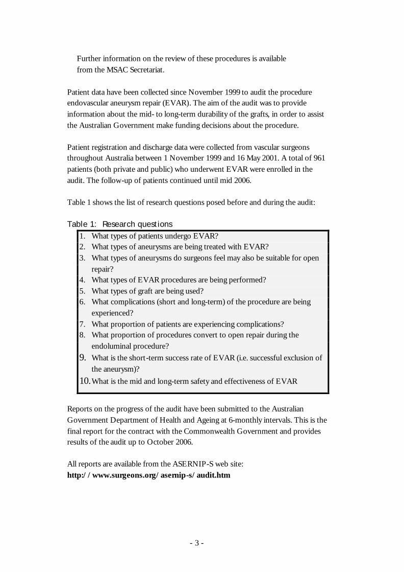
Patient data have been collected since November 1999 to audit the procedure endovascular aneurysm repair (EVAR). The aim of the audit was to provide information about the mid- to long-term durability of the grafts, in order to assist the Australian Government make funding decisions about the procedure. Patient registration and discharge data were collected from vascular surgeons throughout Australia between 1 November 1999 and 16 May 2001. A total of 961 patients (both private and public) who underwent EVAR were enrolled in the audit. The follow-up of patients continued until mid 2006. Table 1 shows the list of research questions posed before and during the audit: Table 1: Research questions
1. What types of patients undergo EVAR? 2. What types of aneurysms are being treated with EVAR? 3. What types of aneurysms do surgeons feel may also be suitable for open
repair? 4. What types of EVAR procedures are being performed? 5. What types of graft are being used? 6. What complications (short and long-term) of the procedure are being
experienced? 7. What proportion of patients are experiencing complications? 8. What proportion of procedures convert to open repair during the
endoluminal procedure? 9. What is the short-term success rate of EVAR (i.e. successful exclusion of
the aneurysm)? 10. What is the mid and long-term safety and effectiveness of EVAR
Reports on the progress of the audit have been submitted to the Australian Government Department of Health and Ageing at 6-monthly intervals. This is the final report for the contract with the Commonwealth Government and provides results of the audit up to October 2006. All reports are available from the ASERNIP-S web site: http://www.surgeons.org/asernip-s/audit.htm

- 4 -
22.. AAuuddiitt iinnffrraassttrruuccttuurree
This section outlines the methods, processes and management infrastructure established to achieve the objectives of the audit and any issues arising since the May 2005 report.
Personnel
The staffing infrastructure, based at ASERNIP-S includes:
• Ms Maggi Boult Morbidity Audit Manager • Miss Claire Miller Project Officer • Dr Wendy Babidge Director, Research and Audit
Division, RACS • Professor Guy Maddern ASERNIP-S Surgical Director
Reference group
An independent reference group of senior vascular surgeons with a high level of expertise in the procedure advises on clinical aspects of the audit. The group comprises the following members:
• Professor Guy Maddern (Chair) (Adelaide, South Australia) • Mr John Anderson (Adelaide, South Australia) • Mr Michael Denton (Melbourne, Victoria) • Associate Professor Robert Fitridge (Adelaide, South Australia) • Professor John Harris (Sydney, New South Wales) • Mr Michael Lawrence-Brown (Perth, Western Australia) • Professor James May (Sydney, New South Wales) • Professor Kenneth Myers (Melbourne, Victoria)
Computing
Data security is a high priority. The ASERNIP-S computer network is run across the Windows 2000/XP platform, with data striped across three SCSI hard drives in an RAID-5 configuration. The system provides network security, with logon access only permitted by password. Password access to the audit information is provided only to staff directly involved in the audit. An additional computer has been purchased to house a back-up copy which is also transferred to CD, ensuring a mirror copy of the database is always available and data loss is

- 5 -
minimised in the event of a system or hardware failure. Back-up files are housed off-site. The ASERNIP-S network is protected from outside intrusion with a hardware firewall, and current virus scan software run daily. A UPS system prevents data corruption caused by sudden power loss to the server.
Confidentiality and Privacy
The audit contains sensitive health information. Advice was sought regarding its handling and housing to ensure that the information was handled appropriately. In 2000, amendments were made to the Privacy Act 1988 (Cwth): The Privacy Amendment (Private Sector) Act 2000 (Cwth), which came into effect 21 December 2001. In order to ensure that the rights of patients were respected by ASERNIP-S, guidance was obtained from the RACS Ethics Committee, and the lawyer acting for the RACS. Recommendations were made that ASERNIP-S provide information for patients about the audit and that patients provide consent for the release of their health information. As a result of these recommendations information brochures and consent forms were given to surgeons to use at patient follow-up (Appendix 1). The patient information brochure, patient consent forms and a privacy statement (which includes information about the EVAR audit) are available for download from the ASERNIP-S web site: http://www.surgeons.org/asernip-s/audit.htm The EVAR audit has been declared a quality assurance activity by the Minister for Health and Ageing, the Hon. Tony Abbott under Part VC of the Health Insurance Act 1973 (QAA No. 2/2004).
Information exchange
Liaison networks have been established or maintained with the following groups during this stage of the audit:
• RACS Council • Board of Professional Development and Standards (RACS) • ASERNIP-S Management Committee • Australian and New Zealand Society of Vascular Surgeons • RACS Ethics Committee • EUROSTAR registry* • National Death Index at the Australian Institute of Health and Welfare
(AIHW) * Links established through the AAA reference group.
- 6 -
Supporting documentation
Documents produced during the audit include: a patient information brochure, patient consent form, project plan, protocol, audit manual, and project reports (produced 6 monthly). The reports, patient consent form and brochure, and patient information sheet are available for download from the ASERNIP-S website or are available on request from the ASERNIP-S office.
Data collection
This section describes the methods used to facilitate accurate and complete data collection.
Participating surgeons
As part of RACS, ASERNIP-S has access to the contact details of vascular surgeons in Australia. During the period of initial data collection around 80 surgeons were performing the EVAR procedure and have continued to submit their data.
Data input
Most data has been submitted using paper-based forms (Appendix 2). Other methods, such as encrypted internet submission or Access databases, were provided earlier in the audit but were not widely used. On arrival at ASERNIP-S the information is entered into a password-protected Access 2003 database. Most of the data is double-checked to ensure data integrity. The date of data entry and checking is logged. Surgeons were asked to provide information using three separate forms:
• Operative form – information obtained in the period immediately prior and during the procedure
• Discharge form – including information obtained post-operatively, up to 30 days from the time of the procedure
• Follow-up form – aimed to collect information at regular follow-up intervals of 3 months, 6 months, 12 months, then on an annual basis.
Copies of each form are included in Appendix 2. The forms do not include any universal identifiers such as the Medicare or Veterans Affairs Numbers as stipulated by the Privacy Amendment (Private Sector) Act 2000, in National Privacy Principle 7.1. Changes were made to the forms in 2004 to identify type III and type IV endoleaks.

- 7 -
Data received
ASERNIP-S was required to collect procedures performed privately in Australia between November 1999 and May 2001. An estimate of the number of private procedures performed was obtained from the Health Insurance Commission (HIC), and a comparison with ASERNIP-S data indicates that around 90% of these cases were submitted. As noted in previous reports, the follow-up of patients varies from surgeon to surgeon. To account for this the following follow-up intervals have been adopted: 1-3 months, 4-8 months, 9-14 months, 15-19 months, 20-29 months, 30-41 months, 42-54 months and 55-67 months. A number of patients are regarded as lost to follow-up when, for instance, their surgeons have retired and we have not been able to establish the new surgeon or GP responsible for these patients Additionally the advanced age and/or increasing frailty of patients may have necessitated their movement to nursing home care, the patient may move to be closer to family members and becomes lost to follow-up or we cannot establish who has taken responsibility for the follow-up of regional patients or the patient refuses further follow-up. In summary, the barriers to follow-up include worsening health, distance, cost and movement between service providers.
National Death Index
The National Death Index (NDI) is a database which lists all deaths that have occurred in Australia since 1980. It is maintained by the Australian Institute of Health and Welfare (AIHW) in Canberra. An application to use the database to track patients enrolled in the audit was first made in 2004. The project received clearance from the AIHW Ethics Committee, which has continued to monitor the project every 12 months. The last application to use NDI data was made in August 2006.
Number of procedures performed by surgeons
Figure 1 shows the number of procedures performed by surgeons during the audit period. In some cases more than one surgeon was listed as having performed the procedure. In this situation the procedure was attributed to the follow-up surgeon.

- 8 -
Figure 1: Number of procedures submitted by surgeons
31
19
13
4 4 46
0
5
10
15
20
25
30
35
=5 10 15 20 25 30 >30
Number of procedures
Nu
mb
er o
f su
rgeo
ns
Reportage
Surgeons participating in the audit receive an updated list of their audit information every three months. This ensures surgeons remain informed if follow-up information is due. The progress of the audit is reported at each ASERNIP-S Management Committee meeting. The most recent took place on the 31st July 2006. An update of audit activities is provided to the Council of the RACS three times each year. During the initial funding period for the audit, reports were submitted to the Australian Government Department of Health and Ageing at six-monthly intervals. For the final phase of the audit (November 2005 – October 2006) a report was submitted in February 2006 and the final report will be submitted in October 2006 (current report). Publications and presentations related to the audit are shown below:
Conference Presentations
2006
• Golledge J, Parr A, Boult M, Maddern G, Fitridge R. The outcome of endovascular repair of small abdominal aortic aneurysms. “Vascular 2006”. The Australian and New Zealand Society for Vascular Surgery. Cairns, QLD Australia. September 2006.
- 9 -
• Fitridge R, Boult M, Babidge W, Maddern G on behalf of the ASERNIP-S EVAR reference group. Effect of pre-operative variables on the mid-term outcomes for patients treated in Australia for endovascular repair. Annual Scientific Congress of the Royal Australasian College of Surgeons. Sydney, Australia. May 2006.
2005
• Fitridge R, Boult M, Babidge W, Maddern G. ASERNIP-S audit of endoluminal repair of abdominal aortic aneurysms in Australia. Annual Scientific Congress of the Royal Australasian College of Surgeons. Perth, Australia. May 2005.
2004
• Fitridge R, Boult M, Babidge W, Maddern G. Endoluminal repair of abdominal aortic aneurysms – contemporary Australian experience. Annual Scientific Congress of the Royal Australasian College of Surgeons. Melbourne, Australia. May 2004.
• Fitridge R, Boult M, Babidge W, Maddern G. Endoluminal repair of abdominal aortic aneurysms – Australian audit. ISCVS World Congress, Hawaii. April 2004.
2003
• Fitridge R, Boult M, Babidge W, Maddern G (for the ASERNIP-S Reference Group for endoluminal graft repair). The Australian Audit of the Safety and Efficacy of Endoluminal Grafts for the Repair Of Abdominal Aortic Aneurysms. Annual Scientific Congress of the Royal Australasian College of Surgeons. Brisbane, Australia. May 2003.
2002
• Harris J. Australian audit of endoluminal and open repair of abdominal aortic aneurysms. 15th Annual International Congress for Endovascular Interventions. Phoenix, Arizona USA. February 2002.
• Fitridge R. ASERNIP-S follow-up data. Annual Scientific Congress of the Royal Australasian College of Surgeons. Adelaide, Australia. May 2002.
• Boult M, Babidge W, Coburn D, Maddern G. Data collection on a new technology to inform funding decision making by the Australian Government. (Poster). 18th Annual Meeting for the International Society of Technology Assessment in Health Care (ISTAHC). Berlin, Germany, June 9-12, 2002.

- 10 -
• Boult M, Babidge W, Maddern G. The role of audit in improving health outcomes. Australasian Health Research Data Managers Association, Brisbane, Australia, 21-22nd August 2002.
• M Denton, R Fitridge, M Boult, W Babidge & G Maddern (for the Endoluminal Reference Group) Highlights from the ASERNIP -S Registry; What are the important findings for clinical practice? International Endovascular Symposium. Sydney, Australia, December 5-7, 2002.
2001
• Maddern G and Fitridge R. Annual Scientific Congress of the Royal Australasian College of Surgeons. Canberra, Australia. May 2001.
• Boult M, Ethics and the Law - Some Issues involved in Data Collection, Australasian Health Research Data Managers Association, Melbourne, Australia, 13-14th September 2001.
Publications
2006
• Golledge J, Parr A, Boult M, Maddern G, Fitridge R. The outcome of endovascular repair of small abdominal aortic aneurysms. Annals of Surgery (in press).
• Boult M, Babidge W, Maddern G, Barnes M, Fitridge R. Predictors of success following endovascular aneurysm repair: mid-term results European Journal of Vascular and Endovascular Surgery 2006; 31(2):123-129.
2004
• Boult M, Babidge W, Maddern G, Fitridge R, on behalf of the Reference Group. Endoluminal repair of abdominal aortic aneurysm – contemporary Australian experience. European Journal of Vascular and Endovascular Surgery. 2004: 28(1); 36-40.
2002
• Boult M, Babidge W, Anderson J, Denton M, Fitridge R, Harris J, Lawrence-Brown M, May J, Myers K, Maddern G. Australian audit for the endoluminal repair of abdominal aortic aneurysm - the first 12-months. Australian and New Zealand Journal of Surgery. 2002; 72(3):190 - 195.
- 11 -
• Boult M, Babidge W, Roder D, Maddern G. Issues of consent and privacy affecting the functioning of ASERNIP-S. Australian and New Zealand Journal of Surgery. 2002; 72(8):580-582.
2001
• Fitridge R. Evaluation of aortic stent grafting – the Australian experience. In: Whittemore A (Ed). Advances in Vascular Surgery. 2001;9:55 – 65.
Accreditation of the ASERNIP-S audit
The RACS Board of Professional Development and Standards has approved the EVAR audit for the purposes of their Continuing Professional Development Programme. The audit is listed on the RACS website under approved audit activities: http://www.surgeons.org/Content/NavigationMenu/FellowshipandStandards/ProfessionalStandards/FAQS/surgical_audit_peer_review_2005.pdf

- 12 -
33.. RReessuullttss
This section summarises patient demographics, procedural and follow-up results and aims to provide answers to the research questions (shown in Table 1). All data for calculating results were received prior to 1 September 2006. Up to 1 September 2006, a total of 961 EVAR procedure patients were enrolled in the audit of which 70% (677/961) were performed in the private sector and the remainder in the public system. The 677 private patients represent around 90% of all procedures performed privately in Australia during the enrolment period. Fifty five percent (495/906) of patients were considered suitable for open repair. Figures showing number of patients enrolled in the audit, amount of follow-up received and mortality are shown in Table 2. Table 2: Enrolments, follow-up and mortality Data Total Percentage Operative data set 961 Public 284 30% Private 677 70% Patients lost to follow-up1 83 9% Patients lost to follow-up2 20 2% Deceased 374 39% Early* 17 2% Late 357 37% 1 Patients lost due to frailty etc 2 Patients lost following retirement or relocation of surgeon * Early death occurs within 30 days of the procedure, and is sometimes referred to as perioperative death. Late death implies death occurring more than 30 days post procedure.
- 13 -
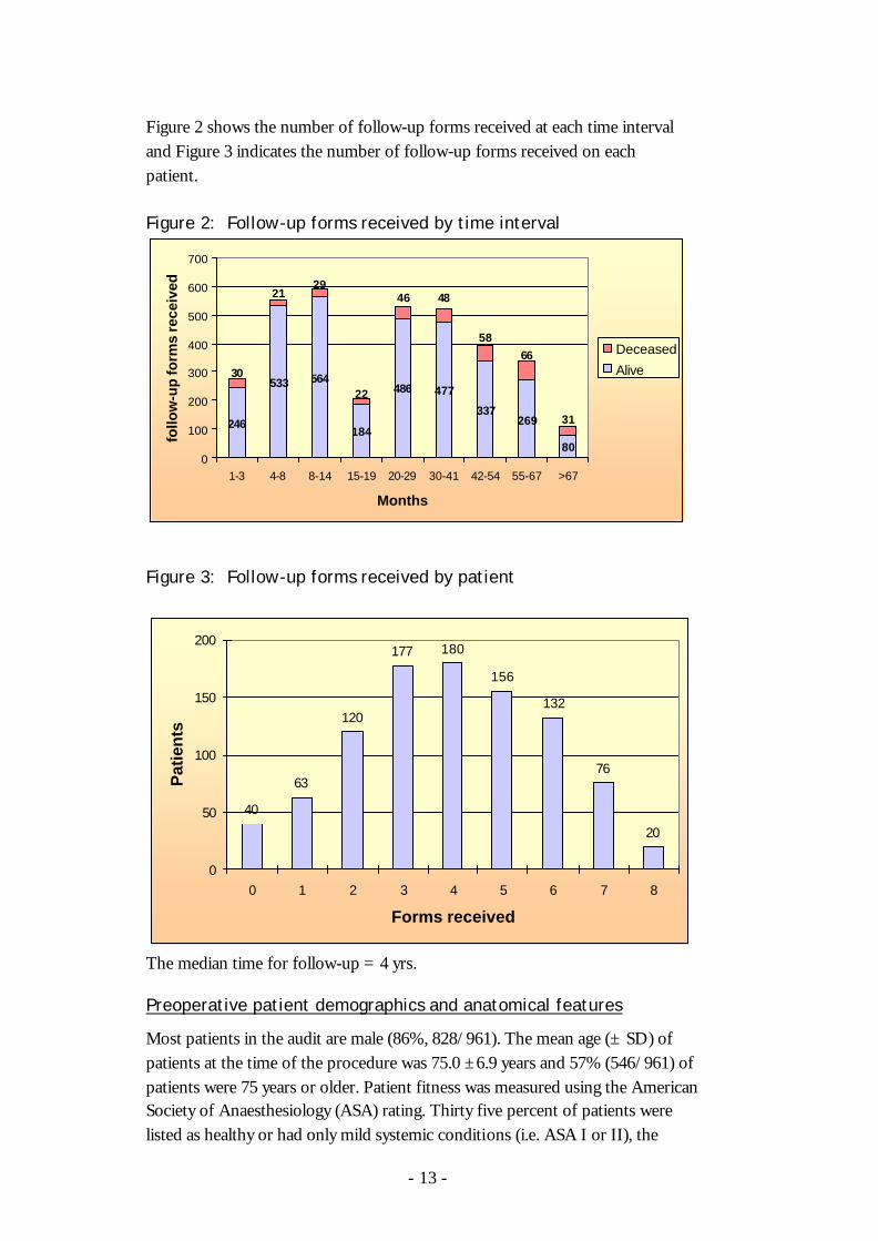
Figure 2 shows the number of follow-up forms received at each time interval and Figure 3 indicates the number of follow-up forms received on each patient. Figure 2: Follow-up forms received by time interval
246
533 564
184
486 477
337269
80
2129
22
30
31
66
58
4846
0
100
200
300
400
500
600
700
1-3 4-8 8-14 15-19 20-29 30-41 42-54 55-67 >67
Months
follo
w-u
p fo
rms
rece
ived
Deceased
Alive
Figure 3: Follow-up forms received by patient
40
63
120
177 180
156
132
76
20
0
50
100
150
200
0 1 2 3 4 5 6 7 8
Forms received
Pat
ien
ts
The median time for follow-up = 4 yrs.
Preoperative patient demographics and anatomical features
Most patients in the audit are male (86%, 828/961). The mean age (± SD) of patients at the time of the procedure was 75.0 ±6.9 years and 57% (546/961) of patients were 75 years or older. Patient fitness was measured using the American Society of Anaesthesiology (ASA) rating. Thirty five percent of patients were listed as healthy or had only mild systemic conditions (i.e. ASA I or II), the

- 14 -
majority of patients (59%) were ASA III (559/945). Figure 4 shows ASA rating for EVAR patients. Figure 4: ASA values for EVAR patients
The number of systemic conditions diagnosed for patients prior to surgery ranged from 0 to more than 10 per patient (55% = 3 conditions). Few patients were listed as current smokers (10%, 89/892), however 71% of patients had smoked at some point (634/892). Mean preoperative aneurysm diameter was 57.5mm (± 10.4mm). Where maximum aneurysm diameter was reported, a total of 44% (411/931) of aneurysms measured less than 55mm in diameter, with 27% (255/931) =50mm in diameter. Ten percent (83/870) of patients had an infrarenal “neck” length less than 15mm. Thirty four percent of patients had a neck length between 15 and 20mm (300/870). An infrarenal neck diameter = 28mm was recorded in 16% of cases (143/877). Significant aortic neck angulation was noted for 229 patients (24%), but the angle size provided varied between 10o and 90o. A neck angle > 45o was noted in 8% of patients (13% were = 45o). Significant aneurysm angulation was noted for 57 patients (6%) with angle size ranging between 5o and 90o. An aneurysm angle of greater than 60o was recorded for only 1% of patients (1.5% were = 60o). Twelve percent (105/884) of patients had thrombi in the aneurysm neck, 22% had a saccular aneurysm (186/869) and 29% had an iliac aneurysm (249/871). Occlusive aorto-iliac disease was noted in 12% of patients (104/851). Iliac tortuosity was severe in 13% of patients (115/898) and moderate in a further 28%
3%
32%
59%
6%
ASA I ASA II
ASA III ASA IV

- 15 -
of patients (247/898); iliac calcification was severe in 5% of patients (41/868) and moderate in 20% of patients (171/868). The aorta was the artery affected by aneurysm in 72% of patients (646/901), whilst a further 26% of aneurysms affected the aorto-iliac arteries (236/901). Forty five percent of patients (411/914) were regarded as unsuitable candidates for open repair. For this group, the main reason given was co-existent morbidity (77%), whilst ‘hostile abdomen’ and ‘unfit for general anaesthetic’ were given as further or additional reasons for 37% of patients. The patients who were considered suitable for open repair were significantly fitter (ASA I/ASA II = 13% not open v 51% open; p<0.05) with fewer comorbidities (3.5 not open v 2.2 open; p<0.05). Patient renal function was measured by pre-operative creatinine level. The mean creatinine among all patients for which it was recorded was 114.9µmol/L, ranging between 41 and 800µmol/L (n=908). Sixty seven percent of patients showed normal renal function with creatinine <120µmol/L (604/908). Mid-range creatinine =120 and =159µmol/L was recorded in 21% of patients (193/908) and high creatinine =160µmol/L was recorded in 12% of patients (111/908).

- 16 -
Summary statistics for females enrolled in audit The average age for the 133 female patients was 75.6 [7.0] years at the time of the procedure. Thirty four percent had an ASA I or II, and 57% had = 3 conditions prior to surgery. The average pre-operative aneurysm diameter was 55 [9.0mm], and 55% of aneurysms measured less than 55mm; 39% measured =50mm. Fourteen percent had an infrarenal neck length <15mm (17/125); 46% had a neck length between 15 and 20mm (n=57). Infrarenal neck diameter was = 28mm in 10% of females treated (13/126). Significant aortic neck angulation was noted in 55 females (41%) with sizes ranging from 15o to 90o; an aortic neck angulation of >450 was noted in 20% of patients (n=26). Significant aneurysm angle was recorded in 17 women (13%) ranging between 30 o and 90 o; an angle >60 o was present in two patients (1.5%). Among the females in the audit, mean pre-operative creatinine was 105.5µmol/L (min 45, max 397µmol/L). The majority of females had normal creatinine level >120µmol/L (75%, 98/130). Thirteen percent had mid-range creatinine =120 and =159µmol/L (17/130) and 12% had high creatinine =160µmol/L (16/130). In the female group, thrombus was noted in 15/129 patients (12%); the aneurysm was reported as saccular in 30/123 patients (24%) and iliac in 18% of patients (23/134). Occlusive aorto-iliac disease was indicated in 12% of women (14/120). Iliac tortuosity was severe in 10% of women, whilst iliac calcification was severe in 9% of women. The aorta was the aneurysm affected artery for 80% of women (102/128). Open repair was not considered suitable for 43% of women (58/134), mostly due to co-existent morbidities (71%).

- 17 -
Imaging
On average, two imaging techniques were used preoperatively (mean number of imaging techniques = 2.1 [0.62]). The most commonly used imaging techniques preoperatively were angiography and spiral CT, used in 83% and 94% of cases respectively. As shown in Table 3, when used in combination together or with other imaging techniques angiography and spiral CT accounted for the majority of patients’ preoperative imaging. Ultrasound was not used as a preoperative imaging technique; however Table 3 shows ultrasound and spiral CT were the most commonly used imaging techniques for later follow-ups and that angiography was not typically used at mid- and long-term follow-up. Ultrasound was used in 59% and spiral CT was used in 42% of long-term follow-ups (55-67 months). For follow-ups that took place after 40 months the mean number of imaging techniques used was 1.3 [0.66]. Table 3: Imaging technique use over time Imaging technique Preoperative
(n=961) Mid-term follow-up 30- 41 months (n=470)
Long-term follow-up 55-67 months (n=256)
Angiography alone 18 (2%) 5 (1%) 0 (0%0 Angiography + other technique(s)
785 (82%) 6 (1%) 8 (3%)
Angiography + spiral CT
538 (56%) 1 (0.2%) 3 (1%)
Spiral / other CT alone 101 (11%) 172 (37%) 63 (25%) Spiral CT + other technique(s)
785 (82%) 104 (22%) 57 (22%)
Spiral CT + x-ray 6 (1%) 64 (14%) 30 (12%) Spiral CT + ultrasound 42 (4%) 16 (3%) 12 (5%) Ultrasound alone 0 (0%) 125 (27%) 72 (28%) Ultrasound + x-ray 0 (0%) 52 (11%) 33 (13%) No imaging listed 18 (2%) 29 (6%) 23 (9%)
- 18 -
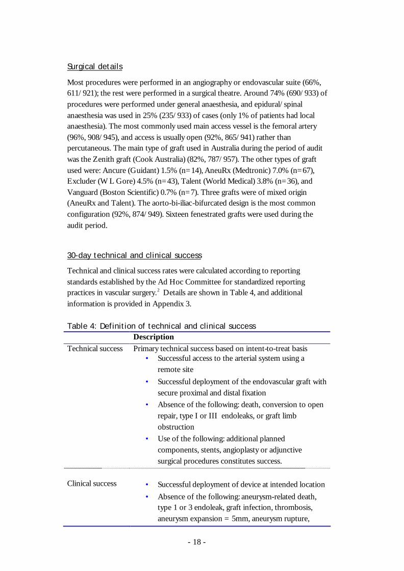
Surgical details
Most procedures were performed in an angiography or endovascular suite (66%, 611/921); the rest were performed in a surgical theatre. Around 74% (690/933) of procedures were performed under general anaesthesia, and epidural/spinal anaesthesia was used in 25% (235/933) of cases (only 1% of patients had local anaesthesia). The most commonly used main access vessel is the femoral artery (96%, 908/945), and access is usually open (92%, 865/941) rather than percutaneous. The main type of graft used in Australia during the period of audit was the Zenith graft (Cook Australia) (82%, 787/957). The other types of graft used were: Ancure (Guidant) 1.5% (n=14), AneuRx (Medtronic) 7.0% (n=67), Excluder (W L Gore) 4.5% (n=43), Talent (World Medical) 3.8% (n=36), and Vanguard (Boston Scientific) 0.7% (n=7). Three grafts were of mixed origin (AneuRx and Talent). The aorto-bi-iliac-bifurcated design is the most common configuration (92%, 874/949). Sixteen fenestrated grafts were used during the audit period.
30-day technical and clinical success
Technical and clinical success rates were calculated according to reporting standards established by the Ad Hoc Committee for standardized reporting practices in vascular surgery.2 Details are shown in Table 4, and additional information is provided in Appendix 3. Table 4: Definition of technical and clinical success Description Technical success Primary technical success based on intent-to-treat basis • Successful access to the arterial system using a
remote site • Successful deployment of the endovascular graft with
secure proximal and distal fixation • Absence of the following: death, conversion to open
repair, type I or III endoleaks, or graft limb obstruction
• Use of the following: additional planned components, stents, angioplasty or adjunctive surgical procedures constitutes success.
Clinical success • Successful deployment of device at intended location • Absence of the following: aneurysm-related death,
type 1 or 3 endoleak, graft infection, thrombosis, aneurysm expansion = 5mm, aneurysm rupture,
- 19 -
conversion to open repair, graft migration, failure of device integrity
Assisted clinical success
• Additional endovascular procedures to achieve ongoing clinical success
Secondary clinical success
• Additional surgical procedures to achieve ongoing clinical success
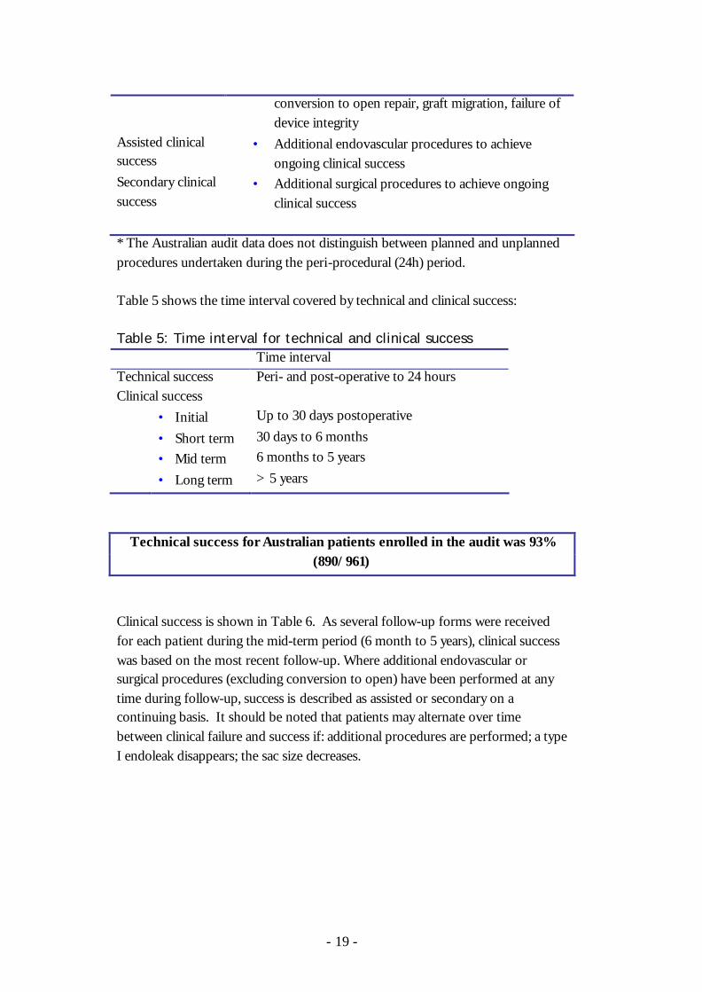
* The Australian audit data does not distinguish between planned and unplanned procedures undertaken during the peri-procedural (24h) period. Table 5 shows the time interval covered by technical and clinical success: Table 5: Time interval for technical and clinical success Time interval Technical success Peri- and post-operative to 24 hours Clinical success • Initial Up to 30 days postoperative • Short term 30 days to 6 months • Mid term 6 months to 5 years • Long term > 5 years
Technical success for Australian patients enrolled in the audit was 93% (890/961)
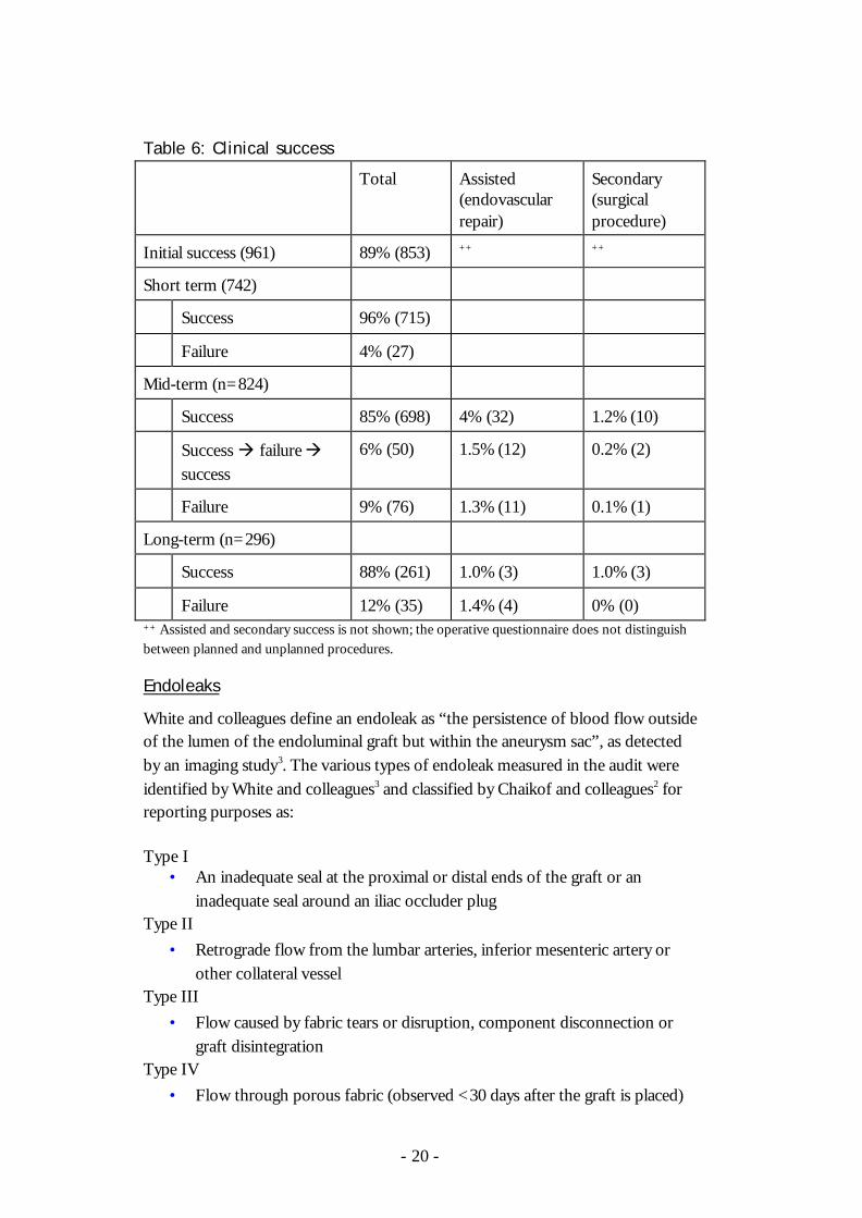
Clinical success is shown in Table 6. As several follow-up forms were received for each patient during the mid-term period (6 month to 5 years), clinical success was based on the most recent follow-up. Where additional endovascular or surgical procedures (excluding conversion to open) have been performed at any time during follow-up, success is described as assisted or secondary on a continuing basis. It should be noted that patients may alternate over time between clinical failure and success if: additional procedures are performed; a type I endoleak disappears; the sac size decreases.
- 20 -
Table 6: Clinical success
Total Assisted (endovascular repair)
Secondary
(surgical procedure)
Initial success (961) 89% (853) ++ ++
Short term (742)
Success 96% (715)
Failure 4% (27)
Mid-term (n=824)
Success 85% (698) 4% (32) 1.2% (10)
Success à failure à success
6% (50) 1.5% (12) 0.2% (2)
Failure 9% (76) 1.3% (11) 0.1% (1)
Long-term (n=296)
Success 88% (261) 1.0% (3) 1.0% (3)
Failure 12% (35) 1.4% (4) 0% (0) ++ Assisted and secondary success is not shown; the operative questionnaire does not distinguish between planned and unplanned procedures.
Endoleaks
White and colleagues define an endoleak as “the persistence of blood flow outside of the lumen of the endoluminal graft but within the aneurysm sac”, as detected by an imaging study3. The various types of endoleak measured in the audit were identified by White and colleagues3 and classified by Chaikof and colleagues2 for reporting purposes as: Type I
• An inadequate seal at the proximal or distal ends of the graft or an inadequate seal around an iliac occluder plug
Type II • Retrograde flow from the lumbar arteries, inferior mesenteric artery or
other collateral vessel Type III
• Flow caused by fabric tears or disruption, component disconnection or graft disintegration
Type IV • Flow through porous fabric (observed <30 days after the graft is placed)

- 21 -
During the perioperative period 24 patients (2.5%) were recorded with type I endoleaks, four patients (0.5%) with both type I and II endoleaks, 64 patients (6.5%) with type II endoleaks only, and one with type II and type IV endoleaks. Table 7 indicates the prognosis of those patients recorded with type I endoleaks. Table 7: Endoleaks recorded < 30 days (n=24) Comments Normal at first follow-up 15 (62.5%) 1 patient had a type I endoleak at
2nd follow-up Type II endoleaks only at
first follow-up 3 (12.5%)
Type I endoleak treated 3 (12.5%) 1 treated following rupture due to contralateral limb separation at 3 years
Deceased 1 (4.2%) septicaemia < 30 days Converted to open 1 (4.2%) following rupture at 6 days Unknown 1 (4.2%) Of the four patients who had type I and type II endoleaks recorded in the perioperative period, one was clear of both types of endoleak at all subsequent follow-ups, one was clear until two years when the type II leak was observed again, and two reported continuing type II endoleaks (but not type I) throughout all follow-ups. Table 8 describes the outcomes for the 36 patients who recorded type I endoleaks during mid-term follow-up. Table 8: Mid-term type I endoleak Type I endoleak during mid-term follow-up 36 Resolved (no treatment) 2 (6%) Additional procedures 25 (69%) Ongoing (declined treatment) 7 (19%) Rupture ð deceased 2 (6%) Additional information: Conversion to open 5 (14%) Total number of ruptures 3 (8%) Migration 11 (31%) Aneurysm-related death 3 (8%)

- 22 -
Five patients recorded type I endoleaks during long term follow-up. Three were new endoleaks and these were treated surgically. The fourth (long-term endoleak) was being treated conservatively and the fifth had an additional procedure planned. Type II endoleaks were noted in 111 patients at some point during their mid-term follow-up. Information relating to these patients is shown in Table 9: Table 9: Mid-term type II endoleaks Type II endoleak at some point during follow-up 111 Resolved 51 (46%) Ongoing (mid-term) 17 (15%) Ongoing (long-term) 12 (11%) Ongoing ð until death 31 (28%) Additional information: Ruptured aneurysm and repaired 1 (1%) Ruptured aneurysm ð deceased 3 (3%) Clinical failure 31 (28%) Converted to open 8 (7%) 21 patients were recorded with type II endoleaks during long term follow-up. For these patients 12 were considered to be clinical failures, mostly (11/12) due to an increase in sac size. Only one of the 21 had subsequently died at the time of this report. Type III endoleaks were not originally specified on the questionnaire, but were added as data points in 2003. To date four type III endoleaks have been reported. One of these patients died at 24 months when an intra-sac injection dislodged the contralateral limb. Two patients had additional endovascular treatment (one underwent a second procedure when the first was unsuccessful), the outcomes for the fourth patient have not yet been received.
Complications (not including endoleaks)
Graft related complications noted immediately following the procedure included failed access, access vessel complications, failed and misplaced deployment of endografts, imperfect seal, twist/kink/obstruction and embolisation. For the 4% of patients whose aneurysms were not successfully excluded (n=41) the following reasons were specified (Table 10):

- 23 -
Table 10: Complications, excluding endoleak where aneurysm not excluded Imperfect seal 18 Failed access 4 Access vessel complications + imperfect seal 2 Failed deployment 1 Misplaced deployment 1 Failed deployment + imperfect seal 1 Misplaced deployment + imperfect seal 1 Misplaced deployment + imperfect seal + embolisation 1 Not specified 12 A further 8% of patients (n=76), whose aneurysms were successfully excluded, had the following complications recorded (Table 11): Table 11: Complications, excluding endoleak where aneurysm excluded Imperfect seal 25 Twist/kink/obstruction 18 Access vessel complications 14 Misplaced deployment 8 Failed access and access vessel complications 3 Embolisation 3 Failed deployment 2 Misplaced deployment + imperfect seal 2 Failed access 1 Prior to discharge 18 patients (2%) had graft related complications (Table 12): Table 12: Graft related complications prior to discharge Thrombosis 6 Stenosis 4 Migration 3 Kinking 2 Migration + thrombosis 1 Migration + kinking 1 Graft infection 1 Surgeons noted a range of systemic complications (excluding pyrexia) prior to discharge in 182 patients (19%); the leading cause was attributed to cardiac-related complications (7%, n=70).
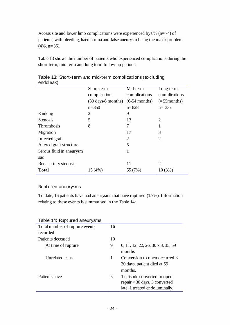
- 24 -
Access site and lower limb complications were experienced by 8% (n=74) of patients, with bleeding, haematoma and false aneurysm being the major problem (4%, n=36). Table 13 shows the number of patients who experienced complications during the short term, mid term and long term follow-up periods. Table 13: Short-term and mid-term complications (excluding endoleak) Short-term
complications (30 days-6 months) n=350
Mid-term complications (6-54 months) n=828
Long-term complications (=55months) n= 337
Kinking 2 9 Stenosis 5 13 2 Thrombosis 8 7 1 Migration 17 3 Infected graft 2 2 Altered graft structure 5 Serous fluid in aneurysm sac
1
Renal artery stenosis 11 2 Total 15 (4%) 55 (7%) 10 (3%)
Ruptured aneurysms
To date, 16 patients have had aneurysms that have ruptured (1.7%). Information relating to these events is summarised in the Table 14: Table 14: Ruptured aneurysms Total number of rupture events recorded
16
Patients deceased 10 At time of rupture 9 0, 11, 12, 22, 26, 30 x 3, 35, 59
months Unrelated cause 1 Conversion to open occurred <
30 days, patient died at 59 months.
Patients alive 5 1 episode converted to open repair <30 days, 3 converted late, 1 treated endoluminally.

- 25 -
Occluded graft limbs
In order to evaluate the proportion of patients who had experienced occluded graft limbs following EVAR, the follow-up table and the discharge and 30 day follow-up table were searched for references to this complication. Cases with ‘thrombosis’ fields ticked for complications prior to discharge or results of follow-up imaging (discharge or follow-up forms) and cases with the words occluded graft, occlusion, thrombus in graft, thrombectomy, embolectomy, graft blocked/blockage or fem-fem crossover graft in any of the text fields were included in the count. To date, 6% of patients (56/961) appear to have experienced occluded graft limb at some point following EVAR.
Changes in aneurysm sac size
Figure 5 shows changes in aneurysm size over time. Aneurysms were deemed to be the same size if they were within 5mm of the original (pre-operative) measurement. Figure 5: Changes in aneurysm size (±5mm)
290336 286 246 284
329182 122 85 67
26 25 18 32 33
0%
20%
40%
60%
80%
100%
year 1(645)
year 2(543)
year 3(426)
year 4(363)
year 5+(384)
Years (n)
Larger
Same (+/-5mm)Smaller
Additional interventions
Three types of intervention are reported: • additional procedures performed at the time of the original procedure
(often referred to as secondary procedures) • interventions performed after the initial procedure but prior to discharge • interventions recorded at follow-up (shown in Table 6 – assisted or
secondary clinical success)
- 26 -
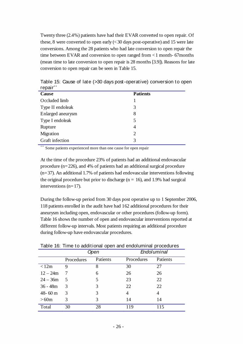
Twenty three (2.4%) patients have had their EVAR converted to open repair. Of these, 8 were converted to open early (<30 days post-operative) and 15 were late conversions. Among the 28 patients who had late conversion to open repair the time between EVAR and conversion to open ranged from <1 month- 67months (mean time to late conversion to open repair is 28 months [3.9]). Reasons for late conversion to open repair can be seen in Table 15. Table 15: Cause of late (>30 days post-operative) conversion to open repair++ Cause Patients Occluded limb 1 Type II endoleak 3 Enlarged aneurysm 8 Type I endoleak 5 Rupture 4 Migration 2 Graft infection 3 ++ Some patients experienced more than one cause for open repair At the time of the procedure 23% of patients had an additional endovascular procedure (n=226), and 4% of patients had an additional surgical procedure (n=37). An additional 1.7% of patients had endovascular interventions following the original procedure but prior to discharge (n = 16), and 1.9% had surgical interventions (n=17). During the follow-up period from 30 days post operative up to 1 September 2006, 118 patients enrolled in the audit have had 162 additional procedures for their aneurysm including open, endovascular or other procedures (follow-up form). Table 16 shows the number of open and endovascular interventions reported at different follow-up intervals. Most patients requiring an additional procedure during follow-up have endovascular procedures. Table 16: Time to additional open and endoluminal procedures Open Endoluminal Procedures Patients Procedures Patients <12m 9 8 30 27 12 – 24m 7 6 26 26 24 – 36m 5 5 23 22 36 - 48m 3 3 22 22 48- 60 m 3 3 4 4 >60m 3 3 14 14 Total 30 28 119 115

- 27 -
Mortality
ASERNIP-S has received notification that 39% (375/961) of the original cohort of patients have died. Information from the National Death Index (AIHW) was last updated in August 2005. The number of patients who died within 30 days of the operation was 17 (1.8%), and 358 (31%) in the time following the 30 day postoperative period. The reasons given for early death and postoperative days to death are shown below: Table 17: Cause of early mortality and days to death Cause of early death Days to death Retro-peritoneal haemorrhage and MI 0 Cardiac and acidosis 1 MI 2 at 3 days Ischaemic bowel 3 Chronic IHD 3 Pulmonary embolus 6 Renal failure 7 Acute bowel rupture 10 Sepsis 11 Cardiac failure + renal failure 13 Rupture 14 Cerebral haemorrhage 14 Brain stem haemorrhage 18 Septicaemia from infected drip 19 Ischaemic gut 21 Myocardial Infarction 21 Dissection thoracic aorta 27 Twenty-one patients died during the “short-term” period (30 days to six months), mostly due to cardiac causes (14/21). Two deaths were related to aneurysm rupture. For patients who died in the mid-term follow up period (6 months-5 years) following EVAR the mean time to death was 33.5 months [16.3]. Mid-term and long-term mortality fell into the following categories:
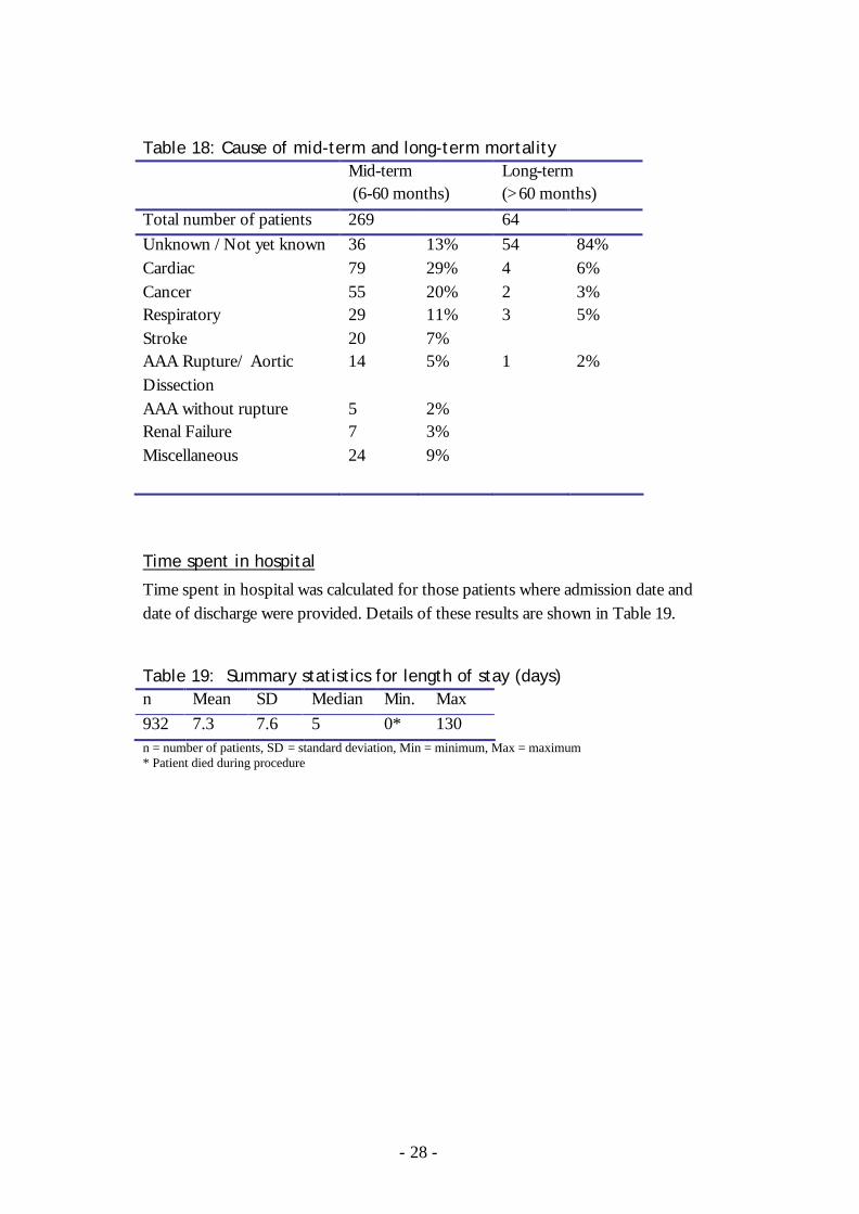
- 28 -
Table 18: Cause of mid-term and long-term mortality Mid-term
(6-60 months) Long-term (>60 months)
Total number of patients 269 64 Unknown /Not yet known 36 13% 54 84% Cardiac 79 29% 4 6% Cancer 55 20% 2 3% Respiratory 29 11% 3 5% Stroke 20 7% AAA Rupture/ Aortic Dissection
14 5% 1 2%
AAA without rupture 5 2% Renal Failure 7 3% Miscellaneous 24 9%
Time spent in hospital
Time spent in hospital was calculated for those patients where admission date and date of discharge were provided. Details of these results are shown in Table 19. Table 19: Summary statistics for length of stay (days) n Mean SD Median Min. Max 932 7.3 7.6 5 0* 130 n = number of patients, SD = standard deviation, Min = minimum, Max = maximum * Patient died during procedure
- 29 -
Comparison of Australian data with Eurostar∗ data registry results
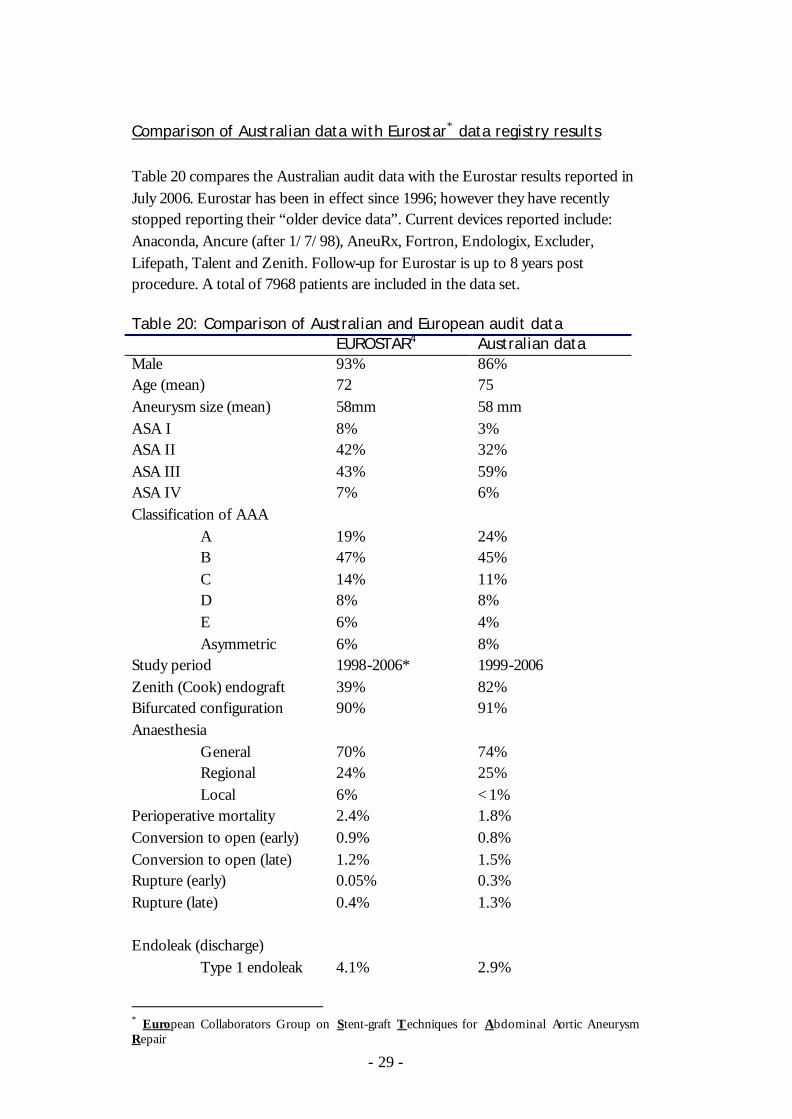
Table 20 compares the Australian audit data with the Eurostar results reported in July 2006. Eurostar has been in effect since 1996; however they have recently stopped reporting their “older device data”. Current devices reported include: Anaconda, Ancure (after 1/7/98), AneuRx, Fortron, Endologix, Excluder, Lifepath, Talent and Zenith. Follow-up for Eurostar is up to 8 years post procedure. A total of 7968 patients are included in the data set. Table 20: Comparison of Australian and European audit data EUROSTAR4 Australian data Male 93% 86% Age (mean) 72 75 Aneurysm size (mean) 58mm 58 mm ASA I 8% 3% ASA II 42% 32% ASA III 43% 59% ASA IV 7% 6% Classification of AAA A 19% 24% B 47% 45% C 14% 11% D 8% 8% E 6% 4% Asymmetric 6% 8% Study period 1998-2006* 1999-2006 Zenith (Cook) endograft 39% 82% Bifurcated configuration 90% 91% Anaesthesia General 70% 74% Regional 24% 25% Local 6% <1% Perioperative mortality 2.4% 1.8% Conversion to open (early) 0.9% 0.8% Conversion to open (late) 1.2% 1.5% Rupture (early) 0.05% 0.3% Rupture (late) 0.4% 1.3% Endoleak (discharge) Type 1 endoleak 4.1% 2.9%
∗ European Collaborators Group on Stent-graft Techniques for Abdominal Aortic Aneurysm Repair

- 30 -
Type 2 endoleak 9.9% 7% Type 3 endoleak 1.9% NA Endoleak 7.5% (at 4-year
follow-up; 63/844) 16% (at any time during mid-term follow-up; 152/961)
Length of stay (mean± standard deviation)
5.98±8.1 days 7.3±7.6 days
n= 7968 (Eurostar), 961 (Australian data) * 2006 Eurostar report excludes “older device data” from analysis. A recent systematic review by Drury et al5 of elective endovascular repair identified 61 studies for inclusion (three randomised controlled trials, 15 non-randomised controlled trials and 43 uncontrolled studies) with 19804 patients undergoing EVAR and 9255 undergoing open repair. Results are shown in Table 21. Only papers published after 2000 were included. Table 21: Safety and Efficacy of elective EVAR (Drury5)
Mean follow-up 7-39 months Male 90.7% Age (mean) 71 Study criteria Papers published between Jan 2000 and Sep
2004. English language articles, RCT’s NRCR’s controlled clinical trials, comparative observational studies, case series and population based registries. Elective EVAR.
Perioperative mortality 1.6% for RCT’s, 1.4% for NRCT’s Conversion to open (early)
1.7% (0-10%)
Conversion to open (late) 2.0% (increasing to 9.1% for 39 month study) Rupture (early) 0.3% (7 studies) Rupture (late) 0.6% (18 studies, n=8552, mean 17 months) Increase in sac size 7.1% (after initial placement) Endoleak 30 days Type 1 endoleak 3.5% 12 months Type 1 endoleak 6.8% 30 days Type 2 endoleak 14% 12 months Type 2 endoleak 10.3% Primary technical success 88.8% (16 studies) Secondary interventions* 16.2% (34 studies) * defined as any surgical or radiological procedure carried out to maintain exclusion of the aneurysm sac from the circulation or to maintain graft patency.

- 31 -
Survival analysis for Eurostar and the Australian audit are shown in Tables 22 and 23. Table 22: Survival table (Eurostar4) Time (months)
Number entering interval
Number of deaths (cumulative)
Proportion surviving (%)
Survival standard error
0 8345 158 98.1 0.001 1 8187 198 97.5 0.002 3 7002 286 96.2 0.002 6 6493 427 94.0 0.003 12 5903 553 91.6 0.003 18 4796 637 89.6 0.004 24 3732 769 85.8 0.005 36 3005 836 81.7 0.006 48 1872 926 76.9 0.008 60 1013 953 73.4 0.010 72 560 970 68.9 0.014 * Methodology not stated in July 2006 report. Table 23: Survival* (Australian audit) Time (months)
Number entering interval
Number of deaths
Proportion surviving (%)
Survival standard error
0 960 10 99.0 0.33 1 950 11 97.8 0.47 3 939 7 97.1 0.54 6 932 13 95.7 0.65 12 919 31 92.5 0.85 18 888 31 89.3 1.00 24 857 29 86.2 1.11 36 828 56 80.4 1.28 48 772 58 74.4 1.41 60 714 52 69.0 1.49 72 365 12 66.7 1.58 * Obtained using right censored Kaplan-Meier survival analysis.

- 32 -
Statistical analyses of outcome and survival following EVAR Three statistical reports have been prepared by a statistician from the CSIRO for the EVAR audit.6-8 The following information is taken from these reports. Pre-operative aneurysm diameter was found to explain more variation in the different success measures than any other predictor variable. ASA, age, open suitability, the number of pre-existing conditions and type of device were the variables with the next highest number of statistically significant p-values for different success measures. ASA, age, aneurysm size and creatinine were found to significantly contribute to predict survival. Predictors of aneurysm-related death were ASA, creatinine, aneurysm size, infrarenal neck length and aortic neck angle. A predictive model of success has also been devised and developed by CSIRO based on logistic generalised linear regression. This model will be trialled by surgeons and aims to help surgeons and patients decide treatment options based on possible outcomes. Currently the model looks at outcomes such as survival, clinical success, endoleak, rupture, conversion to open, migration and reinterventions. The model may be refined following a trial period.
- 33 -
44.. DDiissccuussssiioonn
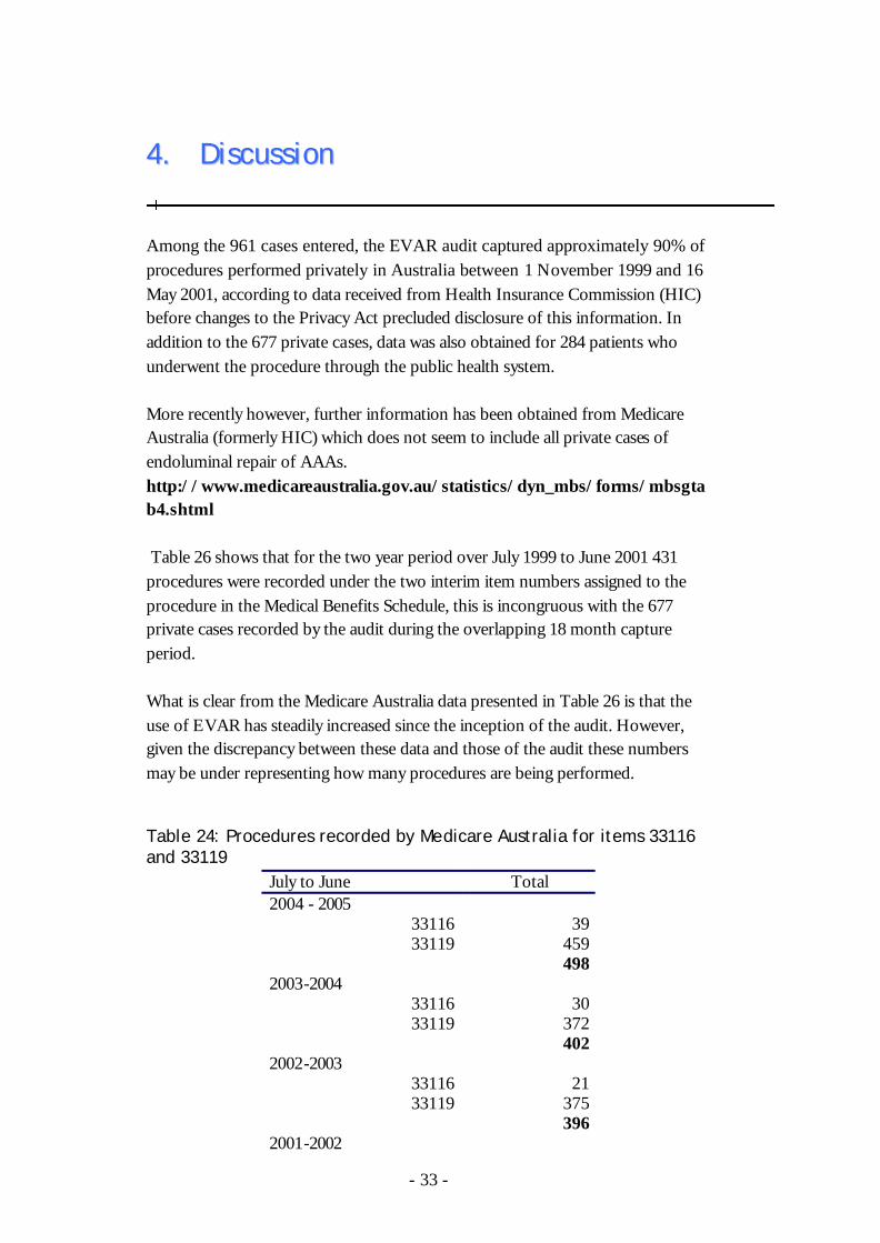
Among the 961 cases entered, the EVAR audit captured approximately 90% of procedures performed privately in Australia between 1 November 1999 and 16 May 2001, according to data received from Health Insurance Commission (HIC) before changes to the Privacy Act precluded disclosure of this information. In addition to the 677 private cases, data was also obtained for 284 patients who underwent the procedure through the public health system. More recently however, further information has been obtained from Medicare Australia (formerly HIC) which does not seem to include all private cases of endoluminal repair of AAAs. http://www.medicareaustralia.gov.au/statistics/dyn_mbs/forms/mbsgtab4.shtml Table 26 shows that for the two year period over July 1999 to June 2001 431 procedures were recorded under the two interim item numbers assigned to the procedure in the Medical Benefits Schedule, this is incongruous with the 677 private cases recorded by the audit during the overlapping 18 month capture period. What is clear from the Medicare Australia data presented in Table 26 is that the use of EVAR has steadily increased since the inception of the audit. However, given the discrepancy between these data and those of the audit these numbers may be under representing how many procedures are being performed. Table 24: Procedures recorded by Medicare Australia for items 33116 and 33119
July to June Total 2004 - 2005
33116 39 33119 459
498 2003-2004
33116 30 33119 372
402 2002-2003
33116 21 33119 375
396 2001-2002
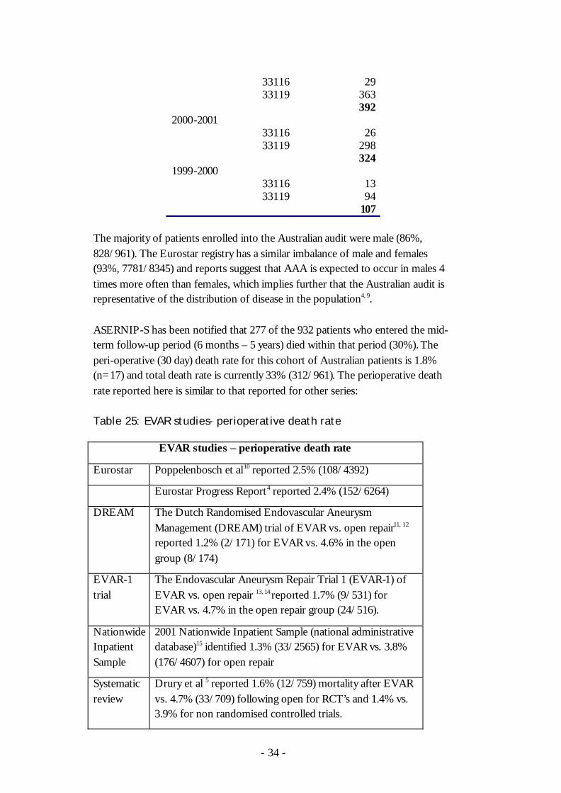
- 34 -
33116 29 33119 363
392 2000-2001
33116 26 33119 298
324 1999-2000
33116 13 33119 94
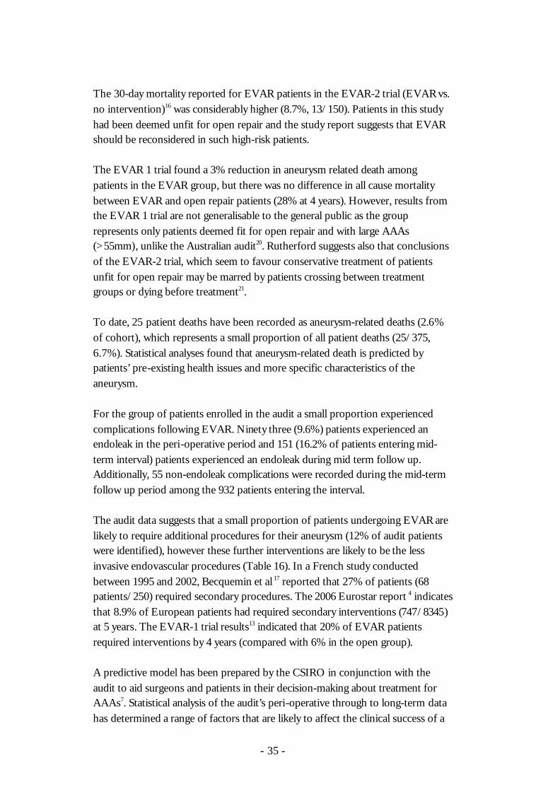
107 The majority of patients enrolled into the Australian audit were male (86%, 828/961). The Eurostar registry has a similar imbalance of male and females (93%, 7781/8345) and reports suggest that AAA is expected to occur in males 4 times more often than females, which implies further that the Australian audit is representative of the distribution of disease in the population4, 9. ASERNIP-S has been notified that 277 of the 932 patients who entered the mid-term follow-up period (6 months – 5 years) died within that period (30%). The peri-operative (30 day) death rate for this cohort of Australian patients is 1.8% (n=17) and total death rate is currently 33% (312/961). The perioperative death rate reported here is similar to that reported for other series: Table 25: EVAR studies- perioperative death rate
EVAR studies – perioperative death rate
Eurostar Poppelenbosch et al10 reported 2.5% (108/4392)
Eurostar Progress Report4 reported 2.4% (152/6264)
DREAM The Dutch Randomised Endovascular Aneurysm Management (DREAM) trial of EVAR vs. open repair11, 12 reported 1.2% (2/171) for EVAR vs. 4.6% in the open group (8/174)
EVAR-1 trial
The Endovascular Aneurysm Repair Trial 1 (EVAR-1) of EVAR vs. open repair 13, 14 reported 1.7% (9/531) for EVAR vs. 4.7% in the open repair group (24/516).
Nationwide Inpatient Sample
2001 Nationwide Inpatient Sample (national administrative database)15 identified 1.3% (33/2565) for EVAR vs. 3.8% (176/4607) for open repair
Systematic review
Drury et al 5 reported 1.6% (12/759) mortality after EVAR vs. 4.7% (33/709) following open for RCT’s and 1.4% vs. 3.9% for non randomised controlled trials.
- 35 -
The 30-day mortality reported for EVAR patients in the EVAR-2 trial (EVAR vs. no intervention)16 was considerably higher (8.7%, 13/150). Patients in this study had been deemed unfit for open repair and the study report suggests that EVAR should be reconsidered in such high-risk patients. The EVAR 1 trial found a 3% reduction in aneurysm related death among patients in the EVAR group, but there was no difference in all cause mortality between EVAR and open repair patients (28% at 4 years). However, results from the EVAR 1 trial are not generalisable to the general public as the group represents only patients deemed fit for open repair and with large AAAs (>55mm), unlike the Australian audit20. Rutherford suggests also that conclusions of the EVAR-2 trial, which seem to favour conservative treatment of patients unfit for open repair may be marred by patients crossing between treatment groups or dying before treatment21. To date, 25 patient deaths have been recorded as aneurysm-related deaths (2.6% of cohort), which represents a small proportion of all patient deaths (25/375, 6.7%). Statistical analyses found that aneurysm-related death is predicted by patients’ pre-existing health issues and more specific characteristics of the aneurysm. For the group of patients enrolled in the audit a small proportion experienced complications following EVAR. Ninety three (9.6%) patients experienced an endoleak in the peri-operative period and 151 (16.2% of patients entering mid-term interval) patients experienced an endoleak during mid term follow up. Additionally, 55 non-endoleak complications were recorded during the mid-term follow up period among the 932 patients entering the interval. The audit data suggests that a small proportion of patients undergoing EVAR are likely to require additional procedures for their aneurysm (12% of audit patients were identified), however these further interventions are likely to be the less invasive endovascular procedures (Table 16). In a French study conducted between 1995 and 2002, Becquemin et al 17 reported that 27% of patients (68 patients/250) required secondary procedures. The 2006 Eurostar report 4 indicates that 8.9% of European patients had required secondary interventions (747/8345) at 5 years. The EVAR-1 trial results13 indicated that 20% of EVAR patients required interventions by 4 years (compared with 6% in the open group). A predictive model has been prepared by the CSIRO in conjunction with the audit to aid surgeons and patients in their decision-making about treatment for AAAs7. Statistical analysis of the audit’s peri-operative through to long-term data has determined a range of factors that are likely to affect the clinical success of a

- 36 -
patient’s treatment by EVAR. ASERNIP-S and the CSIRO intend for this model to be available for surgeons to use to predict patients’ suitability for EVAR and likelihood of mid- to long-term clinical success. Patient and aneurysm factors can be entered into a Microsoft Excel spreadsheet programme at the time of initial consult and again following CT angiography to update the patient’s profile. ASERNIP-S has been notified that 11% (103/961) of surviving patients are considered lost to follow-up. Patients are regarded as ‘lost to follow-up’ for a number of reasons including: whether the patient is non-contactable or has refused further follow-up, or because the treating surgeon has retired or died and we cannot ascertain who has become responsible for the patient. Comments from surgeons suggest that public and rural patients are harder to follow. The problem of being unable to follow patients after EVAR should be recognised by surgeons when they consider patients for the procedure. Regular follow-up is known to be an important aspect of care; especially for certain sub-groups of patients who are at increased risk of complications. Peppelenbosch et al11 identified worse outcomes for patients with larger aneurysms. Boult et al12 also identified larger aneurysms as significant predictors of failure along with ASA rating, increasing age, larger aneurysm angles and number of comorbid conditions diagnosed preoperatively. In an analysis of the audit data relating to smaller aneurysms (<55mm) Golledge et al9 found a re-intervention rate among EVAR patients similar to that reported for larger aneurysms (estimated arterial reintervention rate was 11% and 13% at 3 and 5 years). They also found that survival was reduced in patients with renal impairment and in those considered unfit for general anaesthesia. This warrants ongoing follow-up of these patients. Corriere et al18 concluded that follow-up should not be relaxed for patients whose aneurysms appear stable, due to the potential for late onset endoleaks. In summary, whilst a number of patients would appear to be at higher risk of experiencing problems in the longer term, the need for ongoing and rigorous follow-up of all patients would appear to be vital to the ongoing success of the procedure. However, it may be that sicker patients (eg those considered unfit for open repair), who are most likely to experience post-EVAR complications, may also be the patients who are unable to attend follow-up in the mid to long-term period. The aim of this audit has been to provide information to the Government on the mid- to long-term safety and efficacy of the procedure with a view to informing the decision as to whether it should be permanently introduced into the Medical Benefits Schedule. The audit has been well received by surgeons and it is hoped

- 37 -
that the predictive model will be well regarded and perceived as a useful tool in the armamentarium of vascular surgeons. Nevertheless, evidence surrounding the procedure and potential outcomes for the patient should be discussed by surgeon and patient to gain fully informed consent. In view of the need to collect good long-term data, the audit has secured sufficient funding to enable it to continue for a further two years. Analysis of this data will be ongoing with the aim of contributing to the evidence base of EVAR, and helping to inform decision-making for health professionals and patients.
- 38 -
55.. RReeffeerreenncceess
1 Medicare Services Advisory Committee, May 1999, Endoluminal grafting
for abdominal aortic aneurysm. MSAC application 1006. AusInfo, Canberra, ACT 2601.
2 Chaikof EL, Blankensteijn JD, Harris PL, White GH, Zarins CK Bernhard VM, Matsumura JS, May J, Veith FJ, Fillinger MF, Rutherford RB, Kent KC. Reporting standards for endovascular aortic aneurysm repair. Journal of Vascular Surgery. 2002: 35(5);1048-1060.
3 White GH, Yu W, May J, Chaufour X, Stephen MS. Endoleak as a complication of endoluminal grafting of abdominal aortic aneurysms: classification, incidence, diagnosis and management. Journal of Endovascular Surgery. 1997; 4:152-168.
4 Buth J (executive director data registry centre). Progress Report: Endografts currently in use only (Anaconda, Ancure, AneuRx, Fortron, Endologix, Excluder, Lifepath, Talent & Zenith). Eurostar Data Registry Centre. July 2006. [Online, accessed 20 September 2006]. URL: http://www.eurostar-online.org/
5 Drury D, Michaels JA, Jones L, Ayiku L. Systematic review of recent evidence for the safety and efficacy of elective endovascular repair in the management of infrarenal abdominal aortic aneurysms. British Journal of Surgery. 2005:92; 937-946.
6 Barnes M. Survival analyses for endoluminal repair of abdominal aortic aneurysms. Prepared by CSIRO for Australian Register of Safety and Efficacy of New Interventional Procedures - Surgical. Report number 06/84; 2006
7 Barnes M. Predictive model for endoluminal repair of abdominal artic aneurysms Prepared by CSIRO for Australian Register of Safety and Efficacy of New Interventional Procedures- Surgical. CMIS 06/167; 2006
8 Barnes M. Statistical analyses for the audit of endoluminal repair of abdominal aortic aneurysms. Prepared by CSIRO for the Australian Register of Safety and Efficacy of New Interventional Procedures - Surgical. Report number 06/162; 2006
9 Golledge J, Parr A, Boult M, Maddern G, Fitridge R. The outcome of endovascular repair of small abdominal aortic aneurysms. Annals of Surgery. 2006; 244(6):1-7.
10 Peppelenbosch N, Buth J, Harris PL, van Marrewijk C, Fransen G, for the Eurostar Collaborators. Diameter of abdominal aortic aneurysm and outcome of endovascular aneurysm repair: does size matter? A report from Eurostar. Journal of Vascular Surgery. 2004; 39(2):288-297.
- 39 -
11 Blankenstein JD, de Jong S, Prinssen M, der Ham A, Buth J, van Sterkenburg S, Verhagen H, Buskens E, Grobbee D, for the Dutch Randomised Endovascular Aneurysm Management (DREAM) Trial Group. Two-year outcomes after conventional or endovascular repair of abdominal aortic aneurysms. New England Journal of Medicine. 2005; 352(23):2398-2405.
12 Prinssen M, Verhoeven E, Buth J, Cuypers P, van Sambeek M, Balm R, Buskens E, Grobbee D, Blankenstein J. A randomised trial comparing conventional and endovascular repair of abdominal aortic aneurysms. New England Journal of Medicine. 2004; 351(16):1607-1618.
13 EVAR trial participants. Endovascular aneurysm repair versus open repair in patients with abdominal aortic aneurysm (EVAR trial 1): randomised controlled trial. The Lancet. 2005; 365:2179-2186.
14 EVAR trial participants. Comparison of endovascular aneurysm repair with open repair in patients with abdominal aortic aneurysm (EVAR trial 1), 30 day operative mortality results: randomised controlled trial. The Lancet. 2004: 364; 843-848.
15 Lee WA, Carter JW, Upchurch G, Seeger JM, Juber TS. Perioperative outcomes after open and endovascular repair of intact abdominal aortic aneurysms in the United States during 2001. Journal of Vascular Surgery. 2004:39(3); 491-496.
16 EVAR trial participants. Endovascular aneurysm repair and outcome in patients unfit for open repair of abdominal aortic aneurysm (EVAR trial 2): randomised controlled trial. The Lancet. 2005: 365; 2187-2192.
17 Becqeumin J-P, Kelley L, Zubilewicz T, Desgranges P, Lapeyre M, Kobeiter H. Outcomes of secondary interventions after abdominal aortic aneurysm endovascular repair. Journal of Vascular Surgery. 2004: 39(2); 298-305.
18 Corriere MA, Feurer ID, Becker SY, Dattilo JB, Passman MA, Guzman RJ, Naslund TC. Endoleak following endovascular abdominal aortic aneurysm repair; implications for duration of screening. Annals of Surgery. 2004: 239(6); 800-807
19 White GH, May J, Petrasek P, Weiyun Y, Waugh RC, Chaufour X. A grading scale to predict the degree of difficulty for endovascular AAA graft procedures. Journal of Endovascular Surgery. 1998; 5(4):380-381.
20 Rutherford R. Endovascular aneurysm repair versus open repair in patients with abdominal aortic aneurysm (EVAR 1): randomised controlled trial. Perspectives in Vascular Surgery and Endovascular Therapy. 2006; 18(1): 74-75
21 Rutherford R. Endovascular aneurysm repair and outcome in patients unfit for open repair of abdominal aortic aneurysm (EVAR trial 2): randomised controlled trial. Perspectives in Vascular Surgery and Endovascular Therapy. 2006; 18(1): 76-77

- 40 -
AAppppeennddiixx 11 –– PPaattiieenntt iinnffoorrmmaattiioonn aanndd ccoonnsseenntt ffoorrmmss
- 41 -
CONSENT TO RELEASE OF INFORMATION 1. I………………………………. of (address)
………………………………….
………………………………….
………………………………….
………………………………….
GIVE CONSENT TO:
the Australian Safety and Efficacy Register of New Interventional Procedures – Surgical (“ASERNIP–S”), on behalf of the Royal Australasian College of Surgeons, to obtain information from medical records concerning my endoluminal grafting for abdominal aortic aneurysm, to enable the Medical Services Advisory Committee (“MSAC”) to conduct an evaluation of the procedure.
2. I UNDERSTAND THAT:
• Any report compiled for the purpose of this audit and evaluation will not use information that could identify me.
• MSAC may authorise ASERNIP-S to release information to other organisations, but this will not include any information that could identify me.
• ASERNIP-S is strongly committed to protecting my privacy and maintains high levels of security.
……………………….. Signature ……………………….. Witness signature ………/………/……… Date

- 42 -
Patient information sheet is available from the ASERNIP-S web site

- 43 -
AAppppeennddiixx 22 -- EEVVAARR ddaattaa eennttrryy ffoorrmmss Available from ASERNIP-S web site http://www.surgeons.org/asernip-s/audit.htm

- 45 -
AAppppeennddiixx 33 –– EExxppllaannaattiioonn ooff ‘‘tteecchhnniiccaall’’ aanndd ‘‘cclliinniiccaall’’ ssuucccceessss,, aanndd WWhhiittee’’ss ggrraaddiinngg ssyysstteemm Technical success2
Technical success relates to the first 24 hours after the procedure and implies the following qualifying details:
• Successful access to the arterial system using a remote site • Successful deployment of the endoluminal graft • Absence of type I or III endoleak • Patent endoluminal graft without significant twist, kinks or obstruction.
The standards delimit three subgroups of technical success according to the planned or unplanned use of additional modular components or surgery: primary technical success, assisted primary success and secondary technical success. Primary technical success can include the use of additional modular components, stents, angioplasty or adjunctive surgical procedures. The unplanned use of endovascular components is described as assisted primary technical success and unplanned additional surgical procedures is described as secondary technical success. Clinical success2
“Clinical success requires successful deployment of the endovascular device at the intended location without death (as a result of aneurysm-related treatment), type I or III endoleak, graft infection or thrombosis, aneurysm expansion (diameter >=5mm or volume >=5%), aneurysm rupture, or conversion to open repair. Moreover, the presence of graft dilatation of 20% or more by diameter, graft migration, or a failure of the device integrity classifies a case as a clinical failure. Clinical success can be claimed for those cases with a type II endoleak only in the absence of aneurysm expansion. As long as the significance of a type II endoleak and its implication as a marker for late clinical failure remains an area of active investigation, it is recommended that reports clearly indicate the proportion of patients classified as a clinical success that harbour a type II endoleak. Initial or 30-day clinical success encompasses 30-day data. Short term clinical success includes outcome measures reported within a 30 days to 6 month time frame. Mid term clinical success refers to all outcome measures that are statistically significant up to 5 years after endograft implantation. Long-term clinical success refers to all outcome measure that are statistically significant beyond 5 years. Primary clinical success is clinical success without the need for an additional or secondary surgical or endovascular procedure. Assisted primary success is clinical success achieved with the use of an additional or secondary endovascular procedure. Secondary clinical success is clinical success obtained with the use of an additional surgical procedure.
Clinical success was calculated for the Australian data for discharge and 30 day follow-up.
- 46 -
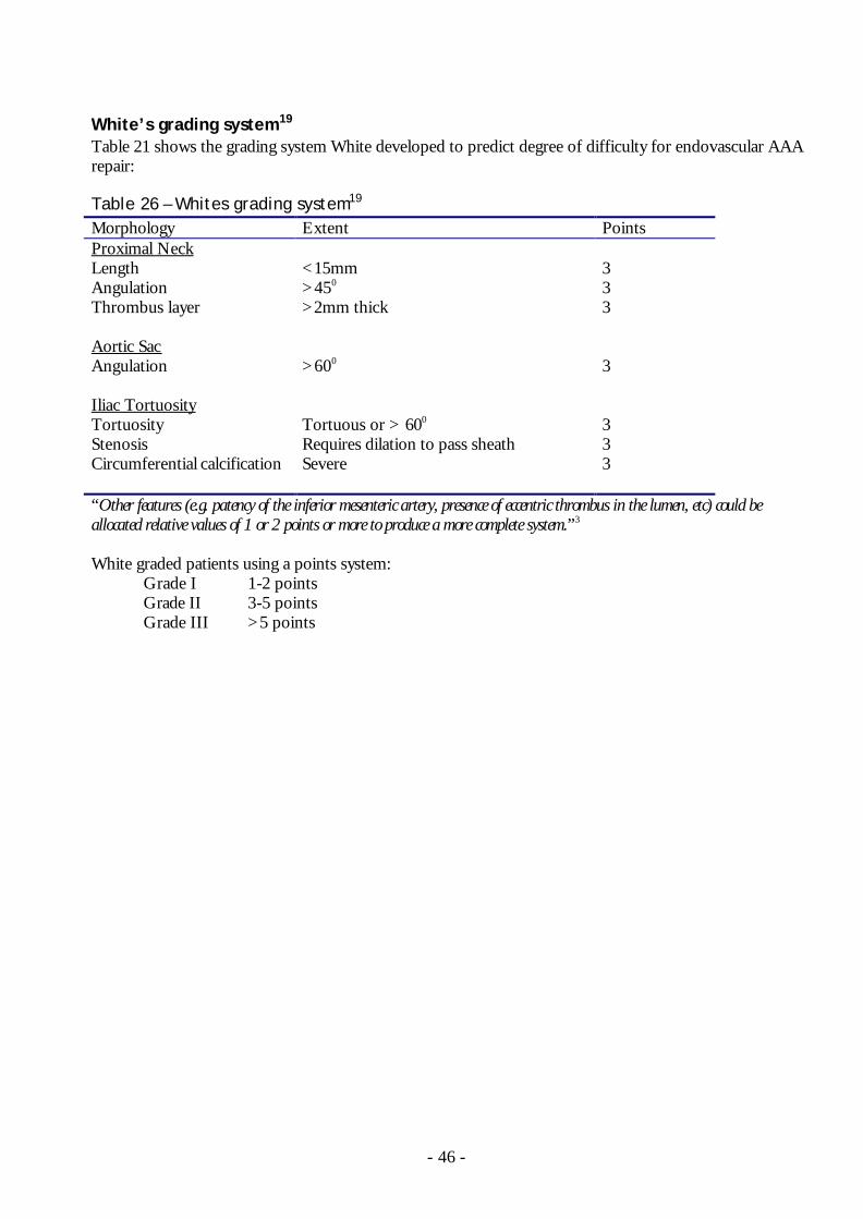
White’s grading system19
Table 21 shows the grading system White developed to predict degree of difficulty for endovascular AAA repair: Table 26 – Whites grading system19 Morphology Extent Points Proximal Neck Length <15mm 3 Angulation >450 3 Thrombus layer
>2mm thick 3
Aortic Sac Angulation
>600 3
Iliac Tortuosity Tortuosity Tortuous or > 600 3 Stenosis Requires dilation to pass sheath 3 Circumferential calcification Severe 3 “Other features (e.g. patency of the inferior mesenteric artery, presence of eccentric thrombus in the lumen, etc) could be allocated relative values of 1 or 2 points or more to produce a more complete system.”3 White graded patients using a points system:
Grade I 1-2 points Grade II 3-5 points Grade III >5 points