austin, e.d., johansen, a.k., alzoubi, a., lahm, t,, …eprints.gla.ac.uk/89792/1/89792.pdf ·...
TRANSCRIPT

Austin, E.D., Johansen, A.K., Alzoubi, A., Lahm, T,, West, J., Tofovic, S.P., MacLean, M.R., and Oka, M. (2013) Gender, sex hormones and pulmonary hypertension. Pulmonary Circulation, 3 (2). pp. 294-314. ISSN 2045-8932 (doi:10.4103/2045-8932.114756) Copyright © 2013 The Authors A copy can be downloaded for personal non-commercial research or study, without prior permission or charge Content must not be changed in any way or reproduced in any format or medium without the formal permission of the copyright holder(s)
When referring to this work, full bibliographic details must be given http://eprints.gla.ac.uk/89792/ Deposited on: 21 January 2014
Enlighten – Research publications by members of the University of Glasgow
http://eprints.gla.ac.uk

Pulmonary Circulation | April-June 2013 | Vol 3 | No 2294
Review Ar t ic les
Gender, sex hormones and pulmonary hypertension
Eric D. Austin1, Tim Lahm2, James West3, Stevan P. Tofovic4, Anne Katrine Johansen5, Margaret R. MacLean5, Abdallah Alzoubi6, and Masahiko Oka6
1Department of Pediatrics, Division of Allergy, Immunology, and Pulmonary Medicine, Vanderbilt University School of Medicine, Nashville, Tennessee, 3Department of Medicine, Division of Allergy, Immunology, and Pulmonary Medicine, Vanderbilt University
School of Medicine, Nashville, Tennessee, 2Division of Pulmonary, Allergy, Critical Care, Occupational, and Sleep Medicine and Richard L. Roudebush Veterans Affairs Medical Center, Department of Medicine, Indiana University School of Medicine,
Indianapolis, Indiana, 4Division of Pulmonary, Allergy and Critical Care Medicine, Department of Medicine, Vascular Medicine Institute, University of Pittsburgh, School of Medicine Pittsburgh, PA, 5Institute of Cardiovascular and Medical Sciences, College of Medical,
Veterinary and Life Sciences, University of Glasgow, Glasgow, Scotland and 6Departments of Medicine and Pharmacology and Center for Lung Biology, University of South Alabama, Mobile, Alabama, USA
All authors contributed equally to this manuscript.
ABSTRACT
Most subtypes of pulmonary arterial hypertension (PAH) are characterized by a greater susceptibility to disease among females, although females with PAH appear to live longer after diagnosis. While this “estrogen paradox” of enhanced female survival despite increased female susceptibility remains a mystery, recent progress has begun to shed light upon the interplay of sex hormones, the pathogenesis of pulmonary hypertension, and the right ventricular response to stress. For example, emerging data in humans and experimental models suggest that estrogens or differential sex hormone metabolism may modify disease risk among susceptible subjects, and that estrogens may interact with additional local factors such as serotonin to enhance the potentially damaging chronic effects of estrogens on the pulmonary vasculature. Regardless, it remains unclear why not all estrogenic compounds behave equally, nor why estrogens appear to be protective in certain settings but detrimental in others. The contribution of androgens and other compounds, such as dehydroepiandrosterone, to pathogenesis and possibly treatment must be considered as well. In this review, we will discuss the recent understandings on how estrogens, estrogen metabolism, dehydroepiandrosterone, and additional susceptibility factors may all contribute to the pathogenesis or potentially to the treatment of pulmonary hypertension, by evaluating current human, cell-based, and experimental model data.
Key Words: pulmonary hypertension, estrogen, dehydroepiandrosterone, serotonin, bone morphogenetic protein receptor type II
Pulmonary hypertension (PH) is the inappropriate elevation of pulmonary artery pressure which can ultimately result in right ventricular (RV) dysfunction and failure. PH may occur due to a wide variety of clinical conditions, the majority of which are manifested histopathologically by medial hypertrophy of the pulmonary arterioles with intimal proliferation.[1,2] In the current classification scheme for PH, PAH is considered Category 1, and is divided into multiple disease subgroups including idiopathic PAH (IPAH), heritable PAH (HPAH), and PAH-associated (APAH) with a variety of other systemic diseases or drug/toxin
exposures.[3] Nearly all subtypes of PAH share a common feature: A higher female than male prevalence among adult patients.[4-6] However, the precise mechanisms of this gender disparity remain elusive, as does the impact of gender upon RV response to PH and, ultimately, survival.
Long recognized,[7] the skewed gender ratio was confirmed in 1987 by a large National Institutes of Health (NIH) registry, which reported a female to male ratio of 1.7:1.[8]
Address correspondence to:Dr. Eric D. AustinVanderbilt University Medical Center, Department of PediatricsDivision of Allergy, Immunology, and Pulmonary MedicineSuite DD-2205, Vanderbilt Medical Center NorthVanderbilt University School of MedicineNashville, TN 37232-2578, USAEmail: [email protected]
Access this article online
Quick Response Code: Website: www.pulmonarycirculation.org
DOI: 10.4103/2045-8932.114756
How to cite this article: Austin ED, Lahm T, West J, Tofovic SP, Johansen AK, MacLean MR, Alzoubi A, Oka M. Gender, sex hormones and pulmonary hypertension. Pulm Circ 2013;3:294-314.
[Downloaded free from http://www.pulmonarycirculation.org on Tuesday, January 21, 2014, IP: 130.209.6.40] || Click here to download free Android application for thisjournal

Austin et al.: Sex hormones in PH
Pulmonary Circulation | April-June 2013 | Vol 3 | No 2 295
However, the precise prevalence and incidence rates of PAH substratified according to gender, as well as clinical outcomes, remain incompletely understood in the current era. Recent epidemiologic studies, such as the Registry to Evaluate Early and Long-term PAH Disease Management (REVEAL), have begun to shed light on the gender inequity and suggest differences between disease initiation and progression. REVEAL, an observational cohort study of PAH cases in North America,[9] recently reported that 79.5% of prevalent adult PAH patients were females, with a female predominance across virtually all subtypes of PAH.[4,10] Investigating incident cases, UK and Ireland investigators recently reported that 69.9% of incident PAH patients from 2001-2009 were females.[11] However, while data from France support the high prevalence of female cases (65.3%) for most subtypes of PAH, among incident cases, the percentage of females was only 57.0%.[6]
The potential difference in prevalence and incidence according to gender, at least in the French cohort, suggests that females live longer than males after diagnosis of PAH. Initial prospectively collected data from France support this conclusion, with improved survival at three years for females compared to males.[12,13] Similarly, REVEAL investigators retrospectively found that men over 60 years of age had poorer survival compared with younger males and compared to all females.[10] While comprehensive prospective data will emerge over the coming years to address specifics with regard to the gender epidemiology of PH, the suggestion that females get disease more frequently but survive longer underscores the complexity of gender in the pathogenesis and course of PH; and, it highlights the need to more precisely understand the pathologic (and potentially protective) consequences of gender. The prospect that gender-associated factors, which promote disease onset may differentially impact disease outcome is particularly interesting given the prevailing opinion that while PAH is a pulmonary vascular disease, survival is dictated by RV adaptation to load stress. This incongruous finding has been termed the “estrogen paradox in PH.”[14]
The epidemiology of PH with regard to gender is but one component of the evolving story around gender in this devastating disease. The issue has attracted renewed focus in the past few years, providing fertile ground for the intersection of in vitro, animal model, and human studies to ultimately improve our understanding of not only the factors which promote disease, but also those which modify survival. While chromosomal differences (XX versus XY) certainly exist between the genders, an obvious source of variation is the production, metabolism, and response to sex hormones, which will be the particular focus of this review.
SEX HORMONES AND HUMAN PAH
The impact of sex hormones on the pulmonary vasculature and right heart is complex and incompletely understood. For example, while acutely vasodilatory, estrogens may have deleterious consequences upon the pulmonary vasculature with chronic excess exposure. Thus, differential exposure to sex hormones is an active area of investigation, particularly in PAH research.
Estrogens and androgens are vasoactive compounds in the pulmonary vasculature with variable effects on cell proliferation and apoptosis. Several previous reports described an association between pharmacologically (and natural increase via pregnancy) enhanced estrogen exposure and PAH.[15-18] More recently, Sweeney and colleagues determined that 81% of women with PAH reported prior use of any female hormone therapy, including 70% reporting exogenous hormone use for greater than 10 years.[19] Meanwhile, in a select cohort of portal hypertension patients, Roberts and colleagues found that both elevated plasma 17β-estradiol (estradiol, E2) levels and genetic variations in estrogen signaling are associated with portopulmonary PAH (PPHTN).[20] However, comprehensive measures of parent compound estrogen and androgen levels in incident and prevalent PH cases are lacking.
While both female and male sex hormones may contribute to pathogenesis (or protection), the metabolic products of sex hormone metabolism are biologically active and may also contribute. As with parent compound sex hormones, the complete effect of the various sex hormone metabolites on the pulmonary vasculature and right ventricle has yet to be determined, but may be highly relevant. For example, variability in metabolism may account for the apparent contradictory influences of estrogens discussed below, perhaps via differential sex hormone receptor activation and signaling.
The majority of estrogen and androgen metabolism occurs via the Cytochrome P450 (CYP) system, although there may be organ-specific differences.[21] CYPs constitute a gene superfamily that plays an essential role in the metabolism of exogenous chemicals present in the diet and environment, as well as endogenous substances such as parent compound estrogens and androgens.[22,23] For the metabolism of estrogens, the initial step is typically an oxidative process, via CYP1B1 and other CYP enzymes (Fig. 1). CYP1B1 and several other CYPs are highly expressed in the lung. CYPs oxidize estrogens to 2-hydroxy (2-OHE1/2) and 4-hydroxy (4-OHE1/2) estrogens in the initial metabolic step[24] to produce “2-estrogens” and “4-estrogens,” or, oxidation of estrogens by hydroxylation can occur at the C-16 position, predominantly resulting in 16α-hydroxyestrone (16α-OHE1)
[Downloaded free from http://www.pulmonarycirculation.org on Tuesday, January 21, 2014, IP: 130.209.6.40] || Click here to download free Android application for thisjournal

Austin et al.: Sex hormones in PH
Pulmonary Circulation | April-June 2013 | Vol 3 | No 2296
for the ultimate generation of “16-estrogens.”[25,26] While full details on the molecular consequences of exposure to the various estrogen metabolites on the pulmonary vasculature are needed, it appears that “2-estrogens” are typically anti-mitogenic, while “16-estrogens” stimulate cellular proliferation by constitutively activating the estrogen receptors. In addition to being more mitogenic, “16-estrogens” may also be more detrimental due to a genotoxic effect via the formation of unstable DNA adducts.[27,28] As a result, one might ultimately hypothesize that individuals who produce more “16-estrogens” are at increased risk of diseases that result from both the mitogenic and genotoxic effects of estrogens.[29-34]
The detection of a germline mutation in the gene bone morphogenetic protein receptor type 2 (BMPR2) gene confers the greatest risk of PAH, although the factors modifying this risk are unclear. Penetrance of PAH among BMPR2 mutation carriers is incomplete and differs according to gender: Female penetrance is ~42%, while male penetrance is ~14%.[35] In an effort to determine genetic modifiers of BMPR2-associated PAH, West et al. studied expression array data, verified by quantitative PCR, in BMPR2 mutation carriers’ EBV-immortalized lymphocytes, and found that CYP1B1 gene expression was reduced 10-fold among female BMPR2 mutation carriers with PAH compared to healthy females.[36] This finding prompted the hypothesis that among female BMPR2 mutation carriers, those with PAH were more likely to possess genetic variants associated with altered CYP1B1 activity, as well as measurable alterations in estrogen metabolites, compared to those without PAH.
To test this hypothesis, targeted functional variants in CYP1B1 were evaluated in a cohort of 140 BMPR2 mutation carriers (86 females, 54 males). Genetic polymorphisms were chosen based upon previous association with alterations in protein function, including CYP1B1 Asn453Ser (N453S).[37] As hypothesized, among females there was a four-fold higher penetrance of PAH (P = 0.005) among subjects homozygous for the wild-type genotype (N/N) of CYP1B1 Asn453Ser (N453S); no difference was detected among males. This genotype had previously been associated with alterations in in vitro CYP1B1 activity and with differences in breast cancer risk in humans.[38] A nested study of a small number of those BMPR2 mutation carriers supported this finding. Specifically, female BMPR2 mutation carriers with PAH had a significantly lower ratio of 2-hydroxyestrogens (2-OHE1/2): 16α-hydroxyestrone (16α-OHE1) compared to unaffected BMPR2 mutation carriers (P = 0.006) carriers.[39] In an effort to explore the biology of this association, it was subsequently demonstrated that parent compound estrogens, and “16-estrogens,” directly reduce BMPR2 gene expression; additionally, this effect occurred at least in part via direct estrogen receptor alpha (ERα) binding to the BMPR2 gene promoter.[40] Taken together, these results support the hypothesis that variations in estrogen metabolism may contribute to the development of PAH among humans at heightened genetic risk (BMPR2 gene mutation), although the precise molecular mechanisms and relevance to variations in estrogen receptor signaling need to be clarified.
ESTROGEN RECEPTOR SUBTYPES, SIGNALING, AND REGULATION
Two classical estrogen receptors (ERs) exist: ERα and ERβ.[41-45] In addition, an orphan G-protein coupled receptor (GPR30) can also bind estrogen, but seems to be primarily responsible for mediating acute (nongenomic) estrogen effects.[46] ERα and ERβ are steroid hormone receptors that are coded by different genes on different chromosomes. ERα is a 66 kDa protein, while ERβ is 59 kDa. Both ERs exhibit a Deoxyribonucleic acid (DNA)-binding domain, a ligand-binding domain, and an amino terminal transcriptional control domain (AF-1), through which they interact with regulatory binding proteins. The DNA-binding domain and the ligand-binding domain are 97% and 60%, respectively, homologous between both ERs. However, significant differences exist between ERα and ERβ in the AF-1 domain. In addition, the two ERs differ in their tissue distribution, and both ERα and -β have been found to have specific and nonredundant functions in the reproductive, cardiovascular, respiratory, central nervous, immune, and skeletal systems. At least two splice variants for ERα (ERα-36 and ERα-46) and four splice variants for ERβ (ERβ2, -4, -5, Δexon5) have been described, thereby, further contributing to the complexity of ER effects.[44]
Figure 1: Simplified schematic of estrogen metabolism. Parent compound estrogens are metabolized via oxidative metabolism to hydroxestradiol forms in the initial step of metabolism. Hydroxylation may occur at position C2, C4, or C16. This step of oxidative metabolism determines the nature of the biologic effects of the metabolites of 17β-estradiol (E2). These products are most commonly cleared from circulation via methylation, as demonstrated in Figure 5.
Circulating C19
Precursors
17β-estradiol
AROMATASE
Oxidative Metabolism CYPs/CYP1B1
2, 4-Hydroxyestradiol
Estrogen Sulfate
SULFATASE
16-Hydroxyestradiol
[Downloaded free from http://www.pulmonarycirculation.org on Tuesday, January 21, 2014, IP: 130.209.6.40] || Click here to download free Android application for thisjournal

Austin et al.: Sex hormones in PH
Pulmonary Circulation | April-June 2013 | Vol 3 | No 2 297
While initially described to be located in the cytoplasm and nucleus, more recent evidence suggests that both ERα and ERβ can also be located in the cell membrane, where they mediate nongenomic estrogen signaling.[47] In addition, ERβ has also been found in the mitochondria, where it is purported to modulate mitochondrial DNA transcription.[48,49] These diverse cellular locations reflect the different existing mechanisms and how ERs mediate their various effects.[41-45] Briefly, four major signaling pathways exist (Fig. 2). The classical (genomic or direct) pathway of ER signaling follows estrogen binding to cytoplasmatic ERα or ERβ, resulting in dissociation of chaperone proteins away from the ER with subsequent ER conformational change and dimerization of the estrogen-ER complex with another estrogen-ER complex. This is followed by translocation of the dimerized estrogen-ER unit to the nucleus, where it then binds to estrogen responsive elements (EREs) and acts as a classical transcription factor, with its function being modified by co-activators and co-repressors. In the tethered pathway, the estrogen-ER-complex dimer—instead of directly binding to EREs—indirectly affects gene regulation by binding to other transcription factors. Thirdly, ER activation, dimerization, and nuclear translocation can also occur in an estrogen-
independent fashion through direct phosphorylation by nonestrogen ligands (usually a growth factor, e.g., epidermal growth factor[1,50]). Lastly, recent evidence suggests that ERα or ERβ can also be located on the cell membrane, where they, upon estrogen binding, directly activate kinases or other second messengers (e.g. phosphoinositide 3-kinase (PI3K), mitogen-activated protein kinase (MAPK)), thereby, leading to rapid cellular affects (e.g. endothelial nitric oxide synthase (eNOS) or ion channel activation) without affecting gene expression (referred to as the non-genomic pathway).[47] In contrast to genomic effects, which occur over several hours, non-genomic effects occur within seconds to minutes. Non-genomic ER signaling appears to be particularly important in the cardiovascular system, and in the cellular response to acute injury.[51-58]
Multiple factors regulate ER expression and activity.[41] For example, estrogen increases ER transcription, while progesterone, vitamin D and the ERα variant ERα-36 have been reported to decrease expression of ER. However, estrogen binding to the ER may also stimulate its ubiquitination and proteosomal degradation. Furthermore, methylation of the ER promoter reduces ER mRNA levels, and transcription factors or enzymes such as myocyte enhancer
Figure 2: Major estrogen receptor (ER)-mediated signaling pathways engaged by 17β-estradiol (E2). (A) and (B) E2 diffuses through the cell membrane and interacts with ER-α or –β, followed by formation of E2-ER homo- or heterodimers. The E2-ER complexes then translocate into the nucleus to interact with estrogen response elements (ERE; A) or with other transcription factors (TF; B). (C) ERs are directly phosphorylated by growth factors (GF). (D) E2 can interact with membrane-bound classical ERs or GPR30, leading to rapid changes in protein function without immediately altering genomic pathways.
[Downloaded free from http://www.pulmonarycirculation.org on Tuesday, January 21, 2014, IP: 130.209.6.40] || Click here to download free Android application for thisjournal

Austin et al.: Sex hormones in PH
Pulmonary Circulation | April-June 2013 | Vol 3 | No 2298
factor 2 or class II histone deacetylase may downregulate ERα expression. In addition, post-translational ER modification occurs, with S-nitrolysation inhibiting, and acteylation enhancing ER transcriptional activity. Consequently, ER expression and activity are significantly affected by sex and variations in endogenous sex hormone levels (e.g., menstrual cycle and menopause), as well as other factors, such as age and disease. Furthermore, ERα or ERβ splice variants are responsible for significant variations in ER effects, and ER splice variants can silence signaling of other ERs. For example, ERβ2 dimerizes with preferentially ERα, thereby, facilitating degradation of the latter.[59]
The role of estrogen receptors in the pulmonary vasculature (from animal to human, and from health to disease)Both ERα and ERβ are present in endothelial cells and smooth muscle cells of the systemic vasculature, where they have been implicated in mediating protective estrogen effects in the setting of acute and chronic vascular injury. Nevertheless, ERα seems to mediate most of the protective effects of estrogen on injured blood vessels.[60,61] Estrogen is also critical for maintaining the homeostasis of the normal endothelium, with effects being mediated by nongenomic activation of both classical ERs as well as G protein-coupled receptor 30 (GPR30).[62-64] However, genomic mechanisms are also involved. Along those lines, several studies have associated single nucleotide polymorphisms (SNPs) in the Estrogen receptor alpha (ESR1) gene (the gene encoding ERα) with myocardial infarction, hypertension, and stroke,[65-67] while, in hypertensive women, SNPs in Estrogen receptor beta (ESR2) (encoding ERβ) have been linked to increases in left ventricular mass and hypertrophy.[68]
In the lung, ERα and -β are expressed in pulmonary artery endothelial cells (PAECs),[69-71] smooth muscle cells,[70,72] airway epithelium,[70,73] type 1 and 2 alveolar cells,[74] and alveolar macrophages.[71,75] Several studies suggest more abundant expression of ERβ over ERα in the lung,[74-77] while others suggest a more predominant role for ERα.[71] It is, therefore, conceivable that the predominance of one ER over the other may depend on the clinical context. Factors such as sex, age, hormonal status, and the presence of disease likely also play a significant role. Lastly, ER expression and function may be species-specific. It should also be noted that divergent results between studies may be due to the fact that some studies look at ER transcript, while others investigate ER protein. In general, the role and expression of ERα and ERβ in the pulmonary vascular system is not as well described as in the systemic vasculature, and most of the currently available information comes from studies using animal tissues. These studies (reviewed below) demonstrate that both ERα and ERβ are present and physiologically active in lung blood vessels.
In vitro and ex vivo studies of lung vascular ER signaling. In studies of simian virus, 40-transformed rat lung vascular endothelial cells (TRLECs), Hisamoto et al. demonstrated that E2-induced eNOS activation through a nongenomic, ERα-mediated, and Akt-dependent mechanism.[51] Another group reported that E2 increases eNOS activity in fetal lamb PAECs through activation of ERβ in caveolae.[52] Also, in fetal PAECs, Sherman et al. demonstrated that E2 enhances prostacyclin synthesis in fetal PAEC through an ERβ- and calcium-dependent, but tyrosine kinase/MAPK-independent, mechanism.[53] Lahm et al. have previously shown that, in isolated rat pulmonary artery rings, administration of the selective ERα-agonist propyl pyrazole triol (PPT) attenuates phenylephrine-induced vasocontraction, while administration of the selective ERβ-agonist diarylpropionitrile (DPN) attenuated hypoxia-induced vasocontraction.[78] Of note, these effects were abolished after administration of the nitric oxide synthase (NOS) inhibitor L-NG-Nitroarginine Methyl Ester (L-NAME), suggesting that the effects of the ER-subtype selective agonists are endothelium- and NO-dependent. Since activation of either ER subtype resulted in rapid (within 20-30 minutes of administration) vasocontraction, the authors concluded that these effects were mediated through the nongenomic pathway. This observation of NO-dependent, nongenomic vasorelaxation is consistent with reports of nongenomic, ER-mediated NO release in the systemic vasculature.[54]
In v ivo studies of lung vascular ER s ignal ing . Immunohistochemical investigations have demonstrated both ERα and -β in PAECs of normal rats and mice, respectively.[70,71] With regards to PAH, a recent study by Umar et al.[79] demonstrated that administration of E2 to male rats in a monocrotaline model attenuated established severe PAH. Interestingly, E2 protection was lost after coadministration of the selective ERβ-antagonist 4-[2-Phenyl-5,7-bis(trifluoromethyl) pyrazolo[1,5-a]pyrimidin-3-yl]phenol (PHTPP), while treatment with an ERβ-agonist (DPN) replicated E2’s effects on the pulmonary vasculature and RV. The authors, therefore, concluded that E2 protection in their model was mediated by an ERβ-mediated mechanism. ER-mediated protection was also demonstrated in a study by Lahm et al. in which E2 was administered to male rats with hypoxia-induced PH (HPH).[80] In this study, E2 attenuated hemodynamic alterations, hypoxia-induced erythrocytosis, and pulmonary vascular and RV remodeling in an ER-dependent fashion, as evidenced by loss of E2 protection after coadministration of the nonselective ER-antagonist fulvestrant. E2 protection was associated with ER-mediated decreases in lung and RV ERK1/2 activation. Similar ER-mediated effects were confirmed in isolated rat PAECs. Importantly, E2 exerted differential effects in normoxic and hypoxic cells. In particular, E2 decreased Extracellular signal-
[Downloaded free from http://www.pulmonarycirculation.org on Tuesday, January 21, 2014, IP: 130.209.6.40] || Click here to download free Android application for thisjournal

Austin et al.: Sex hormones in PH
Pulmonary Circulation | April-June 2013 | Vol 3 | No 2 299
regulated protein kinases 1 and 2 (ERK1/2) activation and increased expression of the cell cycle inhibitor p27Kip1
and the autophagy marker Microtubule-associated protein 1A/1B-light chain 3-phosphatidylethanolamine conjugate (LC3-II) in hypoxic (1% O2 for 48 hours), but not normoxic, rat PAECs. These effects were accompanied by a decrease in PAEC secretion of vascular endothelial growth factor (VEGF) and inhibitory E2 effects on cell proliferation during hypoxia only. Differential, context-specific effects of E2 on proliferative processes may, therefore, explain at least in part the protective effects of estrogens in animal models of PH. Of note, E2 effects on hemodynamics and RV mass in the Lahm study were not attenuated with ERβ-antagonist cotreatment, while coadministration of ERα-antagonist 1-methyl-4-phenyl pyridinium (MPP) lead to a significant decrease in E2 protection. Interestingly, animals cotreated with E2 and ERα-antagonist exhibited a significant decrease in cardiac output, suggesting a predominantly RV-directed mechanism of E2 and ERα action. On the other hand, E2 effects on certain remodeling and signaling parameters (PA muscularization, PAEC ERK1/2 activation) were attenuated after either selective ERα- or selective ERβ-blockade, suggesting that both ERs need to be functional for E2 to exert protective effects on these endpoints. However, neither ERα nor ERβ-blockade attenuated E2 effects on RV capillarization or RV ERK signaling. This suggests a potential redundant role of the two ERs with regards to RV remodeling/capillarization in HPH. The authors of this study also evaluated lung vascular ER expression in normal and HPH rats. While ERα expression was not affected by two weeks of hypobaric hypoxia (Patm = 362 mmHg), there was evidence of hypoxia-induced pulmonary vascular upregulation of ERβ (demonstrated by immunohistochemistry). ERβ expression appeared most pronounced in PAECs.
Studies of ER signaling in humans. With regards to human pulmonary vascular disease, a recent study of genome-wide Ribonucleic acid (RNA) expression profiling in lungs of patients with PAH or PH secondary to idiopathic pulmonary fibrosis (IPF-PH)[81] demonstrated upregulation of ESR1 in PAH subjects relative to normal controls and IPF-PH. Segregation of PAH and normal control samples into females and males showed that both female and male PAH had a higher mean expression of ESR1 than female or male controls. However, the experimental groups in this study were relatively small (n = 4-11). ESR1 has also been involved in the development of PPHTN. In the previously mentioned study of patients with advanced liver disease by Roberts et al.,[20] SNPs in the ESR1 gene were (amongst SNPs in other genes) associated with an increased risk of PPHTN development. While these studies suggest an association between ESR1 and PAH/PPHTN, it is not known if this represents a pathogenic mechanism of PAH/PPHTN development versus a consequence of the disease (e.g., a
compensatory mechanism for impaired ERα or ERβ signaling, or for other abnormalities in E2 signaling pathways).
Taken together, the role of the ERs in the development of PAH at this point remains unclear. While in vitro, ex vivo, and in vivo studies in monocrotaline-PH and HPH suggest potential protective effects of ERα and/or ERβ, the two currently available human studies suggest an association between ESR1 and PAH/PPHTN development. However, cause and effect in human PAH remain unclear. Further mechanistic animal studies in angioproliferative PH models, as well as further epidemiologic studies in PAH patients, will hopefully help shed further light on this issue.
The role of estrogen receptors in the right ventricleVery little is known about the role of the ER in the right ventricle. Multiple studies in different types of acute or chronic left ventricular injury have demonstrated ERα- or ERβ-mediated protective effects of E2 on functional parameters and markers of myocardial metabolism, inflammation, and apoptosis.[82-85] These effects likely occur through E2 action on cardiomyocytes and endothelial cells, as well as progenitor cells and inflammatory cells.[41] Given that RV systolic function in both healthy and PAH-affected women is superior relative to men, and correlates with E2 levels,[86,87] it is conceivable that E2 also has direct, ER-mediated RV cardioprotective effects. Such a notion is supported by the studies by Umar et al.[79] and Lahm et al.,[80] in which E2 exerted beneficial effects on RV function. The first study (using a monocrotaline model of PH) suggested an ERβ-mediated mechanism of action, while involvement of ERs in the second study (performed in an HPH model) was more complex. While the authors of the latter study demonstrated E2-mediated increases in CO that were attenuated after nonselective ER-blockade as well as selective ERα-blockade, E2 effects on RV capillarization as well as RV ERK1/2 activation were only attenuated after nonselective ER-blockade, but not after ERα- or ERβ-selective blockade. This may suggest potential redundant functions of each ER subtype in mediating E2 effects on these endpoints. To date, no study has investigated the role of the ER in the RV in human PAH. Since RV function in healthy individuals correlates with E2 levels,[87] and given the currently existing animal studies of ER-mediated E2 action in both the LV and RV, it is likely that E2 affects function in the human RV at least in part via ER.
In conclusion, ER signaling is very complex, and not only regulated on a transcriptional and translational level, but also affected by the presence or absence of coactivators, corepressors, and splice variants, as well as by epigenetic phenomena, tissue distribution, and factors like age, sex, hormonal status, and disease. The role of the ER in PAH development, though likely important, remains poorly
[Downloaded free from http://www.pulmonarycirculation.org on Tuesday, January 21, 2014, IP: 130.209.6.40] || Click here to download free Android application for thisjournal
Austin et al.: Sex hormones in PH
Pulmonary Circulation | April-June 2013 | Vol 3 | No 2300
defined. Additional investigations of ER signaling in advanced animal models of PH and in PAH patients may help further elucidate the molecular mechanisms of sex differences in PAH, and may facilitate the development of novel nonhormonal, targeted therapies for PAH patients of either sex.
EXPERIMENTAL PH: BMP SIGNALING AND ESTROGENS
BMPR2-estrogen interactionEnhanced estrogenic activity appears to be a risk factor for human PAH, and a BMPR2 gene mutation is a roughly ×100,000 risk factor for PAH; thus, an important question is whether these two factors are independent or interacting. Recent data suggests that estrogen and Bone morphogenetic protein (BMP) signaling interact in complex ways. Broadly, estrogens suppress BMP signaling through transcriptional and nontranscriptional means, while BMP signaling modulates the mode of estrogen signaling.
There is an increasing body of literature from the osteoporosis and breast cancer fields examining interaction between BMP signaling and estrogen. Estrogen suppression of the BMP pathway appears to be through nongenomic estrogen receptor signaling: E2 suppresses BMP2-induced differentiation of osteoblasts; this was true even in mice with an ERα mutated to prevent DNA binding.[88] Estrogen can also suppress BMP4[89,90] and BMP7[91] expression in different tissue types. This consensus of estrogen inhibition of BMP signaling is not perfect; however, E2 may increase BMP6 expression in some breast cancer cell lines.[92]
There is also evidence from the literature that BMP signaling modulates estrogens. In breast cancer cells, BMP2 induces expression of an estrogen splice variant that lacks transcriptional activation domains, and is restricted to noncanonical signaling.[93] In lactotrophs, BMP directly inhibits ER transcription,[94] and induces prolactin expression[95] through direct SMAD binding of the promoters. In oocytes, BMP6 enhances E2 synthesis but not progesterone.[96]
In summary, in the non-PAH literature, there is substantial evidence that the typical role of estrogens is to suppress the BMP pathway, particularly through nongenomic signaling, while the BMP pathway has extensive and complex regulation of estrogens and estrogen receptors.
Effect of BMPR2 mutation on estrogen receptor traffickingBMPR2 normally regulates steroid hormone receptor trafficking,[97] and BMPR2 mutation results in defects in glucocorticoid receptor (GR) trafficking.[98] GR trafficking is regulated by cofilin phosphorylation,[99] and BMPR2
directly binds and regulates the primary protein which phosphorylates cofilin, LIM kinase 1 (LIMK1).[100,101] The most likely mechanism for BMPR2 regulation of steroid hormone receptor trafficking is thus through direct interaction with LIMK1 and consequent regulation of cofilin. However, this mechanism has not been explicitly tested, and BMPR2 may also regulate receptor shuttling through its direct regulation of Src[102] or Dynein light chain Tctex-type 1 (DYNLT1).[103]
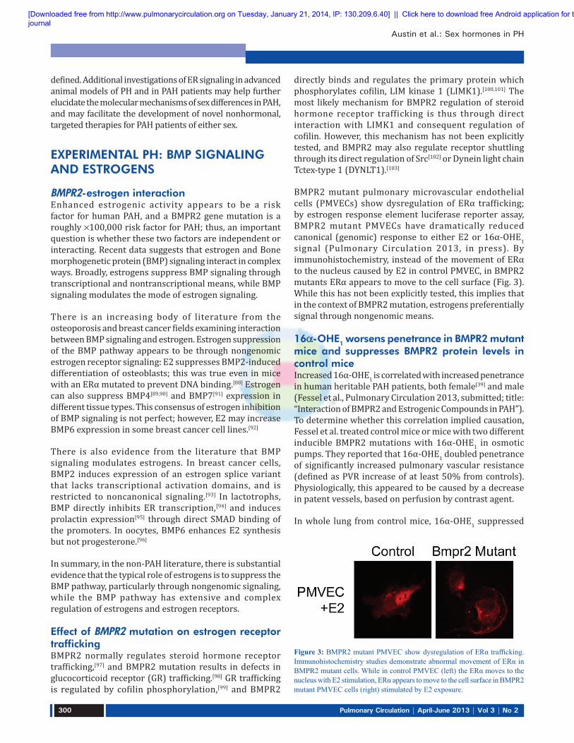
BMPR2 mutant pulmonary microvascular endothelial cells (PMVECs) show dysregulation of ERα trafficking; by estrogen response element luciferase reporter assay, BMPR2 mutant PMVECs have dramatically reduced canonical (genomic) response to either E2 or 16α-OHE1 signal (Pulmonary Circulation 2013, in press). By immunohistochemistry, instead of the movement of ERα to the nucleus caused by E2 in control PMVEC, in BMPR2 mutants ERα appears to move to the cell surface (Fig. 3). While this has not been explicitly tested, this implies that in the context of BMPR2 mutation, estrogens preferentially signal through nongenomic means.
16a-OHE1 worsens penetrance in BMPR2 mutant mice and suppresses BMPR2 protein levels in control miceIncreased 16α-OHE1 is correlated with increased penetrance in human heritable PAH patients, both female[39] and male (Fessel et al., Pulmonary Circulation 2013, submitted; title: “Interaction of BMPR2 and Estrogenic Compounds in PAH”). To determine whether this correlation implied causation, Fessel et al. treated control mice or mice with two different inducible BMPR2 mutations with 16α-OHE1 in osmotic pumps. They reported that 16α-OHE1 doubled penetrance of significantly increased pulmonary vascular resistance (defined as PVR increase of at least 50% from controls). Physiologically, this appeared to be caused by a decrease in patent vessels, based on perfusion by contrast agent.
In whole lung from control mice, 16α-OHE1 suppressed
Figure 3: BMPR2 mutant PMVEC show dysregulation of ERα trafficking. Immunohistochemistry studies demonstrate abnormal movement of ERα in BMPR2 mutant cells. While in control PMVEC (left) the ERα moves to the nucleus with E2 stimulation, ERα appears to move to the cell surface in BMPR2 mutant PMVEC cells (right) stimulated by E2 exposure.
[Downloaded free from http://www.pulmonarycirculation.org on Tuesday, January 21, 2014, IP: 130.209.6.40] || Click here to download free Android application for thisjournal

Austin et al.: Sex hormones in PH
Pulmonary Circulation | April-June 2013 | Vol 3 | No 2 301
Figure 4: The BMP pathway and estrogen have multiple avenues of mutual regulation.
BMPR2 protein levels three-fold and SMAD 1/5/8 phosphorylation two-fold, without a change in BMPR2 expression. These changes were also seen to a lesser extent in kidney. Estrogens regulate the lysosome through a nontransciptional mechanism[104] and BMPR2 protein levels are primarily regulated through lysosomal degradation.[105] While this hasn’t been explicitly shown, the simplest hypothesis for estrogen regulation of BMPR2 protein levels is thus through lysosomal activation.
However, this estrogen-mediated suppression of SMAD phosphorylation and BMPR2 protein levels was not the explanation for worsened penetrance in mice overexpressing BMPR2 dominant negatives; presumably because the dominant negative acts as a decoy for lysosomal degradation, 16α-OHE1 treatment did not change already suppressed SMAD phosphorylation in these mice. There must be an additional mechanism by which 16α-OHE1 worsens BMPR2-mediated PAH.
Gene expression arrays suggest 16a-OHE1 causes vascular injury responseTo try to uncover this additional mechanism, Fessel et al. performed gene expression arrays on lungs from mice with and without BMPR2R899X mutation, and with and without 16α-OHE1 treatment. Only mice with normal right ventricular systolic pressure (RVSP) were used to avoid expression signature caused by high pressure. It was found that, as expected based on the suppression of BMPR2 by 16α-OHE1 in control mice, that gene expression in control mice treated with 16α-OHE1 was shifted somewhat toward the pattern in untreated BMPR2 mutants. Fessel et al. also found that 16α-OHE1 treatment had the positive effects that are normally associated with estrogen treatment, including suppression of traditional cytokine signaling and inflammatory cell recruitment (Fessel et al., Pulmonary Circulation 2013, submitted; title: “Interaction of BMPR2 and Estrogenic Compounds in PAH”). However, it had additional effects, most of which had previously been reported in the literature for other systems but which had not previously been considered in the context of PAH. Chronic 16α-OHE1 treatment resulted in increased angiogenesis-related genes, alterations in metabolic genes (including a 3x increase in the insulin resistance gene Resistin), increased platelet and thrombosis related genes, and alterations in the Wnt pathway consistent with induction of angiogenesis.[106] In sum, it appears that in addition to suppressing BMPR2 protein levels directly, 16α-OHE1 is inducing vascular injury and activation of angiogenesis pathways, potentially through increasing insulin resistance.
2-Methoxyestradiol (2-ME) treatment does not prevent BMPR2-related PAH2-ME ameliorates both hypoxic and monocrotaline-related
PAH through vasodilation and inhibition of cytokines.[14] Estrogens have the same molecular effect in BMPR2-related PAH: Based on array data, elevated cytokine expression is suppressed, and based on tail-cuff data, systemic pressures have statistically significant decrease in the presence of chronic 2-ME. However, despite this, 2-ME was not capable of preventing BMPR2-related PAH.
BMPR2R899X mutant or control mice were implanted with pumps containing vehicle, 2-ME alone, 16α-OHE1 alone, or a mixture of 2-ME and 16α-OHE1 for four weeks, after a prior two weeks of induction of mutation. BMPR2R899X mutant treated with 2-ME alone had a nonsignificant trend toward worse PVR than mutant mice treated with vehicle; those treated with 2-ME in addition to 16α-OHE1 had a nonsignificant trend toward improvement compared with 16α-OHE1 alone (although still worse than mutant mice with vehicle).
In sum, these results suggest that 2-ME effect in the context of BMPR2 mutation is essentially a partial agonist: 2-ME has higher affinity for the estrogen receptors, but 16α-OHE1, when it binds, does so covalently.[107,108] Thus, 2-ME may have a slight beneficial effect in the context of 16α-OHE1 by lowering the rate of covalent binding through competition for receptors. However, on its own, it has the same negative effects as 16α-OHE1 on insulin resistance, even though it continues to have the beneficial effects commonly reported (vasodilation and suppression of inflammation).
In conclusion, the BMP pathway and estrogen have multiple avenues of mutual regulation (Fig. 4). Classical estrogen signaling leads to transcriptional suppression of BMP ligands and receptors.[40,89-91] BMPR2 is an important determinant of steroid hormone sensitivity,[97] and properly functioning BMPR2 promotes classical estrogen signaling through assisting translocation of the complexes to the
[Downloaded free from http://www.pulmonarycirculation.org on Tuesday, January 21, 2014, IP: 130.209.6.40] || Click here to download free Android application for thisjournal
Austin et al.: Sex hormones in PH
Pulmonary Circulation | April-June 2013 | Vol 3 | No 2302
nucleus. Mutant BMPR2 prevents classical estrogen signaling, and promotes cell surface ER, associated with nongenomic signaling (Fig. 4). Nongenomic ER signaling suppresses BMP signaling,[88] most likely through induction of lysosomes[104] which regulate BMPR2 protein level[105] (estrogen reduces BMPR2 protein levels [in press]). Functionally, then increased estrogen signaling leads to decreased BMP signaling through multiple mechanisms. In addition, while estrogen signaling is vasodilatory and anti-inflammatory, it also promotes PAH in the context of BMPR2 mutation through promoting insulin resistance,[109] angiogenesis,[110] and thrombosis.[111,112]
EXPERIMENTAL PH: EFFECTS OF GENDER, ESTRADIOL METABOLITES, AND PROGESTINS
Numerous studies indicate that E2 and female gender have protective effects in experimental PH, mainly hypoxia- and monocrotaline-induced PH (MCT-PH).[2] Yet, as previously noted, an “Estrogen Paradox” exists in PAH as it occurs more frequently in women although women with PAH have better right ventricular function and survival compared to men with PAH.[10,12,14,113] The apparent contradictions posed by the effects of female gender and estrogens in experimental PH versus human PAH may be explained by the complexity of E2 metabolism and limitations of the experimental models used.[14,114]
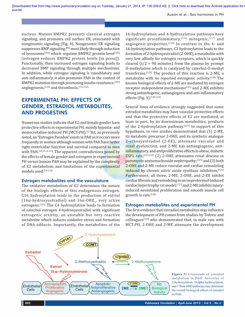
Estrogen metabolites and the vasculatureThe oxidative metabolism of E2 determines the nature of the biologic effects of this endogenous estrogen. C16 hydroxylation leads to the production of estriol (16α-hydroxyestradiol) and 16α-OHE1, very active estrogens.[115] The C4 hydroxylation leads to formation of catechol estrogen 4-hydroxyestradiol with significant estrogenic activity, an unstable but very reactive metabolite which induces oxidative stress and formation of DNA adducts. Importantly, the metabolites of the
16-hydroxylation and 4-hydroxylation pathways have significant proinflammatory,[116] mitogenic,[117] and angiogenic properties.[118] In contrast to the 4- and 16-hydroxylation pathways, C2 hydroxylation leads to the formation of 2-hydroxyestradiol (2-OHE), a metabolite with very low affinity for estrogen receptors, which is quickly cleared (t/2 = 90 minutes) from the plasma by prompt O-methylation which is catalyzed by catechol-O-methyl transferase.[119] The product of this reaction is 2-ME, a metabolite with no reported estrogenic activity.[120] The known biological effects of 2-ME are mediated by estrogen receptor-independent mechanisms[121] and 2-ME exhibits strong antimitogenic, antiangiogenic and anti-inflammatory effects (Fig. 5).[120-122]
Several lines of evidence strongly suggested that some estradiol metabolites may have vascular protective effects and that the protective effects of E2 are mediated, at least in part, by its downstream metabolites, products of the 2-hydroxylation pathway.[121] In support of this hypothesis, in vivo studies demonstrated that (1) 2-ME, its metabolic precursor 2-OHE, and its synthetic analogue 2-ethoxyestradiol (2-EE), attenuate vascular and renal dysfunction, and 2-ME has antiangiogenic, anti-inflammatory, and antiproliferative effects in obese, diabetic ZSF1 rats;[123,124] (2) 2-OHE attenuates renal disease in puromycin aminonucleoside nephropathy;[125] and (3) both 2-OHE and 2-ME reduce vascular and cardiac remodeling induced by chronic nitric oxide synthase inhibition.[126] Furthermore, all three, 2-ME, 2-OHE, and 2-EE inhibit cardiac fibrosis and remodeling in an isoproterenol-induced cardiac hypertrophy rat model,[127] and 2-ME inhibits injury-induced neointimal proliferation and smooth muscle cell growth in rats.[128]
Estrogen metabolites and experimental PHThe first evidence that estradiol metabolites may influence the development of PH comes from studies by Tofovic and colleagues[129] who demonstrated that, in male rats with MCT-PH, 2-OHE and 2-ME attenuate the development
Figure 5: Crossroads of estradiol metabolism in PAH: Activities of 2-hydroxylation, 16alpha-hydroxylation, and 17beta-HSD pathways may determine the overall biological effects of estradiol in PAH.
2-Hydroxyestradiol
COMT
Estradiol
Anti-apoptoticProliferativeAngiogenic
17βHSD
Estriol16α−Hydroxyestrone
ApoptoticAntiproliferativeAntiangiogenic
Endothelial Cells
Endothelial Cells
2-Methoxyestradiol 2-Methoxyestrone
No BiologicalActivity
2ME12ME17βHSD
[Downloaded free from http://www.pulmonarycirculation.org on Tuesday, January 21, 2014, IP: 130.209.6.40] || Click here to download free Android application for thisjournal

Austin et al.: Sex hormones in PH
Pulmonary Circulation | April-June 2013 | Vol 3 | No 2 303
and 2-ME retards the progression of disease, and that both 2-OHE and 2-ME attenuate lungs inflammatory response and right ventricular and pulmonary vascular remodeling. Notably, ovariectomy (OVX) exacerbates MCT-PH or bleomycin-induced PH and pulmonary fibrosis, and treatment of OVX pulmonary hypertensive animals with 2-ME attenuates pulmonary vascular remodeling, RV hypertrophy, and pulmonary inflammation and fibrosis and eliminates the mortality due to ovariectomy.[130,131] Furthermore, 2-ME attenuates the development and retards the progression of hypoxia-induced PH in male rats[132] and has synergistic effects with sildenafil and bosentan in attenuating pulmonary vascular remodeling and pulmonary inflammation and in reducing mortality in MCT-PH male rats.[133] Similar effects in male rats with MCT-PH are seen with synthetic analog 2-EE.[127] The beneficial effects of E2 in MCT-PH are associated with increased lung NO and prostacyclin levels and reduced endothelin levels, and the inhibitory effects on of E2 on vascular remodeling are linked to down-regulated expression of phosphorylated Akt and up-regulated expression of cyclooxygenase-2.[134] 2-ME is decisively more potent than E2 in increasing prostacyclin synthesis and release, in inhibiting endothelin synthesis in endothelial cells (ECs), and in stimulating NO release from endothelium.[14,121] Likewise, in systemic and pulmonary vascular smooth muscle cells, 2-ME exerts stronger antimitotic effects than E2 itself,[14,121] and inhibition of vascular remodeling by 2-ME is also associated with down-regulated expression of phosphorylated Akt and up-regulated expression of cyclooxygenase-2.[128] Thus, at least in part, the beneficial effects of E2 in experimental PH may be mediated by its major non-estrogenic metabolite 2-ME.
However, MCT- and hypoxia-induced PH may not be the best models for studying the complexities of estrogen pharmacology in human PAH.[14,114] The hallmark vascular change in hypoxia- and MCT-induced PH is medial thickening and neomuscularization, with only mild endothelial damage. This is in contrast to the marked endothelial involvement in severe PAH in humans characterized by occlusive proliferation of endothelial cells and formation of plexifom lesions. Estradiol and 2-ME have opposing effects on ECs:E2 is promitogenic, proangiogenic, and antiapoptotic, whereas 2-ME is antimitogenic, antiangiogenic, and proapoptotic. This may have significant ramifications for the overall effects of estrogens in experimental PH, and the overall effects of gender and E2 in experimental PH may depend on the model system used.[14,114] Therefore, it was suggested[129] that the effects of estrogens and their metabolites should be studied in a model that more closely mimics vascular (endothelial) alterations seen in human PAH, i.e., rat model of angioproliferative PH induced by administration of VEGFR2 antagonist SU5416 and exposure to hypoxia (Sugen [Su5416] plus hypoxia [SuHx rats]).[135]
Opposite to the gender effects in hypoxia- and MCT-induced PAH, in this model of experimental PH, female gender does not protect against PH.[136] Female rats rather develop more severe disease and, when compared to males, have increased lung weight and collagen content, as well as more severe nitrosative and oxidative lung injury.[137,138] Plexiform lesions are detected in female animals as early as three weeks after administration of SU5416 and exposure to hypoxia.[138] Male rats develop predominantly occlusive vasculopathy with sporadic presence of plexiform lesions, whereas female rats develop numerous plexiform lesions with sporadic presence of necrotizing arteritis (Grade 6 lesions, Heath-Edwards classification).[137,138] Also, in opposite to the effects in MCT- and hypoxia-induced PH, in the female model of angioproliferative PH, OVX does not exacerbate but rather tends to attenuate the disease; furthermore, in OVX SuHx rats, rescue treatment with E2 exacerbates vascular lesions and PH. Notably, similar to human PAH, in the angioproliferative PH model (SuHx rats), estrogens induce RV-pulmonary vascular uncoupling; despite more severe PH, females had significantly reduced RV hypertrophy compared to males, and OVX exacerbates the increase in RV mass, effects that are reversed by exogenous E2.[137,138]
2 - M E i s e x t e n s i v e l y m e t a b o l i z e d b y t y p e - 2 17β-hydroxysteroid dehydrogenase (17β-HSD-2) to 2-methoxyestrone (2-ME1), a metabolite largely lacking antimitogenic activity in cardiovascular cells. In this regard, in human pulmonary artery smooth muscle cells (hPASMCs) and lung fibroblasts (hLFs), at high pharmacological concentrations (10µM), 2-ME1 had only modest (-10 to -20%) antimitogenic effects.[139] Notably, overexpression and increased activity of 17β-HSD-2 abolishes the antimitogenic effects of 2-ME in tumor cells,[140] suggesting that intracellular conversion to its much less active metabolite may significantly influence the biological effects of 2-ME. Importantly, 2-ME1 can be converted back to 2-ME by 17β-HSD type-1 and thereby exhibit cardiovascular effects. All-trans retinoic acid (atRA) increases 17β-HSD type-1 expression and activity[141,142] and would, thereby, be expected to increase the conversion of 2-ME1 to 2-ME. Indeed, in the presence of 1mM of atRA (a concentration that does not affect cell growth) 2-ME1 strongly and in a concentration-dependent manner (10 ng-10 µM) inhibits growth of hPASMCs and hLFs (-20 to -80%).[139] Initial studies have confirmed the importance of 17β-HSD in experimental PH. In male MCT-PH rats, 2-ME1 attenuates the development of PH, RV hypertrophy, and pulmonary vascular remodeling, suggesting that in vivo significant conversion of 2-ME1 back to 2-ME takes place in PAH.[139] Furthermore, in male rats with MCT-PH, 2-ME and atRA have synergistic effects in ameliorating PH and in inhibiting vascular remodeling.[143] These studies strongly suggest that 2-ME1 to 2-ME conversion takes place in vivo and that the
[Downloaded free from http://www.pulmonarycirculation.org on Tuesday, January 21, 2014, IP: 130.209.6.40] || Click here to download free Android application for thisjournal
Austin et al.: Sex hormones in PH
Pulmonary Circulation | April-June 2013 | Vol 3 | No 2304
17β-hydroxysteroid dehydrogenase metabolic pathway influences the development of experimental PH.
Progestins and experimental PHThe effects of progesterone in experimental PH have not been fully studied. This is somewhat surprising because progesterone receptors are present both in intact human ECs and in modified ECs within plexiform lesions from patients with PAH.[144,145] Furthermore, natural progesterone inhibits proliferation of ECs, and this effect involves a reduction in cyclin-dependent kinase activity, and the altered expression of cyclin E and A in accordance with G1 arrest.[146] Finally, progesterone exerts vasodilatory properties in different vascular beds including the pulmonary circulation of rats.[147] This would suggest that progesterone may be protective in PH. Indeed, in OVX rats with MCT-PH, progesterone attenuates PH and RV hypertrophy, reduces pulmonary inflammation, inhibits pulmonary vascular remodeling, and reduces mortality.[148] Similar effects are seen with medroxyprogesterone, whereas tibolone, a combined progestin/estrogen compound, prevents the development of disease and eliminates MCT-induced late mortality in OVX rats.[149] Although initial studies in estrogen deficient (OVX) rats suggest that progesterone may have beneficial effects in experimental PH, further evaluation of the role of progesterone in experimental PH, and more importantly its interaction with estrogens in angioproliferative PH, are warranted.
EXPERIMENTAL PH: SEROTONERGIC ACTIVITY AND ESTROGENS
Serotonin is a potent inducer of PASMC proliferation and vasoconstriction and has been implicated in the development of both experimental and clinical PAH. This important contributor to the development and pathogenesis of PAH was first recognized by the associated increase in the incidence of PAH in patients prescribed the appetite suppressant drug, dexfenfluramine.[150] Dexfenfluramine acts as an indirect serotonergic agonist as it is a substrate for the serotonin transporter (SERT) through which it enters the cell and causes release of intracellular serotonin (via the SERT).[151] Dexfenfluramine also reduces serotonin reuptake into cells.[152] The development of dexfenfluramine-induced PAH is dependent on serotonin synthesis as PAH is attenuated in mice devoid of tryptophan hydroxylase 1 (TPH1), the rate limiting enzyme in the synthesis of peripheral serotonin.[153] Recently, a similar drug methamphetamine was associated with increased remodeling of the pulmonary vasculature with increased expression of the 5-HT1B receptor and SERT.[154] This indicates the importance of understanding the underlying mechanisms of this family of drugs to prevent future drugs that act by a similar mechanism entering the drug market.
There is substantial evidence suggesting that TPH1 activity contributes to experimental PAH.[155-159] Pulmonary arterial endothelial cell expression of TPH1 is increased in both experimental and clinical PAH, increasing local serotonin production which can act in a paracrine fashion on underlying PASMCs to facilitate proliferation via SERT, the 5-HT1B and 5-HT2A receptors.[158,160] Multiple studies have also implicated the SERT in the pathobiology of PAH. For example, overexpression of the human SERT gene in mice promotes the spontaneous development of a PAH phenotype which is exaggerated by hypoxia.[161] In contrast, mice deficient in SERT develop less severe PAH phenotypes compared to mice that overexpress SERT.[162] Importantly, SERT expression is increased in PASMCs in patients with PAH.[163] Serotonin can mediate PASMC proliferation by transactivation of the platelet-derived growth factor receptor (PDGFRβ) through SERT.[164] Although inhibition of SERT may offer some therapeutic value, extracellular concentrations of serotonin available to bind with the 5-HT1B receptor may increase, which mediates vasoconstriction in human pulmonary arteries and proliferation of human PASMCs.[165,166] Furthermore, there is evidence for cross-talk between SERT and the 5-HT1B receptor. For example, nuclear transport of phosphorylated extracellular regulated kinase (ERK) by the 5-HT1B receptor is mediated by SERT and is necessary for the upregulation of the S100 calcium binding protein, S100A4/mts1, involved in proliferation and migration of PASMCs.[166] Therefore, taken together, a greater therapeutic effect may be offered by a combined inhibition of SERT and the 5-HT1B receptor, as demonstrated in hypoxia-induced PAH and in SERT+ mice.[167]
Both idiopathic pulmonary arterial hypertension (IPAH) and heritable pulmonary arterial hypertension (HPAH) occur more frequently in females compared to males, as discussed previously. For example, in recent epidemiological studies in the UK/Ireland and the USA, ~70% and 80% of the patients studied were female, respectively.[11,168] The reason for this disparity is unclear and is under-investigated. An understanding of this gender difference would certainly lead to novel therapeutic strategies. Paradoxically, mortality is higher in male patients.[12,169] Research into why PAH occur more frequently in females has been hampered by a lack of relevant animal models, as male rodents typically develop a more severe disease phenotype. For example, male rats develop more severe hypoxia-induced PAH compared to their female counterparts.[170] Additionally, there is evidence that E2 attenuates the development of a PAH phenotype in both the hypoxic and monocrotaline models of the disease.[79,80,171]
Recently, MacLean et al. demonstrated that only female SERT+ mice and female mice overexpressing the S100A4/mts1 gene develop a spontaneous PAH phenotype and that dexfenfluramine-induced PAH is also female selective.[172-174]
[Downloaded free from http://www.pulmonarycirculation.org on Tuesday, January 21, 2014, IP: 130.209.6.40] || Click here to download free Android application for thisjournal

Austin et al.: Sex hormones in PH
Pulmonary Circulation | April-June 2013 | Vol 3 | No 2 305
The development of PAH in all these models is dependent on elevated serotonin activity and in all these models, estrogen is also a major contributing factor. This suggests the hypothesis that serotonin is facilitating the damaging effects of estrogens.
Consistent with this, E2 induces proliferation of human PASMCs, increases the expression of TPH1, SERT, and the 5-HT1B receptor, and in line with this, inhibitors of serotonergic signaling have been found to attenuate E2-induced proliferation.[17] This evidence supports an estrogen-serotonin axis mediating the development of PAH. Microarray studies have identified a gene specific to estrogen metabolism that is upregulated in pulmonary arteries of female SERT+ mice, CYP1B1, a major extra-hepatic estrogen metabolizing enzyme.[175] It has subsequently been shown that CYP1B1 expression is upregulated in pulmonary arteries of both IPAH and HPAH, and in two independent animal models, the SUGEN-hypoxic and hypoxic models of PAH, regardless of gender.[176] In contrast, CYP1B1 expression is ten-fold lower in Epstein-Barr virus immortalized B lymphocytes obtained from PAH affected patients harboring a mutation in the BMPR2 gene.[36] The underlying cause of this discrepancy is intriguing and yet to be fully understood but may be attributable to alterations in gene expression induced by the Epstein Barr Virus[177] or attributable to tissue specific differences. Furthermore, a CYP1B1 genetic polymorphism Asn453àSer (N453S), associated with increased degradation of CYP1B1,[178] is more prevalent in unaffected female BMPR2 mutation carriers, compared with affected mutation carriers where the wildtype genotype is more common. However, in males carrying a BMPR2 mutation the wildtype genotype predominates in both unaffected and affected carriers.[39] This may therefore contribute to the decreased penetrance in patients with this mutation. Genetic polymorphisms of CYP1B1 are associated with altered catalytic properties, which may affect its metabolic profile and biological activity.[179] Individual status may, therefore, defer or infer susceptibility.
Aromatase, CYP1B1, and Catechol-O-methyl transferase (COMT) are key enzymes in the synthesis and metabolism of estrogen. The formation of E2 from C19 steroids is catalyzed by aromatase, a cytochrome P450 enzyme encoded by the CYP19A1 gene. A genetic polymorphism of aromatase leading to increased plasma estrogen levels has been associated with an increased risk of PPHTN.[20] As aromatase inhibitors are already widely used as cancer treatment, this makes it a particularly attractive target. Recent unpublished results from MacLean et al. show that the aromatase inhibitor anastrozole can reverse both hypoxia and SUGEN/hypoxic PAH in female mice. A recent, large-scale epidemiological study reported a greater association of PAH among postmenopausal females and in obesity, and at this age comorbidities are increasingly
prevalent.[11] In both men and women, extragonadal sites such as the adipose tissue have the capacity to synthesize estrogen via aromatase from circulating C19 precursors.[180] In postmenopausal women, once the ovaries have stopped producing estrogens, the adipose tissue increases its production of estrogen.[181] Aromatase inhibitors could therefore be effective in a wide range of patients regardless of age or gender.
Local increased aromatase activity provides estrogen as a substrate for metabolizing enzymes. Estrogens are metabolized to both pro- and antiproliferative metabolites by various CYP enzymes. As discussed above, CYP1B1 primarily metabolizes estrogens to the 4-OHE1/2 by hydroxylation at the C4 position and to a lesser extent to 2- and the 16-OHE1/2.[182,183] The catechol estrogens (the 2- and 4-OHE1/2) can then be further metabolized to the methoxyestrogens by the ubiquitously expressed Catechol-O-methyltransferase (COMT). Importantly, hypoxia, serotonin, and estrogen all increase the expression of CYP1B1 and are all factors that have been implicated in PAH. E2 and estrone are present in the lung, and it is, therefore, conceivable that increased estrogen metabolism to proproliferative metabolites by upregulation of CYP1B1 in response to hypoxia, serotonin, and/or estrogen could occur (Fig. 6).[175,176,184] Additionally, CYP1B1 expression and activity is upregulated in response to shear stress, which may implicate a prolonged contribution of CYP1B1 to the pathogenesis of PAH, by persistence of increased CYP1B1 in the face of increased pulmonary arterial pressures, increased circulating estrogens, and locally produced estrogens.[185]
CYP1B1 in PAH may be an attractive therapeutic target and, recently, White et al. established a direct pathological role for CYP1B1 metabolism on the development of PAH.[175] Mice devoid of functional CYP1B1 (CYP1B1-/- mice) and chronic inhibition by a highly selective CYP1B1 inhibitor 2,3’,4,5’-tetramethoxystilbene (TMS) are protected against the development of hypoxia-induced PAH, independent of sex. This effect was also convincingly observed in the SUGEN-hypoxic model and, importantly, TMS also inhibited the formation of occluded pulmonary arteries. To further elucidate potential mechanisms by which CYP1B1 metabolism was causative on disease development, White et al. assessed proliferation to the 2-, 4-, and 2-OHE1/2 in human PASMCs. 16α-OHE1 stimulated the most profound proliferation of PASMCs and this effect was 100-fold more pronounced in PAH-PASMCs. To establish a direct relationship of 16α-OHE1 in vivo, mice were chronically dosed with 16α-OHE1 which induced a PAH phenotype.[175] Although 16α-OHE1 is a minor CYP1B1 metabolite, increased CYP1B1 expression would increase the production of all its metabolites, including 16α-OHE1. There is also the possibility that simultaneous changes in aromatase, COMT, and CYP1B1 activity could drive estrogen metabolism toward an accumulation of damaging
[Downloaded free from http://www.pulmonarycirculation.org on Tuesday, January 21, 2014, IP: 130.209.6.40] || Click here to download free Android application for thisjournal

Austin et al.: Sex hormones in PH
Pulmonary Circulation | April-June 2013 | Vol 3 | No 2306
metabolites. Additionally, 16α-OHE1 levels are increased in affected BMPR2 mutation carriers compared to unaffected carriers.[39] The estrogen metabolism pathway is complex, tissue specific, and dependent on the presence and absence of different enzymes, and more work teasing out its precise function in the pulmonary vasculature is essential.
Estrogen metabolism may be integral to a wide span of diseases and may offer targets for therapeutic intervention in the management of PAH. The exact mechanisms by which aromatase, CYP1B1, and COMT influence disease development and progression merits further investigation. Gender, locally produced estrogens, and serotonin together with increased shear stress may interact to facilitate the development of PAH via CYP1B1 activity and are suggestive as “second hit factors” that may influence the development of PAH in genetically-susceptible individuals and may also be causative in idiopathic cases of PAH.
EXPERIMENTAL PH: DEHYDROEPIANDROSTERONE (DHEA)
DHEA is a naturally occurring C-19 steroid that is synthesized mainly by the adrenal cortex only in humans and close primate relatives (other species, including rodents, have very low levels of DHEA produced by other organs, such as testis, ovary, and brain).[186,187] DHEA and
its sulfated ester, DHEA sulfate (DHEA-S), are produced in larger quantities than any other circulating steroid hormone. Most circulating DHEA is in the form of DHEA-S, which functions as a less active reservoir for DHEA (DHEA-S can be converted to DHEA by a ubiquitous enzyme, steroid sulfatase, in peripheral tissues; Fig. 7). These two steroids show a distinct life history secretion pattern; blood levels of DHEA-S are very high at birth and fall precipitously to near zero by about six months of age, they then rise to peak values in men around age 25, and finally undergo a progressive steep decline over age. Levels in females are lower but follow the same pattern.[188-190] Although DHEA serves as a precursor to both androgens and estrogens (Fig. 7),[191] there is no obvious role for DHEA/DHEA-S in the blood levels of sex steroid hormones in normal men and premenopausal women.[192]
Epidemiological observations and animal experiments indicate that DHEA has a wide variety of beneficial biological and physiological effects, including prevention of obesity, diabetes, cancer, and cardiovascular diseases.[193,194] However, the mechanisms of the cardiovascular protective effects of DHEA, such as antiproliferative, proapoptotic, anti-inflammatory, and antioxidant effects, have not been fully elucidated. There are several known biochemical actions by which some of its beneficial properties might be explained (Table 1). The best-described action of DHEA is inhibition of glucose-6-phosphatase
Figure 6: The influence of serotonin and estrogen in the development of PAH. Estrogens can increase the expression of TPH1, the 5-HT1B receptor and SERT. Serotonin, shear stress, hypoxia, SUGEN + hypoxia and estrogen all increase the expression of CYP1B1 (and perhaps other CYP enzymes) which can metabolize estrogen to both pro- and anti-proliferative metabolites, some via COMT. Changes in this system that favour accumulation of pro-proliferative metabolites in the face of decreased protective metabolites would favour proliferation of PASMCs and contribute to pulmonary vascular remodeling.
ECs, PASMCs, PAFs
Estrogens
2-, 4-hydroxyestrogens
Testosterone
COMT
2-, 4-methoxyestrogens
CYPs/ CYP1B1
TPH1, 5-HT1B, SERT
Serotonin, Estrogen, Hypoxia, SUGEN + Hypoxia, Shear Stress
+/- Proliferation of Pulmonary Arterial Cells
Estrogen-Sulfate
CYP19a1Sulfatase
16α-hydroxyestrogens
[Downloaded free from http://www.pulmonarycirculation.org on Tuesday, January 21, 2014, IP: 130.209.6.40] || Click here to download free Android application for thisjournal

Austin et al.: Sex hormones in PH
Pulmonary Circulation | April-June 2013 | Vol 3 | No 2 307
dehydrogenase (G-6-PDH),[195,196] which decreases NADPH production by the pentose phosphate pathway, and thereby modulates tissue redox state. This mechanism can partly explain its antiproliferative effect via reduced RNA/DNA productions[194,197] and its acute vasodilatory effect via opening of K+ channels.[199] A decrease in NADPH levels plays a major role in DHEA’s antioxidant effect by reducing O2
- production via NADPH oxidase.[196] Another well-described action is activation of endothelial nitric oxide synthase (eNOS),[200] which exerts vasodilatory and antiproliferative effects. DHEA has been reported to bind to a specific endothelial cell membrane receptor coupled to Gai2,3 to activate eNOS.[201] It has also recently been reported that DHEA stimulates Akt-eNOS signaling pathway through activation/upregulation of sigma-1 receptor.[202] Other known actions of DHEA include activation of peroxisome proliferator-activated receptors ([PPARs]; which is involved in fat metabolism and anti-inflammation/-proliferation)[203,204] and inhibition of 3-hydroxy-3-methylglutaryl-coenzyme A (HMG-CoA) reductase[205] (which is involved in cholesterol metabolism and cell cycle arrest and growth inhibition probably through decreased RhoA/Rho kinase signaling activity). Finally, it has been reported that DHEA is a poor agonist for estrogen receptors, and several studies have demonstrated that the antiproliferative effects of DHEA are independent of either the estrogen or androgen receptors.[206] However, since a specific receptor for DHEA has not yet been isolated and fully characterized, it is still not clear whether part of DHEA’s effects are mediated through the androgen/estrogen receptors or through conversion of DHEA to these hormones. Taken together, all these actions of DHEA are closely related to the pathogenesis of PH and could act against its development.
DHEA and pulmonary circulationLittle is known about the effects of DHEA on the pulmonary circulation. Farrukh et al.[206] and Gupte et al.[198] have shown in vitro that DHEA is a pulmonary vasodilator and inhibits acute hypoxic pulmonary vasoconstriction due at least partly to opening of potassium channels. In the latter study, it is shown that vasodilatory effects of pentose phosphate pathway inhibitors, including DHEA, are mediated through opening of voltage-gated K+ channels due to reduction of tissue NADPH levels and a change in redox status.[198] The levels of DHEA/DHEA-S in PH patients over time have not been determined, but the recent MESA-RV Study found that higher DHEA levels were associated with increased RV mass and stroke volume in women.[87]
Effects of chronic DHEA treatment on experimental PHPrompted by the epidemiological and experimental findings in the systemic and pulmonary circulation, studies have been performed to determine whether DHEA is effective for the treatment of PH in various experimental models. There are numerous animal models of PH available for various research purposes, but they are now divided roughly into two groups based on the presence or absence of occlusive neointimal lesions, one of the hallmarks of PAH. One group is conventional, which includes the most frequently used models that essentially lack occlusive lesions, such as chronic hypoxia-induced and monocrotaline-injected models. The other group encompasses relatively newer models that develop occlusive neointimal lesions, including monocrotaline-injected pneumonectomized rats[207] and Sugen5416 injection and hypoxia-exposed rats.[135]
Chronically hypoxic PH. Hampl et al.[208] and Bonnet et al.[209] in the same year reported that chronic treatment with DHEA-S or DHEA inhibited and reversed chronic HPH (increase in pulmonary arterial pressure and RV hypertrophy) in rats. Bonnet et al. showed that the beneficial effects of DHEA treatment were associated with increased expression and function of pulmonary artery Ca2+ activated K+ channels. Oka et al. subsequently reported that dietary DHEA (0.3 and 1% DHEA containing food) dose-dependently, completely prevented and effectively reversed the HPH.[210] In agreement with previous reports in the systemic circulation,[211,212] the chronic DHEA treatment improved endothelium-dependent (acetylcholine-induced) relaxation in isolated hypertensive pulmonary arteries. This improvement was accompanied by activation and upregulation of soluble guanylate cyclase but not eNOS, which is not consistent with the findings in the systemic circulation. It is not clear whether this difference simply reflects the difference between the systemic and pulmonary circulations, but the results suggest that DHEA can modulate activity/expression of soluble guanylate cyclase. It should be noted in this study that while DHEA attenuated
Figure 7: Synthesis and metabolism of dehydroepiandrosterone (DHEA).
Table 1: Major mechanisms of action for dehydroepiandrosteroneInhibition of Glucose 6-Phosphate DehydrogenaseActivation/Upregulation of Endothelial Nitric Oxide Synthase and/or Soluble Guanylate CyclaseActivation of Peroxisome Proliferator-Activated Receptors Inhibition of 3-Hydroxy-3-Methylglutarryl-CoA ReductaseMetabolites, including Estrogens
DHEA-S DHEA Androgens
Estrogens
Cholesterol
Sulfotransferase
Sufatase
[Downloaded free from http://www.pulmonarycirculation.org on Tuesday, January 21, 2014, IP: 130.209.6.40] || Click here to download free Android application for thisjournal
Austin et al.: Sex hormones in PH
Pulmonary Circulation | April-June 2013 | Vol 3 | No 2308
chronic HPH with increased plasma DHEA levels in a dose-dependent fashion, plasma estradiol and testosterone levels were elevated only in the higher dose group (1% DHEA). These findings suggest that protective effects of DHEA against HPH are at least partly independent of its conversion to estrogen/testosterone.
In addition, another study reported that chronic DHEA-S treatment inhibited the development of HPH in older mice, and prevented them from age-related frailty induced by HPH.[213] The authors emphasized the similarity in this and a human study, where a strong inverse correlation between natural DHEA/DHEA-S blood levels and the ten-year mortality in old male smokers and ex-smokers, and suggested clinical trials with DHEA in patients with PH and chronic obstructive pulmonary disease. The same group subsequently showed that DHEA decreased hypoxia inducible factor-1a accumulation under hypoxic conditions in vitro, and suggested this might explain why DHEA was effective in treating chronic HPH.[214] The beneficial effects of DHEA/DHEA-S appear to involve multiple actions including K+ channel activation, upregulation of soluble guanylate cyclase, and downregulation of hypoxia inducible factor-1a. Collectively, there is an increasing body of experimental evidence that supports that DHEA/DHEA-S is an effective agent in preventing and reversing CH-PH. In fact, a recent preliminary report of an ongoing clinical trial in chronic obstructive lung disease-associated PH patients revealed that chronic DHEA treatment (200 mg/day for 12 weeks) improved their exercise tolerance and hemodynamic parameters.[215]
Other models. The drugs currently used to treat PH patients were first tested and found effective in conventional PH models, i.e., chronically hypoxic and/or monocrotaline-injected pulmonary hypertensive rats, and many new agents continue to be evaluated in these models. While the information acquired from studies of these models advances our understanding of the molecular mechanisms of pulmonary vasoconstriction and vascular remodeling, a major limitation is that these animals do not develop the occlusive neointimal and plexiform pulmonary arterial lesions that are the hallmarks of severe human pulmonary arterial hypertension. There are newer models of PH that develop a closer phenotype to human pulmonary arterial hypertension than the conventional models. These models are usually induced by multiple insults/hits. In contrast to the conventional models, these models develop occlusive neointimal lesions in distal pulmonary arteries. Oka et al. tested effects of chronic DHEA treatment on two of these models, i.e., monocrotaline-injected pneumonectmized (MCT-PN) and Sugen 5416 (SU, a vascular endothelial growth factor receptor blocker)-injected followed by exposure to hypoxia and subsequent re-exposure to normoxia (SuHx) rats:(a) MCT-PN: This model was first introduced by Okada
et al. in 1997 based on a two-hit theory, i.e., endothelial injury by MCT combined with increased blood flow by unilateral pneumonectomy.[207] In contrast to its effects in normal rats, MCT injection causes severe PH accompanied not only by medial wall thickening but also by neointimal lesions in small pulmonary arteries in PN rats. Homma et al. reported that a high dose of three-week dietary DHEA treatment (1% DHEA containing food) completely prevented the development of MCT-induced severe PH, RV hypertrophy, and pulmonary vascular remodeling (medial wall thickening and occlusive neointimal formation) in left PN rats with no adverse systemic hemodynamic effects.[216] Protein analyses of lungs from PN rats revealed that MCT injection caused the following: (1) Increased RhoA activity accompanied by markedly increased 3-hydroxy-3-methylglutaryl-coenzyme A (HMG-CoA) reductase and slightly decreased soluble guanylyl cyclase (sGC) expression; (2) constitutive activation (cleavage) of Rho-associated coiled-coil protein kinase 1 (ROCK I) associated with caspase-3 activation; (3) increased ROCK II expression; and (4) increased ROCK activity. Chronic DHEA treatment nearly normalized the increased RhoA/ROCK signaling pathway activity, which was associated with inhibition of caspase-3 activation, HMG-CoA reductase overexpression, and preservation of sGC expression. DHEA, when started after the PH was fully developed, also completely inhibited the progression of MCT-induced severe PH, and more importantly, strikingly improved survival (30 vs. 100% for DHEA-untreated vs. -treated). These results showed that chronic DHEA treatment nearly completely prevented the development of MCT-induced severe PH, and that inhibition of RhoA/ROCK signaling, apparently by multiple mechanisms including downregulation of HMG-CoA reductase and inhibition of ROCK I cleavage, was involved in the impressive protective effects of DHEA. In addition, a recent study suggests that downregulation of Src/STAT3 signaling may be involved in the inhibitory effect of DHEA against RhoA/ROCK signaling[217]
(b) SuHx: In 2001, Taraseviciene-Stewart et al. first described a rat model of severe PH induced by a combination of vascular endothelial growth factor (VEGF) receptor blockade (by Sugen 5416) and chronic hypoxic exposure.[135] A single injection of Sugen 5416 followed by a three-week exposure to hypoxia (10% O2) resulted in the development of severe PH that was accompanied by occlusive neointimal lesions in precapillary small pulmonary arteries. Importantly, the PH is progressive even when the animals are re-exposed to normoxia. Recent work with this model shows that after three weeks of exposure to hypoxia and an additional two to 10 weeks in normoxia (5–13 weeks after Sugen 5416 injection), the severe pulmonary hypertensive animals develop over time a variety of pulmonary arterial lesions including medial wall thickening (Heath-Edwards Grade 1) and neointimal (Grades 2 and 3) and complex plexiform lesions (Grade 4)
[Downloaded free from http://www.pulmonarycirculation.org on Tuesday, January 21, 2014, IP: 130.209.6.40] || Click here to download free Android application for thisjournal

Austin et al.: Sex hormones in PH
Pulmonary Circulation | April-June 2013 | Vol 3 | No 2 309
that are histologically indistinguishable from those found in human PH.[218] In addition, hemodynamic data show that RVSP appears to reach its maximum (~100 mmHg) around three to five weeks after Sugen 5416 injection and stays at about the same high level thereafter. At the five-week time point, cardiac output decreases to approximately 50% of that of the normal, and is further reduced at the 13-week time point. Reflecting this reduction in cardiac input, calculated total pulmonary vascular resistance increases further at this very late time point, even though the RVSP does not increase, suggesting that the vascular disorder is still progressing. This hemodynamic time-course pattern is very similar to that observed in patients with PAH, and notably, the PH in these animals is also resistant to treatment with drugs conventionally used to treat the human disease.
Alzoubi et al. have tested effects of DHEA in this severe PAH model. The authors found that a five-week dietary treatment with a high dose DHEA (1%), starting from the three-week time point when RVSP is near the maximum level (>80 mmHg), slowed the progression but did not reverse the severe PH (RVSP = 70 ± 7 and 97 ± 5 mmHg, at eight-week time point with and without DHEA treatment, respectively). Likewise, DHEA treatment significantly reduced the severity of RV hypertrophy and the neointimal lesion formation in small pulmonary arteries, but it did not normalize them. It is, however, notable that DHEA treatment impressively improved the impaired cardiac index to near normal levels, and the calculated total pulmonary vascular resistance was dramatically reduced. The authors hypothesized that this impressive improvement of cardiac function might be due not only to decreases in RVSP and RV pressure overload, but also to direct effects of DHEA on cardiomyocytes. Because it has been reported that increased reactive oxygen species (ROS) are involved in the impaired RV function in this model,[219] and because DHEA is a potent antioxidant, the authors looked at ROS signals in the heart. They found that ROS signals were markedly increased in RV but not LV from PH rats (at the eight-week time point), and DHEA treatment effectively reduced the signal. These results suggest that DHEA is effective in treating this severe PH model, not only by improving abnormalities in the hypertensive pulmonary arteries but also by directly protecting cardiomyocytes as an antioxidant.[220]
In conclusion, high doses of DHEA are effective against various experimental models of PH. Multiple mechanisms, including upregulation of Nitric oxide cyclic guanosine monophosphate (NO-cGMP) and inhibition of ROCK signaling pathways, are involved in its beneficial effects, and DHEA may have a direct protective effect on impaired RV function. Taken together, DHEA appears to be a promising agent for the treatment of PH as a monotherapy. DHEA is
an over-the-counter food supplement and generally has no major side effects, but its safety at very high doses is not known. Because DHEA can be metabolized to androgens and estrogens, sex hormone-dependent cancer development may be a concern for its high dose chronic treatment. Although, effective doses in humans might be much lower because of very high basal levels of DHEA/DHEA-S compared to those of rodents, and in fact a recent preliminary clinical trial shows that a relatively low dose (200 mg/day) improves 6-Minute Walk Distance as well as hemodynamic parameters in patients with PH owing to chronic obstructive lung disease.[215] One may need to evaluate synthetic derivatives of DHEA such as HE3286 (Triolex) that is not metabolized to sex steroids and retains potent anti-inflammatory, and possibly other properties, of DHEA.[221]
CONCLUSIONS AND FUTURE DIRECTION
Despite improvements in our understanding of sex hormone physiology and estrogen metabolism in PH, it remains unclear why estrogens appear to be protective in certain settings (HPH, MCT-PH) but detrimental in others (SERT+, S100A4/mts1+). Effects of E2, its receptors, and its metabolites may be context- and/or compartment-specific, and vary depending on oxygen tension, ER distribution, estrogen metabolism, duration of exposure, and biochemical milieu. The emerging paradigm of “good” and “bad” estrogens and altered estrogen metabolism suggests that PAH may be a condition with an imbalance between beneficial and detrimental estrogens. Such alterations may be provoked or aggravated in the setting of genetic disposition (BMPR2 mutation) or other “first hits” (PPHTN, altered serotonin homeostasis); this could explain why protective E2 effects in classical PH models are not recapitulated in others. Further research will need to decipher the molecular basis of these disparate estrogen effects. Detailed investigations of estrogen metabolites in human PAH using mass spectroscopy would be helpful, but are currently limited by lack of standardized techniques. The “estrogen paradox” of increased female susceptibility, yet better survival, still remains a mystery. It appears that women—though being more prone to developing the disease—tolerate PAH better than men, and recent research suggests that female PAH patients may have better RV function,[86] more favorable baseline hemodynamics,[169] and a better response to certain therapeutic interventions.[222] Further studies of the mechanisms underlying these phenomena are needed. DHEA appears uniformly protective in PAH models, while the effects of male sex hormones are only poorly characterized. The investigations of sex hormone effects reviewed here, as well as results of future studies
[Downloaded free from http://www.pulmonarycirculation.org on Tuesday, January 21, 2014, IP: 130.209.6.40] || Click here to download free Android application for thisjournal
Austin et al.: Sex hormones in PH
Pulmonary Circulation | April-June 2013 | Vol 3 | No 2310
in PAH models and patients, will hopefully allow for the development of targeted therapies that ultimately will benefit PAH patients of either sex.
REFERENCES
1. Rich S, Rabinovitch M. Diagnosis and treatment of secondary(non-category1)pulmonaryhypertension.Circulation2008;118:2190-9.
2. TuderRM,AbmanSH,BraunT,CapronF,StevensT,ThistlethwaitePA,etal.Developmentandpathologyofpulmonaryhypertension.JAmCollCardiol2009;54:S3-9.
3. SimonneauG, Robbins IM, BeghettiM, Channick RN,DelcroixM,DentonCP,etal.Updatedclinicalclassificationofpulmonaryhypertension.JAmCollCardiol2009;54:S43-54.
4. BadeschDB,RaskobGE,ElliottCG,KrichmanAM,FarberHW,FrostAE,etal.Pulmonaryarterialhypertension:Baselinecharacteristics fromtherevealregistry.Chest2010;137:376-87.
5. ChinKM,RubinLJ.Pulmonaryarterialhypertension.JAmCollCardiol2008;51:1527-38.
6. HumbertM,SitbonO,ChaouatA,BertocchiM,HabibG,GressinV,etal.Pulmonaryarterialhypertensioninfrance:Resultsfromanationalregistry.AmJRespirCritCareMed2006;173:1023-30.
7. DresdaleDT,SchultzM,MichtomRJ.Primarypulmonaryhypertension.I.Clinicalandhemodynamicstudy.AmJMed1951;11:686-705.
8. RichS,DantzkerDR,AyresSM,BergofskyEH,BrundageBH,DetreKM,et al. Primarypulmonaryhypertension.Anationalprospective study.AnnInternMed1987;107:216-23.
9. McGoonMD,KrichmanA,FarberHW,BarstRJ,RaskobGE,LiouTG,etal.Designoftherevealregistryforuspatientswithpulmonaryarterialhypertension.MayoClinProc2008;83:923-31.
10. BenzaRL,MillerDP,Gomberg-MaitlandM, FrantzRP, ForemanAJ,CoffeyCS,etal.Predictingsurvivalinpulmonaryarterialhypertension:Insights from the registry to evaluate early and long-termpulmonaryarterial hypertension disease management (reveal). Circulation2010;122:164-72.
11. Ling Y, JohnsonMK, Kiely DG, Condliffe R, Elliot CA, Gibbs JS,et al.Changingdemographics, epidemiology, and survival of incidentpulmonaryarterialhypertension:Resultsfromthepulmonaryhypertensionregistryoftheunitedkingdomandireland.AmJRespirCritCareMed2012;186:790-6.
12. HumbertM,SitbonO,ChaouatA,BertocchiM,HabibG,GressinV,etal.Survival inpatientswithidiopathic, familial,andanorexigen-associatedpulmonary arterial hypertension in themodernmanagement era.Circulation2010;122:156-63.
13. HumbertM,SitbonO,YaiciA,MontaniD,O’CallaghanDS,JaisX,etal.Survival in incident andprevalent cohortsofpatientswithpulmonaryarterialhypertension.EurRespirJ2010;36:549-55.
14. Tofovic SP. Estrogens anddevelopment of pulmonary hypertension:Interaction of estradiolmetabolism andpulmonary vascular disease.JCardiovascPharmacol2010;56:696-708.
15. KleigerRE,BoxerM,InghamRE,HarrisonDC.Pulmonaryhypertensioninpatientsusingoralcontraceptives.Areportofsixcases.Chest1976;69:143-7.
16. MorseJH,HornEM,BarstRJ.Hormonereplacementtherapy:Apossibleriskfactorincarriersoffamilialprimarypulmonaryhypertension.Chest1999;116:847.
17. IreyNS,ManionWC,TaylorHB.Vascularlesionsinwomentakingoralcontraceptives.ArchPathol1970;89:1-8.
18. IreyNS,NorrisHJ. Intimal vascular lesions associatedwith femalereproductivesteroids.ArchPathol1973;96:227-34.
19. Sweeney L, Voelkel NF. Estrogen exposure, obesity and thyroiddisease inwomenwithseverepulmonaryhypertension.Eur JMedRes2009;14:433-42.
20. RobertsKE,FallonMB,KrowkaMJ,BrownRS,TrotterJF,PeterI,etal.Genetic risk factors forportopulmonaryhypertension inpatientswithadvancedliverdisease.AmJRespirCritCareMed2009;179:835-42.
21. ParlFF,DawlingS,RoodiN,CrookePS.Estrogenmetabolismandbreastcancer:Ariskmodel.AnnNYAcadSci2009;1155:68-75.
22. GonzalezFJ.Themolecularbiologyofcytochromep450s.PharmacolRev1988;40:243-88.
23. NebertDW,RussellDW.Clinical importanceof the cytochromesp450.Lancet2002;360:1155-62.
24. TsuchiyaY,NakajimaM,YokoiT.Cytochromep450-mediatedmetabolismofestrogensanditsregulationinhuman.CancerLett2005;227:115-24.
25. YagerJD,DavidsonNE.Estrogencarcinogenesisinbreastcancer.NEnglJMed2006;354:270-82.
26. NebertDW.Elevated estrogen 16 alpha-hydroxylase activity: Is thisa genotoxic or nongenotoxic biomarker in humanbreast cancer risk?JNatlCancerInst1993;85:1888-91.
27. RoyD,CaiQ,FeltyQ,NarayanS.Estrogen-inducedgenerationofreactiveoxygen andnitrogen species, genedamage, and estrogen-dependentcancers.JToxicolEnvironHealthBCritRev2007;10:235-57.
28. Bolton JL, ThatcherGR. Potentialmechanisms of estrogen quinonecarcinogenesis.ChemResToxicol2008;21:93-101.
29. MutiP,BradlowHL,MicheliA,KroghV,FreudenheimJL,SchunemannHJ,etal.Estrogenmetabolismandriskofbreastcancer:Aprospectivestudyofthe2:16alpha-hydroxyestroneratioinpremenopausalandpostmenopausalwomen.Epidemiology2000;11:635-40.
30. BelousAR,HacheyDL,DawlingS,RoodiN,ParlFF.Cytochromep4501b1-mediatedestrogenmetabolismresultsinestrogen-deoxyribonucleosideadductformation.CancerRes2007;67:812-7.
31. EliassenAH,Missmer SA, Tworoger SS,Hankinson SE.Circulating2-hydroxy-and16alpha-hydroxyestronelevelsandriskofbreastcanceramongpostmenopausalwomen.Cancer Epidemiol Biomarkers Prev2008;17:2029-35.
32. Kaaks R, Rinaldi S, Key TJ, Berrino F, Peeters PH, BiessyC, et al.Postmenopausal serumandrogens, oestrogens andbreast cancer risk:The european prospective investigation into cancer and nutrition.EndocrRelatCancer2005;12:1071-82.
33. MissmerSA,EliassenAH,BarbieriRL,HankinsonSE.Endogenousestrogen,androgen,andprogesteroneconcentrationsandbreastcancerriskamongpostmenopausalwomen.JNatlCancerInst2004;96:1856-65.
34. MutiP,WesterlindK,WuT,GrimaldiT,DeBerryJ3rd, Schunemann H, et al. Urinaryestrogenmetabolitesandprostatecancer:Acase-controlstudyintheunitedstates.CancerCausesControl2002;13:947-55.
35. LarkinEK,NewmanJH,AustinED,HemnesAR,WheelerL,RobbinsIM,etal.Longitudinalanalysiscastsdoubtonthepresenceofgeneticanticipationinheritablepulmonaryarterialhypertension.AmJRespirCritCareMed2012;186:892-6.
36. WestJ,CoganJ,GeraciM,RobinsonL,NewmanJ,PhillipsJA,etal.Geneexpression in bmpr2mutation carrierswith andwithout evidence ofpulmonaryarterialhypertension suggestspathways relevant todiseasepenetrance.BMCMedGenomics2008;1:45.
37. Agundez JA. Cytochrome p450 gene polymorphism and cancer.CurrDrugMetab2004;5:211-24.
38. GreenleeH, Chen Y, Kabat GC,WangQ, KibriyaMG, Gurvich I,etal.Variantsinestrogenmetabolismandbiosynthesisgenesandurinaryestrogenmetabolites inwomenwith a familyhistoryof breast cancer.BreastCancerResTreat2007;102:111-7.
39. AustinED,CoganJD,WestJD,HedgesLK,HamidR,DawsonEP,etal.Alterationsinoestrogenmetabolism:Implicationsforhigherpenetranceof familial pulmonary arterial hypertension in females. EurRespir J2009;34:1093-9.
40. AustinED,HamidR,HemnesAR,LoydJE,BlackwellT,YuC,etal.BMPR2expression is suppressedby signaling through the estrogen receptor.BiolSexDiffer2012;3:6.
41. Murphy E. Estrogen signaling and cardiovascular disease. Circ Res2011;109:687-96.
42. MendelsohnME,KarasRH.Molecularandcellularbasisofcardiovasculargenderdifferences.Science2005;308:1583-7.
43. MendelsohnME,KarasRH.Theprotective effects of estrogenon thecardiovascularsystem.NEnglJMed1999;340:1801-11.
44. HeldringN, PikeA,Andersson S,Matthews J,ChengG,Hartman J,etal.Estrogenreceptors:Howdotheysignalandwhataretheirtargets.PhysiolRev2007;87:905-31.
45. SimonciniT,MannellaP,FornariL,CarusoA,VaroneG,GenazzaniAR.Genomicandnon-genomiceffectsofestrogensonendothelialcells.Steroids2004;69:537-42.
46. HsiehYC,YuHP,FrinkM,SuzukiT,ChoudhryMA,SchwachaMG,etal.Gprotein-coupled receptor 30-dependentproteinkinase apathway iscriticalinnongenomiceffectsofestrogeninattenuatingliverinjuryaftertrauma-hemorrhage.AmJPathol2007;170:1210-8.
47. Simoncini T, Hafezi-MoghadamA, Brazil DP, Ley K, ChinWW,LiaoJK.Interactionofoestrogenreceptorwiththeregulatorysubunitofphosphatidylinositol-3-ohkinase.Nature2000;407:538-41.
48. PedramA,RazandiM,WallaceDC,LevinER.Functionalestrogenreceptors
[Downloaded free from http://www.pulmonarycirculation.org on Tuesday, January 21, 2014, IP: 130.209.6.40] || Click here to download free Android application for thisjournal

Austin et al.: Sex hormones in PH
Pulmonary Circulation | April-June 2013 | Vol 3 | No 2 311
inthemitochondriaofbreastcancercells.MolBiolCell2006;17:2125-37.49. Simpkins JW, Yang SH, Sarkar SN, Pearce V. Estrogen actions
on mitochondria--physiological and pathological implications.MolCellEndocrinol2008;290:51-9.
50. CurtisSW,WashburnT,SewallC,DiAugustineR,Lindzey J,Couse JF,etal.Physiologicalcouplingofgrowthfactorandsteroidreceptorsignalingpathways:Estrogenreceptorknockoutmicelackestrogen-likeresponsetoepidermalgrowthfactor.ProcNatlAcadSciUSA1996;93:12626-30.
51. HisamotoK,OhmichiM,KurachiH,HayakawaJ,KandaY,NishioY,etal.EstrogeninducestheAkt-dependentactivationofendothelialnitric-oxidesynthaseinvascularendothelialcells.JBiolChem2001;276:3459-67.
52. ChamblissKL,Yuhanna IS,AndersonRG,MendelsohnME,ShaulPW.Erbetahasnongenomicactionincaveolae.MolEndocrinol2002;16:938-46.
53. ShermanTS,ChamblissKL,Gibson LL, PaceMC,MendelsohnME,Pfister SL, et al. Estrogen acutely activates prostacyclin synthesis inovine fetalpulmonary artery endothelium.Am JRespirCellMolBiol2002;26:610-6.
54. ChenZ,Yuhanna IS,Galcheva-GargovaZ,KarasRH,MendelsohnME,ShaulPW.Estrogenreceptoralphamediatesthenongenomicactivationofendothelialnitricoxidesynthasebyestrogen.JClinInvest1999;103:401-6.
55. ShaulPW.Rapidactivationofendothelialnitricoxidesynthasebyestrogen.Steroids1999;64:28-34.
56. Yu HP, Hsieh YC, Suzuki T, Choudhry MA, Schwacha MG,BlandKI, et al.Mechanismof the nongenomic effects of estrogen onintestinalmyeloperoxidase activity following trauma-hemorrhage:Up-regulationofthepi-3k/Aktpathway.JLeukocBiol2007;82:774-80.
57. SuzukiT,YuHP,HsiehYC,ChoudhryMA,BlandKI,ChaudryIH.Estrogen-mediated activation of non-genomicpathway improvesmacrophagescytokine production following trauma-hemorrhage. J Cell Physiol2008;214:662-72.
58. BaZF,LuA,ShimizuT,SzalayL,SchwachaMG,RueLW3rd,etal.17beta-estradiolmodulatesvasoconstrictioninducedbyendothelin-1followingtrauma-hemorrhage.AmJPhysiolHeartCircPhysiol2007;292:H245-50.
59. OgawaS,InoueS,WatanabeT,OrimoA,HosoiT,OuchiY,etal.Molecularcloningandcharacterizationofhumanestrogenreceptorbetacx:Apotentialinhibitorofestrogenactioninhuman.NucleicAcidsRes1998;26:3505-12.
60. PareG,KrustA,KarasRH,DupontS,AronovitzM,ChambonP, et al.Estrogenreceptor-alphamediatestheprotectiveeffectsofestrogenagainstvascularinjury.CircRes2002;90:1087-92.
61. ToutainCE,FilipeC,BillonA,FontaineC,BrouchetL,Guery JC,etal.Estrogenreceptoralphaexpressioninbothendotheliumandhematopoieticcellsisrequiredfortheaccelerativeeffectofestradiolonreendothelialization.ArteriosclerThrombVascBiol2009;29:1543-50.
62. ChamblissKL,WuQ,OltmannS,KonaniahES,UmetaniM,KorachKS,etal.Non-nuclearestrogenreceptoralphasignalingpromotescardiovascularprotectionbutnotuterineorbreastcancergrowthinmice.JClinInvest2010;120:2319-30.
63. WuQ,ChamblissK,UmetaniM,MineoC,ShaulPW.Non-nuclearestrogenreceptorsignalingintheendothelium.JBiolChem2011;286:14737-43.
64. HaasE,Bhattacharya I,BrailoiuE,DamjanovicM,BrailoiuGC,GaoX,etal.Regulatoryroleofgprotein-coupledestrogenreceptorforvascularfunctionandobesity.CircRes2009;104:288-91.
65. Peter I, Shearman AM, Zucker DR, Schmid CH, Demissie S,CupplesLA,etal.Variationinestrogen-relatedgenesandcross-sectionalandlongitudinalbloodpressureintheframinghamheartstudy.JHypertens2005;23:2193-200.
66. ShearmanAM,CooperJA,KotwinskiPJ,HumphriesSE,MendelsohnME,HousmanDE,etal.Estrogenreceptoralphagenevariationandtheriskofstroke.Stroke2005;36:2281-2.
67. ShearmanAM,Cooper JA,Kotwinski PJ,MillerGJ,Humphries SE,ArdlieKG,etal.Estrogenreceptoralphagenevariationisassociatedwithriskofmyocardialinfarctioninmorethanseventhousandmenfromfivecohorts.CircRes2006;98:590-2.
68. Peter I , Shearman AM, Vasan RS, Zucker DR, Schmid CH,DemissieS,etal.Associationofestrogenreceptorbetagenepolymorphismswithleftventricularmassandwallthicknessinwomen.AmJHypertens2005;18:1388-95.
69. VenkovCD,RankinAB,VaughanDE.Identificationofauthenticestrogenreceptorinculturedendothelialcells.Apotentialmechanismforsteroidhormoneregulationofendothelialfunction.Circulation1996;94:727-33.
70. Hamidi SA,DickmanKG,BerishaH, SaidSI. 17beta-estradiolprotectsthelungagainstacuteinjury:Possiblemediationbyvasoactiveintestinalpolypeptide.Endocrinology2011;152:4729-37.
71. VegetoE,Cuzzocrea S,CrisafulliC,MazzonE, SalaA,KrustA, et al.
Estrogen receptor-alphaasadrug target candidate forpreventing lunginflammation.Endocrinology2010;151:174-84.
72. KarasRH,PattersonBL,MendelsohnME.Humanvascularsmoothmusclecellscontainfunctionalestrogenreceptor.Circulation1994;89:1943-50.
73. JiaS,ZhangX,HeDZ,SegalM,BerroA,GersonT,etal.Expressionandfunctionofanovelvariantofestrogenreceptor-alpha36inmurineairways.AmJRespirCellMolBiol2011;45:1084-9.
74. PatroneC,CasselTN,PetterssonK,PiaoYS,ChengG,CianaP, et al.Regulationofpostnatal lungdevelopmentandhomeostasisbyestrogenreceptorbeta.MolCellBiol2003;23:8542-52.
75. SuzukiT,ShimizuT,YuHP,HsiehYC,ChoudhryMA,SchwachaMG,etal.Tissuecompartment-specificroleofestrogenreceptorsubtypesinimmunecell cytokineproduction following trauma-hemorrhage. JApplPhysiol2007;102:163-8.
76. Couse JF, Lindzey J,GrandienK,Gustafsson JA,KorachKS. Tissuedistributionandquantitativeanalysisofestrogenreceptor-alpha(eralpha)andestrogenreceptor-beta(erbeta)messengerribonucleicacidinthewild-typeanderalpha-knockoutmouse.Endocrinology1997;138:4613-21.
77. MollerupS, JorgensenK,BergeG,HaugenA.Expressionof estrogenreceptorsalphaandbetainhumanlungtissueandcelllines.LungCancer2002;37:153-9.
78. LahmT,CrisostomoPR,Markel TA,WangM,WangY, Tan J, et al.Selectiveestrogenreceptor-{alpha}andestrogenreceptor-{beta}agonistsrapidlydecreasepulmonaryarteryvasoconstrictionbyanitricoxide-dependentmechanism.Am J Physiol Regul Integr Comp Physiol2008;295:R1486-93.
79. UmarS,IorgaA,MatoriH,NadadurRD,LiJ,MalteseF,etal.Estrogenrescues preexisting severe pulmonary hypertension in rats.Am JRespirCritCareMed2011;184:715-23.
80. LahmT,AlbrechtM,FisherAJ,SelejM,PatelNG,BrownJA,etal.17beta-estradiolattenuateshypoxicpulmonaryhypertensionviaestrogenreceptor-mediatedeffects.AmJRespirCritCareMed2012;185:965-980.
81. Rajkumar R, Konishi K, Richards TJ, IshizawarDC,WiechertAC,KaminskiN,etal.GenomewideRNAexpressionprofilinginlungidentifiesdistinct signatures in idiopathic pulmonary arterial hypertension andsecondarypulmonaryhypertension.Am J PhysiolHeartCircPhysiol2010;298:H1235-48.
82. WangM,WangY,WeilB,AbarbanellA,HerrmannJ,TanJ,etal.Estrogenreceptor betamediates increased activationofpi3k/Akt signaling andimprovedmyocardialfunctioninfemaleheartsfollowingacuteischemia.AmJPhysiolRegulIntegrCompPhysiol2009;296:R972-8.
83. Wang M, Crisostomo P, Wairiuko GM, Meldrum DR. Estrogenreceptor-alphamediates acutemyocardialprotection in females.Am JPhysiolHeartCircPhysiol2006;290:H2204-9.
84. FliegnerD,SchubertC,PenkallaA,WittH,KararigasG,DworatzekE,etal.Femalesexandestrogenreceptor-betaattenuatecardiacremodelingandapoptosisinpressureoverload.AmJPhysiolRegulIntegrCompPhysiol2010;298:R1597-606.
85. LinJ,SteenbergenC,MurphyE,SunJ.Estrogenreceptor-betaactivationresults in s-nitrosylation of proteins involved in cardioprotection.Circulation2009;120:245-54.
86. KawutSM,Al-NaamaniN,AgerstrandC,RosenzweigEB,RowanC,BarstRJ,et al.Determinants of right ventricular ejection fraction inpulmonaryarterialhypertension.Chest2009;135:752-9.
87. VentetuoloCE,OuyangP,BluemkeDA,TandriH,BarrRG,BagiellaE,etal.Sexhormonesareassociatedwithrightventricularstructureandfunction:Themesa-rightventriclestudy.AmJRespirCritCareMed2011;183:659-67.
88. AlmeidaM,Martin-MillanM,Ambrogini E, BradsherR 3rd, Han L, ChenXD,etal.EstrogensattenuateoxidativestressandthedifferentiationandapoptosisofosteoblastsbyDNA-binding-independentactionsoftheeralpha.JBoneMinerRes2010;25:769-81.
89. MinHJ,LeeMJ,KimJY,ChoSW,ParkHD,LeeSI,etal.Alterationofbmp-4and runx2expressionpatterns inmouse temporomandibular joint afterovariectomy.OralDis2007;13:220-7.
90. van den Wijngaard A, Mulder WR, Dijkema R, Boersma CJ,MosselmanS,vanZoelenEJ,etal.Antiestrogensspecificallyup-regulatebonemorphogeneticprotein-4promoter activity inhumanosteoblasticcells.MolEndocrinol2000;14:623-33.
91. MonroeDG,JinDF,SandersMM.Estrogenopposestheapoptoticeffectsof bonemorphogenetic protein 7 on tissue remodeling.MolCell Biol2000;20:4626-34.
92. ZhangM,YanJD,ZhangL,WangQ,LuSJ,ZhangJ,etal.Activationofbonemorphogeneticprotein-6genetranscriptioninmcf-7cellsbyestrogen.ChinMedJ(Engl)2005;118:1629-36.
[Downloaded free from http://www.pulmonarycirculation.org on Tuesday, January 21, 2014, IP: 130.209.6.40] || Click here to download free Android application for thisjournal

Austin et al.: Sex hormones in PH
Pulmonary Circulation | April-June 2013 | Vol 3 | No 2312
93. WangD,HuangP,ZhuB,SunL,HuangQ,WangJ.Inductionofestrogenreceptoralpha-36expressionbybonemorphogeneticprotein2inbreastcancercelllines.MolMedReport2012;6:591-6.
94. WuL,WuY,GathingsB,WanM,LiX,GrizzleW, et al. Smad4 as atranscription corepressor for estrogen receptor alpha. J Biol Chem2003;278:15192-200.
95. GiacominiD, Paez-PeredaM, Stalla J, StallaGK,Arzt E.Molecularinteractionofbmp-4,tgf-beta,andestrogensinlactotrophs:Impactontheprlpromoter.MolEndocrinol2009;23:1102-14.
96. EbelingS,TopferD,WeitzelJM,MeineckeB.Bonemorphogeneticprotein-6(bmp-6):mRNAexpressionandeffectonsteroidogenesisduringinvitromaturation of porcine cumulus oocyte complexes.ReprodFertilDev2011;23:1034-42.
97. DonnR,BerryA,StevensA,FarrowS,Betts J, StevensR, et al.Useofgene expressionprofiling to identify anovelglucocorticoid sensitivitydetermininggene,bmprii.FASEBJ2007;21:402-14.
98. West J,NiswenderKD, Johnson JA, PughME,Gleaves L, Fessel JP,et al.Apotential role for insulin resistance in experimentalpulmonaryhypertension.EurRespirJ2012.
99. RueggJ,HolsboerF,TurckC,ReinT.Cofilin1isrevealedasaninhibitorofglucocorticoidreceptorbyanalysisofhormone-resistantcells.MolCellBiol2004;24:9371-82.
100. FolettaVC,LimMA,SoosairajahJ,KellyAP,StanleyEG,ShannonM,etal.Directsignalingbythebmptypeiireceptorviathecytoskeletalregulatorlimk1.JCellBiol2003;162:1089-98.
101. Johnson JA, HemnesAR, Perrien DS, SchusterM, Robinson LJ,GladsonS, et al.Cytoskeletal defects inBmpr2-associatedpulmonaryarterialhypertension.AmJPhysiolLungCellMolPhysiol2012;302:L474-84.
102. WongWK,KnowlesJA,MorseJH.Bonemorphogeneticproteinreceptortypeiic-terminusinteractswithc-src:Implicationforaroleinpulmonaryarterialhypertension.AmJRespirCellMolBiol2005;33:438-46.
103. MachadoRD,RudarakanchanaN,AtkinsonC,FlanaganJA,HarrisonR,MorrellNW,etal.Functionalinteractionbetweenbmpr-iiandtctex-1,alightchainofdynein,isisoform-specificanddisruptedbymutationsunderlyingprimarypulmonaryhypertension.HumMolGenet2003;12:3277-86.
104. Burlando B,Marchi B, Panfoli I, ViarengoA. Essential role ofCa2+-dependentphospholipase a2 in estradiol-induced lysosomeactivation.AmJPhysiolCellPhysiol2002;283:C1461-8.
105. DurringtonHJ,UptonPD,HoerS,BonameJ,DunmoreBJ,YangJ,etal.Identificationofalysosomalpathwayregulatingdegradationofthebonemorphogeneticproteinreceptortypeii.JBiolChem2010;285:37641-9.
106. deJesusPerezVA,AlastaloTP,WuJC,AxelrodJD,CookeJP,AmievaM,etal.Bonemorphogeneticprotein2inducespulmonaryangiogenesisviawnt-beta-cateninandwnt-rhoa-rac1pathways.JCellBiol2009;184:83-99.
107. SwaneckGE,FishmanJ.Covalentbindingoftheendogenousestrogen16alpha-hydroxyestronetoestradiolreceptorinhumanbreastcancercells:Characterizationand intranuclear localization.ProcNatlAcadSciUSA1988;85:7831-5.
108. MiyairiS,MaedaK,OeT,KatoT,NaganumaA.Effectofmetalionsonthestableadductformationof16alpha-hydroxyestronewithaprimaryamineviatheheynsrearrangement.Steroids1999;64:252-8.
109. CajaS,PuertaM.Controlby reproduction-relatedhormonesof resistinexpressionandplasmaconcentration.HormMetabRes2007;39:501-6.
110. HelmestamM,AnderssonH,Stavreus-EversA,BritteboE,OlovssonM.Tamoxifenmodulatescellmigrationandexpressionofangiogenesis-relatedgenesinhumanendometrialendothelialcells.AmJPathol2012;180:2527-35.
111. Tchaikovski SN, Rosing J.Mechanisms of estrogen-induced venousthromboembolism.ThrombRes2010;126:5-11.
112. Anderson JA,Weitz JI. Hypercoagulable states. Clin ChestMed2010;31:659-73.
113. Escribano-Subias P, Blanco I, Lopez-MeseguerM, Lopez-GuarchCJ,RomanA,MoralesP,etal.SurvivalinpulmonaryhypertensioninSpain:Insightsfromthespanishregistry.EurRespirJ2012;40:596-603.
114. Tofovic SP, JacksonEK.Complexities of oestradiol pharmacology inpulmonaryarterialhypertension.EurRespirJ2012.
115. FishmanJ,MartucciC.Biologicalpropertiesof16alpha-hydroxyestrone:Implications in estrogen physiology and pathophysiology.JClinEndocrinolMetab1980;51:611-5.
116. Zuckerman SH,Ahmari SE, Bryan-Poole N, Evans GF, Short L,GlasebrookAL.Estriol:ApotentregulatoroftnfandIL-6expressioninamurinemodelofendotoxemia.Inflammation1996;20:581-97.
117. LippertC, SeegerH,MueckAO. The effect of endogenous estradiolmetabolites on theproliferation of humanbreast cancer cells. Life Sci2003;72:877-83.
118. Cullinan-BoveK,KoosRD.Vascularendothelialgrowth factor/vascularpermeability factor expression in the ratuterus:Rapid stimulationbyestrogencorrelateswithestrogen-induced increases inuterine capillarypermeabilityandgrowth.Endocrinology1993;133:829-37.
119. BallP,EmonsG,KayserH,Teichmann J.Metabolic clearance rates ofcatecholestrogensinrats.Endocrinology1983;113:1781-3.
120. LakhaniNJ,SarkarMA,VenitzJ,FiggWD.2-methoxyestradiol,apromisinganticanceragent.Pharmacotherapy2003;23:165-72.
121. DubeyRK,Tofovic SP, JacksonEK.Cardiovascular pharmacology ofestradiolmetabolites.JPharmacolExpTher2004;308:403-9.
122. Sutherland TE,AndersonRL,Hughes RA,Altmann E, SchuligaM,ZiogasJ,etal.2-methoxyestradiol--auniqueblendofactivitiesgeneratinganewclassofanti-tumour/anti-inflammatoryagents.DrugDiscovToday2007;12:577-84.
123. Tofovic SP,DubeyRK, JacksonEK. 2-hydroxyestradiol attenuates thedevelopmentofobesity,themetabolicsyndrome,andvascularandrenaldysfunctioninobesezsf1rats.JPharmacolExpTher2001;299:973-7.
124. Zhang X, Jia Y, Jackson EK, Tofovic SP. 2-methoxyestradiol and2-ethoxyestradiolretardtheprogressionofrenaldiseaseinaged,obese,diabeticzsf1rats.JCardiovascPharmacol2007;49:56-63.
125. TofovicSP,DubeyR,SalahEM,JacksonEK.2-hydroxyestradiolattenuatesrenal disease in chronic puromycin aminonucleoside nephropathy.JAmSocNephrol2002;13:2737-47.
126. Tofovic SP, Salah EM, Dubey RK, Melhem MF, Jackson EK.Estradiolmetabolitesattenuate renalandcardiovascular injury inducedby chronic nitric oxide synthase inhibition. J Cardiovasc Pharmacol2005;46:25-35.
127. Tofovic SP,ZhangX,ZhuH, JacksonEK,RafikovaO,PetrusevskaG.2-ethoxyestradiolisantimitogenicandattenuatesmonocrotaline-inducedpulmonaryhypertension andvascular remodeling.Vascul Pharmacol2008;48:174-83.
128. BarchiesiF,JacksonEK,FingerleJ,GillespieDG,OdermattB,DubeyRK.2-methoxyestradiol,anestradiolmetabolite,inhibitsneointimaformationandsmoothmusclecellgrowthviadoubleblockadeofthecellcycle.CircRes2006;99:266-74.
129. Tofovic SP, SalahEM,MadyHH, JacksonEK,MelhemMF.Estradiolmetabolitesattenuatemonocrotaline-inducedpulmonaryhypertensioninrats.JCardiovascPharmacol2005;46:430-7.
130. TofovicSP,ZhangX,JacksonEK,DacicS,PetrusevskaG.2-methoxyestradiolmediates theprotective effects of estradiol inmonocrotaline-inducedpulmonaryhypertension.VasculPharmacol2006;45:358-67.
131. TofovicSP,ZhangX,JacksonEK,ZhuH,PetrusevskaG.2-methoxyestradiolattenuatesbleomycin-inducedpulmonaryhypertensionandfibrosis inestrogen-deficientrats.VasculPharmacol2009;51:190-7.
132. Tofovic SP, Zhang X, Jones T, Jackson EK, Petrusevska G, 2005.2-methoxyestradiolattenuatesthedevelopmentandretardstheprogressionofchronichypoxia-inducedpulmonaryhypertensioninrats.Circulation2005;112:II98-9.
133. Tofovic SP, JonesTJ,BilanVP, JacksonEK,PetrusevskaG. Synergistictherapeuticeffectsof2-methoxyestradiolwitheithersildenafilorbosentanonameliorationofmonocrotaline-inducedpulmonaryhypertensionandvascularremodeling.JCardiovascPharmacol2010;56:475-83.
134. YuanPW,WuWH,GaoL,ZhengZQ,LiuD,MeiHY,etal.OestradiolamelioratesmonocrotalinepulmonaryhypertensionviaNO,PGI2ANDET-1pathways.EurRespirJ2012.
135. Tarasev i c i ene -S tewar t L , Kasahara Y, A lger L , Hi r th P ,McMahonG,Waltenberger J, et al. Inhibitionof theVEGF receptor 2combinedwithchronichypoxiacausescelldeath-dependentpulmonaryendothelialcellproliferationandseverepulmonaryhypertension.FASEBJ2001;15:427-38.
136. Tofovic SP, Rafikova O. Preventive and therapeutic effects of2-methoxyestradiol, but not estradiol, in severe occlusivepulmonaryarterialhypertensioninfemalerats.AmJRespirCritCareMed2009;179.1_MeetingAbstracts.A1802.
137. TofovicSP,RafikovaO,ChampionH.Estrogensexacerbatesdevelopmentofocclusivepulmonaryarterialhypertensionandformationofplexiformlesions.AmJCritCareMed2012;185.1_MeetingAbstracts.A6803.
138. RafikovaO,RafikovR,KumarS,SharmaS,AggarwalS,SchneiderF,etal.Bosentan inhibitsoxidativeandnitrosative stress and rescuesocclusivepulmonaryhypertension.FreeRadicBiolMed2013;56:28-43.
139. TofovicSP,ZhuH, JacksonEK,RafikovaO. 2-methoxyestrone inhibitsvascular remodelingandattenuatesmonocrotaline-inducedpulmonaryhypertension.EurRespirJ2008;32:S164.
140. NewmanSP,IresonCR,TutillHJ,DayJM,ParsonsMF,LeeseMP,etal.Theroleof17beta-hydroxysteroiddehydrogenasesinmodulatingtheactivity
[Downloaded free from http://www.pulmonarycirculation.org on Tuesday, January 21, 2014, IP: 130.209.6.40] || Click here to download free Android application for thisjournal
Austin et al.: Sex hormones in PH
Pulmonary Circulation | April-June 2013 | Vol 3 | No 2 313
of2-methoxyestradiolinbreastcancercells.CancerRes2006;66:324-30.141. BayardF,ClamensS,MeggettoF,BlaesN,DelsolG,Faye JC.Estrogen
synthesis,estrogenmetabolism,andfunctionalestrogenreceptorsinratarterialsmoothmusclecellsinculture.Endocrinology1995;136:1523-9.
142. Piao YS, PeltoketoH, JouppilaA, VihkoR. Retinoic acids increase17 beta-hydroxysteroid dehydrogenase type 1 expression in JEG-3andT47Dcells,but thestimulation ispotentiatedbyepidermalgrowthfactor, 12-o-tetradecanoylphorbol-13-acetate, and cyclic adenosine3’,5’-monophosphateonlyinjeg-3cells.Endocrinology1997;138:898-904.
143. TofovicSP,JacksonEK,RafikovaO.Synergisticeffectsof2-methoxyestradiolandretinoicacidonameliorationofmonocrotaline-inducedpulmonaryhypertension.AmJResCritCareMed2008;177:A592.
144. WelterBH,HansenEL, SanerKJ,WeiY,PriceTM.Membrane-boundprogesterone receptor expression in human aortic endothelial cells.JHistochemCytochem2003;51:1049-55.
145. BarberisMC,VeroneseS,BauerD,DeJuliE,HarariS.Immunocytochemicaldetectionofprogesterone receptors.A study inapatientwithprimarypulmonaryhypertension.Chest1995;107:869-72.
146. Vazquez F, Rodriguez-Manzaneque JC, Lydon JP, Edwards DP,O’MalleyBW,Iruela-ArispeML.Progesteroneregulatesproliferationofendothelialcells.JBiolChem1999;274:2185-92.
147. EnglishKM, Jones RD, Jones TH,MoriceAH,ChannerKS.Genderdifferences in thevasomotoreffectsofdifferentsteroidhormones inratpulmonaryandcoronaryarteries.HormMetabRes2001;33:645-52.
148. Tofovic PS, ZhangX, PetrusevskaG. Progesterone inhibits vascularremodelingandattenuatesmonocrotaline-inducedpulmonaryhypertensioninestrogen-deficientrats.Prilozi2009;30:25-44.
149. JonesT,ZhangX,JacksonEK,TofovicSP.Medroxyprogesteroneacetateattenuatesandtibolonepreventsthedevelopmentofmonocrotaline-inducedpulmonaryhypertension.FASEBJ2006;20:A402.
150. RochefortGY,LemaireMC,EderV,HantonG,Hyvelin JM,BonnetP,et al.Dexfenfluraminedoesnotworsenbutmoderatesprogressionofchronic hypoxia-induced pulmonary hypertension. Eur J Pharmacol2006;550:149-54.
151. Rothman RB,AyestasMA,Dersch CM, BaumannMH.Aminorex,fenfluramine,andchlorphentermineareserotonintransportersubstrates.Implications for primary pulmonary hypertension. Circulation1999;100:869-75.
152. Eddahibi S,Adnot S, Frisdal E, LevameM,HamonM, Raffestin B.Dexfenfluramine-associatedchangesin5-hydroxytryptaminetransporterexpressionanddevelopmentofhypoxicpulmonaryhypertensioninrats.JPharmacolExpTher2001;297:148-54.
153. Dempsie Y, Morecroft I, Welsh DJ, MacRitchie NA, Herold N,LoughlinL,etal.Convergingevidenceinsupportoftheserotoninhypothesisofdexfenfluramine-inducedpulmonaryhypertensionwithnoveltransgenicmice.Circulation2008;117:2928-37.
154. LiuM,WangY,WangHM,BaiY,ZhangXH,SunYX,etal.Fluoxetineattenuates chronicmethamphetamine-induced pulmonary arterialremodeling:Possibleinvolvementofserotonintransporterandserotonin1Breceptor.BasicClinPharmacolToxicol2013;112:77-82.
155. AbidS,HoussainiA,ChevarinC,MarcosE,TissotCM,Gary-BoboG,etal.Inhibitionofgut-andlung-derivedserotoninattenuatespulmonaryhypertensioninmice.AmJPhysiolLungCellMolPhysiol2012;303:L500-8.
156. IzikkiM,HanounN,MarcosE, SavaleL, Barlier-MurAM, Saurini F,etal.Tryptophanhydroxylase1knockoutandtryptophanhydroxylase2polymorphism:Effectsonhypoxicpulmonaryhypertensioninmice.AmJPhysiolLungCellMolPhysiol2007;293:L1045-52.
157. Morecroft I,DempsieY, BaderM,WaltherDJ,KotnikK,LoughlinL,etal.Effectoftryptophanhydroxylase1deficiencyonthedevelopmentofhypoxia-inducedpulmonaryhypertension.Hypertension2007;49:232-6.
158. MorecroftI,WhiteK,CarusoP,NilsenM,LoughlinL,AlbaR,etal.Genetherapy by targeted adenovirus-mediated knockdown of pulmonaryendothelialTPH1attenuateshypoxia-inducedpulmonaryhypertension.MolTher2012;20:1516-28.
159. CiuclanL,HusseyMJ,BurtonV,GoodR,DugganN,BeachS, J et al.Imatinib attenuateshypoxia-inducedpulmonary arterial hypertensionpathology via reduction in 5-hydroxytryptamine through inhibitionof tryptophanhydroxylase 1 expression.Am JRespirCritCareMed2013;187:78-89.
160. Eddahibi S, Guignabert C, Barlier-MurAM,Dewachter L, Fadel E,DartevelleP,etal.Crosstalkbetweenendothelialandsmoothmusclecellsinpulmonaryhypertension:Critical role for serotonin-induced smoothmusclehyperplasia.Circulation2006;113:1857-64.
161. MacLeanMR,DeucharGA,HicksMN,MorecroftI,ShenS,ShewardJ,etal.Overexpressionof the5-hydroxytryptamine transportergene:Effecton
pulmonaryhemodynamicsandhypoxia-inducedpulmonaryhypertension.Circulation2004;109:2150-5.
162. EddahibiS,HanounN,LanfumeyL,LeschKP,RaffestinB,HamonM,et al.Attenuatedhypoxicpulmonaryhypertension inmice lacking the5-hydroxytryptaminetransportergene.JClinInvest2000;105:1555-62.
163. MarcosE,FadelE,SanchezO,HumbertM,DartevelleP,SimonneauG,etal.Serotonin-inducedsmoothmusclehyperplasiainvariousformsofhumanpulmonaryhypertension.CircRes2004;94:1263-70.
164. LiuY,LiM,WarburtonRR,HillNS,FanburgBL.The5-ht transportertransactivatesthepdgfbetareceptorinpulmonaryarterysmoothmusclecells.FASEBJ2007;21:2725-34.
165. Morecroft I, Heeley RP, Prentice HM, Kirk A, MacLean MR.5-hydroxytryptamine receptorsmediating contraction inhuman smallmuscularpulmonary arteries: Importance of the 5-ht1b receptor. Br JPharmacol1999;128:730-4.
166. LawrieA,SpiekerkoetterE,MartinezEC,AmbartsumianN,ShewardWJ,MacLeanMR, et al. Interdependent serotonin transporter and receptorpathwaysregulates100a4/mts1,ageneassociatedwithpulmonaryvasculardisease.CircRes2005;97:227-35.
167. Morecroft I,PangL,BaranowskaM,NilsenM,LoughlinL,DempsieY,et al. Invivo effects of a combined5-HT1B receptor/sert antagonist inexperimentalpulmonaryhypertension.CardiovascRes2010;85:593-603.
168. McGoonMD,MillerDP.REVEAL:AcontemporaryUSpulmonaryarterialhypertensionregistry.EurRespirRev2012;21:8-18.
169. Fessel JP, Loyd JE,Austin ED. The genetics of pulmonary arterialhypertensioninthepost-bmpr2era.PulmCirc2011;1:305-19.
170. RabinovitchM,GambleWJ,MiettinenOS,ReidL.Ageandsexinfluenceonpulmonaryhypertensionofchronichypoxiaandonrecovery.AmJPhysiol1981;240:H62-72.
171. XuDQ,LuoY,LiuY,WangJ,ZhangB,XuM,etal.Beta-estradiolattenuateshypoxicpulmonaryhypertensionbystabilizingtheexpressionofp27kip1inrats.RespirRes2010;11:182.
172. WhiteK,DempsieY,NilsenM,WrightAF,LoughlinL,MacLeanMR.Theserotonintransporter,gender,and17betaoestradiolinthedevelopmentofpulmonaryarterialhypertension.CardiovascRes2011;90:373-82.
173. DempsieY,NilsenM,WhiteK,MairKM,LoughlinL,AmbartsumianN,etal.Developmentofpulmonaryarterialhypertensioninmiceover-expressings100a4/mts1isspecifictofemales.RespirRes2011;12:159.
174. DempsieY,MorecroftI,NilsenM,LoughlinL,MacLeanMR.Theeffectsofgenderonthedevelopmentofdexfenfluramine-inducedpulmonaryarterialhypertensioninmice.AmJRespirCritCareMed2009;179:A1808.
175. WhiteK, Loughlin L,MaqboolZ,NilsenM,McClure J,DempsieY,etal.Serotonintransporter,sex,andhypoxia:MicroarrayanalysisinthepulmonaryarteriesofmiceidentifiesgeneswithrelevancetohumanPAH.PhysiolGenomics2011;43:417-37.
176. White K, JohansenAK,NilsenM, Ciuclan L,Wallace E, Paton L,etal.Activityoftheestrogen-metabolizingenzymecytochromep4501b1influencesthedevelopmentofpulmonaryarterialhypertension.Circulation2012;126:1087-98.
177. CarterKL,Cahir-McFarlandE,KieffE.Epstein-barrvirus-inducedchangesinb-lymphocytegeneexpression.JVirol2002;76:10427-36.
178. BandieraS,WeidlichS,HarthV,BroedeP,KoY,FriedbergT.ProteasomaldegradationofhumanCYP1B1:Effectoftheasn453serpolymorphismonthepost-translational regulationof cyp1b1 expression.MolPharmacol2005;67:435-43.
179. Hanna IH,Dawling S,RoodiN,GuengerichFP,Parl FF.Cytochromep450 1b1 (CYP1B1) pharmacogenetics:Association of polymorphismswithfunctionaldifferencesinestrogenhydroxylationactivity.CancerRes2000;60:3440-4.
180. S impson ER. Sources o f e s t rogen and the i r impor tance .JSteroidBiochemMolBiol2003;86:225-30.
181. MissoML, JangC,Adams J, Tran J,MurataY, BellR, et al.Adiposearomatasegeneexpressionisgreaterinolderwomenandisunaffectedbypostmenopausalestrogentherapy.Menopause2005;12:210-5.
182. BadawiAF,Cavalieri EL,RoganEG.Role ofhuman cytochromeP4501A1,1A2,1B1,and3A4inthe2-,4-,and16alpha-hydroxylationof17beta-estradiol.Metabolism2001;50:1001-3.
183. LeeAJ,CaiMX,ThomasPE,ConneyAH,ZhuBT.Characterizationoftheoxidativemetabolitesof 17beta-estradiol andestrone formedby15selectively expressedhumancytochromep450 isoforms.Endocrinology2003;144:3382-98.
184. MeirelesSI,EstevesGH,HirataR,PeriS,DevarajanK,SlifkerM,etal.Early changes ingeneexpression inducedby tobacco smoke:Evidencefortheimportanceofestrogenwithinlungtissue.CancerPrevRes(Phila)
[Downloaded free from http://www.pulmonarycirculation.org on Tuesday, January 21, 2014, IP: 130.209.6.40] || Click here to download free Android application for thisjournal

Austin et al.: Sex hormones in PH
Pulmonary Circulation | April-June 2013 | Vol 3 | No 2314
Source of Support: Career Development Award Support NIH 5K23HL098743 (EDA), Conflict of Interest: None declared.
2010;3:707-17.185. ConwayDE,SakuraiY,WeissD,VegaJD,TaylorWR,JoH,etal.Expression
ofCYP1A1andCYP1B1inhumanendothelialcells:Regulationbyfluidshearstress.CardiovascRes2009;81:669-77.
186. Tariq SH, Kamel H, Morley JE. Dehydroepiandrosterone andpregnenolone. In:MeikleAW, ed. Endocrine Replacement TherapyinClinicalPractice.Totowa,NJ;HumanaPress;2003:307-368.
187. JohnsonMD,BebbRA,SirrsSM.Usesofdheainagingandotherdiseasestates.AgeingResRev2002;1:29-41.
188. OrentreichN,Brind JL,RizerRL,Vogelman JH.Age changes and sexdifferences in serumdehydroepiandrosterone sulfate concentrationsthroughoutadulthood.JClinEndocrinolMetab1984;59:551-5.
189. OrentreichN,BrindJL,VogelmanJH,AndresR,BaldwinH.Long-termlongitudinalmeasurementsofplasmadehydroepiandrosteronesulfateinnormalmen.JClinEndocrinolMetab1992;75:1002-4.
190. Baulieu EE.Dehydroepiandrosterone (DHEA):A fountain of youth?JClinEndocrinolMetab1996;81:3147-51.
191. LabrieF,Luu-TheV,LabrieC,Simard J.DHEAand its transformationintoandrogensandestrogensinperipheraltargettissues:Intracrinology.FrontNeuroendocrinol2001;22:185-212.
192. MacDonaldPC,EdmanCD,Kerber IJ, SiiteriPK.Plasmaprecursorsofestrogen.III.Conversionofplasmadehydroisoandrosteronetoestrogeninyoungnonpregnantwomen.GynecolInvest1976;7:165-75.
193. Barrett-ConnorE,Goodman-GruenD.The epidemiologyofdheasandcardiovasculardisease.AnnNYAcadSci1995;774:259-70.
194. Williams JR.The effects ofdehydroepiandrosteroneon carcinogenesis,obesity,theimmunesystem,andaging.Lipids2000;35:325-31.
195. L e vy HR . G l u c o s e - 6 - pho spha t e d ehyd r og en a s e s . Adv EnzymolRelatAreasMolBiol1979;48:97-192.
196. SchwartzAG,PashkoLL.Dehydroepiandrosterone,glucose-6-phosphatedehydrogenase,andlongevity.AgeingResRev2004;3:171-87.
197. TianWN,BraunsteinLD,Pang J, StuhlmeierKM,XiQC,TianX, et al.Importanceofglucose-6-phosphatedehydrogenaseactivityforcellgrowth.JBiolChem1998;273:10609-17.
198. GupteSA,LiKX,OkadaT,SatoK,OkaM.Inhibitorsofpentosephosphatepathway cause vasodilation: Involvement of voltage-gatedpotassiumchannels.JPharmacolExpTher2002;301:299-305.
199. SimonciniT,MannellaP,FornariL,VaroneG,CarusoA,GenazzaniAR.Dehydroepiandrosteronemodulates endothelial nitric oxide synthesisvia direct genomic and nongenomicmechanisms. Endocrinology2003;144:3449-55.
200. LiuD,Dillon JS.Dehydroepiandrosterone activates endothelial cellnitric-oxide synthasebya specificplasmamembrane receptor coupledtoGalpha(i2,3).JBiolChem2002;277:21379-88.
201. BhuiyanMS, TagashiraH, FukunagaK. Dehydroepiandrosterone-mediatedstimulationofsigma-1receptoractivatesakt-enossignalinginthethoracicaortaofovariectomizedratswithabdominalaorticbanding.CardiovascTher2011;29:219-30.
202. KarbowskaJ,KochanZ.Effectofdheaonendocrinefunctionsofadiposetissue,theinvolvementofppargamma.BiochemPharmacol2005;70:249-57.
203. PoynterME,DaynesRA. Peroxisomeproliferator-activated receptoralphaactivationmodulatescellularredoxstatus,repressesnuclearfactor-kappabsignaling,andreducesinflammatorycytokineproductioninaging.JBiolChem1998;273:32833-41.
204. Schulz S,Nyce JW. Inhibition of protein isoprenylation and p21rasmembrane association bydehydroepiandrosterone in human colonicadenocarcinomacellsinvitro.CancerRes1991;51:6563-7.
205. WilliamsMR, Ling S,Dawood T,HashimuraK,DaiA, LiH, et al.Dehydroepiandrosterone inhibits humanvascular smoothmuscle cellproliferation independent of ars and ers. J Clin EndocrinolMetab2002;87:176-81.
206. FarrukhIS,PengW,OrlinskaU,HoidalJR.Effectofdehydroepiandrosterone
onhypoxicpulmonaryvasoconstriction:ACa(2+)-activatedK(+)-channelopener.AmJPhysiol1998;274:L186-95.
207. OkadaK,TanakaY,BernsteinM,ZhangW,PattersonGA,BotneyMD.Pulmonaryhemodynamicsmodify the ratpulmonary artery responseto injury.Aneointimalmodelofpulmonaryhypertension.AmJPathol1997;151:1019-25.
208. HamplV, Bibova J, PovysilovaV,Herget J.Dehydroepiandrosteronesulphate reduces chronic hypoxic pulmonary hypertension in rats.EurRespirJ2003;21:862-5.
209. Bonnet S,Dumas-de-La-Roque E, BegueretH,MarthanR, FayonM,DosSantosP,etal.Dehydroepiandrosterone(DHEA)preventsandreverseschronic hypoxic pulmonary hypertension. ProcNatlAcad SciUSA2003;100:9488-93.
210. OkaM,KaroorV,HommaN,NagaokaT,SakaoE,GolembeskiSM,etal.Dehydroepiandrosteroneupregulatessolubleguanylatecyclaseandinhibitshypoxicpulmonaryhypertension.CardiovascRes2007;74:377-87.
211. Hayashi T, Esaki T,Muto E, KanoH,Asai Y, Thakur NK, et al.Dehydroepiandrosterone retards atherosclerosis formation through itsconversion to estrogen: The possible role of ni tr ic oxide.ArteriosclerThrombVascBiol2000;20:782-92.
212. KawanoH,YasueH,KitagawaA,HiraiN,YoshidaT,SoejimaH,etal.Dehydroepiandrosteronesupplementationimprovesendothelialfunctionandinsulinsensitivityinmen.JClinEndocrinolMetab2003;88:3190-5.
213. DebonneuilEH,QuillardJ,BaulieuEE.Hypoxiaanddehydroepiandrosteroneinoldage:Amousesurvivalstudy.RespirRes2006;7:144.
214. DessourouxA,Akwa Y, Baulieu EE. DHEA decreasesHIF-1alphaaccumulation under hypoxia in human pulmonary artery cells:Potential role in the treatment of pulmonary arterial hypertension.JSteroidBiochemMolBiol2008;109:81-9.
215. DumasdeLaRoqueE,SavineauJP,MetivierAC,BillesMA,KraemerJP,DoutreleauS,etal.Dehydroepiandrosterone(DHEA)improvespulmonaryhypertensioninchronicobstructivepulmonarydisease(COPD):Apilotstudy.AnnEndocrinol(Paris)2012;73:20-5.
216. Homma N, Nagaoka T, Karoor V, Imamura M, Taraseviciene-Stewart L,Walker LA, et al. Involvement of RHOA/RHO kinasesignaling in protection againstmonocrotaline-induced pulmonaryhypertensioninpneumonectomizedratsbydehydroepiandrosterone.AmJPhysiolLungCellMolPhysiol2008;295:L71-8.
217. PaulinR,Meloche J, JacobMH,BisserierM,CourboulinA,Bonnet S.Dehydroepiandrosterone inhibits the src/stat3 constitutiveactivation inpulmonary arterial hypertension.Am J PhysiolHeart Circ Physiol2011;301:H1798-809.
218. AbeK,TobaM,AlzoubiA,ItoM,FaganKA,CoolCD,etal.Formationofplexiformlesionsinexperimentalseverepulmonaryarterialhypertension.Circulation2010;121:2747-54.
219. BogaardHJ,NatarajanR,Henderson SC, LongCS,KraskauskasD,SmithsonL,etal.Chronicpulmonaryarterypressureelevationisinsufficienttoexplainrightheartfailure.Circulation2009;120:1951-60.
220. AlzoubiA, TobaM,AbeK,O’Neill KD, Rocic P, FaganKA, et al.Dehydroepiandrosteronerestoresrightventricularstructureandfunctioninratswithseverepulmonaryarterialhypertension.AmJPhysiolHeartCircPhysiol2013Apr12.[Epubaheadofprint]
221. AhlemC,AuciD,ManganoK,ReadingC,Frincke J, StickneyD, et al.HE3286:Anovelsyntheticsteroidasanoral treatmentforautoimmunedisease.AnnNYAcadSci2009;1173:781-90.
222. GablerNB,FrenchB,StromBL,LiuZ,PalevskyHI,TaichmanDB,etal.Raceandsexdifferencesinresponsetoendothelinreceptorantagonistsforpulmonaryarterialhypertension.Chest2012;141:20-6.
[Downloaded free from http://www.pulmonarycirculation.org on Tuesday, January 21, 2014, IP: 130.209.6.40] || Click here to download free Android application for thisjournal