athreattoavirtualhandelicitsmotorcortex...
TRANSCRIPT

1
A Threat to a Virtual Hand Elicits Motor Cortex
Activation
Mar González-Franco 1,5, Tabitha C. Peck 1,
Antoni Rodríguez-Fornells 2,3, Mel Slater 1, 2, 4,5
1 Event Lab, Faculty of Psychology, University of Barcelona, Spain
2 Institució Catalana de Recerca i Estudis Avançats (ICREA), Spain
3 Cognition and Brain Plasticity Group, Bellvitge Biomedical Research Institute
(IDIBELL) – Dept. Basic Psychology, University of Barcelona, Spain
4 Department of Computer Science, University College London, UK
5 IR3C Institute for Brain, Cognition and Behaviour, University of Barcelona, Spain
Running Title : Threat to a Virtual Hand
Corresponding author: Mel Slater, [email protected]

2
Abstract
We report an experiment where participants observed an attack on their virtual body as
experienced in an immersive virtual reality (IVR) system. Participants sat by a table with their
right hand resting upon it. In IVR they saw a virtual table that was registered with the real
one, and they had a virtual body that substituted their real body seen from a first person
perspective. The virtual right hand was collocated with their real right hand. Event-related
brain potentials (ERPs) were recorded in two conditions, one where the participant’s virtual
hand was attacked with a knife and a control condition where the knife only struck the virtual
table. Significantly greater P450 potentials were obtained in the attack condition confirming
our expectations that participants had a strong illusion of the virtual hand being their own,
which was also strongly supported by questionnaire responses. Higher levels of subjective
virtual hand ownership correlate with larger P450 amplitudes. Mu-rhythm Event Related
Desynchronization (ERD) in the motor cortex , and Readiness Potential (C3-C4) negativity were
clearly observed when the virtual hand was threatened –as would be expected if the real hand
was threatened and the participant tried to avoid harm. Our results support the idea that event-
related potentials (ERPs) may provide a promising non-subjective measure of virtual
embodiment. They also support previous experiments on pain observation and are placed into
context of similar experiments and studies of body-perception and body-ownership within
cognitive neuroscience.
Key words: body ownership; rubber hand illusion; virtual reality; motor cortex; pain; ERPs

3
1. Introduction
When someone anticipates that a knife might stab their hand that is resting on a table
they would be likely to attempt to move the threatened hand out of the way. They
would expect to feel considerable pain should the knife stab it. In this paper we
consider what happens when a person’s real body is visually substituted by a life-
sized virtual body, and they see a threat or attack to a hand of this virtual body. Our
experiment investigates brain activity in response to events that would cause pain to
the observer were these ‘pain observation’ events to occur in reality. Our contribution
lies in introducing a new technique for the study of such pain observation, by using
immersive virtual reality (IVR) for the scenario and stimulation, while recording brain
activity with EEG. Our work contributes to the growing field of body representation,
how the brain represents the body, as well as presenting results on pain observation.
Several brain imaging techniques have used pain observation experiments to
understand the associated mental processes. Methods that employ Magnetic
Resonance Imaging (fMRI) have found that the anterior cingulate cortex and the right
insula brain regions are associated with such pain observation (Jackson et al., 2005,
Gu and Han, 2007). Studies that have examined the Event Related Potential (ERP)
temporal dynamics involved in empathy, measured as the response to observation of
pain in others, especially prominent in the motor cortex area, have found greater P450
responses for painful images compared to neutral images (Fan and Han, 2008, Li and
Han, 2010, Meng et al., 2012, Meng et al., 2013). These effects were modulated by
the realism of the presentation and were stronger with greater realism (Fan and Han,
2008). Similarly, studies using Transcranial Magnetic Stimulation (TMS), participants
have shown a reduction in Motor Evoked Potentials (MEPs) resulting from watching

4
a hand undergoing a painful situation (Avenanti et al., 2005). Experiments combining
both pain observation and electrical stimulation have shown modulations in the
somatosensory evoked potentials (SEP), particularly prominent in the centroparietal
locations, with larger amplitudes for the P450 component when observing a painful
situation (Bufalari et al., 2007).
This automatic empathy response is elicited involuntarily (Preston and de Waal,
2002) through a bottom-up process. However it can also be modulated consciously
(top-down), for example under instructions of subjective pain estimation, generating
stronger P450 responses (Fan and Han, 2008).
Interestingly, pain observation studies that have focused on Frequency Power Spectra
(FPS) decomposition have shown a depression in the mu-rhythm during painful
conditions, using magnetoencephalography (MEG) and EEG (Cheng et al., 2008,
Yang et al., 2009). This abolition or suppression of the mu-rhythm when observing
painful situations has been interpreted to be in agreement with previous observations
about the involvement of this oscillatory activity in the execution of voluntary
movements (Neuper et al., 2005). The mu-ERD is described as a circumscribed
desynchronization in the upper alpha frequency band (in the range of about 9-12Hz)
when a participant performs a motor action (Pfurtscheller and Lopes da Silva, 1999)
or motor action observation (Muthukumaraswamy and Johnson, 2004). Moreover,
previous studies have found that mu-ERD can also be triggered as an unconscious
mechanism to avoid painful events (Babiloni et al., 2008). When a sound alerted
participants 2.5 seconds prior to an electrical painful stimulation at the left index
finger, a suppression of the mu-rhythm was elicited, as if the participant had tried to
move the hand to avoid harm. This effect was not elicited during non-painful
stimulation.

5
We present a pain observation experiment in the context of a whole body ownership
illusion in immersive virtual reality (IVR). The IVR was delivered through a wide
field-of-view head-tracked stereo head-mounted display (HMD). This setup
substitutes a person’s own body by a virtual body seen from a first person perspective
(1PP), such that when participants look down towards their body they would see a
virtual body replacing their own (Figure 1). In the experiment the participant’s
stationary right virtual hand, which was collocated with the real right hand resting on
a table, was repeatedly threatened by a virtual knife, thereby reproducing in IVR
previously conducted pain observation experiments (Avenanti et al., 2006, Bufalari et
al., 2007, Gu and Han, 2007, Fan and Han, 2008, Li and Han, 2010, Meng et al.,
2012, Meng et al., 2013). The painful stimulation was compared to a control where
the same knife attacked only the virtual table that was spatially registered with the
real table on which the hand was resting (Figure 1). In short we measured EEG
responses, which resulted in similar ERPs compared to previous experiments, with
greater P450 effects in CP3 for the painful condition compared to the control
condition (Bufalari et al., 2007, Fan and Han, 2008, Li and Han, 2010, Meng et al.,
2013).
We used IVR for the study of pain observation due to recent results that show that
virtual reality can be used to induce an illusion of ownership over a virtual body. This
work has its origin in the rubber hand illusion, where it has been shown that
synchronous tactile stimulation of a visible rubber hand and the experimental
subject’s corresponding hidden real hand, results in an illusion of ownership over the
rubber hand (Botvinick and Cohen, 1998, Armel and Ramachandran, 2003, Ehrsson et
al., 2004, Tsakiris and Haggard, 2005). Here the rubber hand is placed on a table in
front of the subject in an anatomically plausible position, with the corresponding real

6
hand out of sight behind a screen. When the real and rubber hand are synchronously
brushed in the same location on each hand then there is typically and quickly an
illusion of ownership over the rubber hand. This result has been extended to a virtual
hand in virtual reality (Slater et al., 2008) including, but less strongly, a table-top
video projection of a hand (Ijsselsteijn et al., 2006), and the illusion is also reproduced
when visuomotor synchrony is used rather than visuotactile (Sanchez-Vives et al.,
2010, Kalckert and Ehrsson, 2012).
Similar multisensory techniques have been used for whole body ownership illusions –
both illusions of displacement (or out of the body illusions) (Ehrsson, 2007,
Lenggenhager et al., 2007), and illusions of body substitution (Petkova and Ehrsson,
2008). Evidence suggests that the dominant factor in such whole body illusions may
be first person perspective (Slater et al., 2010, Petkova et al., 2011, Maselli and Slater,
2013), though it is likely that additional multisensory stimulation such as visuotactile
and visuomotor synchrony would also play a role. For a review of the field see
(Blanke, 2012).
Typically, however, pain observation experiments present a series of pictures with
hands or other extremities undergoing painful situations, and they compare the brain
response of the participants to the activation produced by pictures where the same
extremities do not undergo painful situations (Avenanti et al., 2006, Bufalari et al.,
2007, Fan and Han, 2008, Li and Han, 2010). Many of these experiments present
scissors and needles perforating the extremities as painful stimuli. A potential
advantage of IVR, however, is that there is greater ecological validity, going beyond
the presentation of two-dimensional, static stimuli. With IVR there is a life-sized,
three dimensional virtual body seen in stereo, that visually substitutes the obscured
real body of the participant, which can be virtually attacked. Hence the level of

7
realism can be greatly enhanced. In the present study participants saw a knife
attacking the hand of their virtual body, the virtual body therefore acting as a
surrogate for the real body in the context of pain observation.
2. Materials and methods
2.1 Apparatus
Participants were fitted with a stereo NVIS nVisor SX111 head-mounted display
(HMD). This has dual SXGA displays with 76°H×64°V degrees field of view (FOV)
per eye, totaling a wide field-of-view 111° horizontal and 60º vertical, with a
resolution of 1280×1024 per eye displayed at 60Hz. Head tracking was by a 6-DOF
Intersense IS-900 device. The virtual environment was programmed in the XVR
system (Tecchia et al., 2010) and the virtual character rendered using the HALCA
library (Gillies and Spanlang, 2010).
2.2 Procedures
Nineteen healthy volunteers - 9 male, 10 female; aged 25 ± 4.0 (S.D.) years - all right-
handed - participated in the experiment. The experimental protocol was approved by
the Universitat de Barcelona Ethics Committee (Spain), and all the participants gave
written informed consent and were paid 10€ for their participation.
Participants entered the virtual reality, and saw a virtual body (avatar) from 1PP that
was consistent with their gender and skin color. The virtual scene consisted of the
avatar seated on a chair with its virtual right hand placed on a desk. In the laboratory,
the participant was seated with his/her real right hand collocated with the avatar’s
hand and resting on a table. The left hand was placed comfortably on the participant’s

8
lap. Participants were asked to relax and keep their arms and hand still throughout the
experiment (Figure 1).
Participants were encouraged to freely look around for 60 seconds to familiarize
themselves with the environment while keeping their arms still and collocated with
those of the virtual body. After the familiarization time, we told participants several
times to fix their gaze on the virtual hand resting on the table and to keep their real
hand still. We did not ask them to perform any other task at all, such as pain
judgment, but only to fixate on the virtual hand.
2.3 Stimuli
Participants repeatedly experienced two conditions in a within-group design:
condition HAND where the knife stabbed the virtual right hand, and condition
TABLE where the knife stabbed the table, 15 cm away from the right hand (Figure 1).
The experiment consisted of 70 trials repeating the HAND and TABLE conditions
(30 HAND and 40 TABLE). A trial consisted of three parts:
1. Pre-stimulus: the participant looked at the virtual hand (5-8 seconds).
2. Stimulus: a knife appeared in the HAND or TABLE (2 seconds).
3. Black screen: a black screen appeared (2 seconds).
During the first 10 trials only the TABLE condition was presented to acclimatize
participants to the trial evolution and the black screen. Then, there were 6 predefined
blocks of 10 trials each, each block had 5 HAND and 5 TABLE, with the order
randomized within each block. The order in which these blocks were presented to the
participants was randomized for each participant. After the 70 trials the screen went
black and the experiment ended. This virtual reality exposure lasted for 15 minutes.

9
(See Electronic Supplementary Material Video for an overview of the whole
experiment). Participants then completed a questionnaire about their virtual
experience, in which they answered the following questions:
1. Ownership: I felt as if the hand I saw in the virtual world might be my hand.
2. Harm Hand: I had the feeling that I might be harmed when I saw the knife
inside the hand.
3. Harm Table: I had the feeling that I might be harmed when I saw the knife
outside the hand.
4. No Ownership: The hand I saw was the hand of another person.
5. Body Threat: I saw the knife as a threat to my body.
Responses to these statements were on a 5-point Likert-scale where 1 was anchored to
strong disagreement and 5 to strong agreement. Questions 1 and 4 were related to the
sense of ownership of the hand, with question 1 expected to record high scores while
question 4 expected to record low scores. These two questions are similar to those
used in previous studies to measure ownership illusions (Banakou et al., 2013,
Llobera et al., 2013) for example: “How much did you feel that the virtual body was
your body” for the ownership question, and “How much did you feel that the virtual
body was another person?” as a control for the no ownership, or “How much did you
feel that the virtual body you saw when you looked down at yourself was your own
body”, versus “How much did you feel as if you had two bodies”. Moreover question
1 is similar to that used by the original Botvinick and Cohen (1998) paper “I felt as if
the rubber hand were my hand”. Questions 2 and 3 were to examine whether there
was any feeling of harm in response to the knife being in the condition HAND or

10
TABLE. Question 5 was a consistency check to control questions 2 and 3; we expect
similar responses to Harm Hand.
2.4 Electrophysiological Recording
Both EEG and electromyography (EMG) were recorded using an gUSBamp1
amplifier with a resolution of 30nV; the electrodes were set to cover the motor cortex
area and surrounding: FC3, FC4, C3, C4, CP3, CP4 located according to the 10/20
standard EEG recording; the reference was set with an ear clip on the left ear lobe; the
ground was positioned on the forehead; electrodes in the face measured ocular
activity (EOG). Three EMG electrodes were placed in the flexor carpi ulinaris muscle
of the right arm to measure whether participants moved their hand. All the electrodes
were kept to impedances below 10 kΩ. The data was recorded using Matlab with a
sampling frequency of 512 Hz. Trials that were contaminated, i.e., exceeding
amplitudes of ±100 µV by any electrode, or by the EOG were rejected off-line; 3.8%
of the trials were excluded due to artifacts (2.26 ± 2.42 trials per participant).
2.5 EEG Data analysis
In order to study the effects of the stimuli on the pain sensitivity, Event Related
Potential (ERP) components were analyzed as in (Fan and Han, 2008, Li and Han,
2010, Meng et al., 2012). The stimulus locked ERP helped us determine the pain
related levels of the participants with respect to the HAND condition, where a higher
P450 activity was expected in case of a pain response.
The ERPs in both conditions HAND and TABLE were averaged separately for each
subject. The ERPs were also used to better study the lateralization part of the
1 The EEG equipment was supplied by Guger Technologies, www.gtec.at

11
Readiness Potential in order to detect which hemisphere was more active. The
Readiness Potential has been previously related to movement preparation and it is
generally calculated as the double subtraction of C3 - C4 (Eimer, 1998), considering
right and left hand movements. As in our case we only used right hand manipulations
we report only one side C3 - C4 subtraction. An increase in negativity is expected
when a movement is prepared with the contra lateral hemisphere. Thus, negative
amplitude might reflect a right hand movement preparation.
Apart from the ERPs, frequency bands were also evaluated. To account for variations
we used short-time power spectra as described in (Pfurtscheller and Lopes da Silva,
1999). Power Spectral Density (PSD) was calculated as the superimposed 1-s power
spectra calculated over the event-related EEG for the HAND and TABLE conditions
for both the reference and activity periods.
2.6 EMG Data analysis
EMG data was filtered with a band pass of 20 to 250 Hz selected according to the
recommendations of (Fridlund and Cacioppo, 1986) and keeping the frequency range
where the primary energy in the surface EMG signal is located. As is common
practice the Root Mean Squared (RMS) processing technique was used (Fridlund and
Cacioppo, 1986). The RMS of the signal was computed with a sliding window of 500
ms in order to detect if right arm muscles were activated at any moment. For the
purpose of this experiment subjects were asked not to move their hand under any
circumstance, and the plan was that trials showing EMG activation would be
discarded.

12
3. Results
3.1 Hand Movements
A critical question for this experiment was whether participants did actually move
their threatened hand or not. Real hand movement had to be negligible otherwise it
would increase activation in the motor cortex. This was assessed using the EMG data.
The RMS was calculated for the pre-stimuli reference period (-1 to 0 seconds) and the
post-stimuli activation period (0.7 to 1.7 seconds), these periods correspond to the
time when the motor cortex was found activated. Using a repeated measures ANOVA
comparing (HAND-BASELINE vs. TABLE-BASELINE) no significant difference
nor effects were found in the RMS, F(1,18)=2.685, P=0.119. Other timings also did
not show any activation, and the same ANOVA analysis was later used to analyze the
mu-ERD. These results suggest that the participants did not move their real hand
during the experimental period (see Discussion where this issue is revisited).
3.2 Questionnaire
Here we consider whether the setup did induce an illusion of ownership over the
virtual hand, and whether the stabbing knife was subjectively experienced as a threat.
Figure 2 shows the box plot (n = 19) for the questionnaire responses that were
designed to assess this. It is clear that the illusion of ownership was high (the median
level of ownership is 5), and the no ownership score was comparatively low (the
median is 2). The Wilcoxon matched pairs sign-rank test (two-sided) comparing
Ownership with No Ownership shows that this difference is significant (z = 3.89, P <
0.0001). The illusion of harm to the hand (Harm Hand) has median 3, and Harm
Table has median 1. The paired sign rank test again shows these to be significantly
13
different (z = 3.74, P < 0.0002). The threat to the body as a whole (Body Threat) also
has median 3, and is significantly different from Harm Table (z = 3.59, P < 0.0003).
Although the medians of Harm Hand and Body Threat are the same, the greater range
of the former leads to it being significantly greater overall (z = 2.36, P < 0.018).
Table 1 shows that Ownership is positively correlated with Harm Hand which is
positively correlated with Body Threat. Body Threat is also positively correlated with
Ownership. There are no other significant correlations. This is important since
illusory ownership of the hand should go along with the feeling of threat to that hand
or to the body, since without illusory ownership there is no actual threat. These results
are consistent with the original hypothesis that the stronger the illusion of ownership
the greater the tendency of participants to give higher ratings to the Harm questions.
We consider these relationships in greater depth in Section 3.6.
3.3 ERP Stimulus-locked activity
The pain sensitivity levels of the participants for the HAND and TABLE conditions
were assessed using stimulus-locked ERPs depicted in Figure 3. A repeated measures
ANOVA P450 [condition (HAND/TABLE) ⋅ electrode (Frontal/Central/Centro-
parietal) ⋅ hemisphere (Left, Right)] in the time window 420 to 620 ms on the original
real voltage data showed a significant main within subjects effect for Condition
(F(2,18)=6.977, P=0.017) and for Electrode position (F(2,36)=21.401, P<0.001). A
centroparietal distribution was observed for the P450 component as reflected by the
significant interaction between Condition and Electrode (F(2, 36)=7.640, P=0.002)
(the peak value was observed at CP3, see Figure 3). We conducted further post-hoc
pairwise comparisons between both conditions (hand and table) at parietal and central
electrodes; the paired samples t-test were significant for the P450 at C3 and CP3

14
electrodes (t(18)=3.438, P=0.003 and t(18)=3.637, P=0.002, respectively). These
results are consistent with the P450 effects induced when a pain estimation task was
performed in previous studies (Fan and Han, 2008, Li and Han, 2010, Meng et al.,
2012).
3.4 Frequency Power Spectral Density
To determine whether participants showed a different frequency response to the attack
(HAND) versus the control stimulus (TABLE), we performed a 1-s power spectra
analysis (see Figure 4abc). In Figure 4a the Time Frequency evolution of the two
conditions and the difference in the spectral activity can be observed; further
representation of the Mu-rhythm evolution can be found in Figure 4c; and the 1-s
power spectral differences between the reaction (0.7 to 1.7 seconds) and the baseline
(-1to 0 seconds) can be found in Figure 4b. The three visualizations show a clear
attenuation in the mu-rhythm during the HAND condition.
The 1-s power spectrum of the mu-rhythm (9-12Hz) in both conditions (hand-baseline
vs. table-baseline) was used for the statistical analysis. A repeated measures ANOVA
with three factors [condition (HAND/TABLE) ⋅ electrode (Frontal/Central/Centro-
parietal) ⋅ hemisphere (Left, Right)] was run to analyze the desynchronization. We
found a significant main within-subject effect for the condition (F(1,18)=12.235,
P=0.003). The distribution of this component was dependent on the electrode position
as reflected by the significant interaction (condition ⋅ electrode F(2, 36)=8.751, P
=0.001). Further post-hoc tests comparing the conditions in the parietal and central
electrodes showed most prominent desynchronizations during the HAND condition in
C3 (t(18)=-3.482, P=0.003) and CP3 (t(18)=-3.670, P=0.002). These results are

15
similar to the mu-ERD effects induced when an imaginary hand movement is
performed (Pfurtscheller and Lopes da Silva, 1999, Neuper et al., 2005).
3.5 Readiness Potential
To detect which hemisphere was more activated, and thus if there was any movement
preparation (Eimer, 1998), we calculated the Readiness Potential as C3 - C4. An
increase in negativity is expected if a movement is prepared with the contra lateral
hemisphere.
Figure 4d depicts the response-locked Readiness Potential (C3-C4) activity that was
analyzed via a paired-samples t-test for time-window 300 to 500 ms. A significant
difference between conditions was found (t(19)=-2.237, p=0.038). This result shows
negativity in the contralateral hemisphere (left, C3 electrode) during the HAND
condition (Mean=-0.455 Std=1.183), which indicates right hand pre-movement
activity versus a more positive response during the TABLE condition (Mean = 0.419
Std=1.221).
3.6 Relationship Between Questionnaire Scores, P450 and mu
Here we examine the relationship between the EEG response variables (P450, mu),
the condition (TABLE, HAND), and the subjective responses from the questionnaire.
Table 2 shows strong positive correlations between Ownership and each of P450 and
mu, and a positive correlation between Harm Hand and P450. There is a negative
correlation between No Ownership and mu.
Correlations do not imply causality, but the fact that there are very strong correlations
between variables obtained in totally different ways (questionnaire and electrical
recordings from the scalp) suggests that there is something to be explained. It would

16
be surprising indeed if these were just coincidental, especially given the underlying
supposition of this paper that the level of ownership would be reflected in brain
activity in just the way that these correlations suggest. In particular given the setup
and based on previous results showing that body ownership is likely to be induced as
a result of 1PP (Slater et al., 2010), we would expect that the level of ownership
would be positively associated with the feelings of threat to the hand and the body,
which in turn would influence the P450 and mu values. These would also be
influenced by the manipulated condition (i.e., whether the knife penetrated the hand
or was close to it but did not penetrate).
Conventional approaches would have to treat these different relationships in separate
linear models (for example, using regression) that cannot assess multiple
simultaneous effects. To address this we turned to path analysis - for example
(Kaplan, 2009) - which can model multiple simultaneous stochastic equations.
Although not conventional in this domain of research it is a powerful method that we
have used before in the context of body ownership studies (Kilteni et al., 2013,
Llobera et al., 2013, Maselli and Slater, 2013, Steptoe et al., 2013).
Path analysis is particularly appropriate in the case when there are several strong
correlations between variables, and a hypothesized model specifying potential causal
relationships amongst them. The model is expressed as a set of stochastic equations
(not necessarily linear) with the dependent variables on the left hand sides and the
functional specifications of the model relationships on the right (plus random error).
Path analysis estimates the resulting covariance matrix of this model (and the
parameters involved in the equations) typically using maximum likelihood estimation.
It can unravel spurious correlations, for example, when x and y are apparently highly
correlated but where actually they are each influenced separately by another variable

17
z. A model that consisted of (z→x, z→y, x→y) where ‘u→w’ represents a potential
causal relationship from u to w (e.g. an equation of the form w = α +βu + ε) would
find that the path x→y was not significant (in spite of a high correlation between x
and y). We used path analysis to try to isolate potential causal paths from correlations.
For the path analyses we used Maximum Likelihood estimation, with robust standard
errors, available in Stata 13 (www.stata.com), and the questionnaire responses were
modeled as ordinal logistic variables.
We fitted the model allowing Ownership to influence Harm Hand, Harm Table and
Body Threat. In turn these could influence P450 and mu, which were also influenced
by condition. We fitted the path model and deleted paths with significance levels less
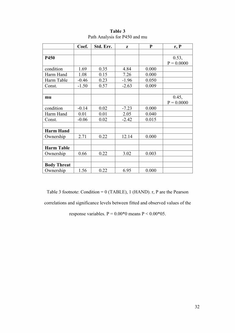
than 0.05. The resulting path model is shown in Figure 5 and associated Table 3. It
can be seen that Ownership is very strongly positively associated with the three harm
variables. Harm Hand is very strongly positively associated with P450 and weakly
with mu. Harm Table is weakly negatively associated with P450. Condition is
strongly positively associated with P450 and negatively with mu. The overall fit of
the model is good as shown by the last column of Table 3 which presents the
correlations between values fitted by the model and the observed values of the
response variables P450 and mu.
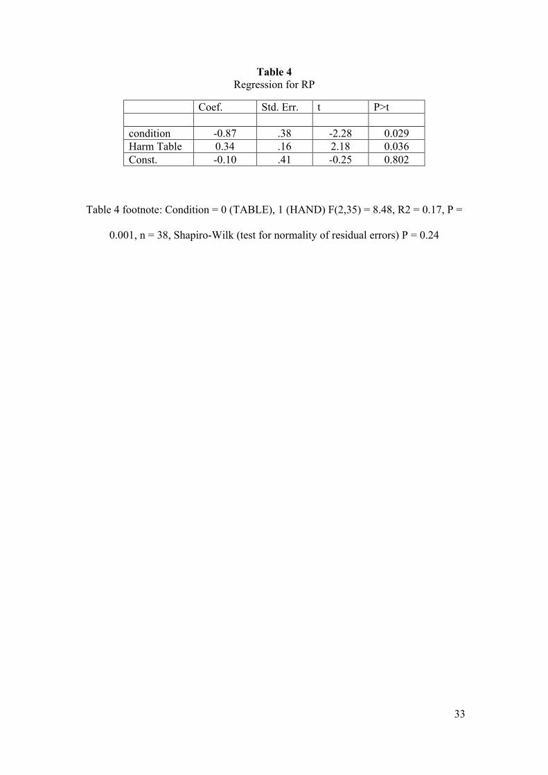
Now turning attention to the Readiness Potential (RP), this is based on a different set
of data (n = 38) since RP is a bipolar difference of the activity between the C3 and
C4 electrodes in the motor cortex so cannot be considered at the same time as P450
and mu. Applying path analysis to this data, only condition and Harm Table are
significantly related to RP. Hence an ordinary regression can be used (although still
we allow robust standard errors). The result is shown in Table 4, where condition

18
(Knife in Hand) is negatively associated with RP but positively associated with Harm
Table. This is consistent with a lateralization between hemispheres occurring during
the preparation of a motor action, the RP (C3-C4) is more negative when there is
preparation to move the right hand (Eimer, 1998), which in the current experiment is
the attacked hand. Therefore, a reduction in RP for higher scores in Harm Hand
question indicates stronger preparation of movement.
4. Discussion
Our results suggest that participants instinctively avoided a virtual knife stab to their
virtual body, thus activating the motor cortex and generating a mu-ERD, and a
Readiness Potential, as would be expected if their real hand were threatened. Our
study reproduced the results of (Fan and Han, 2008, Li and Han, 2010, Meng et al.,
2012, Meng et al., 2013) in terms of ERP correlates, showing significant evidence that
pain effects were found, with the mean P450 showing greater amplitudes at the CP3
electrode location in the HAND compared to the TABLE condition.
Importantly, participants had been instructed not to move their hand during the whole
experiment – and this was verified by the EMG analysis. However, it is important to
note that measurements of the flexor carpi alone could not have detected very subtle
movements, a reason for caution. Nevertheless, when doing the ERP study we found
motor cortex activation in the HAND condition with a significantly greater negative
Readiness Potential (C3-C4), associated with the intention of moving the right hand,
and this Readiness Potential was probably an instinctive reaction to the harm that
could not be controlled consciously by the participants.

19
Additionally, we found that when the virtual hand was attacked with the virtual knife,
it elicited significant motor cortex activation. A significant mu-ERD was found when
the knife attacked the hand - especially prominent in the C3 electrode - as if the
participant were trying to avoid harm. This suppression of the mu-rhythm in the
HAND condition could be interpreted as being in agreement with previous
observations about the involvement of this oscillatory desynchronization when a
participant performs a hand motor action (Pfurtscheller and Lopes da Silva, 1999).
Besides, this effect reproduces the results of (Yang et al., 2009, Perry et al., 2010,
Whitmarsh et al., 2011), although we believe that the illusory feeling of ownership
over the virtual body was likely much greater than in any previous pain observation
experiment. Furthermore, this illusory ownership provoked more prominent responses
with greater similarity to those described by (Babiloni et al., 2008) in preparation for
an electrical painful stimulation of the left index finger.
A recent paper (Evans and Blanke, 2013) showed that synchronous visual-tactile
feedback during the hand ownership illusion generates mu-ERD in the sensorimotor
cortex similar to the one produced during motor imagery BCI. Although, in our
experiment no tactile feedback was provided, we postulate that their results are
compatible with our findings and suggest that the correlations found in the current
experiment between the mu-ERD and P450 with the ownership illusion question may
be related by a similar mechanism to the one they describe. Future research could
assess whether tactile feedback would enhance the experience in the current scenario
and inhibit any existing sensory mismatch. In our study tactile feedback was avoided
to prevent overlaying activities in the sensorimotor cortex between the interpretation
of tactile sensory information and the efferent motor reactions (Yetkin et al., 1995). It
would have been very difficult to dissociate the effects of the tactile stimulation from

20
the subconscious motor reaction to the harm. However, regarding the sensory
mismatch, some participants reported a strange feeling in their finger at the end of the
experiment that would indicate that they were having illusions of tactile stimulation
through a top down mechanism.
According to (Pfurtscheller and Lopes da Silva, 1999) an ERS in the beta-rhythm
would typically be found in hand motor-imagery when the movement finishes.
Nevertheless, in the current experiment we could not find significant beta rebound.
We have shown that automatic neural mechanisms, such as pain responses, that occur
in reality occurred in this case in response to events in the virtual reality scenario of
this study. This is in line with previous findings that people do tend to have similar
responses in IVR as they would to similar situations in reality (Sanchez-Vives and
Slater, 2005), and this study seems to confirm this at the level of brain activity as
measured by EEG.
Additionally the results are useful for understanding the neural and cognitive
mechanisms of body perception. We have shown that neural responses (P450, mu and
RP) are correlated with the subjective level of the ownership illusion and the
subjective illusions of harm and threat to the body. It seems quite remarkable that
these variables, being in principle totally unrelated to one another (electrical brain
signals measured from the scalp compared with scores in a questionnaire) are
nevertheless apparently strongly related. This correlation provides a cross validation
that both the questionnaire responses and the electrical signals relate to the same
underlying brain processes. However, correlations should not be confused with
causation, and the path analysis proved useful for investigating a causal model
between the observed variables. For example, although there is a positive correlation
21
between Body Threat and P450 (Table 2), which might simplistically be interpreted as
a direct causal relation, in the context of the path model this can be seen as spurious.
The model provides an alternative interpretation that Ownership is positively
associated with Harm Hand, which in turn is positively associated with P450. But also
Ownership is positively associated with Body Threat. Overall the path analysis was
able to unravel possible relationships that would otherwise not be apparent, and
provide a quantitative assessment of a model.
From the path model P450 is higher in the HAND compared to the TABLE condition,
and it is also higher the stronger the subjective feeling that the hand might be harmed.
But whose hand? A possible caveat in the interpretation of the results is that we
cannot easily dissociate some of the intrinsic factors that may be modulating the
pattern of ERP responses observed, for example, between empathy and body
ownership. Previous empathy related studies (Fan and Han, 2008, Li and Han, 2010,
Meng et al., 2012, Meng et al., 2013) suggest that the P450 component is mostly
associated to empathy processing. Here, however, it appears to be related to
ownership, given that Harm Hand specifically refers to harm to the participant (“… I
might be harmed …”) a statement that is not compatible with an interpretation that
only favours empathy.
Empathy refers to the capacity to respond and understand experiences of another
person (Decety and Jackson, 2004). Brain activity associated with empathic responses
occurs, for example, in the context of pain observation of the (even violet coloured)
hand of a stranger (Avenanti et al., 2010). However, the same study shows that it is
not generated when the hand belongs to racial outgroup members (specifically white
individuals observing black hands). However, recent evidence suggests that
ownership by white individuals over a black rubber arm reduces implicit racial bias

22
(Maister et al., 2013) as does ownership of a dark skinned virtual body (Peck et al.,
2013). Since (Avenanti et al., 2010) found that the degree of implicit racial bias and
empathy responses were negatively correlated, we could conclude that embodiment in
the body of another might be likely to increase empathy towards that person or the
stereotype or group that the person represents (other things being equal). So although
empathy and body ownership are not the same, they are related - for body ownership
may be used to manipulate the degree of empathy.
Perhaps the common factor between empathy and body ownership is perspective
taking (Lamm et al., 2007). Perspective taking denotes the ability to see the world
from the eyes of another, and metaphorically to put yourself in the shoes of another. It
has been shown, for example, that perspective taking can improve attitudes towards
others such as racial or ethnic outgroups (Swart et al., 2010). However, virtual
embodiment provides a technological method for actually realising perspective taking
- when embodied in a virtual body it is literally the case that you see through the eyes
of a (virtual) other, so it is not especially surprising that virtual embodiment can lead
to a change in empathy, since it well realises perspective taking. However, we would
argue that in the present study empathy plays less of a role - except in the tautological
sense that you might have ‘maximal’ empathy towards yourself, your own body. We
suggest that this ‘maximal’ level of empathy may be a reason why stronger reactions
were found in the motor cortex in our experiment in comparison to previous pain
observation studies.
In our study we observed new effects (mu-ERD and lateralization) that have not been
reported before in previous empathy studies. We believe that these strong effects were
observed due to the strong embodiment illusion. Previous research has shown that
embodiment can be modulated by different combinations of self-location and body

23
ownership (Longo et al., 2008, Kilteni et al., 2012). In our setup the control condition
TABLE in which the knife did not appear where the hand was located, but 15cm
away, did not trigger the brain activation, indicating therefore that the possibility of
harm to the own body played an important role. Our results show that the exploitation
of virtual body ownership illusions could be useful for further understanding the
underlying neural mechanisms involved in cognitive processes of perception. Besides,
the measurements of cognitive processes provide a promising tool to measure virtual
embodiment.
This may also have implications not only for the measurement of virtual body
ownership but also to discriminate the strength of this illusion, so that people reacting
with a stronger EEG activation – greater P450 amplitude the virtual hand is attacked –
may have a stronger illusion than people with weaker P450 amplitude. This is
indicated in the path diagram (Figure 5) where the subjective level of ownership is
seen to be indirectly associated with both P450 and mu.
The questionnaire responses indicated generally a very strong illusion of ownership
over the virtual body. This could explain why the brain responses observed – P450,
Readiness Potential and mu-ERD – were larger in comparison to previous
experiments reported in the literature as observed above. A future experiment could
explicitly test this by reducing the level of ownership through a third person
perspective rather than a first person perspective condition (Slater et al., 2010,
Petkova et al., 2011, Peck et al., 2013). For example, this would involve observing the
reactions to seeing somebody else being attacked in an immersive virtual
environment. These results could also be further studied by focusing on the effects of
self-location with respect to the threat-stimuli. In general the neural responses by
themselves may provide a non-subjective measure of embodiment, however the

24
current findings are based on correlations of both objective and subjective measures.
Further studies may explore the extent in which ERPs may be exploited as an
objective measure of embodiment.
5. Conclusions
The present study suggests that, when a person is in an immersive virtual reality, and
has body ownership illusion towards a virtual body that apparently substitutes their
own body, there are autonomic responses that correspond to what would be observed
were the events to take place in reality. Overall automatic brain mechanisms –P450–
were found in this variation of the classical pain observation experiment, which is
consistent with what (Bufalari et al., 2007, Fan and Han, 2008, Li and Han, 2010,
Meng et al., 2012, Meng et al., 2013) previously reported. However, our setup was
not one concerned with participants experiencing empathy towards another person but
rather experienced direct attacks to their own body, since both subjective and
objective data point in that direction. The results support our initial hypothesis that a
threat to a virtual hand, towards which the participant has an illusion of ownership,
would significantly produce a harm prevention effect (measures using the Readiness
Potential (C3-C4) and oscillatory movement-related components, the mu-ERD), such
as trying to move it away from the source of the harm. The questionnaire also
confirmed high levels of ownership over the virtual body (see Figure 2). In addition,
the correlation between the automatic brain mechanisms –P450– and the subjective
illusion of ownership opens the door for a new promising measure of virtual
embodiment.

25
Acknowledgements
This study was funded by the European Union FP7 Integrated Project VERE (No.
257695). MGF’s research was supported by the FI-DGR predoctorate grant from the
Catalan Government co-founded by the European Social Found (EC-ESF). ARF has
been supported by a research grant from the Spanish government (PSI2011-29219).
The ERC project TRAVERSE (#227985) also contributed towards this research.
References
Armel KC, Ramachandran VS (2003) Projecting sensations to external objects: evidence from skin conductance response. Proceedings Biological sciences / The Royal Society 270:1499-1506.
Avenanti A, Bueti D, Galati G, Aglioti SM (2005) Transcranial magnetic stimulation highlights the sensorimotor side of empathy for pain. Nature neuroscience 8:955-960.
Avenanti A, Paluello IM, Bufalari I, Aglioti SM (2006) Stimulus-driven modulation of motor-evoked potentials during observation of others' pain. Neuroimage 32:316-324.
Avenanti A, Sirigu A, Aglioti SM (2010) Racial Bias Reduces Empathic Sensorimotor Resonance with Other-Race Pain. Current biology : CB 20:1018-1022.
Babiloni C, Capotosto P, Brancucci A, Del Percio C, Petrini L, Buttiglione M, Cibelli G, Romani GL, Rossini PM, Arendt-Nielsen L (2008) Cortical Alpha Rhythms Are Related to the Anticipation of Sensorimotor Interaction Between Painful Stimuli and Movements: A High-Resolution EEG Study. The journal of pain : official journal of the American Pain Society 9:902-911.
Banakou D, Groten R, Slater M (2013) Illusory ownership of a virtual child body causes overestimation of object sizes and implicit attitude changes. Proceedings of the National Academy of Sciences.
Blanke O (2012) Multisensory brain mechanisms of bodily self-consciousness. Nature Reviews Neuroscience 13:556-571.
Botvinick M, Cohen J (1998) Rubber hands' feel'touch that eyes see. Nature 391:756-756.
Bufalari I, Aprile T, Avenanti A, Di Russo F, Aglioti SM (2007) Empathy for Pain and Touch in the Human Somatosensory Cortex. Cerebral Cortex 17:2553-2561.
Cheng Y, Yang C-Y, Lin C-P, Lee P-L, Decety J (2008) The perception of pain in others suppresses somatosensory oscillations: A magnetoencephalography study. Neuroimage 40:1833-1840.
Decety J, Jackson PL (2004) The functional architecture of human empathy. Behavioral and cognitive neuroscience reviews 3:71-100.

26
Ehrsson HH (2007) The Experimental Induction of Out-of-Body Experiences. Science 317:1048.
Ehrsson HH, Spence C, Passingham RE (2004) That's My Hand! Activity in Premotor Cortex Reflects Feeling of Ownership of a Limb. Science 305:875-877.
Eimer M (1998) The lateralized readiness potential as an on-line measure of central response activation processes. Behavior Research Methods, Instruments, & Computers 30:146-156.
Evans N, Blanke O (2013) Shared electrophysiology mechanisms of body ownership and motor imagery. Neuroimage 64:216-228.
Fan Y, Han S (2008) Temporal dynamic of neural mechanisms involved in empathy for pain: An event-related brain potential study. Neuropsychologia 46:160-173.
Fridlund AJ, Cacioppo JT (1986) Guidelines for human electromyographic research. Psychophysiology 23:567-589.
Gillies M, Spanlang B (2010) Comparing and evaluating real time character engines for virtual environments. Presence: Teleoperators and Virtual Environments 19:95-117.
Gu X, Han S (2007) Attention and reality constraints on the neural processes of empathy for pain. NeuroImage 36:256-267.
Ijsselsteijn WA, de Kort YAW, Haans A (2006) Is This My Hand I See Before Me? The Rubber Hand Illusion in Reality, Virtual Reality, and Mixed Reality. Presence: Teleoperators and Virtual Environments 15:455-464.
Jackson PL, Meltzoff AN, Decety J (2005) How do we perceive the pain of others? A window into the neural processes involved in empathy. Neuroimage 24:771-779.
Kalckert A, Ehrsson HH (2012) Moving a rubber hand that feels like your own: dissociation of ownership and agency. Frontiers in Human Neuroscience 6.
Kaplan D (2009) Structural Equation Modeling: Foundations and Extensions: SAGE Publications.
Kilteni K, Bergstrom I, Slater M (2013) Drumming in immersive virtual reality: the body shapes the way we play. Visualization and Computer Graphics, IEEE Transactions on 19:597-605.
Kilteni K, Normand J-M, Sanchez-Vives MV, Slater M (2012) Extending body space in immersive virtual reality: a very long arm illusion. PloS one 7:e40867.
Lamm C, Batson CD, Decety J (2007) The neural substrate of human empathy: effects of perspective-taking and cognitive appraisal. Journal of Cognitive Neuroscience 19:42-58.
Lenggenhager B, Tadi T, Metzinger T, Blanke O (2007) Video Ergo Sum: Manipulating Bodily Self-Consciousness. Science 317:1096-1099.
Li W, Han S (2010) Perspective taking modulates event-related potentials to perceived pain. Neuroscience Letters 469:328-332.
Llobera J, Sanchez-Vives MV, Slater M (2013) The relationship between virtual body ownership and temperature sensitivity. Journal of The Royal Society Interface 10.
Longo MR, Schüür F, Kammers MPM, Tsakiris M, Haggard P (2008) What is embodiment? A psychometric approach. Cognition 107:978-998.
Maister L, Sebanz N, Knoblich Gn, Tsakiris M (2013) Experiencing ownership over a dark-skinned body reduces implicit racial bias. Cognition 128:170-178.
Maselli A, Slater M (2013) The building blocks of the full body ownership illusion. Frontiers in Human Neuroscience 7.

27
Meng J, Hu L, Shen L, Yang Z, Chen H, Huang X, Jackson T (2012) Emotional primes modulate the responses to others’ pain: an ERP study. Exp Brain Res 220:277-286.
Meng J, Jackson T, Chen H, Hu L, Yang Z, Su Y, Huang X (2013) Pain perception in the self and observation of others: An ERP investigation. NeuroImage 72:164-173.
Muthukumaraswamy SD, Johnson BW (2004) Primary motor cortex activation during action observation revealed by wavelet analysis of the EEG. Clinical neurophysiology : official journal of the International Federation of Clinical Neurophysiology 115:1760-1766.
Neuper C, Scherer R, Reiner M, Pfurtscheller G (2005) Imagery of motor actions: Differential effects of kinesthetic and visual–motor mode of imagery in single-trial EEG. Cognitive Brain Research 25:668-677.
Peck T, Seinfeld S, Aglioti S, Slater M (2013) Putting yourself in the skin of a black avatar reduces implicit racial bias. Consciousness and Cognition 22:779–787.
Perry A, Bentin S, Bartal I-A, Lamm C, Decety J (2010) “Feeling” the pain of those who are different from us: Modulation of EEG in the mu/alpha range. Cognitive, Affective, & Behavioral Neuroscience 10:493-504.
Petkova VI, Ehrsson HH (2008) If I were you: perceptual illusion of body swapping. PLoS One 3:e3832.
Petkova VI, Khoshnevis M, Ehrsson HH (2011) The perspective matters! Multisensory integration in ego-centric reference frames determines full-body ownership. Frontiers in psychology 2.
Pfurtscheller G, Lopes da Silva FH (1999) Event-related EEG/MEG synchronization and desynchronization: basic principles. Clinical neurophysiology : official journal of the International Federation of Clinical Neurophysiology 110:1842-1857.
Preston SD, de Waal FBM (2002) Empathy: Its ultimate and proximate bases. Behavioral and Brain Sciences 25:1-20.
Sanchez-Vives MV, Slater M (2005) From presence to consciousness through virtual reality. Nature Reviews Neuroscience 6:332-339.
Sanchez-Vives MV, Spanlang B, Frisoli A, Bergamasco M, Slater M (2010) Virtual Hand Illusion Induced by Visuomotor Correlations. PLoS ONE 5:e10381.
Slater M, Pérez Marcos D, Ehrsson H, Sanchez-Vives MV (2008) Towards a digital body: the virtual arm illusion. Frontiers in Human Neuroscience 2.
Slater M, Spanlang B, Sanchez-Vives MV, Blanke O (2010) First Person Experience of Body Transfer in Virtual Reality. PLoS ONE 5:e10564.
Steptoe W, Steed A, Slater M (2013) Human tails: ownership and control of extended humanoid avatars. Ieee Transactions on Visualization and Computer Graphics 19:in press.
Swart H, Hewstone M, Christ O, Voci A (2010) The impact of crossgroup friendships in South Africa: Affective mediators and multigroup comparisons. Journal of Social Issues 66:309-333.
Tecchia F, Carrozzino M, Bacinelli S, Rossi F, Vercelli D, Marino G, Gasparello P, Bergamasco M (2010) A Flexible Framework for Wide-Spectrum VR Development. Presence: Teleoperators and Virtual Environments 19:302-312.
Tsakiris M, Haggard P (2005) The rubber hand illusion revisited: visuotactile integration and self-attribution. Journal of Experimental Psychology-Human Perception and Performance 31:80-91.
28
Whitmarsh S, Nieuwenhuis ILC, Barendregt H, Jensen O (2011) Sensorimotor alpha activity is modulated in response to the observation of pain in others. Frontiers in Human Neuroscience 5.
Yang C-Y, Decety J, Lee S, Chen C, Cheng Y (2009) Gender differences in the mu rhythm during empathy for pain: An electroencephalographic study. Brain Research 1251:176-184.
Yetkin FZ, Mueller WM, Hammeke TA, Morris GLI, Haughton VM (1995) Functional Magnetic Resonance Imaging Mapping of the Sensorimotor Cortex with Tactile Stimulation. Neurosurgery 36:921-925.
List of Figures
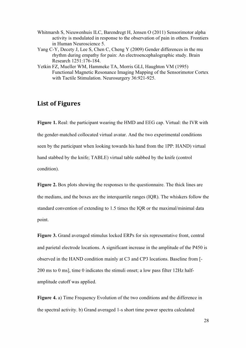
Figure 1. Real: the participant wearing the HMD and EEG cap. Virtual: the IVR with
the gender-matched collocated virtual avatar. And the two experimental conditions
seen by the participant when looking towards his hand from the 1PP: HAND) virtual
hand stabbed by the knife; TABLE) virtual table stabbed by the knife (control
condition).
Figure 2. Box plots showing the responses to the questionnaire. The thick lines are
the medians, and the boxes are the interquartile ranges (IQR). The whiskers follow the
standard convention of extending to 1.5 times the IQR or the maximal/minimal data
point.
Figure 3. Grand averaged stimulus locked ERPs for six representative front, central
and parietal electrode locations. A significant increase in the amplitude of the P450 is
observed in the HAND condition mainly at C3 and CP3 locations. Baseline from [-
200 ms to 0 ms], time 0 indicates the stimuli onset; a low pass filter 12Hz half-
amplitude cutoff was applied.
Figure 4. a) Time Frequency Evolution of the two conditions and the difference in
the spectral activity. b) Grand averaged 1-s short time power spectra calculated

29
from EEG data (electrode C3) recorded. The baseline corresponds to the range [-1 to
0] seconds before the stimuli and the activity period corresponds to the range [0.7 to
1.7] seconds after the stimuli. Both the Baseline and TABLE frequency spectra show
a peak in the mu-rhythm that is attenuated in the HAND condition. c) Grand averaged
Mu-rhythm (9-12Hz) Event Related Desynchronization for the C3 electrode. d) Grand
averaged Readiness Potential (C3-C4) subtraction between the brain activity in the
two hemispheres shows movement preparation effects. Low pass filter 8Hz, half-
amplitude cutoff.
Figure 5. Path analysis for P450 and mu and in relation to questionnaire variables
Harm Hand, Harm Table, Body Threat, Ownership and condition (Knife out = 0,
Knife in = 1). The values on the paths are the path coefficients and the corresponding
significance levels. The epsilon terms represent the random error term. The diagram
can be interpreted as a set of simultaneous linear prediction equations, e.g. from
Table 3 we can see that P450 = -1.50 + 1.69*condition + 1.08*(Harm Hand) -
0.46*(Harm Table) + epsilon. The circles are the random error terms and values by
the epsilon circles are their variances. The curved path represents a covariance.

30
List of Tables
Table 1 Spearman Correlation Coefficients Between the Questionnaire Scores
Ownership Harm Hand Harm Table No Ownership Body Threat Ownership 1.000 Harm Hand 0.726 1.000 (0.000) Harm Table 0.162 0.302 1.000 (0.508) (0.209) No Ownership -0.048 0.079 -0.125 1.000 (0.844) (0.749) (0.611) Body Threat 0.481 0.774 0.418 -0.179 1.000 (0.037) (0.000) (0.075) (0.463)
Table 1 footnote: (P values for test of 0 correlation). P = 0.000 means P < 0.0005, n = 19

31
Table 2 Spearman Correlation Coefficients Between the Questionnaire Scores and EEG
Variables
Ownership Harm Hand
Harm Table
No Ownership
Body Threat
P450 mu
p450 0.287 0.389 0.113 -0.021 0.289 1.0000 (0.0000) (0.0000) (0.089) (0.751) (0.0000) mu 0.266 0.093 -0.035 -0.169 0.040 0.029 1.0000 (0.0000) (0.160) (0.601) (0.011) (0.545) (0.658)
Table 2 footnote: n = 228. Overall R2 = 0.26, F(5,222) = 15.59, n = 228.
Shapiro-Wilk test for normality of residuals: P = 0.10.
P = 0.000 means P < 0.0005
32
Table 3 Path Analysis for P450 and mu
Coef. Std. Err. z P r, P P450 0.53,
P = 0.0000 condition 1.69 0.35 4.84 0.000 Harm Hand 1.08 0.15 7.26 0.000 Harm Table -0.46 0.23 -1.96 0.050 Const. -1.50 0.57 -2.63 0.009 mu 0.45,
P = 0.0000 condition -0.14 0.02 -7.23 0.000 Harm Hand 0.01 0.01 2.05 0.040 Const. -0.06 0.02 -2.42 0.015 Harm Hand Ownership 2.71 0.22 12.14 0.000 Harm Table Ownership 0.66 0.22 3.02 0.003 Body Threat Ownership 1.56 0.22 6.95 0.000
Table 3 footnote: Condition = 0 (TABLE), 1 (HAND). r, P are the Pearson
correlations and significance levels between fitted and observed values of the
response variables. P = 0.00*0 means P < 0.00*05.
33
Table 4 Regression for RP
Coef. Std. Err. t P>t condition -0.87 .38 -2.28 0.029 Harm Table 0.34 .16 2.18 0.036 Const. -0.10 .41 -0.25 0.802
Table 4 footnote: Condition = 0 (TABLE), 1 (HAND) F(2,35) = 8.48, R2 = 0.17, P =
0.001, n = 38, Shapiro-Wilk (test for normality of residual errors) P = 0.24