asympatheticneuronautonomousroleforegr3-mediated ... · 2003; howard, 2005). once precursors...
TRANSCRIPT
Development/Plasticity/Repair
A Sympathetic Neuron Autonomous Role for Egr3-MediatedGene Regulation in Dendrite Morphogenesis and TargetTissue Innervation
David H. Quach,1 Michelle Oliveira-Fernandes,1 Katherine A. Gruner,1 and Warren G. Tourtellotte1,2
1Department of Pathology (Division of Neuropathology) and 2Department of Neurology, Feinberg School of Medicine, Northwestern University, Chicago,Illinois 60611
Egr3 is a nerve growth factor (NGF)-induced transcriptional regulator that is essential for normal sympathetic nervous system develop-ment. Mice lacking Egr3 in the germline have sympathetic target tissue innervation abnormalities and physiologic sympathetic dysfunc-tion similar to humans with dysautonomia. However, since Egr3 is widely expressed and has pleiotropic function, it has not been clearwhether it has a role within sympathetic neurons and if so, what target genes it regulates to facilitate target tissue innervation. Here, weshow that Egr3 expression within sympathetic neurons is required for their normal innervation since isolated sympathetic neuronslacking Egr3 have neurite outgrowth abnormalities when treated with NGF and mice with sympathetic neuron-restricted Egr3 ablationhave target tissue innervation abnormalities similar to mice lacking Egr3 in all tissues. Microarray analysis performed on sympatheticneurons identified many target genes deregulated in the absence of Egr3, with some of the most significantly deregulated genes havingroles in axonogenesis, dendritogenesis, and axon guidance. Using a novel genetic technique to visualize axons and dendrites in asubpopulation of randomly labeled sympathetic neurons, we found that Egr3 has an essential role in regulating sympathetic neurondendrite morphology and terminal axon branching, but not in regulating sympathetic axon guidance to their targets. Together, theseresults indicate that Egr3 has a sympathetic neuron autonomous role in sympathetic nervous system development that involves modu-lating downstream target genes affecting the outgrowth and branching of sympathetic neuron dendrites and axons.
IntroductionThe sympathetic nervous system (SNS) is a subdivision of theautonomic nervous system that regulates organ homeostasis andit is the target of many developmental and degenerative diseasesin vertebrates, including humans. SNS development is a complexprocess mediated by multiple transcriptional regulators, mor-phogens, and target tissue-derived growth factors that direct pre-cursor migration, noradrenergic specification, differentiation,and target tissue innervation (Goridis and Rohrer, 2002; Rohrer,2003; Howard, 2005). Once precursors migrate to form discretesympathetic ganglia, axons extend along blood vessels, whichproduce diffusible factors such as Artemin (Honma et al., 2002)and neurotrophin 3 (Francis et al., 1999; Kuruvilla et al., 2004)that are required for normal axon growth along blood vessels.Additional target tissue-derived trophic factors such as nerve
growth factor (NGF) (Crowley et al., 1994; Glebova and Ginty,2004) and NGF-induced morphogens such as Wnt5a (Bodmer etal., 2009; Ho et al., 2012) are required for normal patterning ofsympathetic innervation within target tissues.
NGF has long been known as an essential target tissue-derivedsurvival factor for sympathetic neurons (Levi-Montalcini andBooker, 1960; Levi-Montalcini and Cohen, 1960; Crowley et al.,1994); however, more recent studies have demonstrated its addi-tional role in target tissue innervation (Albers et al., 1994; Has-sankhani et al., 1995; Patel et al., 2000; Glebova and Ginty, 2004).NGF function is mediated by tyrosine kinase receptor TrkA sig-naling, which activates Ras/mitogen activated protein kinase(MAPK), Src/protein kinase C (PKC), and phosphotidylinositoltriphosphate (PI3) kinase signaling pathways (Skaper, 2008). Al-though there is considerable cross-talk among the different sig-naling pathways, PI3 kinase signaling has a major role in neuronsurvival, whereas Ras/MAPK signaling is primarily involved inneuronal differentiation and neurite outgrowth (Klesse andParada, 1999; Atwal et al., 2000; Huang and Reichardt, 2001).However, very little is known about how NGF signaling pathwaysregulate gene expression within sympathetic neurons to facilitateits major functions in axon and dendrite outgrowth during targettissue innervation.
Egr3 is an example of a transcriptional regulator that is in-duced by NGF and coupled to MAPK signaling in sympatheticneurons. Mice lacking Egr3 in all tissues have sympathetic inner-vation abnormalities and dysautonomia, but whether its expres-
Received Nov. 27, 2012; revised Jan. 4, 2013; accepted Jan. 25, 2013.Author contributions: D.H.Q. and W.G.T. designed research; D.H.Q., M.O.-F., K.A.G., and W.G.T. performed re-
search; D.H.Q., M.O.-F., and W.G.T. analyzed data; D.H.Q. and W.G.T. wrote the paper.This work was supported by National Institutes of Health Grants (NIH) R01-NS040748, K02-NS046468, and
K26-OD026099 (W.G.T.). D.H.Q. was supported by NIH Grant F31-NS066721. We thank X. Gao for assistance ingenerating the DC�lZ transgenic mice and L. Eldredge, A. Hosanoge, and Q. Chen for technical assistance. G. Schutzgenerously provided the CaMKII-iCre transgenic mice.
Correspondence should be addressed to Dr. Warren G. Tourtellotte, Northwestern University, FeinbergSchool of Medicine, Department of Pathology, W-127, 303 East Chicago Avenue, Chicago, IL 60611. E-mail:[email protected].
DOI:10.1523/JNEUROSCI.5481-12.2013Copyright © 2013 the authors 0270-6474/13/334570-14$15.00/0
4570 • The Journal of Neuroscience, March 6, 2013 • 33(10):4570 – 4583

sion in sympathetic neurons is required for normal target tissueinnervation and/or what target genes it regulates is not known(Eldredge et al., 2008; Li et al., 2011). Here, we show that loss ofEgr3 expression specifically in sympathetic neurons leads to ab-normal neurite outgrowth in response to NGF signaling in vitroand sympathetic neuron dendrite and axon abnormalities in vivo.We identified several Egr3-regulated target genes with knownroles in axonogenesis and dendritogenesis, and using a geneticlabeling strategy to visualize a random subset of sympathetic neu-rons, we found that Egr3 is important for dendrite developmentand axon innervation, but not for axon guidance to target tissues.Thus, Egr3 has a sympathetic neuron autonomous role in generegulation involved in the outgrowth and branching of dendritesand axons, which likely explains the diminished sympatheticinnervation and physiologic dysautonomia that occurs in itsabsence.
Materials and MethodsAnimalsMice with Bax and Egr3 germline loss-of-function mutations were gen-erated as previously described (Knudson et al., 1995; Tourtellotte andMilbrandt, 1998). CaMKII-iCre BAC transgenic mice were obtainedfrom G. Schutz (Casanova et al., 2001). D�H-Cre BAC transgenic micewere recovered from the GENSAT sperm cryorepository (MMRRCStock# 032081-UCD) using in vitro fertilization and oocytes derivedfrom C57BL/6 female mice. Isogenic C57BL/6J CAG-FlpE mice wereobtained from the RIKEN animal resource (#RBRC01834) (Kanki et al.,2006), and Rosa-eGFP (JAX# 005670), Ai14 (JAX# 007908), and StLa(JAX# 010633) Cre-recombinase reporter mice were obtained from TheJackson Laboratory (Soriano, 1999; Belteki et al., 2005; Nam andBenezra, 2009; Madisen et al., 2010). All mice were back-crossed at leastfour generations to the C57BL/6 genetic background.
Egr3-Cre knockin mice. An 8.3 kb HindIII genomic DNA fragment(spanning 50605023–50613323, GenBank NT_039606.8) isolated from129Sv/J mouse genomic DNA that contained both coding exons, theintron, and the 3�UTR of Egr3 was used to generate the targeting vector(Tourtellotte and Milbrandt, 1998). The endogenous translation startcodon was converted to an NcoI site and the coding sequence for Cre-recombinase was exchanged with the endogenous exon 1, intron, andexon 2 coding sequence of Egr3 using homologous recombination inEscherichia coli (recombineering) (Liu et al., 2003). A PGK-Neo (Neo)positive-selection cassette containing a novel BglII restriction site andflanked by forward-oriented Frt (F) sites was inserted downstream of thenlsCre coding sequence. The targeting construct was electroporated into129/ola-derived HM-1 embryonic stem (ES) cells and G418-resistantclones were screened for homologous recombination. ES cell clones werescreened using long-range PCR flanking the 3� recombination arm of thetargeting construct with primers OT1182 (TTGCATCGCATTGTCTGA) and OT1114 (AAGCACCCCTGTCAAGTTTC). PCR-positiveclones were additionally screened by Southern blotting with a 700 ntprobe located 5� of the 5� recombination arm (spanning 50604041–50604741, GenBank, NT_039606.8), which was hybridized to genomicDNA restricted with BglII endonuclease to yield a 7.1 kb wild-type bandand a 4.3 kb targeted band. Three appropriately targeted clones wereinjected into isogenic C57BL/6J blastocysts using standard methods(Magin et al., 1992); chimeric mice were mated to C57BL/6J female miceand germline heterozygote mice (Egr3 �/Neo) were mated to CAG-FlpEmice (Kanki et al., 2006) to remove the frt-flanked Neo positive-selectioncassette from the germline and to generate the final Egr3-Cre allele (des-ignated Egr3 �/Cre).
Egr3-flx mice. The Egr3-flx mice were generated using the same 8.3 kbHindIII genomic DNA fragment used to generate the Egr3-Cre knockintargeting construct. A forward-oriented loxP site containing a novel BglIIrestriction site was targeted into the intron using homologous recombi-nation in E. coli (insertion site at position 778, GenBank, NT_039606). APGK-Neo (Neo) positive selection cassette flanked by forward-orientedFrt (F) sites and a single forward-oriented loxP site positioned 3� to the
F-Neo-F cassette was targeted immediately downstream of the TGAtranslation stop codon in exon 2 using homologous recombination in E.coli (insertion site at 2463 nt, GenBank, NT_039606). The targeting con-struct was electroporated into 129/ola-derived HM-1 ES cells and G418-resistant clones were subjected to screening by long-range PCR andSouthern blotting. Targeted clones were identified by 3� long-rangePCR using primers OT1111 (TACGAAGTTATTAGGTGGATCC) andOT1114 that spanned the 3� recombination arm. Correctly targetedclones were screened by Southern blotting using the same probe that wasused for generating Egr3-Cre mice, which yielded a 7.2 kb fragment forthe wild-type allele and a 4.5 kb fragment for the Neo allele on BglIIrestricted genomic DNA. Three targeted clones were injected into iso-genic C57BL/6J blastocysts using standard methods (Magin et al., 1992).Germline heterozygous Egr3 �/Neo mice were mated to CAG-FlpE miceto excise the frt-flanked Neo positive-selection cassette (Kanki et al.,2006) to generate the final Egr3 �/f allele (designated Egr3 �/f).
D�H-CreIRES�lacZ (DC�lZ) transgenic mice: The transgenic micewere generated using B6SJL embryos and are similar to sympathetic re-porter mice that were previously generated by us (Eldredge et al., 2008).However, these new sympathetic reporter mice express Cre-recombinaseand the axon localized �lacZ reporter (Callahan and Thomas, 1994) tran-scribed as a single bicistronic mRNA under control of the human dopa-mine �-hydroxylase (D�H) promoter (Mercer et al., 1991).
All mice were genotyped by PCR with genomic DNA isolated from tailbiopsy tissue. The sequences of the genotyping primers and PCR condi-tions are available upon request. All experimental procedures involvingmice complied with the Public Health Service Policy on Humane Careand Use of Laboratory Animals and all animal-related protocols wereapproved by the Northwestern University Institutional Animal Care andUse Committee.
Tissue preparationAnesthetized mice were perfused through the heart with 0.1 M phosphatebuffered, pH 7.2, 4% paraformaldehyde (PFA) and tissues were postfixedat 4°C for 1– 4 h. For frozen sections, tissues were cryoprotected over-night at 4°C in graded (15–30%) sucrose, embedded in OCT and sec-tioned at 12 �m thickness. For quantitative studies involving stereologicneuron counting, PFA fixed tissues were embedded in paraffin and serialsectioned at 16 �m. For some experiments, tissues were isolated fresh forRNA extraction.
Ganglion neuron countsSCG neuron numbers from adult (�8 weeks) DC�lZ �; Egr3 �/� andDC�lZ �; Egr3 f/� mice were determined using unbiased stereology andoptical dissector methods (StereoInvestigator; Microbrightfield) on ev-ery fifth serial section as previously described (Albert et al., 2005; El-dredge et al., 2008).
ImmunohistochemistryImmunofluorescence staining for tyrosine hydroxylase (TH; rabbitanti-TH, 1:5000; Millipore Bioscience Research Reagents), Renin(goat anti-renin, 1:150; Santa Cruz Biotechnology), and PECAM (hamsteranti-PECAM (2H8), 1:250; a gift from William A. Muller, NorthwesternUniversity, Chicago, IL) were performed on frozen tissue sections. Im-munohistochemistry staining for Egr3 (rabbit anti-Egr3, 1:1000; SantaCruz Biotechnology) was performed on paraffin tissue sections. �IIItubulin staining (mouse anti-tubulin, TU-20, 1:1000; Millipore Biosci-ence Research Reagents) was used to label neurons grown in culture.Species-appropriate secondary antibodies conjugated to Cy3 (JacksonImmunoResearch) or Alexa488 (Invitrogen) were used to localize pri-mary antibody binding.
Semiquantitative sympathetic axon terminal innervationQuantification of target tissue innervation on adult DC�lZ �; Egr3 �/�
and DC�lZ �; Egr3 f/� mice was performed using fluorescence densitom-etry. Fluorescent images were captured with a Zeiss LS510 confocalmicroscope using identical aperture and photomultiplier tube voltagesettings to ensure accurate comparison between tissues from bothconditions. The density of TH-positive terminals was calculated usingMetaMorph software (Molecular Devices) as the ratio of total immuno-
Quach et al. • Egr3-Mediated Gene Regulation in Sympathetic Neurons J. Neurosci., March 6, 2013 • 33(10):4570 – 4583 • 4571

fluorescence in a specified area and averaged for six separate confocalareas per section. Representative sympathetic target tissues, such asspleen, pineal gland, heart, and kidney, were analyzed in areas with thehighest density of axons identified in control tissues. The “relative inner-vation” in DC�lz �; Egr3 f/� mice was expressed as a mean percentage ofcontrol (DC�lZ �; Egr3 �/�) from 3 to 4 animals of each genotype asindicated.
LacZ enzyme histochemistryWhole mount lacZ enzyme histochemistry was performed on adolescent(�4 weeks) D�H-Cre �; Egr3 �/�; StLa �/f and D�H-Cre �; Egr3 �/�;StLa �/f mice. Adolescent mice were examined since adult tissues hadhigh nonspecific lacZ background staining in whole-mounted tissuesand SNS development is complete by this age. Tissues were dissected andpostfixed in 2% PFA, 0.2% glutaraldehyde, 5 mM EGTA, and 0.01%NP-40 in PBS-Mg at 4°C and reacted overnight at 37°C in reaction buffer(1 mg/ml X-gal, 5 mM potassium ferrocyanide, 5 mM potassium ferricya-nide). Whole tissues were cleared by stepping them through increasingconcentrations of glycerol (20, 50, 80, and 100%; 4 d each) mixed in 1%potassium hydroxide (KOH). The tissues were imaged with a NikonSMZ1500 stereomicroscope and a Nikon DS-Ri1 digital camera. Focalimage planes for superior cervical ganglion (SCG) neurons were pro-jected to flat-field using the Zerene Stacker software program (ZereneSystems). Neuron tracings were drawn using the DrawingSlate II (GTCO
CalComp Peripherals) drawing pad with TabletWorks software. Quan-tification of dendrite morphology was done using MetaMorph softwareon calibrated images. The axon was identified as the only neurite thatextended out of the ganglia while all other neurites were classified asdendrites. Only dendrites longer than the diameter of the soma werecounted as primary dendrites. The maximum extent of the dendriticarbor was calculated as the diameter of the largest concentric circle thatcould encompass the entire dendritic arbor. Calculation of divergentaxons emanating from the external carotid artery was measured bycounting axons that crossed a defined border that was parallel to andthree times the width of the artery. Quantification of sympathetic axonbranching in the submandibular gland was calculated by counting thebranch points of axons within the target tissue once they breached thetissue edge.
Primary sympathetic neuron culturesSCG neurons from newborn (P0) Egr3 �/� and Egr3 �/� mice were usedfor neuron culture. SCG neurons were dissociated with type IV collage-nase (1 mg/ml, Sigma), followed by 0.25% trypsin-EDTA and plated onpoly-L-lysine- and laminin-coated coverslips with media containingDMEM, 10% fetal bovine serum, 1% penicillin/streptomycin, and 2ng/ml NGF. Neurons were differentiated for 2 days as previously de-scribed (Eldredge et al., 2008). Neurite outgrowth parameters were mea-
Figure 1. Design and validation of Egr3-Cre knockin mice to trace historical Egr3 expression in tissues. A, Gene targeting in ES cells was used to replace both exons and the intron of Egr3 with theopen reading frame encoding nuclear localized Cre-recombinase (nlsCre). The Egr3-Cre (Egr3 �/Cre) allele was generated by removing the frt-site flanked Neo-positive selection cassette from thegermline using CAG-FlpE mice. B, Left, Electroporated G418 selected ES cell clones were screened using long-range PCR to amplify across the 3� recombination arm (primers: OT1182 and OT1114).Right, PCR confirmed ES cell clones were screened by Southern blotting using a 713 nt probe located upstream of the 5� recombination arm (5� ext), which was hybridized to BglII restricted genomicDNA. C, To validate the appropriate expression and function of Cre-recombinase, Egr3 �/Cre mice were mated to Ai14 Cre-recombinase reporter mice that irreversibly express fluorescent tdTomatoprotein after Cre-recombinase mediated loxP-site recombination (Madisen et al., 2010). Dentate gyrus granule neurons expressed Egr3 protein in (left) Egr3 �/Cre; Ai14 �/f and Egr3 �/�; Ai14 �/f
mice, but only in (right) Egr3 �/Cre; Ai14 �/f mice did they also express the tdTomato reporter. D, In Egr3 �/Cre; Ai14 �/f double heterozygous mice, Egr3 reporter (T) was observed in arteries alongwhich sympathetic nerves travel (splenic artery shown here), which colocalized in endothelial cells with the vascular endothelial marker PECAM (arrowheads). The tdTomato reporter is localizedprimarily in the endothelial nucleus whereas the PECAM protein is expressed on the endothelial membrane surface. E, Left, Sympathetic nerves labeled with TH and T (data not shown) were observedcoursing along the adventitia of arteries (arrowheads). Right, Within nerves (sciatic nerve shown here) T was colocalized in S100� Schwann cells (data not shown) and a fraction (28%) of TH�unmyelinated axons was closely associated with T� Schwann cells (TH�; T�) in sectioned nerves. Scale bars: C, 200 �m; D, E, left, 50 �m; E, right, 5 �m.
4572 • J. Neurosci., March 6, 2013 • 33(10):4570 – 4583 Quach et al. • Egr3-Mediated Gene Regulation in Sympathetic Neurons

sured using MetaMorph software on calibrated digital images of �IIItubulin-positive neurons.
Illumina BeadChip gene expression profilingMicroarray studies were performed on total cellular RNA isolated fromP0 SCG from Egr3 �/�; Bax �/� and Egr3 �/�; Bax �/� mice. As neuronsdo not innervate target tissues properly in Egr3 �/� mice and hence someneurons die, the Bax-deficient genetic background (Bax �/�) was used toinhibit neuron apoptosis and therefore avoid detecting apoptosis-related genes that are a secondary consequence of Egr3 loss. Total RNAwas isolated from SCG using Trizol extraction (Invitrogen) and the in-tegrity of the RNA samples was verified using an Agilent 2100 Bioana-lyzer. Microarray analysis was performed at the National Institutes ofHealth Neuroscience Microarray Consortium using the Illumina Bead-Chip platform and MouseWG-6 v.2.0 BeadChip microarrays. Briefly,cRNA for array hybridization was generated using 500 ng of total cellularRNA from each experimental condition using the TotalPrep RNA Am-plification kit (Applied Biosystems). Then 1500 ng of amplified heatdenatured biotin-labeled cRNA was mixed with hybridization buffer (Il-lumina), the mixture was hybridized to the arrays at 58°C for 20 h, thearrays were washed and stained according to the manufacturer’s specifi-cations, and they were scanned using the Illumina iScan. RNA isolatedfrom three individual mice of each genotype was used for hybridizationto a MouseWG-6 v.2.0 slide with six independent arrays each containing45,281 probe targets. Using the BeadStudio software, the raw dataset wasprocessed by subtracting background noise and normalizing using anaverage normalization parameter. The error model used to determinesignificantly differentially expressed genes was Illumina custom. Thedataset was filtered using the cutoff of difference p value � 0.01. The listof differentially expressed genes was analyzed using the Gene Ontology(GO) software program Database for Annotation, Visualization and In-tegrated Discovery (DAVID). The background reference list used wasMouseWG-6_V1_1_R4_11234304_A. The microarray data have beendeposited in NCBI’s Gene Expression Omnibus and are accessiblethrough NCBI GEO Series accession number GSE41290 (Edgar et al.,2002).
Quantitative real-time PCRQuantitative real-time PCR (qPCR) was performed as previously described(Albert et al., 2005). Briefly, total cellular RNA was isolated from P0 SCG
from control Egr3�/�; Bax�/� mice and exper-imental Egr3�/�; Bax�/� mice. cDNA was syn-thesized using Superscript III reversetranscriptase (Invitrogen) and random octamer/oligo-dT priming according to recommenda-tions by the manufacturer. Relative geneexpression was determined using Sybr Green (In-vitrogen) fluorescence with non-intron spanningprimers. The primers spanned and amplifiedthe following coding regions of target genes:apbb1 (amplified 2184 –2304 nt, GenBank,NM_001253885), b3galt6 (amplified 466–712 nt,GenBank, NM_080445), efnb1 (amplified 1467–1663 nt, GenBank, NM_010110), esd (amplified352–731 nt, GenBank, NM_016903), gins2 (am-plified 506 – 814 nt, GenBank, NM_178856),hs3st2 (amplified 996 –1300 nt, GenBank,NM_001081327), insc (amplified 410–670 nt,GenBank, NM_173767), irak1 (amplified 1234–1383 nt, GenBank, NM_001177973), lmo7 (am-plified 2754–2971 nt, GenBank, NM_201529),mxra8 (amplified 300 –511 nt, GenBank,NM_024263), nf1 (amplified 5041–5316 nt, Gen-Bank, NM_010897), ngfr (amplified 723–904 nt,GenBank, NM_033217), notch1 (amplified6532–6836 nt, GenBank, NM_008714), pabpc1l(amplified 1029 –1188 nt, GenBank,NM_001114079), pde3a (amplified 728–993 nt,GenBank, NM_018779), plxnd1 (amplified3584–3800 nt, GenBank, NM_026376), ptpn11
(amplified 564–679 nt, GenBank, NM_001109992), rccd1 (amplified 1152–1439 nt, GenBank, NM_173445), ret (amplified 965–1206 nt, GenBank,NM_001080780), slc15a2 (amplified 2343–2649 nt, GenBank,NM_021301), slit2 (amplified 5249–5407 nt, GenBank, NM_178804), sox9(amplified 807–1016 nt, GenBank, NM_011448), and zfp292 (amplified2973–3235 nt, GenBank, NM_013889). Standard curves were generated foreach primer pair using mouse genomic DNA and the target gene expressionresults were normalized to GAPDH expression for each sample analyzed.Results are represented as fold-change expression relative to the control con-dition and represent 3–4 replicates from different animals.
Statistical analysisValues are expressed as mean � SEM and p values are determined byStudent’s t test unless otherwise designated. For the � 2 test the expectedrandom distribution was calculated by dividing the total N evenly amongthe number of given conditions.
ResultsEgr3 has a sympathetic neuron autonomous role in targettissue innervationEgr3 expression is regulated by NGF signaling in sympatheticneurons and in the absence of Egr3 mice have physiologic dysau-tonomia caused by a failure of sympathetic neurons to properlyinnervate target tissues (Eldredge et al., 2008). In addition, micelacking both Egr3 and Bax (the latter, which prevents apoptoticneuron death) have innervation abnormalities that resemblethose occurring in mice lacking both NGF and Bax (Glebova andGinty, 2004; Li et al., 2011), further suggesting that Egr3 regu-lates, at least in part, gene expression induced by NGF signaling insympathetic neurons to influence target tissue innervation. How-ever, previous studies indicate that Egr3 is also expressed in bloodvessels along which sympathetic axons travel (Shneider et al.,2009) and in Schwann cells that myelinate some axons in nervesthrough which unmyelinated sympathetic axons pass during de-velopment (Gao et al., 2007). Moreover, Egr3 may be expressedin other tissues that sympathetic axons encounter during devel-opment, and its expression in those tissues may be transient,
Figure 2. Egr3 is required for normal neurite outgrowth and branching in NGF-treated sympathetic neurons. A, DissociatedEgr3 �/� (KO) sympathetic neurons have impaired neurite outgrowth in response to NGF treatment. Neurites from Egr3 KOneurons have significantly fewer branch points (B), decreased total neurite length (C), and decreased longest neurites comparedwith Egr3 �/� wild-type (WT) neurons (D). *p � 0.05, N � 60 neurons of each genotype analyzed. Scale bar: A, 50 �m.
Quach et al. • Egr3-Mediated Gene Regulation in Sympathetic Neurons J. Neurosci., March 6, 2013 • 33(10):4570 – 4583 • 4573

raising the possibility that Egr3 has a complex role in target tissueinnervation that may not solely depend on its expression in sym-pathetic neurons. To identify cells that express Egr3 at any timeduring their development, we generated Egr3-Cre knockin miceusing gene targeting to replace the endogenous Egr3 coding se-quence with the coding sequence for nuclear localized Cre-recombinase (nlsCre) (Fig. 1A,B). Egr3�/Cre mice were matedwith Ai14�/f reporter mice, which constitutively express the redfluorescent protein tdTomato (T) in cells that express Cre-recombinase (Madisen et al., 2010). In the hippocampal dentategyrus, where Egr3 is robustly and transiently expressed by neu-ronal activity (Li et al., 2007), T fluorescence was constitutivelycoexpressed with Egr3 in Egr3�/Cre; Ai14�/f double heterozygousdentate gyrus granule neurons as expected (Fig. 1C). We exam-ined T fluorescence in peripheral tissues known to interact withsympathetic axons from newborn and adult Egr3�/Cre; Ai14�/flx
double heterozygous mice and found strong labeling in arterialvascular endothelial cells (identified by colocalization with theendothelial cell marker, PECAM; Fig. 1D), which are in closeproximity with sympathetic axons that course along the surfaceof arterial vessels (identified by the sympathetic marker, TH; Fig.1E, left). Within perivascular nerves, the Egr3 reporter was pres-
ent in 100% of Schwann cells (identified by colocalization withthe Schwann cell marker, S100; data not shown and Gao et al.,2007). Although sympathetic axons are unmyelinated, they arebundled with myelinated axons in mixed motor and sensorynerves, where they often come in close contact with Schwanncells. For example, within the sciatic nerve we found that 28% ofTH� axons were closely associated with Schwann cells that ex-pressed the Egr3 tracer (Fig. 1E). The association frequency isvery likely higher since the analysis only assessed association insingle tissue sections and not along entire individual axons. To-gether, these results indicate that Egr3 is expressed in cells thatcould influence SNS development apart from its presumed au-tonomous function within sympathetic neurons.
Unlike NGF, which is required for sympathetic neuron sur-vival and neurite outgrowth, Egr3 is not required for neuronsurvival in isolated sympathetic neurons treated with NGF (El-dredge et al., 2008). Thus, if Egr3 has a role in mediating generegulation induced by NGF signaling, it may have a role in neuriteoutgrowth. To test whether Egr3 has an autonomous role in sym-pathetic neurite outgrowth in response to NGF, sympathetic neu-rons were isolated from the SCG of newborn Egr3�/� (wild-type)and Egr3�/� (Egr3 knock-out; KO) mice and grown in dissoci-
Figure 3. Design and validation of Egr3-flx conditional knock-out and sympathetic neuron Cre-driver mice. A, The mouse Egr3 genomic locus was modified to contain forward-oriented loxP sitesin intron 1 and downstream of the stop codon using homologous recombination in E. coli. A frt-flanked Neo selection cassette was inserted upstream of the 3� loxP site and the Egr3-flx (Egr3 �/f)allele was generated by removing the Neo selection cassette from the germline using CAG-FlpE mice. B, Left, Electroporated G418 selected ES cell clones were screened using long-range PCR withprimers that flanked the 3� recombination arm (primers: OT1111 and OT1114). Right, PCR confirmed ES cell clones were screened by Southern blotting analysis using a 713 nt probe (5� ext) locatedupstream of the 5� recombination arm that was hybridized to BglII restricted genomic DNA. C, To validate the conditional knock-out allele, CaMKII-iCre transgenic mice (Casanova et al., 2001) weremated to Egr3 �/f mice to ablate Egr3 in postnatal forebrain and hippocampal dentate gyrus neurons. As expected, Egr3 protein was robustly expressed in CaMKII-iCre �; Egr3 f/f (left; Ctl) dentategyrus neurons but was completely absent in CaMKII-iCre �; Egr3 f/f (right; cKO) neurons. D, The human D�H promoter was used to generate transgenic mice that express a Cre-recombinase and axonlocalized �-galactosidase (�lacZ) bicistronic message in noradrenergic neurons (designated DC�lZ transgenic mice). E, Cre-recombinase and lacZ expression were detectable in all TH� sympatheticneurons. F, To validate the efficiency of DC�lZ mice to recombine loxP sites in sympathetic neurons, they were mated to Rosa-eGFP reporter mice (Belteki et al., 2005). DC�lZ �; Rosa-eGFP �/f miceshowed no expression of eGFP, whereas DC�lZ �; Rosa-eGFP �/f mice showed expression of eGFP in all sympathetic neurons (SCG shown here). Scale bars: C, 200 �m; E, F, 100 �m.
4574 • J. Neurosci., March 6, 2013 • 33(10):4570 – 4583 Quach et al. • Egr3-Mediated Gene Regulation in Sympathetic Neurons
ated cultures treated with NGF (Fig. 2A). Compared with wild-type SCG neurons, those lacking Egr3 had significantly fewerneurite branch points (Fig. 2B), decreased total neurite length(Fig. 2C), and a reduction in the length of the longest neurite (Fig.2D). These data indicate that Egr3 has a moderate, but significantautonomous role in neurite extension and branching in responseto NGF, which may be correlated with the abnormal sympathetictarget tissue innervation abnormalities observed in germlineEgr3 KO mice.
To further examine whether Egr3 has a sympathetic neuronautonomous role in target tissue innervation, we generated twonovel mouse strains. We generated mice with a conditional Egr3allele by inserting loxP sites flanking exon 2 (flx allele), whichcontains the zinc-finger DNA binding domain required for tran-scriptional activation by the Egr3 protein. When loxP sites arerecombined, the resulting mutation is similar to the germlineloss-of-function mutation we generated in Egr3 KO mice (Tour-tellotte and Milbrandt, 1998) (Fig. 3A,B). Homozygous Egr3 f/f
mice were indistinguishable from wild-type mice, they expressednormal levels of Egr3, and removal of exon2 by loxP-site recom-bination led to complete loss of Egr3 protein in Cre-recombinase-expressing cells as expected (Fig. 3C). In addition,we generated transgenic mice (DC�lZ) using the human D�Hpromoter (Mercer et al., 1991) to drive the expression of a bicis-tronic transcript in noradrenergic neurons that encodes Cre-recombinase and axon localized lacZ (�lacZ) (Fig. 3D). DC�lZtransgenic mice expressed high levels of Cre-recombinase and
lacZ in sympathetic neurons (Fig. 3E),and functional tests demonstrated effi-cient loxP-site recombination by activa-tion of eGFP expression in sympatheticneurons from reporter mice (Fig. 3F).
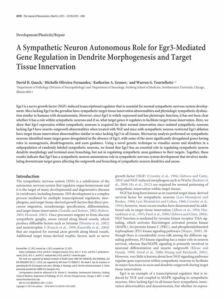
Using DC�lZ transgenic mice andEgr3-flx mice, we ablated Egr3 expressionin sympathetic neurons to examine itsneuron-specific role in target tissue inner-vation (Fig. 4A). Mice with the genotypeDC�lZ�; Egr3 f/f showed loxP-site recom-bination specifically in sympathetic neu-rons and adrenal chromaffin cells, but notin blood vessels, nerves, or other tissuesexamined (Fig. 4B). Since Egr3 heterozy-gous mice have no identifiable abnormal-ities, and to minimize mosaicism inrecombined sympathetic neurons, wemated Egr3�/f mice to Egr3�/� mice togenerate Egr3 f/� mice. Mice with specificablation of Egr3 in sympathetic neurons(DC�lZ�; Egr3 f/�, subsequently referredto as cKO mice) lacked the characteristicscoliosis and gait ataxia seen in germlineEgr3 KO mice, the latter which is causedby loss of Egr3 in muscle spindle stretchreceptors (Tourtellotte and Milbrandt,1998; Tourtellotte et al., 2001; Chen et al.,2002b). However, the cKO mice hadprominent SNS abnormalities includingblepharoptosis and significant neuronloss in the SCG compared with DC�lZ�;Egr3�/� (referred to as Ctl) mice, andsimilar to the blepharoptosis and neuronloss identified in germline Egr3 KO mice(Fig. 4C). In addition, Egr3 cKO mice had
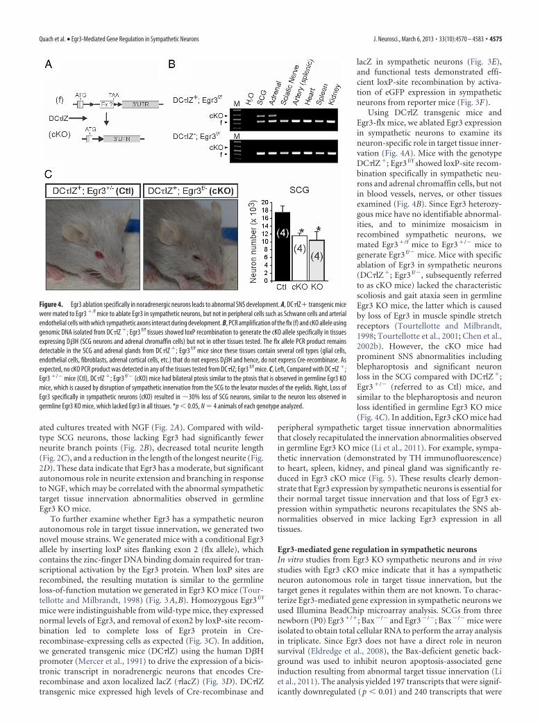
peripheral sympathetic target tissue innervation abnormalitiesthat closely recapitulated the innervation abnormalities observedin germline Egr3 KO mice (Li et al., 2011). For example, sympa-thetic innervation (demonstrated by TH immunofluorescence)to heart, spleen, kidney, and pineal gland was significantly re-duced in Egr3 cKO mice (Fig. 5). These results clearly demon-strate that Egr3 expression by sympathetic neurons is essential fortheir normal target tissue innervation and that loss of Egr3 ex-pression within sympathetic neurons recapitulates the SNS ab-normalities observed in mice lacking Egr3 expression in alltissues.
Egr3-mediated gene regulation in sympathetic neuronsIn vitro studies from Egr3 KO sympathetic neurons and in vivostudies with Egr3 cKO mice indicate that it has a sympatheticneuron autonomous role in target tissue innervation, but thetarget genes it regulates within them are not known. To charac-terize Egr3-mediated gene expression in sympathetic neurons weused Illumina BeadChip microarray analysis. SCGs from threenewborn (P0) Egr3�/�; Bax�/� and Egr3�/�; Bax�/� mice wereisolated to obtain total cellular RNA to perform the array analysisin triplicate. Since Egr3 does not have a direct role in neuronsurvival (Eldredge et al., 2008), the Bax-deficient genetic back-ground was used to inhibit neuron apoptosis-associated geneinduction resulting from abnormal target tissue innervation (Liet al., 2011). The analysis yielded 197 transcripts that were signif-icantly downregulated (p � 0.01) and 240 transcripts that were
Figure 4. Egr3 ablation specifically in noradrenergic neurons leads to abnormal SNS development. A, DC�lZ� transgenic micewere mated to Egr3 �/f mice to ablate Egr3 in sympathetic neurons, but not in peripheral cells such as Schwann cells and arterialendothelial cells with which sympathetic axons interact during development. B, PCR amplification of the flx (f) and cKO allele usinggenomic DNA isolated from DC�lZ �; Egr3 f/f tissues showed loxP recombination to generate the cKO allele specifically in tissuesexpressing D�H (SCG neurons and adrenal chromaffin cells) but not in other tissues tested. The flx allele PCR product remainsdetectable in the SCG and adrenal glands from DC�lZ �; Egr3 f/f mice since these tissues contain several cell types (glial cells,endothelial cells, fibroblasts, adrenal cortical cells, etc.) that do not express D�H and hence, do not express Cre-recombinase. Asexpected, no cKO PCR product was detected in any of the tissues tested from DC�lZ; Egr3 f/f mice. C, Left, Compared with DC�lZ �;Egr3 �/� mice (Ctl), DC�lZ �; Egr3 f/� (cKO) mice had bilateral ptosis similar to the ptosis that is observed in germline Egr3 KOmice, which is caused by disruption of sympathetic innervation from the SCG to the levator muscles of the eyelids. Right, Loss ofEgr3 specifically in sympathetic neurons (cKO) resulted in �30% loss of SCG neurons, similar to the neuron loss observed ingermline Egr3 KO mice, which lacked Egr3 in all tissues. *p � 0.05, N � 4 animals of each genotype analyzed.
Quach et al. • Egr3-Mediated Gene Regulation in Sympathetic Neurons J. Neurosci., March 6, 2013 • 33(10):4570 – 4583 • 4575
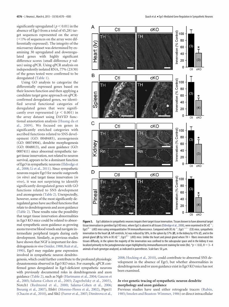
significantly upregulated (p � 0.01) in theabsence of Egr3 from a total of 45,281 tar-get sequences represented on the array(�1% of sequences on the array were dif-ferentially expressed). The integrity of themicroarray dataset was determined by ex-amining 30 upregulated and downregu-lated genes with highly significantdifference scores (small difference p val-ues) using qPCR. Using qPCR analysis onindependently isolated RNA, 77% (23/30)of the genes tested were confirmed to bederegulated (Table 1).
Using GO analysis to categorize thedifferentially expressed genes based ontheir known function and then applying acandidate target gene approach on qPCR-confirmed deregulated genes, we identi-fied several functional categories ofderegulated genes that were signifi-cantly over represented ( p � 0.001) inthe array dataset using DAVID func-tional annotation analysis (Huang da etal., 2009). We focused on genes insignificantly enriched categories withascribed functions related to SNS devel-opment (GO: 0048485), axonogenesis(GO: 00074904), dendrite morphogenesis(GO: 0048813), and axon guidance (GO:0007411) since abnormal sympathetic tar-get tissue innervation, not related to neuronsurvival, appears to be a dominant functionof Egr3 in sympathetic neurons (Eldredge etal., 2008; Li et al., 2011). Since sympatheticneurons require Egr3 for neurite outgrowth(in vitro) and target tissue innervation (invivo), it was not surprising to identifysignificantly deregulated genes with GOfunctions related to SNS developmentand axonogenesis (Table 2). Unexpectedly,however, some of the most significantly de-regulated genes have ascribed functions thatrelate to dendritogenesis and axon guidance(Table 2). These results raise the possibilitythat target tissue innervation abnormalitiesin Egr3 KO mice could be related to abnor-mal sympathetic axon guidance as growingaxons traverse blood vessels and navigate in-termediate peripheral targets during earlydevelopment. Similarly, as previous studieshave shown that NGF is important for den-dritogenesis in vivo (Snider, 1988; Ruit et al.,1990), Egr3 may regulate gene expressioninvolved in sympathetic neuron dendrito-genesis, which could further contribute to the profound physiologicdysautonomia observed in Egr3 KO mice. For example, qPCR con-firmed genes deregulated in Egr3-deficient sympathetic neuronswith previously documented roles in dendritogenesis and axonguidance (Table 2), such as Ngfr (Hartmann et al., 2004; Gascon etal., 2005; Salama-Cohen et al., 2005; Zagrebelsky et al., 2005),Notch1 (Redmond et al., 2000; Salama-Cohen et al., 2006;Breunig et al., 2007), Efnb1 (Moreno-Flores et al., 2002), Ptpn11(Chacon et al., 2010), and Slit2 (Furrer et al., 2007; Dimitrova et al.,
2008; Hocking et al., 2010), could contribute to abnormal SNS de-velopment in the absence of Egr3, but whether abnormalities indendritogenesis and/or axon guidance exist in Egr3 KO mice has notbeen examined.
In vivo genetic tracing of sympathetic neuron dendritemorphology and axon guidancePrevious studies have used either retrograde tracers (Rubin,1985; Smolen and Beaston-Wimmer, 1986) or direct intracellular
Figure 5. Egr3 ablation in sympathetic neurons impairs their target tissue innervation. Tissues known to have abnormal targettissue innervation in germline Egr3 KO mice, where Egr3 is absent in all tissues (Eldredge et al., 2008), were examined in DC�lZ �;Egr3 f/� (cKO) mice using semiquantitative TH immunofluorescence. Compared with DC�lz �; Egr3 �/� (Ctl) mice, sympatheticinnervation to the heart (A, left ventricle, lv) was reduced by 58%, in the spleen by 57% (B), in the kidney by 41% (C), and in thepineal gland (D) by 56% in DC�lZ �; Egr3 f/� (cKO) mice. Unlike the heart and pineal gland where TH� fibers innervated thetissues diffusely, in the spleen the majority of the innervation was confined to the subcapsular space and in the kidney it waslocalized primarily to the juxtaglomerular organ highlighted by immunofluorescent staining for renin (Rn). *p � 0.02, N � 3– 4animals of each genotype analyzed, as indicated in parentheses. Scale bars: 50 �m.
4576 • J. Neurosci., March 6, 2013 • 33(10):4570 – 4583 Quach et al. • Egr3-Mediated Gene Regulation in Sympathetic Neurons
sympathetic neuron injections with fluorescent dyes (Yawo,1987; Voyvodic, 1989) to label dendrites and axons in vivo, butthe yield of analyzable neurons is often low. As some of the iden-tified target genes may have a role in dendritogenesis, dendritemorphology was examined in vivo using a genetic tracing tech-nique that generates mosaic loxP recombination to sparsely labelneurons and their processes in Cre-recombinase reporter mice.D�H-Cre BAC transgenic mice (recovered from the GENSATrepository; MMRRC Stock# 032081-UCD and previously un-characterized) were found to inefficiently recombine loxP sites insympathetic neurons. For example, when they were mated toStLa reporter mice, which express axon localized lacZ (�lacZ)after Cre-mediated loxP recombination (Nam and Benezra,2009), �1% of sympathetic neurons were labeled in sympatheticganglia (Fig. 6A; SCG shown). The labeling pattern was similar toGolgi-stained neurons or neurons labeled by a Thy1 driven trans-gene reporter (Feng et al., 2000) in that the entire neuron and itsprocesses were labeled. This sparse labeling pattern made it pos-sible to compare in detail individual sympathetic neuron den-drite and axon morphology between wild-type and Egr3 KOmice.
Although there was never a visually similar pattern of labeledneurons in sympathetic ganglia in any of the mice examined, todetermine whether sympathetic neurons were randomly labeled,we first quantified sympathetic neurons that were lacZ labeled inD�H-Cre�; Egr3�/�; StLa�/f (referred to as Ctl) and D�H-Cre�; Egr3�/�; StLa�/f (referred to as Exp) mice and we foundno significant difference between them (Fig. 6B). To determinewhether the loxP-site recombination in sympathetic neurons wasstochastic, and hence whether labeling of SCG neurons was ran-dom, we examined the percentage of neurons labeled in differentregions of the SCG. SCG neurons that give rise to internal carotid
axons (ICA) generally reside in the rostral half of the ganglion,whereas those giving rise to the external carotid axons (ECA)generally reside in the caudal half of the ganglion (Bowers andZigmond, 1979). The SCGs were divided into rostral and caudalportions (Fig. 6A) and the percentage of lacZ-stained neurons inboth halves was determined. No significant difference from theexpected random distribution of neurons (50% in rostral and50% in caudal) was identified between rostral and caudal por-tions of the SCG in Ctl (� 2, p � 0.39) or Exp mice (� 2, p � 0.75)(Fig. 6C). Similarly, since SCG neurons give rise to more eCAaxons that innervate target tissues compared with iCA axons, thedistribution frequency of labeled eCA axons was greater than iniCA axons in both Ctl (� 2, p � 0.03) and Exp (� 2, p � 0.02) miceas expected (Fig. 6D). However, there was no significant differ-ence in the frequency distribution of axons between Ctl and Expmice (� 2, p � 0.66). These results indicate that sympathetic neu-rons are randomly labeled in these reporter mice and that therandom labeling pattern is retained in Egr3 KO mice.
Abnormal sympathetic neuron dendritogenesis in Egr3KO miceChemical clearing of whole lacZ-stained sympathetic gangliamade it possible to visualize dendrites in detail and to comparethem between Ctl and Exp mice (Fig. 7A). Neuron tracings fromflat-field projected images of labeled sympathetic neurons dem-onstrated that neurons from Exp mice had significantly fewerprimary dendrites compared with neurons from Ctl mice in boththe SCG (p � 0.00001) and stellate ganglion (STG) (p �0.00001) (Fig. 7B). Similarly, labeled neurons had decreased totaldendrite length in Exp mice in the SCG (p � 0.00001) and STG(p � 0.00001) (Fig. 7C) as well as a reduction in the maximumextent of the dendritic arbor in SCG (p � 0.00001) and STG (p �0.00001) compared with neurons from Ctl mice (Fig. 7D). Fi-nally, in agreement with previously published results indicatingthat sympathetic neurons are atrophic in the absence of Egr3(Eldredge et al., 2008; Li et al., 2011), we observed that labeledneurons in Exp mice were also significantly atrophic comparedwith Ctl mice (mean soma area in Ctl � 769.9 � 45.6 �m 2 and inExp � 560.5 � 32.7 �m 2, p � 0.0004). Thus, in addition to targettissue innervation abnormalities, Egr3 has a role in dendrite mor-phogenesis that may further contribute to physiological sympa-thetic dysfunction in Egr3 KO mice. Since germline Egr3 KOmice have �30% neuron loss that occurs during late embryogen-esis and early postnatal life (Eldredge et al., 2008), these data donot rule out the possibility that neuron loss may influence thedendritogenesis of remaining neurons. Similarly, since thesestudies were performed using germline KO mice, the data do notdefinitively demonstrate that dendritogenesis abnormalities aredue to loss of Egr3 expression within sympathetic neurons.
Role of Egr3 in axon guidance and target tissue innervationGene expression analysis indicated that several genes well estab-lished to have a role in axon guidance such as Plxnd1 (Chauvet etal., 2007), Efnb1 (Orioli and Klein, 1997; Flanagan and Vander-haeghen, 1998; Bush and Soriano, 2009), and Slit2 (Nguyen Ba-Charvet et al., 1999; Brose and Tessier-Lavigne, 2000; Plump etal., 2002) and some genes less well recognized to be involved inaxon guidance including Ngfr (Wong et al., 2002; Lim et al.,2008) and Ret (Enomoto et al., 2001; Kramer et al., 2006; Honmaet al., 2010) are deregulated in sympathetic neurons that lackEgr3 (Table 2). This raises the possibility that target tissue inner-vation abnormalities may result from abnormal axon guidance asthey traverse the periphery to innervate target tissues during de-
Table 1. qPCR-confirmed deregulated genes sampled from 30 genes with thesmallest difference p value in the microarray dataset of differentially expressedgenes in Egr3-deficient sympathetic neurons. (23 confirmed genes shown)
Official gene symbolNCBI geneID (mouse)
MicroarrayqPCRfold change(� SEM)
Difference pvalue Fold change
DownregulatedSox9 20682 2.74 10 �6 �2.25 �2.32 � 0.08Plxnd1 67784 5.07 10 �6 �1.82 �2.28 � 0.05Esd 13885 5.99 10 �6 �3.80 �1.64 � 0.12Gins2 272551 2.22 10 �5 �1.88 �1.51 � 0.07Ret 19713 5.28 10 �5 �1.63 �1.94 � 0.06Insc 233752 2.94 10 �4 �2.32 �2.77 � 0.48Notch1 18128 3.92 10 �4 �1.63 �2.07 � 0.30Efnb1 13641 2.75 10 �3 �1.40 �2.24 � 0.34Lmo7 380928 3.00 10 �3 �2.90 �2.59 � 0.53Hs3st2 195646 5.42 10 �3 �2.68 �1.60 � 0.25Ngfr 18053 8.50 10 �3 �1.48 �2.86 � 0.19
UpregulatedB3galt6 117592 3.68 10 �8 7.47 2.58 � 0.11Mxra8 74761 6.73 10 �7 2.20 1.60 � 0.17Nf1 18015 2.05 10 �5 2.87 2.26 � 0.37Apbb1 11785 7.44 10 �5 1.54 1.78 � 0.16Pabpc1l 381404 2.90 10 �4 4.77 3.67 � 0.23Slit2 20563 5.32 10 �4 1.70 1.63 � 0.20Slc15a2 57738 8.59 10 �4 3.72 2.24 � 0.55Irak1 16179 1.57 10 �3 2.80 2.00 � 0.17Zfp292 30046 1.95 10 �3 2.79 1.50 � 0.08Pde3a 54611 6.81 10 �3 3.91 1.97 � 0.20Rccd1 269955 7.72 10 �3 3.59 4.43 � 0.45Ptpn11 19247 8.65 10 �3 1.55 1.73 � 0.27
Results represent qPCR-confirmed deregulated genes from three replicate cDNA samples ( p � 0.05).
Quach et al. • Egr3-Mediated Gene Regulation in Sympathetic Neurons J. Neurosci., March 6, 2013 • 33(10):4570 – 4583 • 4577

velopment. However, as sympathetic target tissue innervation isdiminished, but not completely absent in Egr3 KO mice (El-dredge et al., 2008; Li et al., 2011), axon guidance abnormalitieswould likely exist in only a subset of axons, if at all. Previousanalysis using sympathetic axon reporter mice to label all sympa-thetic axons in the absence of Egr3 was not informative because itwas impossible to visualize a small number of potentially mis-guided axons among the entire labeled axon population (El-dredge et al., 2008). However, using mosaic neuron/axon labelingin Ctl and Exp mice, where only a small subset of sympatheticaxons are randomly labeled, it was possible to carefully examineindividual axon trajectories in Exp mice.
To determine whether sympathetic axon guidance was dis-rupted in Egr3 KO mice, a whole-mount preparation was devel-oped to examine axons projecting along their entire trajectory,from the SCG along the external carotid artery to the subman-dibular gland (Smg) (Fig. 8A). The Smg was chosen as a repre-sentative target tissue to study since it receives robust sympatheticinnervation from the SCG and innervation to it is highly abnor-mal in Egr3 KO mice (Eldredge et al., 2008). In Ctl and Exp mice,labeled sympathetic ECAs followed the course of the externalcarotid artery (Fig. 8B, arrowheads) and only occasional axonswere observed to diverge from the artery en route to the Smg (Fig.8B, arrow). There was no statistical difference between the num-ber of divergent axons arising from the SCG to innervate the Smgbetween Ctl and Exp mice (p � 0.39, N � 12 Ctl and N � 12 Expmice analyzed) (Fig. 8B). These results indicate that loss of Egr3does not result in axon misguidance to the Smg and thereforedoes not explain the diminished sympathetic target tissue inner-vation observed in Egr3 KO mice.
Egr3 has a clear neuron autonomous role in target tissue in-nervation that is unrelated to neuron survival or axon guidance,but how sympathetic target tissue innervation is impaired in Egr3KO mice remains unclear. Based on in vitro results demonstrat-ing that Egr3 has a role in neurite extension and branching, targettissue innervation could be impaired if axons from Egr3 KO neuronsfail to extend and branch properly within tissues. Interestingly, micelacking both NGF and Bax have innervation abnormalities thatare characterized by ineffective axon invasion and branchingwithin most tissues (Glebova and Ginty, 2004) and similar resultswere observed in mice lacking both Egr3 and Bax (Li et al., 2011).To determine whether Egr3 has a role in axon branching withintarget tissues, SCG-Smg whole-mount preparations were used toexamine axon innervation to the gland. A significant decrease inaxon branching within the gland in Exp relative to Ctl mice was
observed (Fig. 8C). Thus, terminal axon branching within targettissues, and not neuron loss or axon guidance, appears to explainthe relatively diminished target tissue innervation that occurs insympathetic neurons lacking Egr3.
DiscussionThe SNS is an essential part of the autonomic nervous system,which is impaired in many human neurodegenerative and meta-bolic diseases such as diabetes. Since the underlying cause of sym-pathetic neuron loss in these diseases is largely unknown and theylead to substantial suffering and premature death (Vinik et al.,2011), a greater understanding of critical signaling pathways in-volved in establishing and maintaining sympathetic target tissueinnervation homeostasis is of considerable interest. NGF and sig-naling through its high-affinity TrkA receptor is essential forsympathetic neuron survival, target tissue innervation, and post-natal viability in mice (Crowley et al., 1994; Smeyne et al., 1994;Glebova and Ginty, 2004) so it is not surprising that rare muta-tions in NGF� (HSAN type V, OMIM #608654) and TrkA(HSAN type IV, OMIM #256800) also lead to severe develop-mental abnormalities of the SNS in humans (Rotthier et al.,2012). How these essential mediators regulate downstream sig-naling in sympathetic neurons or whether any downstream effec-tors are involved in human disease is not known.
In previous studies we found that Egr3 is regulated by NGFsignaling in sympathetic neurons and that it has an essential rolein SNS development in mice (Eldredge et al., 2008; Li et al., 2011).However, the studies were performed in germline Egr3 knock-out mice, where Egr3 was absent in all tissues. In addition tosympathetic neurons, Egr3 is expressed in a variety of cell typeswhere it is induced by signaling molecules that mediate its sub-stantial pleiotropism. For example, Egr3 expression is regulatedby neuregulin-1 (Nrg1) in developing myoblasts, where it has animportant role in regulating skeletal muscle stretch receptormorphogenesis (Tourtellotte and Milbrandt, 1998; Hippen-meyer et al., 2002), it is regulated by factors such as brain-derivedneurotrophic factor in cortical neurons, where it has a role inregulating NMDA receptor and the plasticity associated Arc geneexpression involved in learning and memory (Li et al., 2005,2007; Kim et al., 2012), and it is regulated by T-cell receptorsignaling, where it has a role in immune function (Carter et al.,2007; Li et al., 2012). To determine how Egr3 expression mayspecifically influence sympathetic neuron development, we ex-amined its expression pattern in tissues known to interact withdeveloping sympathetic axons. Using Egr3-Cre knockin mice
Table 2. Selected qPCR-confirmed deregulated genes in Egr3-deficient sympathetic neurons and their GO determined functional associations
Official gene symbolNCBI geneID (mouse)
MicroarrayqPCR foldchange(�SEM) SNS development Axonogenesis Dendritogenesis Axon guidance
Difference pvalue Fold change
DownregulatedPlxnd1 67784 5.07 10 �6 �1.82 �2.28 � 0.05 XRet 19713 5.28 10 �5 �1.63 �1.94 � 0.06 X X XNotch1 18128 3.92 10 �4 �1.63 �2.0 � 0.30 X X XEfnb1 13641 2.75 10 �3 �1.40 �2.24 � 0.34 X X XNgfr 18053 8.50 10 �3 �1.48 �2.86 � 0.19 X X X X
UpregulatedNf1 18015 2.05 10 �5 2.87 2.26 � 0.37 X XApbb1 11785 7.44 10 �5 1.54 1.78 � 0.16 XSlit2 20563 5.32 10 �4 1.70 1.63 � 0.20 X X XPtpn11 19247 8.65 10 �3 1.55 1.73 � 0.27 X X X
Fold change compared to control with negative fold-change values representing genes downregulated in the absence of Egr3 and positive fold-change values representing genes upregulated in the absence of Egr3. All qPCR fold changesare significant, p � 0.05, three replicate cDNA samples.
4578 • J. Neurosci., March 6, 2013 • 33(10):4570 – 4583 Quach et al. • Egr3-Mediated Gene Regulation in Sympathetic Neurons

(Egr3�/Cre) and Cre-dependent reporter mice (Ai14) it was pos-sible to trace the historical expression of Egr3, even if expressionis transient and/or present in lineage precursors during develop-ment. In newborn and adult mice, we found reporter expressionlocalized in Schwann cells and arterial vascular endothelial cells,but not in other tissues that may be encountered by developingsympathetic axons during their outgrowth to target tissues. Thus,Egr3 may have a functional role in Schwann cells or endothelialcells, which could influence sympathetic target tissue innerva-tion. We confirmed a cell autonomous function for Egr3 withinsympathetic neurons using isolated Egr3-deficient sympatheticneurons, which showed significant neurite outgrowth andbranching abnormalities compared with wild-type neurons inresponse to NGF treatment. Similarly, using Egr3 conditionalknock-out and D�H-CreIRES�LacZ (DC�lZ) cre-driver trans-
genic mice to ablate Egr3 function in sympathetic neurons invivo, but not in other cells such as Schwann cells or vascularendothelial cells, we found that Egr3 cKO mice had sympathetictarget tissue innervation abnormalities that were indistinguish-able from Egr3 KO mice. Our studies did not address the role ofEgr3 expression in Schwann cells or vascular endothelial cells.Nevertheless, previous studies indicate that Egr3 modulates theexpression of the low-affinity NGF receptor (Ngfr) in Schwanncells where it has a role in peripheral axon myelination (Cosgayaet al., 2002; Gao et al., 2007) and it appears to mediate vascularendothelial growth factor signaling in vascular endothelial cells(Suehiro et al., 2010). Thus, a sympathetic neuron autonomousphenotype that is similar to the phenotype observed in germlineKO mice indicates that Egr3 function in Schwann and vascularendothelial cells is not likely related to SNS development.
To identify target genes that are regulated by Egr3 in sympa-thetic neurons we used microarray analysis on SCG isolated fromnewborn wild-type and Egr3 knock-out mice. We identified sev-eral hundred genes that were significantly upregulated anddownregulated in the absence of Egr3 (p � 0.01 and 1.2-foldcutoff). The list of deregulated genes appeared to be relativelyreliable since �75% of the 30 most significantly deregulatedgenes were confirmed by qPCR analysis and all of them werefound to be deregulated in the same direction (upregulated ordownregulated) as indicated by the microarray data (Table 1).Our current results indicate that Egr3 has a role in neurite out-growth that appears to correlate with abnormal dendrite devel-opment and terminal axon branching in vivo. Thus, the large list
Figure 6. In vivo genetic tracing highlights randomly labeled sympathetic neurons and theirdendrite morphology and axon trajectories. A, Sympathetic neurons are labeled in a mosaicpattern in D�H-Cre �; StLa �/f SCG where �1% of the neurons are labeled by the �lacZ re-porter (neurons and their processes labeled by lacZ histochemistry shown). iCA axons ariseprimarily from neurons located in the rostral 50% of the ganglion (black dashed line) and eCAaxons arise primarily from neurons located in the caudal 50% of the ganglion (red dashed line).B, No significant difference in the number of lacZ-labeled SCG neurons was detected betweenD�H-Cre �; Egr3 �/�; StLa �/f (Ctl) and D�H-Cre �; Egr3 �/�; StLa �/f (Exp) SCG (N � 6mice of each genotype analyzed). C, To examine whether lacZ-labeled neurons were randomlydistributed, the distribution frequency of neurons located in rostral and caudal regions wasdetermined. No difference in their distribution frequency relative to an expected random 50%distribution was detected between rostral and caudal regions of the SCG in either Ctl or Exp SCG.(N � 6 mice of each genotype analyzed; n.s., not significant, � 2 test). D, Since the SCG providesgreater innervation to target tissues innervated by axons that travel along the external carotidartery, the frequency distribution of lacZ-stained axons was significantly different from a ran-dom (50%) distribution, as expected in both Ctl (� 2, *p � 0.03) and Exp (� 2, *p � 0.02) mice.However, no significant difference was seen in the ECA–ICA frequency distributions between Ctland Exp mice (n.s., not significant; � 2 test; frequency distributions are shown as percentage oftotal lacZ-stained axons for each genotype, N � 6 animals of each genotype analyzed). Scalebar: A, 200 �m.
Figure 7. Abnormal dendrite morphogenesis in sympathetic neurons lacking Egr3. A, Top,lacZ-stained neurons in Ctl (D�H-Cre �; Egr3 �/�; StLa �/f) and Exp (D�H-Cre �; Egr3 �/�;StLa �/f) sympathetic ganglia were labeled in a sparse mosaic pattern with their axons anddendrites clearly visible. In labeled neurons, processes that projected out of the ganglia weredefined as axons (arrowhead), while the remainder of the processes confined to the gangliawere defined as dendrites. Bottom, Representative tracings from neuron profiles of flat-field(arrowheads) projected images demonstrated attenuated dendritic processes in Exp sympa-thetic neurons compared with those from Ctl mice. B–D, Dendrite morphology was quantifiedin Exp and Ctl neurons from SCG and STG. Compared with Ctl neurons those from Exp miceshowed significantly decreased total number of dendrites per neuron (B) significantly de-creased total dendrite length (C) and a reduction in maximum extent of the dendritic arbor insympathetic neurons from both SCG and STG (D). *p � 0.00001, N � 3– 4 animals analyzed ofeach genotype. Scale bars: A, 50 �m.
Quach et al. • Egr3-Mediated Gene Regulation in Sympathetic Neurons J. Neurosci., March 6, 2013 • 33(10):4570 – 4583 • 4579

of deregulated genes was refined using GOanalysis on qPCR confirmed deregulatedgenes to organize them by relevant func-tion (Table 2). Some of the genes that weresignificantly deregulated in the absence ofEgr3 have been previously shown to beinvolved in SNS development includingNgfr (Lee et al., 1994; Bamji et al., 1998;Brennan et al., 1999), Notch1 (Tsarovinaet al., 2008), Ret (Enomoto et al., 2001;Tsui-Pierchala et al., 2002), Nf1 (Vogel etal., 1995; Vogel and Parada, 1998), andPtpn11 (shp-2) (Chen et al., 2002a). Morespecifically, many of the genes regulatedby Egr3 in sympathetic neurons have rolesin neurite outgrowth such as Ngfr(Yamashita et al., 1999; Bentley andLee, 2000; Domeniconi et al., 2005),Ephrin-B1 (Tanaka et al., 2004), Notch1(Sestan et al., 1999; Huang et al., 2005),Ret (Enomoto et al., 2001; Zhang et al.,2006), Nf1 (Romero et al., 2007), Apbb1(Ikin et al., 2007), Ptpn11 (shp-2) (Chenet al., 2002a; Perrinjaquet et al., 2010), andSlit2 (Wang et al., 1999; Ozdinler and Er-zurumlu, 2002). For example, Ret is a re-ceptor for the glial cell line-derivedneurotrophic factor (GDNF) family of li-gands, which include GDNF, neurturin,artemin (ARTN), and persephin (Airaksi-nen et al., 1999; Baloh et al., 2000; Airak-sinen and Saarma, 2002). Ret knock-outmice have sympathetic neuron migrationabnormalities (Enomoto et al., 2001) andone of its ligands, ARTN has an essentialrole in mediating sympathetic axongrowth along blood vessels (Honma et al.,2002). Since Egr3 is first expressed inpostmigratory sympathetic neurons, it isnot surprising that loss of Egr3 does notresult in neuron migration abnormalities(Eldredge et al., 2008). However, de-creased Ret expression in postmigratory Egr3-deficient neuronscould result in abnormalities in GDNF and ARTN signaling.GDNF and ARTN knock-out mice have partial loss of sympa-thetic neurons and target tissue innervation (Moore et al., 1996;Honma et al., 2002), most likely caused by their well establishedrole in sympathetic neurite outgrowth and branching (Ebendal etal., 1995; Honma et al., 2002; Niwa et al., 2002; Yan et al., 2003).In addition, Ngfr is also required for normal sympathetic targettissue innervation (Lee et al., 1994). Thus, Egr3-mediated regu-lation of Ngfr expression in sympathetic neurons could modulatesome aspects of NGF signaling that may be essential for modu-lating a feedforward signaling loop that is dependent on NGF-induced Egr3 expression. Negative regulators of sympatheticneurite outgrowth, which were upregulated in the absence ofEgr3, such as Ptpn11 (shp-2), Apbb1, and Nf1 may further con-tribute to decreased sympathetic axon and dendrite outgrowthin vivo (Chen et al., 2002a; Ikin et al., 2007; Romero et al.,2007). NGF has a clear role in sympathetic neuron dendritedevelopment in vivo (Snider, 1988; Ruit et al., 1990) althoughour studies did not determine which particular target genesmay predominate.
The microarray analysis also identified several genes with arole in axon guidance such as Plxnd1 and Efnb1 (downregulated)and Slit2 (upregulated), suggesting that Egr3 could modulateaxon guidance. While we cannot rule out that Egr3 has a role inearly sympathetic axon guidance, our analysis showed only axonswith abnormal branching and no evidence of axon guidance ab-normalities along blood vessels. Many molecules with axon guid-ance functions have been shown to have other functions inneuronal processes including synapse formation and dendriticspine formation as well as axon and dendritic growth (Miller andKaplan, 2003). For example, Slit2, which was upregulated inEgr3-deficient sympathetic neurons, can increase axon branch-ing of sensory neurons (Wang et al., 1999; Ozdinler and Er-zurumlu, 2002). Slit proteins along with their Robo receptors arealso involved in dendrite development in cortical and retinal gan-glion neurons (Whitford et al., 2002; Dimitrova et al., 2008).Therefore, this molecule could influence axon and dendrite de-velopment in sympathetic neurons apart from any specific role inaxon guidance.
In summary, Egr3 is an NGF-induced transcriptional reg-ulator with a sympathetic neuron autonomous function in
Figure 8. Egr3 has a role in target tissue innervation that is independent of axon guidance. A, Chemical clearing and whole-mountpreparations using genetic axon tracing with D�H-Cre driver and StLa reporter mice made it possible to visualize a random subset of SCGneurons and their entire axon trajectories as they innervate the Smg. The Smg has profound innervation abnormalities in Egr3 KO mice(Eldredge et al., 2008) making this an informative preparation for testing whether innervation abnormalities are due to axon guidancedefects or branching within the target tissue. B, Sparsely labeled axons were traced along the external carotid artery (eCA; white dashedlines) in Ctl (D�H-Cre �; Egr3 �/�; StLa �/f) and Exp (D�H-Cre �; Egr3 �/�; StLa �/f) mice. In both Ctl and Exp mice sympathetic axonscoursed along the artery (arrowheads) to innervate the Smg, but occasional axons diverged from the artery to innervate tissues other thanthe Smg (arrow). There was no significant difference in the number of sympathetic axons traveling from the SCG to the Smg that divergedfrom the eCA between Ctl and Exp mice. C, In the Smg from Ctl and Exp mice lacZ-stained sympathetic axons entered the target tissuenormally. However, the extent of axon branching within the target tissue was decreased in Exp relative to Ctl mice. *p�0.015; N�6 –12animals studied of each genotype as indicated. Scale bars: A, 500�m; B, 250�m; C, 100�m. Dashed lines highlight the common carotidartery, internal carotid artery (iCA) and external carotid artery (eCA).
4580 • J. Neurosci., March 6, 2013 • 33(10):4570 – 4583 Quach et al. • Egr3-Mediated Gene Regulation in Sympathetic Neurons

dendrite morphogenesis and axon branching. Mice lackingEgr3 have physiologic sympathetic dysautonomia that appearsto be caused by disrupted expression of a relatively small net-work of genes, some of which have functions related to den-drite and axon development. These results raise the possibilitythat similar regulatory networks may be impaired in humanswith SNS disease, the majority of which are not understoodand poorly treated.
ReferencesAiraksinen MS, Saarma M (2002) The GDNF family: signalling, biological
functions and therapeutic value. Nat Rev Neurosci 3:383–394. CrossRefMedline
Airaksinen MS, Titievsky A, Saarma M (1999) GDNF family neu-rotrophic factor signaling: four masters, one servant? Mol Cell Neu-rosci 13:313–325. CrossRef Medline
Albers KM, Wright DE, Davis BM (1994) Overexpression of nerve growthfactor in epidermis of transgenic mice causes hypertrophy of the periph-eral nervous system. J Neurosci 14:1422–1432. Medline
Albert Y, Whitehead J, Eldredge L, Carter J, Gao X, Tourtellotte WG (2005)Transcriptional regulation of myotube fate specification and intrafusalmuscle fiber morphogenesis. J Cell Biol 169:257–268. CrossRef Medline
Atwal JK, Massie B, Miller FD, Kaplan DR (2000) The TrkB-Shc site signalsneuronal survival and local axon growth via MEK and P13-kinase. Neu-ron 27:265–277. CrossRef Medline
Baloh RH, Enomoto H, Johnson EM Jr, Milbrandt J (2000) The GDNFfamily ligands and receptors-implications for neural development. CurrOpin Neurobiol 10:103–110. CrossRef Medline
Bamji SX, Majdan M, Pozniak CD, Belliveau DJ, Aloyz R, Kohn J, CausingCG, Miller FD (1998) The p75 neurotrophin receptor mediates neuro-nal apoptosis and is essential for naturally occurring sympathetic neurondeath. J Cell Biol 140:911–923. CrossRef Medline
Belteki G, Haigh J, Kabacs N, Haigh K, Sison K, Costantini F, Whitsett J,Quaggin SE, Nagy A (2005) Conditional and inducible transgene ex-pression in mice through the combinatorial use of Cre-mediated recom-bination and tetracycline induction. Nucleic Acids Res 33:e51. CrossRefMedline
Bentley CA, Lee KF (2000) p75 is important for axon growth and Schwanncell migration during development. J Neurosci 20:7706 –7715. Medline
Bodmer D, Levine-Wilkinson S, Richmond A, Hirsh S, Kuruvilla R (2009)Wnt5a mediates nerve growth factor-dependent axonal branching andgrowth in developing sympathetic neurons. J Neurosci 29:7569 –7581.CrossRef Medline
Bowers CW, Zigmond RE (1979) Localization of neurons in the rat superiorcervical ganglion that project into different postganglionic trunks.J Comp Neurol 185:381–391. CrossRef Medline
Brennan C, Rivas-Plata K, Landis SC (1999) The p75 neurotrophin receptorinfluences NT-3 responsiveness of sympathetic neurons in vivo. Nat Neu-rosci 2:699 –705. CrossRef Medline
Breunig JJ, Silbereis J, Vaccarino FM, Sestan N, Rakic P (2007) Notch regu-lates cell fate and dendrite morphology of newborn neurons in the post-natal dentate gyrus. Proc Natl Acad Sci U S A 104:20558 –20563. CrossRefMedline
Brose K, Tessier-Lavigne M (2000) Slit proteins: key regulators of axonguidance, axonal branching, and cell migration. Curr Opin Neurobiol10:95–102. CrossRef Medline
Bush JO, Soriano P (2009) Ephrin-B1 regulates axon guidance by reversesignaling through a PDZ-dependent mechanism. Genes and development23:1586 –1599. CrossRef Medline
Callahan CA, Thomas JB (1994) Tau-beta-galactosidase, an axon-targetedfusion protein. Proc Natl Acad Sci U S A 91:5972–5976. CrossRef Medline
Carter JH, Lefebvre JM, Wiest DL, Tourtellotte WG (2007) Redundant rolefor early growth response transcriptional regulators in thymocyte differ-entiation and survival. J Immunol 178:6796 – 6805. Medline
Casanova E, Fehsenfeld S, Mantamadiotis T, Lemberger T, Greiner E, StewartAF, Schutz G (2001) A CaMKIIalpha iCre BAC allows brain-specificgene inactivation. Genesis 31:37– 42. CrossRef Medline
Chacon PJ, Arevalo MA, Tebar AR (2010) NGF-activated protein tyrosinephosphatase 1B mediates the phosphorylation and degradation ofI-kappa-Balpha coupled to NF-kappa-B activation, thereby controllingdendrite morphology. Mol Cell Neurosci 43:384 –393. CrossRef Medline
Chauvet S, Cohen S, Yoshida Y, Fekrane L, Livet J, Gayet O, Segu L, BuhotMC, Jessell TM, Henderson CE, Mann F (2007) Gating of Sema3E/Plex-inD1 signaling by neuropilin-1 switches axonal repulsion to attractionduring brain development. Neuron 56:807– 822. CrossRef Medline
Chen B, Hammonds-Odie L, Perron J, Masters BA, Bixby JL (2002a) SHP-2mediates target-regulated axonal termination and NGF-dependent neu-rite growth in sympathetic neurons. Dev Biol 252:170 –187. CrossRefMedline
Chen HH, Tourtellotte WG, Frank E (2002b) Muscle spindle-derived neu-rotrophin 3 regulates synaptic connectivity between muscle sensory andmotor neurons. J Neurosci 22:3512–3519. Medline
Cosgaya JM, Chan JR, Shooter EM (2002) The neurotrophin receptorp75NTR as a positive modulator of myelination. Science 298:1245–1248.CrossRef Medline
Crowley C, Spencer SD, Nishimura MC, Chen KS, Pitts-Meek S, ArmaniniMP, Ling LH, McMahon SB, Shelton DL, Levinson AD, et al. (1994)Mice lacking nerve growth factor display perinatal loss of sensory andsympathetic neurons yet develop basal forebrain cholinergic neurons.Cell 76:1001–1011. CrossRef Medline
Dimitrova S, Reissaus A, Tavosanis G (2008) Slit and Robo regulate den-drite branching and elongation of space-filling neurons in Drosophila.Dev Biol 324:18 –30. CrossRef Medline
Domeniconi M, Zampieri N, Spencer T, Hilaire M, Mellado W, Chao MV,Filbin MT (2005) MAG induces regulated intramembrane proteolysisof the p75 neurotrophin receptor to inhibit neurite outgrowth. Neuron46:849 – 855. CrossRef Medline
Ebendal T, Tomac A, Hoffer BJ, Olson L (1995) Glial cell line-derived neu-rotrophic factor stimulates fiber formation and survival in cultured neu-rons from peripheral autonomic ganglia. J Neurosci Res 40:276 –284.CrossRef Medline
Edgar R, Domrachev M, Lash AE (2002) Gene Expression Omnibus: NCBIgene expression and hybridization array data repository. Nucleic AcidsRes 30:207–210. CrossRef Medline
Eldredge LC, Gao XM, Quach DH, Li L, Han X, Lomasney J, Tourtellotte WG(2008) Abnormal sympathetic nervous system development and physi-ological dysautonomia in Egr3-deficient mice. Development 135:2949 –2957. CrossRef Medline
Enomoto H, Crawford PA, Gorodinsky A, Heuckeroth RO, Johnson EM Jr,Milbrandt J (2001) RET signaling is essential for migration, axonalgrowth and axon guidance of developing sympathetic neurons. Develop-ment 128:3963–3974. Medline
Feng G, Mellor RH, Bernstein M, Keller-Peck C, Nguyen QT, Wallace M,Nerbonne JM, Lichtman JW, Sanes JR (2000) Imaging neuronal subsetsin transgenic mice expressing multiple spectral variants of GFP. Neuron28:41–51. CrossRef Medline
Flanagan JG, Vanderhaeghen P (1998) The ephrins and Eph receptors inneural development. Annu Rev Neurosci 21:309 –345. CrossRef Medline
Francis N, Farinas I, Brennan C, Rivas-Plata K, Backus C, Reichardt L, LandisS (1999) NT-3, like NGF, is required for survival of sympathetic neu-rons, but not their precursors. Dev Biol 210:411– 427. CrossRef Medline
Furrer MP, Vasenkova I, Kamiyama D, Rosado Y, Chiba A (2007) Slit andRobo control the development of dendrites in Drosophila CNS. Develop-ment 134:3795–3804. CrossRef Medline
Gao X, Daugherty RL, Tourtellotte WG (2007) Regulation of low affinityneurotrophin receptor (p75(NTR)) by early growth response (Egr) tran-scriptional regulators. Mol Cell Neurosci 36:501–514. CrossRef Medline
Gascon E, Vutskits L, Zhang H, Barral-Moran MJ, Kiss PJ, Mas C, Kiss JZ(2005) Sequential activation of p75 and TrkB is involved in dendriticdevelopment of subventricular zone-derived neuronal progenitors invitro. Eur J Neurosci 21:69 – 80. CrossRef Medline
Glebova NO, Ginty DD (2004) Heterogeneous requirement of NGF forsympathetic target innervation in vivo. J Neurosci 24:743–751. CrossRefMedline
Goridis C, Rohrer H (2002) Specification of catecholaminergic and seroto-nergic neurons. Nat Rev Neurosci 3:531–541. CrossRef Medline
Hartmann M, Brigadski T, Erdmann KS, Holtmann B, Sendtner M, Narz F,Lessmann V (2004) Truncated TrkB receptor-induced outgrowth ofdendritic filopodia involves the p75 neurotrophin receptor. J Cell Sci117:5803–5814. CrossRef Medline
Hassankhani A, Steinhelper ME, Soonpaa MH, Katz EB, Taylor DA,Andrade-Rozental A, Factor SM, Steinberg JJ, Field LJ, Federoff HJ(1995) Overexpression of NGF within the heart of transgenic mice causes
Quach et al. • Egr3-Mediated Gene Regulation in Sympathetic Neurons J. Neurosci., March 6, 2013 • 33(10):4570 – 4583 • 4581

hyperinnervation, cardiac enlargement, and hyperplasia of ectopic cells.Dev Biol 169:309 –321. CrossRef Medline
Hippenmeyer S, Shneider NA, Birchmeier C, Burden SJ, Jessell TM, Arber S(2002) A role for neuregulin1 signaling in muscle spindle differentiation.Neuron 36:1035–1049. CrossRef Medline
Ho HY, Susman MW, Bikoff JB, Ryu YK, Jonas AM, Hu L, Kuruvilla R,Greenberg ME (2012) Wnt5a-Ror-Dishevelled signaling constitutes acore developmental pathway that controls tissue morphogenesis. ProcNatl Acad Sci U S A 109:4044 – 4051. Medline
Hocking JC, Hehr CL, Bertolesi GE, Wu JY, McFarlane S (2010) Distinctroles for Robo2 in the regulation of axon and dendrite growth by retinalganglion cells. Mech Dev 127:36 – 48. CrossRef Medline
Honma Y, Araki T, Gianino S, Bruce A, Heuckeroth R, Johnson E, MilbrandtJ (2002) Artemin is a vascular-derived neurotropic factor for developingsympathetic neurons. Neuron 35:267–282. CrossRef Medline
Honma Y, Kawano M, Kohsaka S, Ogawa M (2010) Axonal projections ofmechanoreceptive dorsal root ganglion neurons depend on Ret. Develop-ment 137:2319 –2328. CrossRef Medline
Howard MJ (2005) Mechanisms and perspectives on differentiation of au-tonomic neurons. Dev Biol 277:271–286. CrossRef Medline
Huang da W, Sherman BT, Lempicki RA (2009) Systematic and integrativeanalysis of large gene lists using DAVID bioinformatics resources. NatProtoc 4:44 –57. Medline
Huang EJ, Reichardt LF (2001) Neurotrophins: roles in neuronal develop-ment and function. Annu Rev Neurosci 24:677–736. CrossRef Medline
Huang EJ, Li H, Tang AA, Wiggins AK, Neve RL, Zhong W, Jan LY, Jan YN(2005) Targeted deletion of numb and numblike in sensory neuronsreveals their essential functions in axon arborization. Genes Dev 19:138 –151. CrossRef Medline
Ikin AF, Sabo SL, Lanier LM, Buxbaum JD (2007) A macromolecular com-plex involving the amyloid precursor protein (APP) and the cytosolicadapter FE65 is a negative regulator of axon branching. Mol Cell Neurosci35:57– 63. CrossRef Medline
Kanki H, Suzuki H, Itohara S (2006) High-efficiency CAG-FLPe deletermice in C57BL/6J background. Exp Anim 55:137–141. CrossRef Medline
Kim JH, Roberts DS, Hu Y, Lau GC, Brooks-Kayal AR, Farb DH, Russek SJ(2012) Brain-derived neurotrophic factor uses CREB and Egr3 to regu-late NMDA receptor levels in cortical neurons. J Neurochem 120:210 –219. CrossRef Medline
Klesse LJ, Parada LF (1999) Trks: signal transduction and intracellular path-ways. Microsc Res Tech 45:210 –216. CrossRef Medline
Knudson CM, Tung KS, Tourtellotte WG, Brown GA, Korsmeyer SJ (1995)Bax-deficient mice with lymphoid hyperplasia and male germ cell death.Science 270:96 –99. CrossRef Medline
Kramer ER, Knott L, Su F, Dessaud E, Krull CE, Helmbacher F, Klein R(2006) Cooperation between GDNF/Ret and ephrinA/EphA4 signals formotor-axon pathway selection in the limb. Neuron 50:35– 47. CrossRefMedline
Kuruvilla R, Zweifel LS, Glebova NO, Lonze BE, Valdez G, Ye H, Ginty DD(2004) A neurotrophin signaling cascade coordinates sympathetic neu-ron development through differential control of TrkA trafficking andretrograde signaling. Cell 118:243–255. CrossRef Medline
Lee KF, Bachman K, Landis S, Jaenisch R (1994) Dependence on p75 forinnervation of some sympathetic targets. Science 263:1447–1449.CrossRef Medline
Levi-Montalcini R, Booker B (1960) Destruction of the sympathetic gangliain mammals by an antiserum to a nerve-growth protein. Proc Natl AcadSci U S A 46:384 –391. CrossRef Medline
Levi-Montalcini R, Cohen S (1960) Effects of the extract of the mouse sub-maxillary salivary glands on the sympathetic system of mammals. AnnN Y Acad Sci 85:324 –341. Medline
Li L, Carter J, Gao X, Whitehead J, Tourtellotte WG (2005) Theneuroplasticity-associated arc gene is a direct transcriptional target ofearly growth response (Egr) transcription factors. Mol Cell Biol 25:10286 –10300. CrossRef Medline
Li L, Yun SH, Keblesh J, Trommer BL, Xiong H, Radulovic J, Tourtellotte WG(2007) Egr3, a synaptic activity regulated transcription factor that is es-sential for learning and memory. Mol Cell Neurosci 35:76 – 88. CrossRefMedline
Li L, Eldredge LC, Quach DH, Honasoge A, Gruner K, Tourtellotte WG(2011) Egr3 dependent sympathetic target tissue innervation in the ab-sence of neuron death. PLoS One 6:e25696. CrossRef Medline
Li S, Miao T, Sebastian M, Bhullar P, Ghaffari E, Liu M, Symonds AL, WangP (2012) The transcription factors egr2 and egr3 are essential for thecontrol of inflammation and antigen-induced proliferation of B and Tcells. Immunity 37:685– 696. CrossRef Medline
Lim YS, McLaughlin T, Sung TC, Santiago A, Lee KF, O’Leary DD (2008)p75(NTR) mediates ephrin-A reverse signaling required for axon repul-sion and mapping. Neuron 59:746 –758. CrossRef Medline
Liu P, Jenkins NA, Copeland NG (2003) A highly efficient recombineering-based method for generating conditional knockout mutations. GenomeRes 13:476 – 484. CrossRef Medline
Madisen L, Zwingman TA, Sunkin SM, Oh SW, Zariwala HA, Gu H, Ng LL,Palmiter RD, Hawrylycz MJ, Jones AR, Lein ES, Zeng H (2010) A robustand high-throughput Cre reporting and characterization system for thewhole mouse brain. Nat Neurosci 13:133–140. CrossRef Medline
Magin TM, McWhir J, Melton DW (1992) A new mouse embryonic stemcell line with good germ line contribution and gene targeting frequency.Nucleic Acids Res 20:3795–3796. CrossRef Medline
Mercer EH, Hoyle GW, Kapur RP, Brinster RL, Palmiter RD (1991) Thedopamine beta-hydroxylase gene promoter directs expression of E. colilacZ to sympathetic and other neurons in adult transgenic mice. Neuron7:703–716. CrossRef Medline
Miller FD, Kaplan DR (2003) Signaling mechanisms underlying dendriteformation. Curr Opin Neurobiol 13:391–398. CrossRef Medline
Moore MW, Klein RD, Farinas I, Sauer H, Armanini M, Phillips H, ReichardtLF, Ryan AM, Carver-Moore K, Rosenthal A (1996) Renal and neuronalabnormalities in mice lacking GDNF. Nature 382:76 –79. CrossRefMedline
Moreno-Flores MT, Martín-Aparicio E, Avila J, Díaz-Nido J, Wandosell F(2002) Ephrin-B1 promotes dendrite outgrowth on cerebellar granuleneurons. Mol Cell Neurosci 20:429 – 446. CrossRef Medline
Nam HS, Benezra R (2009) High levels of Id1 expression define B1 typeadult neural stem cells. Cell Stem Cell 5:515–526. CrossRef Medline
Nguyen Ba-Charvet KT, Brose K, Marillat V, Kidd T, Goodman CS, Tessier-Lavigne M, Sotelo C, Chedotal A (1999) Slit2-Mediated chemorepul-sion and collapse of developing forebrain axons. Neuron 22:463– 473.CrossRef Medline
Niwa H, Hayakawa K, Yamamoto M, Itoh T, Mitsuma T, Sobue G (2002)Differential age-dependent trophic responses of nodose, sensory, andsympathetic neurons to neurotrophins and GDNF: potencies for neuriteextension in explant culture. Neurochem Res 27:485– 496. CrossRefMedline
Orioli D, Klein R (1997) The Eph receptor family: axonal guidance by con-tact repulsion. Trends Genet 13:354 –359. CrossRef Medline
Ozdinler PH, Erzurumlu RS (2002) Slit2, a branching-arborization factorfor sensory axons in the mammalian CNS. J Neurosci 22:4540 – 4549.Medline
Patel TD, Jackman A, Rice FL, Kucera J, Snider WD (2000) Development ofsensory neurons in the absence of NGF/TrkA signaling in vivo. Neuron25:345–357. CrossRef Medline
Perrinjaquet M, Vilar M, Ibanez CF (2010) Protein-tyrosine phosphataseSHP2 contributes to GDNF neurotrophic activity through direct bindingto phospho-Tyr687 in the RET receptor tyrosine kinase. J Biol Chem285:31867–31875. CrossRef Medline
Plump AS, Erskine L, Sabatier C, Brose K, Epstein CJ, Goodman CS, MasonCA, Tessier-Lavigne M (2002) Slit1 and Slit2 cooperate to prevent pre-mature midline crossing of retinal axons in the mouse visual system.Neuron 33:219 –232. CrossRef Medline
Redmond L, Oh SR, Hicks C, Weinmaster G, Ghosh A (2000) NuclearNotch1 signaling and the regulation of dendritic development. Nat Neu-rosci 3:30 – 40. CrossRef Medline
Rohrer H (2003) The role of bone morphogenetic proteins in sympatheticneuron development. Drug News Perspect 16:589 –596. CrossRefMedline
Romero MI, Lin L, Lush ME, Lei L, Parada LF, Zhu Y (2007) Deletion of Nf1in neurons induces increased axon collateral branching after dorsal rootinjury. J Neurosci 27:2124 –2134. CrossRef Medline
Rotthier A, Baets J, Timmerman V, Janssens K (2012) Mechanisms of dis-ease in hereditary sensory and autonomic neuropathies. Nat Rev Neurol8:73– 85. CrossRef Medline
Rubin E (1985) Development of the rat superior cervical ganglion: ganglioncell maturation. J Neurosci 5:673– 684. Medline
Ruit KG, Osborne PA, Schmidt RE, Johnson EM Jr, Snider WD (1990)
4582 • J. Neurosci., March 6, 2013 • 33(10):4570 – 4583 Quach et al. • Egr3-Mediated Gene Regulation in Sympathetic Neurons

Nerve growth factor regulates sympathetic ganglion cell morphology andsurvival in the adult mouse. J Neurosci 10:2412–2419. Medline
Salama-Cohen P, Arevalo MA, Meier J, Grantyn R, Rodríguez-Tebar A(2005) NGF controls dendrite development in hippocampal neurons bybinding to p75NTR and modulating the cellular targets of Notch. MolBiol Cell 16:339 –347. Medline
Salama-Cohen P, Arevalo MA, Grantyn R, Rodríguez-Tebar A (2006)Notch and NGF/p75NTR control dendrite morphology and the balanceof excitatory/inhibitory synaptic input to hippocampal neurones throughNeurogenin 3. J Neurochem 97:1269 –1278. CrossRef Medline
Sestan N, Artavanis-Tsakonas S, Rakic P (1999) Contact-dependent inhibi-tion of cortical neurite growth mediated by notch signaling. Science 286:741–746. CrossRef Medline
Shneider NA, Mentis GZ, Schustak J, O’Donovan MJ (2009) Functionallyreduced sensorimotor connections form with normal specificity despiteabnormal muscle spindle development: the role of spindle-derived neu-rotrophin 3. J Neurosci 29:4719 – 4735. CrossRef Medline
Skaper SD (2008) The biology of neurotrophins, signalling pathways, andfunctional peptide mimetics of neurotrophins and their receptors. CNSNeurol Disord Drug Targets 7:46 – 62. CrossRef Medline
Smeyne RJ, Klein R, Schnapp A, Long LK, Bryant S, Lewin A, Lira SA, Barba-cid M (1994) Severe sensory and sympathetic neuropathies in mice car-rying a disrupted Trk/NGF receptor gene. Nature 368:246 –249. CrossRefMedline
Smolen AJ, Beaston-Wimmer P (1986) Dendritic development in the ratsuperior cervical ganglion. Brain Res 394:245–252. Medline
Snider WD (1988) Nerve growth factor enhances dendritic arborizationof sympathetic ganglion cells in developing mammals. J Neurosci8:2628 –2634. Medline
Soriano P (1999) Generalized lacZ expression with the ROSA26 Cre re-porter strain. Nat Genet 21:70 –71. CrossRef Medline
Suehiro J, Hamakubo T, Kodama T, Aird WC, Minami T (2010) Vascularendothelial growth factor activation of endothelial cells is mediated byearly growth response-3. Blood 115:2520 –2532. CrossRef Medline
Tanaka M, Ohashi R, Nakamura R, Shinmura K, Kamo T, Sakai R, SugimuraH (2004) Tiam1 mediates neurite outgrowth induced by ephrin-B1 andEphA2. EMBO J 23:1075–1088. CrossRef Medline
Tourtellotte WG, Milbrandt J (1998) Sensory ataxia and muscle spindleagenesis in mice lacking the transcription factor Egr3. Nat Genet 20:87–91. CrossRef Medline
Tourtellotte WG, Keller-Peck C, Milbrandt J, Kucera J (2001) The tran-scription factor Egr3 modulates sensory axon-myotube interactions dur-ing muscle spindle morphogenesis. Dev Biol 232:388 –399. CrossRefMedline
Tsarovina K, Schellenberger J, Schneider C, Rohrer H (2008) Progenitor cell
maintenance and neurogenesis in sympathetic ganglia involves Notchsignaling. Mol Cell Neurosci 37:20 –31. CrossRef Medline
Tsui-Pierchala BA, Milbrandt J, Johnson EM Jr (2002) NGF utilizesc-Ret via a novel GFL-independent, inter-RTK signaling mechanismto maintain the trophic status of mature sympathetic neurons. Neuron33:261–273. CrossRef Medline
Vinik AI, Maser RE, Ziegler D (2011) Autonomic imbalance: prophet ofdoom or scope for hope? Diabet Med 28:643– 651. CrossRef Medline
Vogel KS, Parada LF (1998) Sympathetic neuron survival and proliferationare prolonged by loss of p53 and neurofibromin. Mol Cell Neurosci 11:19 –28. CrossRef Medline
Vogel KS, Brannan CI, Jenkins NA, Copeland NG, Parada LF (1995) Loss ofneurofibromin results in neurotrophin-independent survival of embry-onic sensory and sympathetic neurons. Cell 82:733–742. CrossRefMedline
Voyvodic JT (1989) Peripheral target regulation of dendritic geometry inthe rat superior cervical ganglion. J Neurosci 9:1997–2010. Medline
Wang KH, Brose K, Arnott D, Kidd T, Goodman CS, Henzel W, Tessier-Lavigne M (1999) Biochemical purification of a mammalian slit proteinas a positive regulator of sensory axon elongation and branching. Cell96:771–784. CrossRef Medline
Whitford KL, Marillat V, Stein E, Goodman CS, Tessier-Lavigne M, ChedotalA, Ghosh A (2002) Regulation of cortical dendrite development by Slit-Robo interactions. Neuron 33:47– 61. CrossRef Medline
Wong ST, Henley JR, Kanning KC, Huang KH, Bothwell M, Poo MM (2002)A p75(NTR) and Nogo receptor complex mediates repulsive signaling bymyelin-associated glycoprotein. Nat Neurosci 5:1302–1308. CrossRefMedline
Yamashita T, Tucker KL, Barde YA (1999) Neurotrophin binding to thep75 receptor modulates Rho activity and axonal outgrowth. Neuron24:585–593. CrossRef Medline
Yan H, Newgreen DF, Young HM (2003) Developmental changes in neuriteoutgrowth responses of dorsal root and sympathetic ganglia to GDNF,neurturin, and artemin. Dev Dyn 227:395– 401. CrossRef Medline
Yawo H (1987) Changes in the dendritic geometry of mouse superior cervicalganglion cells following postganglionic axotomy. J Neurosci 7:3703–3711.Medline
Zagrebelsky M, Holz A, Dechant G, Barde YA, Bonhoeffer T, Korte M (2005)The p75 neurotrophin receptor negatively modulates dendrite complex-ity and spine density in hippocampal neurons. J Neurosci 25:9989 –9999.CrossRef Medline
Zhang Y, Zhu W, Wang YG, Liu XJ, Jiao L, Liu X, Zhang ZH, Lu CL, He C(2006) Interaction of SH2-Bbeta with RET is involved in signaling ofGDNF-induced neurite outgrowth. J cell science 119:1666 –1676.CrossRef Medline
Quach et al. • Egr3-Mediated Gene Regulation in Sympathetic Neurons J. Neurosci., March 6, 2013 • 33(10):4570 – 4583 • 4583