asymmetric extraction of adult orthodontic treatment...
TRANSCRIPT

90
IJOI 36 iAOI CASE REPORT Asymmetric Extraction of Adult Orthodontic Treatment IJOI 36
History and Etiology
A 50-year-old female was referred by her dentist for orthodontic consultation (Fig. 1). Her chief concerns were crowding and protrusion of the maxillary anterior teeth (Figs. 2 and 3). There were no contributory medical problems. The clinical exam revealed: 1. maxillary incisor protrusion with an overjet of about 8 mm, 2. two three-unit bridges to replace missing 1st molars, 3. crown on the lower left 1st molar, and 4. three missing teeth (maxillary
left 1st molar, mandibular right 1st molar and left central
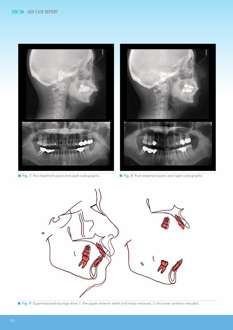
incisor). The patient was treated to an acceptable result as documented in Figs. 4-9. The cephalometric and panoramic radiographs document the pre-treatment conditions (Fig. 7) and the post-treatment results (Fig. 8). The cephalometric tracings before and after treatment are superimposed in Fig. 9. The details for diagnosis and treatment will be discussed below.
Diagnosis
Skeletal:
• Skeletal Class II (SNA 77°, SNB 69.5°, ANB 7.5°)
• Mandibular plane angle (SN-MP 38°, FMA 31°)
Dental:
• Molar relationships: Right Class ll; Left Class l; 8mm
Asymmetric Extraction of Adult Orthodontic Treatment
█ Fig. 2: Pre-treatment intraoral photographs
█ Fig. 1: Pre-treatment facial photographs
█ Fig. 3: Pre-treatment study models

IJOI 36 iAOI CASE REPORT
91
Asymmetric Extraction of Adult Orthodontic Treatment IJOI 36
overjet ; 6mm overbite (Fig. 10). Labially inclined
mandibular incisors (112°)
• Missing teeth: maxillary left 1st molar, mandible right
1st molar and left central incisor
• Unesthetic prostheses: three-unit bridges to replaced
missing molars, and a metal crown on the lower left 1st
molar
Facial:
• Maxillary protrusion with upper lip strain.
The ABO Discrepancy Index (DI) was 38 as shown in the subsequent worksheet.
Specific Objectives of Treatment
Maxilla (all three planes):
• A - P: Retract.
• Vertical: Maintain.
• Transverse: Maintain.
Mandible (all three planes):
• A - P: Maintain.
• Vertical: Maintain.
• Transverse: Maintain.
Maxillary Dentition
• A- P: Retract incisors, protract posterior segments
bilaterally.
Dr. Ming-Jen Chang,Lecturer, Beethoven Orthodontic Course (Left)
Dr. Chris Chang, Founder, Beethoven Orthodontic Center
Publisher, International Journal of Orthodontics& Implantology (middle)
W. Eugene Roberts,Consultant, International Journal of Orthodontics & Implantology (right)
█ Fig. 4: Post-treatment facial photographs
█ Fig. 5: Post-treatmentintraoral photographs
█ Fig. 6: Post-treatment study models
92
IJOI 36 iAOI CASE REPORT Asymmetric Extraction of Adult Orthodontic Treatment IJOI 36
█ Fig. 8: Post-treatment pano and ceph radiographs █ Fig. 7: Pre-treatment pano and ceph radiographs
█ Fig. 9: Superimposed tracings show 1. the upper anterior teeth and molar retraced. 2. the lower anterior intruded.

IJOI 36 iAOI CASE REPORT
93
Asymmetric Extraction of Adult Orthodontic Treatment IJOI 36
• Vertical: Maintain.
• Inter-molar Width: Maintain.
Mandibular Dentition:
• A - P: Maintain.
• Vertical: Maintain.
• Inter-molar / Inter-canine Width: Maintain.
Facial Esthetics:
• Reduce upper lip protrusion.
Treatment Plan
Extraction treatment with a full fixed orthodontic appliance was indicated to retract and level the upper dentition and align the lower arch. In the initial stage of the treatment, the upper right first
█ Fig. 11:The right first premolar was extracted, the three-unit bridge from the left 2nd premolar to 2nd molar was removed, and the temporary crowns were place on both abutments.
█ Fig. 12:The black triangle between the maxillary central incisors was corrected with interproximal stripping and power tube traction to close the resulting space.
█ Fig. 10: The maxillary incisor was protrusion with an overjet of about 8 mm and 6 mm overbite.
premolar was extracted to relieve upper anterior crowding (Fig. 11), and OrthoBoneScrew®(OBS) anchorage was used to assist in anterior protrusion correction. Power chains were used to close the extraction spaces. Detail bending and settling elastics produced the final occlusion. The bonded appliances were removed and the corrected dentition was retained with fixed retainers from the maxillary right lateral incisor to the left lateral incisor, and from the mandibular right canine to the left canine. Clear overlay retainers were constructed for both arches.
0M
3M

94
IJOI 36 iAOI CASE REPORT Asymmetric Extraction of Adult Orthodontic Treatment IJOI 36
█ Fig. 14:The maxillary anterior segment was ligated with a Figure-eight tie using a .012” stainless steel ligature, and the mandibular arch was bonded with standard torque brackets.
█ Fig. 15:A bony defect was noted distal to the upper left 2nd premolar.
█ Fig. 13:Open coil springs were used to open spaces between the upper left canine and left 1st premolar, as well as between the left 1st premolar and 2nd premolar.
Appliances and Treatment Progress
The right first premolar was extracted, the three-unit bridge from the left 2nd premolar to 2nd molar was removed, and the temporary crowns were place on both abutments (Fig. 11). A .022” slot Damon D3MX bracket system (Ormco) was used, and the maxillary incisions were bonded with high torque brackets. The initial archwire was .014” CuNiTi.
After one and half months of initial alignment and leveling, the archwire was changed to .014x.025“ CuNiTi. Meanwhile, the black triangle between the maxillary central incisors was corrected with interproximal stripping and power tube traction to close the resulting space (Fig. 12). In the 4th month, the archwire was changed to .017x.025" low friction TMA in the upper arch. Open coil springs were used to open spaces between the upper left canine and left 1st premolar, as well as between the left 1st premolar and 2nd premolar (Fig. 13). Opening space facilitated the restoration of caries on the upper left 1st premolar. In the 8th month of active treatment, the maxillary anterior segment was ligated with a
Figure-eight tie using a .012” stainless steel ligature, and the mandibular arch was bonded with standard torque brackets (Fig. 14). After fourteen months of treatment, a bony defect was noted distal to the upper left 2nd premolar. Periodontal therapy was indicated and closely monitored with follow-up checks (Fig. 15). In the 23th month, the lower arch archwire was changed to .017x.025” TMA and the anterior segment was ligated with a Figure-eight tie. At the same time, two miniscrews (2x12
mm OrthoBoneScrew®, Newton’s A Ltd, Taiwan.) were inserted into the infrazygomatic crests bilaterally. The elastometric chains were attached from upper right and left canines to the screws (Fig. 16). During the active treatment period, the brackets
5M
8M
14M
IJOI 36 iAOI CASE REPORT
95
Asymmetric Extraction of Adult Orthodontic Treatment IJOI 36
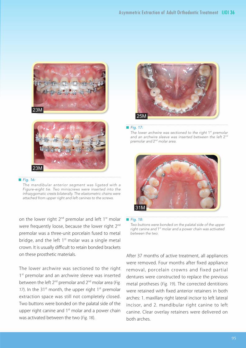
on the lower right 2nd premolar and left 1st molar were frequently loose, because the lower right 2nd premolar was a three-unit porcelain fused to metal bridge, and the left 1st molar was a single metal crown. It is usually difficult to retain bonded brackets on these prosthetic materials.
The lower archwire was sectioned to the right 1st premolar and an archwire sleeve was inserted between the left 2nd premolar and 2nd molar area (Fig.
17). In the 31st month, the upper right 1st premolar extraction space was still not completely closed. Two buttons were bonded on the palatal side of the upper right canine and 1st molar and a power chain was activated between the two (Fig. 18).
After 37 months of active treatment, all appliances were removed. Four months after fixed appliance removal , porcelain crowns and f ixed part ial dentures were constructed to replace the previous metal protheses (Fig. 19). The corrected dentitions were retained with fixed anterior retainers in both arches: 1. maxillary right lateral incisor to left lateral incisor, and 2. mandibular right canine to left canine. Clear overlay retainers were delivered on both arches.
█ Fig. 16:The mandibular anterior segment was ligated with a Figure-eight tie. Two miniscrews were inserted into the infrazygomatic crests bilaterally. The elastometric chains were attached from upper right and left canines to the screws.
█ Fig. 17:The lower archwire was sectioned to the right 1st premolar and an archwire sleeve was inserted between the left 2nd premolar and 2nd molar area.
█ Fig. 18:Two buttons were bonded on the palatal side of the upper right canine and 1st molar and a power chain was activated between the two.
25M
31M
23M
23M

96
IJOI 36 iAOI CASE REPORT Asymmetric Extraction of Adult Orthodontic Treatment IJOI 36
█ Fig. 19: Four months after fixed appliance removal, porcelain crowns and fixed partial dentures were constructed to replace the previous metal protheses.
Results Achieved
Maxilla (all three planes):
• A - P: Retracted.
• Vertical: Maintained
• Transverse: Maintained
Mandible (all three planes):
• A - P: Maintained
• Vertical: Increased ~2mm
• Transverse: Maintained
Maxillary Dentition
• A - P: Decreased axial inclination and retraction of
central incisors, extraction spaces were closed.
• Vertical: Maintained.
• Inter-molar / Inter-canine Width: Maintained.
Mandibular Dentition
• A - P: Alignment and intrusion of anterior teeth.
• Vertical: Maintained.
• Inter-molar / Inter-canine Width: Maintained.
Facial Esthetics:
• Protrusive upper lip was retracted, decreased bimaxillary lip prominence.
Retention
The fixed retainers were bonded on all maxillary incisors and from canine to canine in the mandibular arch. The upper and lower clear overlay retainers were delivered with instructions for full time wear for the first 6 months and nights only thereafter. The patient was carefully instructed in the home care and maintenance of the retainers.
Final Evaluation of Treatment
The American Board of Orthodontics (ABO) Cast-Radiograph Evaluation (CRE) score was 26 points. The major discrepancy was an occlusal relationship problem (10 points), which reflected an inadequate correction of the Class II buccal segments. The final interdigitation of the buccal segments was a compensated Class II occlusion, due to severe mandibular retrusion (SNB 69.50). The IBOI pink and white esthetic score was 3.
The upper anterior incisors were retracted and upper extraction spaces were closed to resolve the
37M
37M

IJOI 36 iAOI CASE REPORT
97
Asymmetric Extraction of Adult Orthodontic Treatment IJOI 36
patient’s chief complaints. Pleasing dental esthetics were achieved by correcting the excessive overjet, overbite and extraction space. However, close follow-up is indicated to monitor the tendency for extraction spaces to reopen.
Overall, there was a significant improvement in both dental esthetics and occlusion. The facial esthetics, associated with a decreased lip profile and excessive nasolabial angle, were acceptable considering the occlusal compromise necessitated by the severe mandibular retrusion.
Discussion
Skeletal Class II malocclusions should be treated according to the anteroposterior discrepancy, age of the patient, and expected compliance. Orthopedic methodology inc lude extraora l anchorage, functional appliances, and temporary anchorage devices (TADs). Dentoalveolar compensation can be accomplished with fixed appliances and Class II inter-maxillary elastics. Extraction space is helpful for correcting overjet and a midline discrepancy. In addition to correcting the dental Class II relationship, an important objective of dentofacial orthopedic treatment is to produce a good facial balance.
The extraction pattern can involve maxillary and/or mandibular premolars. The extraction of only 2 maxillary premolars is generally indicated when there is no crowding or cephalometric discrepancy in the mandibular arch. Extraction of a premolar in each quadrant is indicated primarily for crowding in the mandibular arch, and/or a cephalometric discrepancy in growing patients. Correction of Class █ Fig. 20: Post-treatment intra-oral frontal photo
II malocclusion with excessive overjet in an adult usually requires maximum anchorage, when only 2 maxillary premolars are extracted. Anchorage can be supplemented with an extraoral appliances, but that require rigorous patient compliance. However, when a Class I I malocclusion is treated with premolar extractions in all four quadrants, there is an even greater need for anchorage. Consequently, successful treatment increasingly depends on patient compliance, so the result may compromised.¹ Overall, treatment of Class II malocclusions with maxillary extractions only, or with extractions of premolars in both arches, has similar long-term post-treatment stability. ²
For the present patient, the overjet was 8 mm and the overbite was 6mm. Correction of a large overjet and deep-bite is difficult in adult patients. The treatment plan for these patients usually involves extraction of the maxillary first premolars. As shown in Fig. 7, the upper left first molar was missing, so the asymmetric extraction of the upper right first premolar was indicated. Closing the extraction spaces to improve the overjet and overbite is a relatively simple approach, but posterior anchorage

98
IJOI 36 iAOI CASE REPORT Asymmetric Extraction of Adult Orthodontic Treatment IJOI 36
CEPHALOMETRIC
SKELETAL ANALYSIS
PRE-Tx POST-Tx DIFF.
SNA° 77° 74.5° -2.5°SNB° 69.5° 69.5° 0° ANB° 7.5° 5° -2.5° SN-MP° 38° 39° 1° FMA° 31° 32° 1°
DENTAL ANALYSIS
U1 TO NA mm 9 mm 5 mm -4 mm U1 TO SN° 113° 97° -16°
L1 TO NB mm 9 mm 9 mm 0mm L1 TO MP° 112° 110° -2°
FACIAL ANALYSIS
E-LINE UL 2 mm -5 mm -7 mm E-LINE LL 0 mm -2 mm -2 mm
██ Table 1: Cephalometric summary
can be a problem, requiring headgear, orthodontic bone screws, or intermaxillary elastics.³
As a general rule, orthodontics only is not indicated for a positive overjet greater than 8 mm, a negative overjet of 4 mm or greater, and/or a transverse discrepancy greater than 3 mm. However, deep overbite patients can usually be treated without extractions or surgery.⁴
Patient with Class II malocclusions may be Class I on one side and Class II on the other, resulting in an asymmetric occlusal relationship that complicates orthodontic treatment. Depending on the degree of asymmetry, treatment approaches by quadrant include symmetric extraction of 4 premolars and asymmetric extraction of 3 premolars. The 4-premolar-extraction approach has the potential
to produce a final occlusion with bilateral Class I molar and canine relationships. On the other hand, asymmetric extraction of 3 premolars (2
maxillary premolars and 1 mandibular premolar on
the Class I side) will produce Class I canine and molar relationships on one side, with a Class II molar and Class I canine relationships on the Class II side. With either approach, the maxillary and mandibular dental midlines can be corrected to coincide with the midsagittal plane (facial midline).⁵
Orthodontic treatment combined with either miniscrew anchorage or headgear can achieve acceptable results with overjet reduction and improvement of facial profile in patients with skeletal Class II malocclusion. However, miniscrew anchorage does not require patient cooperation, so the treatment prognosis is more predictable.⁶
According to the A-line of Alvarez et al.,⁷ there was a severe anterior position of the maxillary incision roots, indicating the use of high-torque brackets and bilateral miniscrews in the infrazygomatic crests. This approach allowed for the correction of the maxillary incisor inclination without compromising the anterio-posterior position of the maxilla.
M i n i s c r e w s h a v e a h i g h s u c c e s s r a t e o f approximately 90% and they provided sufficient anchorage immediately after placement surgery for orthodontic tooth movement. In addition, miniscrews placed without a mucoperiosteal incision or flap surgery result in significantly reduced pain and discomfort after implantation. Miniscrews have suitable characteristics for orthodontics anchorage.⁸ When a midline discrepancy is present (Figs. 1-3), the incisors can be aligned and moved to their optimum

IJOI 36 iAOI CASE REPORT
99
Asymmetric Extraction of Adult Orthodontic Treatment IJOI 36
location with a fixed appliance, supplemented by intermaxillary elastics.
The CRE score was 24, with most of the points reflecting a problem in the sagittal occlusal relationship (interdigitation). The etiology of the malocclusion involved asymmetric extractions, so treatment was directed at achieving the best occlusal alignment by utilizing extraction spaces supplemented with posterior maxillary miniscrews. Fortunately, it was possible to correct the midline, close space and achieve an optimal posterior interdigitation. The Pink & White esthetic score was 3, reflecting problematic areas in the maxillary anterior: inadequate soft tissue papilla between the central incisors (black triangle) and irregular incisal edges.
Conclusion
Extraction in only one quadrant is a common approach for resolving asymmetric malocclusions in adults. If there is excessive overjet and/or a midline discrepancy, it is important to optimally manage the space with supplemental anchorage, such as bilateral infrazygomatic miniscrews. Palatal buttons for attachment of power chains are helpful for efficient space closure and control of rotations.
The present difficult malocclusion (DI =38) was treated to an acceptable result as documented by a CRE = 24, and a Pink and White esthetic score of 3. The patient was pleased with the dental and facial result, although her lips were relatively flat and the nasolabial angle was excessive. Considering the patient’s severely retrusive mandible, this was an optimal facial result.
Acknowledgment
Thanks to Mr. Paul Head for proofreading this article.
References
1. GuilhermeJ,AdrianaCB,JoseFCH,MarcosRF,LenianaSN.ClassIItreatmentsuccessratein2-and4-premolarextractionprotocols.AmJOrthodDentofacialOrthop2004;125:472-9.
2. GuilhermeJ,VladimirLS,RubenLS,MarcosJ,andMarcosRF.Long-termstabilityofClassIImalocclusiontreatedwith2-and4-premolarextractionprotocols.AmJOrthodDentofacialOrthop2009;136:154.e.1-154.e.10.
3. WuS,ChangCH,RobertsWE.ImpingingOverbiteandLargeOverjet.iAOICaseReport2011;18:192-9.
4. DouglasS,AlMB,StevenJL,andDanielML.Determiningthe limitsoforthodontictreatmentofoverbite,overjet,andtransversediscrepancy:Apilotstudy.AmJOrthodDentofacialOrthop2006;129:804-8.
5. GuilhermeJ,EduardoAD,Jose'FCH,MarcosRF,andKarinaJeroˆnimoRSL.Class II subdivisiontreatmentsuccessratewithsymmetricandasymmetricextractionprotocols.AmJOrthodDentofacialOrthop2003;124:257-64.
6. KurodaS,YamadaK,DeguchiT,KyungHM,andTerukoTY.ClassIImalocclusiontreatedwithminiscrewanchorage:Comparisonwithtraditionalorthodonticmechanicsoutcomes.AmJOrthodDentofacialOrthop2009;135:302-9.
7. Alvarez, et al.TheALine: anewguide fordiagnosis andtreatmentplanning.JCO2001;35:556-569.
8. KurodaS,YamadaK,DeguchiT,KyungHM,TerukoTY.Clinicaluseofminiscrewimplantsasorthodonticanchorage:Success ratesandpostoperativediscomfort.AmJOrthodDentofacialOrthop2007;131:9-15.
9. ChangCH.AdvancedDamonCourseNo.8:Excellence inFinishing,BeethovenPodcastEncyclopedia inOrthodontics[podcast].Hsinchu:Newton’sALtd;2011.
10. ChangCH.AdvancedDamonCourseNo.4,5 :DI&CREWorkshop (1)(2)., Beethoven Podcast Encyclopedia inOrthodontics[podcast].Hsinchu:Newton’sALtd;2011.
11. Chang CH. Basic Damon Course No.5: Finish Bending ,BeethovenPodcastEncyclopedia inOrthodontics[podcast].Hsinchu:Newton’sALtd;2011.
12. HuangS.TomPitts’:SecretsofExcellentFinishing.News&TrendsinOrthodontics2009;14:6-23.

100
IJOI 36 iAOI CASE REPORT Asymmetric Extraction of Adult Orthodontic Treatment IJOI 36
DISCREPANCY INDEX WORKSHEET
(Rev. 9/22/08)
OVERJET
0 mm. (edge-to-edge) = 1 pt.1 Ð 3 mm. = 0 pts.3.1 Ð 5 mm. = 2 pts.5.1 Ð 7 mm. = 3 pts.7.1 Ð 9 mm. = 4 pts.> 9 mm. = 5 pts.
Negative OJ (x-bite) 1 pt. per mm. per tooth =
OVERBITE
0 Ð 3 mm. = 0 pts.3.1 Ð 5 mm. = 2 pts.5.1 Ð 7 mm. = 3 pts.Impinging (100%) = 5 pts.
ANTERIOR OPEN BITE
0 mm. (edge-to-edge), 1 pt. per tooth
then 1 pt. per additional full mm. per tooth
LATERAL OPEN BITE
2 pts. per mm. per tooth
CROWDING (only one arch)
1 Ð 3 mm. = 1 pt.3.1 Ð 5 mm. = 2 pts.5.1 Ð 7 mm. = 4 pts.> 7 mm. = 7 pts.
OCCLUSION
Class I to end on = 0 pts.End on Class II or III = 2 pts. per side pts.
Full Class II or III = 4 pts. per side pts.
Beyond Class II or III = 1 pt. per mm. pts. additional
LINGUAL POSTERIOR X-BITE
1 pt. per tooth Total = 0
BUCCAL POSTERIOR X-BITE
2 pts. per tooth Total = 2
CEPHALOMETRICS (See Instructions)
ANB ≥ 6¡ or ≤ -2¡ = 4 pts.
SN-MP
≥ 38¡ = 2 pts.
Each degree > 38¡ x 2 pts. =
≤ 26¡ = 1 pt.
Each degree < 26¡ 4 x 1 pt. = 4
1 to MP ≥ 99¡ = 1 pt.
Each degree > 99¡ 2 x 1 pt. = 2
OTHER (See Instructions)
Supernumerary teeth x 1 pt. =
Ankylosis of perm. teeth x 2 pts. =
Anomalous morphology x 2 pts. =
Impaction (except 3rd molars) x 2 pts. =
Midline discrepancy (≥3mm) @ 2 pts. =
Missing teeth (except 3rd molars) x 1 pts. =
Missing teeth, congenital x 2 pts. =
Spacing (4 or more, per arch) x 2 pts. = 2
Spacing (Mx cent. diastema ≥ 2mm) @ 2 pts. = 2
Tooth transposition x 2 pts. =
Skeletal asymmetry (nonsurgical tx) @ 3 pts. =
Addl. treatment complexities x 2 pts. =
Identify:
Total = 1
Total = 5
Total = 0
Total = 0
Total = 5
Total = 0
Each degree > 6¡ x 1 pt. =
Each degree < -2¡ x 1 pt. =
Total = 8
CASE # 1 PATIENT CHAO-YUEN CHIU PATIENT CHAO-YUEN CHIU PATIENT CHAO-YUEN CHIU
TOTAL D.I. SCORETOTAL D.I. SCORETOTAL D.I. SCORE 25
Total = 4
EXAM YEAR 2009
ABO ID# 96112
0
0
8
2
0
63
DISCREPANCY INDEX WORKSHEET
3.1 Ð 5 mm. = 2 pts.5.1 Ð 7 mm. = 3 pts.7.1 Ð 9 mm. = 4 pts.> 9 mm. = 5 pts.
Negative OJ (x-bite) 1 pt. per mm. per tooth =
OVERBITE
0 Ð 3 mm. = 0 pts.3.1 Ð 5 mm. = 2 pts.5.1 Ð 7 mm. = 3 pts.Impinging (100%) = 5 pts.
ANTERIOR OPEN BITE
0 mm. (edge-to-edge), 1 pt. per tooth
then 1 pt. per additional full mm. per tooth
LATERAL OPEN BITE
2 pts. per mm. per tooth
CROWDING (only one arch)
1 Ð 3 mm. = 1 pt.3.1 Ð 5 mm. = 2 pts.5.1 Ð 7 mm. = 4 pts.> 7 mm. = 7 pts.
OCCLUSION
Class I to end on = 0 pts.End on Class II or III = 2 pts. per side pts.
Full Class II or III = 4 pts. per side pts.
Beyond Class II or III = 1 pt. per mm. pts. additional
LINGUAL POSTERIOR X-BITE
1 pt. per tooth Total = 0
BUCCAL POSTERIOR X-BITE
2 pts. per tooth Total = 2
CEPHALOMETRICS (See Instructions)
ANB ≥ 6¡ or ≤ -2¡ = 4 pts.
SN-MP
≥ 38¡ = 2 pts.
Each degree > 38¡ x 2 pts. =
≤ 26¡ = 1 pt.
Each degree < 26¡ 4 x 1 pt. = 4
1 to MP ≥ 99¡ = 1 pt.
Each degree > 99¡ 2 x 1 pt. = 2
OTHER (See Instructions)
Supernumerary teeth x 1 pt. =
Ankylosis of perm. teeth x 2 pts. =
Anomalous morphology x 2 pts. =
Impaction (except 3rd molars) x 2 pts. =
Midline discrepancy (≥3mm) @ 2 pts. =
Missing teeth (except 3rd molars) x 1 pts. =
Missing teeth, congenital x 2 pts. =
Spacing (4 or more, per arch) x 2 pts. = 2
Spacing (Mx cent. diastema ≥ 2mm) @ 2 pts. = 2
Tooth transposition x 2 pts. =
Skeletal asymmetry (nonsurgical tx) @ 3 pts. =
Addl. treatment complexities x 2 pts. =
Identify:
Total = 1
Total = 5
Total = 0
Total = 0
Total = 5
Total = 0
Each degree > 6¡ x 1 pt. =
Each degree < -2¡ x 1 pt. =
Total = 8
CHIU CHIU CHIU
Total = 4
EXAM YEAR 2009
ABO ID# 96112
38
4
3
0
0
1
4
0
0
21
5IMPLANT SITELip line : Low (0 pt), Medium (1 pt), High (2 pts) = Gingival biotype : Low-scalloped, thick (0 pt), Medium-scalloped, medium-thick (1 pt), High-scalloped, thin (2 pts) = Shape of tooth crowns : Rectangular (0 pt), Triangular (2 pts) = Bone level at adjacent teeth : ≦ 5 mm to contact point (0 pt), 5.5 to 6.5 mm to contact point (1 pt), ≧ 7mm to contact point (2 pts) = Bone anatomy of alveolar crest : H&V sufficient (0 pt), Deficient H, allow simultaneous augment (1 pt), Deficient H, require prior grafting (2 pts), Deficient V or Both
H&V (3 pts) = Soft tissue anatomy : Intact (0 pt), Defective ( 2 pts) = Infection at implant site : None (0 pt), Chronic (1 pt), Acute( 2 pts) =
0
3 3
4
2
1 2
1 1
4
11313
Discrepancy Index Worksheet
Total Score:
4
2
1
11
20
2
1
2
1
2
10
3
Alignment/Rotations
Marginal Ridges
Buccolingual Inclination
Overjet
Occlusal Contacts
Occlusal Relationships
Interproximal Contacts
INSTRUCTIONS: Place score beside each deficient tooth and enter total score for each parameter in the white box. Mark extracted teeth with ÒXÓ. Second molars should be in occlusion.
24
Case #
1
1
Marginal Ridges
Buccolingual Inclination
Overjet
Occlusal Contacts
Occlusal Relationships
Interproximal Contacts
INSTRUCTIONS: Place score beside each deficient tooth and enter total score for each parameter in the white box. Mark extracted teeth with ÒXÓ. Second molars should be in occlusion.
IBOI Cast-Radiograph Evaluation
Root Angulation
1
2
11
2
2
11
1 1
22
21

IJOI 36 iAOI CASE REPORT
101
Asymmetric Extraction of Adult Orthodontic Treatment IJOI 36
Cast-Radiograph Evaluation
Total Score:
4
2
1
11
20
2
1
2
1
2
10
3
Alignment/Rotations
Marginal Ridges
Buccolingual Inclination
Overjet
Occlusal Contacts
Occlusal Relationships
Interproximal Contacts
INSTRUCTIONS: Place score beside each deficient tooth and enter total score for each parameter in the white box. Mark extracted teeth with ÒXÓ. Second molars should be in occlusion.
24
Case #
1
1
Marginal Ridges
Buccolingual Inclination
Overjet
Occlusal Contacts
Occlusal Relationships
Interproximal Contacts
INSTRUCTIONS: Place score beside each deficient tooth and enter total score for each parameter in the white box. Mark extracted teeth with ÒXÓ. Second molars should be in occlusion.
IBOI Cast-Radiograph Evaluation
Root Angulation
1
2
11
2
2
11
1 1
22
21

102
IJOI 36 iAOI CASE REPORT
12 34
56
2. White Esthetic Score ( for Micro-esthetics )
5
1
2
34 6
211 34
562
1
1
2
12 34
56
2. White Esthetic Score ( for Micro-esthetics )
5
1
2
34 6
211 34
562
1
1
2
12 34
56
2. White Esthetic Score ( for Micro-esthetics )
5
1
2
34 6
211 34
562
1
1
2
IBOI Pink & White Esthetic Score
12 34
56
2. White Esthetic Score ( for Micro-esthetics )
5
1
2
34 6
211 34
562
1
1
2
1. M & D Papillae 0 1 2
2. Keratinized Gingiva 0 1 2
3. Curvature of Gingival Margin 0 1 2
4. Level of Gingival Margin 0 1 2
5. Root Convexity ( Torque ) 0 1 2
6. Scar Formation 0 1 2
1. Tooth Form 0 1 2
2. Mesial & Distal Outline 0 1 2
3. Crown Margin 0 1 2
4. Translucency ( Incisal thrid ) 0 1 2
5. Hue & Value ( Middle third ) 0 1 2
6. Tooth Proportion 0 1 2
1. M & D Papillae 0 1 2
2. Keratinized Gingiva 0 1 2
3. Curvature of Gingival Margin 0 1 2
4. Level of Gingival Margin 0 1 2
5. Root Convexity (Torque) 0 1 2
6. Scar Formation 0 1 2
1. Midline 0 1 2
2. Incisor Curve 0 1 2
3. Axial Inclination (5°, 8°, 10°) 0 1 2
4. Contact Area (50%, 40%, 30%) 0 1 2
5. Tooth Proportion (1:0.8) 0 1 2
6. Tooth to Tooth Proportion 0 1 2
1. Pink Esthetic Score
Total Score: = 3
2. White Esthetic Score ( for Micro-esthetics )
Total = 1
Total = 2