association of increased ccl5 and cxcl7 chemokine ... · association of increased ccl5 and cxcl7...
TRANSCRIPT
Association of increased CCL5 and CXCL7chemokine expression with neutrophil activation insevere stable COPD
A Di Stefano,1 G Caramori,2 I Gnemmi,1 M Contoli,2 L Bristot,2 A Capelli,1
F L M Ricciardolo,3 F Magno,1 S Ennio D’Anna,4 A Zanini,1 M Carbone,1 F Sabatini,5
C Usai,6 P Brun,7 K F Chung,8 P J Barnes,8 A Papi,2 I M Adcock,8 B Balbi1
c Additional details arepublished online only at http://thorax.bmj.com/content/vol64/issue11
1 Division of Pulmonary Diseaseand Laboratory ofCytoimmunopathology of Heartand Lung, Salvatore MaugeriFoundation, IRCCS, Veruno, Italy;2 Centro di Ricerca su Asma eBPCO, Universita di Ferrara,Italy; 3 Department of PulmonaryDisease, University of Torino,Italy; 4 Divisione di Pneumologia,Fondazione San Raffaele, IRCCS,G Giglio, Cefalu, Italy; 5 Unit ofRespiratory Disease, IRCCSGaslini Institute, Genoa, Italy;6 Istituto di Cibernetica eBiofisica, CNR, Genoa, Italy;7 Department of Histology,Microbiology and MedicalBiotechnology, University ofPadova, Italy; 8 Airways DiseaseSection, National Heart andLung Institute, Imperial CollegeLondon, UK
Correspondence to:Dr A Di Stefano, Fondazione SMaugeri, IRCCS, Laboratorio diCitoimmunopatologia ApparatoCardio Respiratorio, Via perRevislate 13, 28010 Veruno(NO), Italy; [email protected]
ADS and GC contributed equallyto this work.
Received 12 January 2009Accepted 22 July 2009Published Online First23 August 2009
ABSTRACTBackground: Increased numbers of activated neutrophilshave been reported in the bronchial mucosa of patientswith stable chronic obstructive pulmonary disease(COPD), particularly in severe disease.Objectives: To investigate the expression of neutrophilicchemokines and adhesion molecules in bronchial biopsiesfrom patients with stable COPD of different severity (GOLDstages I–IV) compared with age-matched control subjects,smokers with normal lung function and never smokers.Methods: The expression of CCL5, CXCL1, 5, 6, 7 and 8,CXCR1, CXCR2, CD11b and CD44 was measured in thebronchial mucosa using immunohistochemistry, confocalimmunofluorescence, real-time quantitative polymerasechain reaction (RT-QPCR) and Western blotting (WB).Results: The numbers of CCL5+ epithelial cells and CCL5+and CXCL7+ immunostained cells were increased in thebronchial submucosa of patients with stable severe COPDcompared with control never smokers and smokers withnormal lung function. This was also confirmed at the levelof mRNA expression. The numbers of CCL5+ cells in thesubmucosa of patients with COPD were 2–15 times higherthan any other chemokines. There was no correlationbetween the number of these cells and the number ofneutrophils in the bronchial submucosa. Compared withcontrol smokers, the percentage of neutrophils co-expressing CD11b and CD44 receptors was significantlyincreased in the submucosa of patients with COPD.Conclusion: The increased expression of CCL5 andCXCL7 in the bronchial mucosa of patients with stableCOPD, together with an increased expression of extra-cellular matrix-binding receptors on neutrophils, may beinvolved in the pathogenesis of COPD.
Inflammation occurs in the central peripheralairways (bronchioles) and lung parenchyma ofpatients with COPD.1–4 Studies have emphasisedthe potential pathological role of many inflamma-tory cells including macrophages and T lympho-cytes, whereas fewer investigations have examinedneutrophil granulocytes despite increased numbersbeing present in the bronchial mucosa, particularlyin severe disease.4–9 Furthermore, the need for morepathological investigations in patients with severeCOPD has been highlighted.3
Several chemokines of the CXC and CC familyare involved in neutrophil chemotaxis.10–35 CXCchemokines were principally investigated usingbronchoalveolar lavage (BAL) fluid or sputumsamples, and increased levels of CXCL112 and
CXCL821 in sputum or CXCL5 in BAL fluid14 havebeen reported in patients with COPD.
Fewer studies have investigated the expression ofCC chemokines in COPD. CCL5 levels are increasedin the sputum of patients with COPD.23 Theleukocyte aMb2 integrin (also known as CD11b/CD18) functions as an adhesion molecule facilitat-ing diapedesis. Overexpression of CD11b has beenreported in peripheral blood neutrophils26 27 andsputum28 of patients with COPD. Finally, hyalur-onan, an extracellular matrix component, is themain ligand for CD44 and its expression isupregulated in neutrophils co-cultured with humanprimary bronchial epithelial cells and granulocyte-macrophage colony stimulating factor (GM-CSF).31
The expression of CD11b and CD44 on neutrophilsin the bronchial mucosa of patients with COPD hasnot been previously reported (see online datasupplement for a more detailed background).
The mechanisms responsible for tissue neutrophiliain COPD have not been fully clarified. The prevalenceof neutrophils can be related to increased chemotaxis,increased adhesion to collagens or to prolongedsurvival. A partial loss of neutrophilic chemotacticresponse to CXCL8 stimuli has recently been reportedfor sputum neutrophils in patients with COPD aftermigration and residence in the bronchial lumen.10
Furthermore, a comprehensive analysis of chemokineexpression in COPD bronchi is lacking.
The aim of the present study was to investigate thepresence of relevant CXC and CC chemokines,cytokines and chemokine receptors involved in neu-trophilic migration andactivation inbronchialbiopsiesfrom patients with increasing disease severity. Sinceincreased adhesion of neutrophils to extracellularmatrix components may play a role in neutrophilpermanence in the bronchial mucosa, the expression oftwo neutrophil receptors, CD11b and CD44, poten-tially involved in this process was also studied.
METHODSSubjectsAll subjects were recruited from the Section ofRespiratory Medicine of the Fondazione SalvatoreMaugeri (Veruno, Italy). Table 1 and Tables E1–E3 (inthe online data supplement) show the clinicalcharacteristics of the subjects used for the immuno-histochemistry (n = 49), confocal microscopy (n = 8),real-time PCR (RT-PCR, n = 31) and Western blot-ting (n = 12) studies. The severity of the airflowobstruction was staged according to the GOLDcriteria.1
Chronic obstructive pulmonary disease
968 Thorax 2009;64:968–975. doi:10.1136/thx.2009.113647
on 14 February 2019 by guest. P
rotected by copyright.http://thorax.bm
j.com/
Thorax: first published as 10.1136/thx.2009.113647 on 23 A
ugust 2009. Dow
nloaded from

Lung function tests and volumesPulmonary function tests were performed as previouslydescribed6 according to published guidelines (see online datasupplement).
Fibreoptic bronchoscopy, collection and processing of bronchialbiopsiesA standardised procedure, previously reported,6 was followedfor fibreoptic bronchoscopy and collection of bronchial biopsies.Bronchial biopsies for immunohistochemistry, Western blotanalysis and RT-QPCR were processed as previously described6
(see online data supplement for details).
ImmunohistochemistryA panel of antibodies was used (table 2) and primary antibodieswere applied at optimal dilutions in TRIS-buffered saline and
revealed with the use of appropriate secondary antibodies andfast-red substrate (see online data supplement for details).
Immunofluorescence staining with confocal microscopySections were fixed with 4% paraformaldehyde, washed withphosphate buffered saline (PBS) and incubated with blockingserums followed by incubation with primary antibodies and therevealing antibody system, as previously described32 (see onlinedata supplement for details).
Scoring system for immunohistochemistry and confocal microscopyLight microscopic analysis was performed at a magnification of6306. Immunostained cells in the bronchial submucosa werequantified as previously described.6 The immunostaining wasalso scored in the bronchial epithelium. The quantitativeestimation of co-localised proteins was performed calculating
Table 1 Characteristics of subjects for the optical immunohistochemical study
Subjects n Age (years) M/FSmoking history(pack-years)
Ex/current-smokers
FEV1 pre(% pred)
FEV1 post(% pred) FEV1/FVC%{
Healthy non-smokers 11 62 (4) 12/1 0 0 115 (4) ND 85 (3)
Healthy smokers 13 60 (2) 9/3 46 (12) 2/11 107 (4) ND 82 (2)
Mild/moderate COPD 12 64 (2) 11/2 35 (6) 4/8 66 (4)* 72 (4) 60 (2)*
Severe/very severe COPD 13 65 (3) 4/7 49 (11) 9/4 30 (2)*{ 33 (3) 42 (3)*{
Data are presented as mean (SEM).COPD, chronic obstructive pulmonary disease; F, female; FEV1, forced expiratory volume in 1 s; FVC, forced vital capacity; M, male; ND, not determined.*p,0.001 (ANOVA) significantly different from control smokers with normal lung function (healthy smokers) and control never-smokers.{p,0.001 (ANOVA) significantly different from mild/moderate COPD.{For patients with COPD, FEV1/FVC% are post-bronchodilator values.The GOLD classification of severity for COPD has been followed where patients are classified as mild (GOLD stage I), moderate (stage II), severe (stage III) and very severe (stage IV).
Table 2 Antibodies used to characterise inflammatory cells and CXC, CC chemokines, their receptors and other molecules involved in the neutrophiliaof the bronchial mucosa
Antibodyspecificity Company Catalogue number Source/host
Dilution of theprimary antibody
Biotinylatedsecondaryantibody Revealing kit Positive control
CXCL1 R&D Systems AF-275 Goat 1:50 Vector, BA 5000;1:200
Dako,StreptABComplex/AP; 1:100
Nasal polyp
CXCL5 R&D Systems MAB-254 Mouse 1:50 Vector, BA 2000;1:200
Dako,StreptABComplex/AP; 1:100
Nasal polyp
CXCL6 Santa Cruz SC-5813 Goat 1:50 Vector, BA 5000;1:200
Dako,StreptABComplex/AP; 1:100
Nasal polyp
CXCL7 R&D Systems MAB393 Mouse 1:50 Vector, BA 2000;1:200
Dako,StreptABComplex/AP; 1:100
Nasal polyp
CXCL8 R&D Systems AF-208NA Goat 1:50 Vector, BA 5000;1:200
Dako,StreptABComplex/AP; 1:100
Nasal polyp
CCL5 R&D Systems AF-278NA Goat 1:100 Vector, BA 5000;1:200
Dako,StreptABComplex/AP; 1:100
Nasal polyp
CXCR1 R&D Systems MAB330 Mouse 1:200 Vector, BA 2000;1:200
Dako,StreptABComplex/AP; 1:100
Nasal polyp
CXCR2 R&D Systems MAB331 Mouse 1:200 Vector, BA 2000;1:200
Dako,StreptABComplex/AP; 1:100
Nasal polyp
CD44 AMS-Biotech 211-JM144A Mouse 1:100 Vector, BA 2000;1:200
Dako,StreptABComplex/AP; 1:100
Nasal polyp
CD11b R&D Systems MAB16991 Mouse 1:50 Vector, BA 2000;1:200
Dako,StreptABComplex/AP; 1:100
Nasal polyp
Neutrophilelastase
Dako M752 Mouse 1:100 Vector, BA 2000;1:200
Dako,StreptABComplex/AP; 1:100
Human tonsil
Chronic obstructive pulmonary disease
Thorax 2009;64:968–975. doi:10.1136/thx.2009.113647 969
on 14 February 2019 by guest. P
rotected by copyright.http://thorax.bm
j.com/
Thorax: first published as 10.1136/thx.2009.113647 on 23 A
ugust 2009. Dow
nloaded from
the ‘‘co-localisation coefficients’’32 (see online data supplementfor details).
Quantification of chemokine and cytokine mRNA levels inbronchial biopsiesTotal RNA was extracted (Micro RNeasy Kit, Qiagen, Milan,Italy) from 30 mm thick cryostat sections of bronchial biopsiesand 1 mg used for cDNA synthesis. Relative levels of mRNAswere expressed as the ratio of the Ct value for the gene ofinterest Ct/housekeeping gene Ct (see online data supplementfor details).
Western blot analysis for CCL5, CXCL7 and CXCL8 in bronchialbiopsiesWhole cell protein extraction from bronchial biopsies, gelelectrophoresis, nitrocellulose filters transfer, incubation withappropriate primary antibodies, detection on nitrocellulosefilters and protein quantification were performed as previouslydescribed6 (see online data supplement for details).
Data analysisGroup data were expressed as mean (SEM) for functional dataor median (range) for morphological data. Differences betweengroups were analysed using analysis of variance (ANOVA orKruskal-Wallis as appropriate) for functional data or non-parametric tests for morphological parameters (see online datasupplement for details).
RESULTS
Clinical findingsThe characteristics of subjects used for immunohistochemistry areshown in table 1. Tables E1–E3 (see online data supplement) showthe characteristics of subjects used for confocal immunofluores-cence, RT-QPCR and Western blotting, respectively. The fourgroups of subjects were of similar age (table 1). Smoking historywas similar in patients with mild/moderate COPD, severe COPDand healthy smokers with normal lung function. As expected fromthe selection criteria, the values of forced expiratory volume in 1 s(FEV1 % predicted) and FEV1/forced vital capacity (FVC) (%) weresignificantly different in the groups with mild/moderate andsevere/very severe COPD compared with both control groups(healthy smokers and healthy never-smokers). Patients withsevere/very severe COPD also differed significantly from thosewith mild/moderate COPD (for overall groups, p,0.001(ANOVA) for FEV1% predicted and FEV1/FVC% values).
ImmunohistochemistryNeutrophils in bronchial submucosaThe number of neutrophils, as previously reported,6 was sig-nificantly higher in the bronchial submucosa of patients withsevere/very severe COPD (274 (range 47–500)) compared withcontrol healthy smokers (124 (17–308), p = 0.008) and healthynever smokers (110 (59–270), p = 0.009; table 3). Patients withmild/moderate COPD did not differ significantly from bothcontrol groups, nor did the two control groups differ significantlyfrom each other.
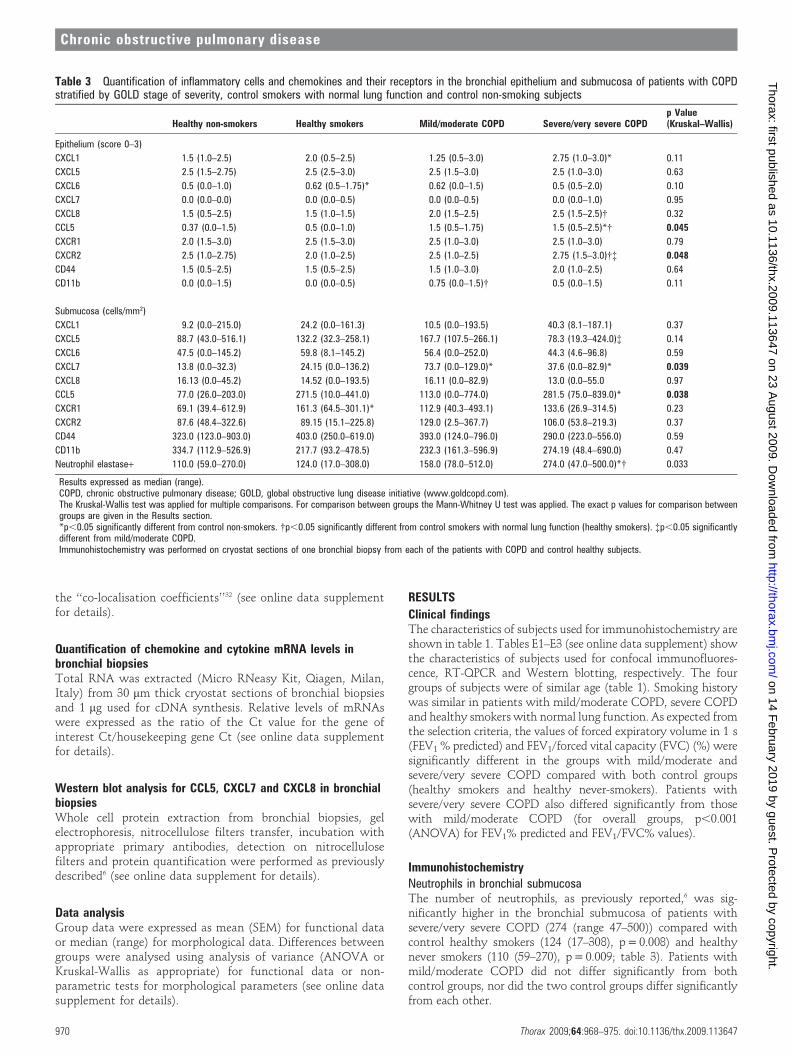
Table 3 Quantification of inflammatory cells and chemokines and their receptors in the bronchial epithelium and submucosa of patients with COPDstratified by GOLD stage of severity, control smokers with normal lung function and control non-smoking subjects
Healthy non-smokers Healthy smokers Mild/moderate COPD Severe/very severe COPDp Value(Kruskal–Wallis)
Epithelium (score 0–3)
CXCL1 1.5 (1.0–2.5) 2.0 (0.5–2.5) 1.25 (0.5–3.0) 2.75 (1.0–3.0)* 0.11
CXCL5 2.5 (1.5–2.75) 2.5 (2.5–3.0) 2.5 (1.5–3.0) 2.5 (1.0–3.0) 0.63
CXCL6 0.5 (0.0–1.0) 0.62 (0.5–1.75)* 0.62 (0.0–1.5) 0.5 (0.5–2.0) 0.10
CXCL7 0.0 (0.0–0.0) 0.0 (0.0–0.5) 0.0 (0.0–0.5) 0.0 (0.0–1.0) 0.95
CXCL8 1.5 (0.5–2.5) 1.5 (1.0–1.5) 2.0 (1.5–2.5) 2.5 (1.5–2.5){ 0.32
CCL5 0.37 (0.0–1.5) 0.5 (0.0–1.0) 1.5 (0.5–1.75) 1.5 (0.5–2.5)*{ 0.045
CXCR1 2.0 (1.5–3.0) 2.5 (1.5–3.0) 2.5 (1.0–3.0) 2.5 (1.0–3.0) 0.79
CXCR2 2.5 (1.0–2.75) 2.0 (1.0–2.5) 2.5 (1.0–2.5) 2.75 (1.5–3.0){{ 0.048
CD44 1.5 (0.5–2.5) 1.5 (0.5–2.5) 1.5 (1.0–3.0) 2.0 (1.0–2.5) 0.64
CD11b 0.0 (0.0–1.5) 0.0 (0.0–0.5) 0.75 (0.0–1.5){ 0.5 (0.0–1.5) 0.11
Submucosa (cells/mm2)
CXCL1 9.2 (0.0–215.0) 24.2 (0.0–161.3) 10.5 (0.0–193.5) 40.3 (8.1–187.1) 0.37
CXCL5 88.7 (43.0–516.1) 132.2 (32.3–258.1) 167.7 (107.5–266.1) 78.3 (19.3–424.0){ 0.14
CXCL6 47.5 (0.0–145.2) 59.8 (8.1–145.2) 56.4 (0.0–252.0) 44.3 (4.6–96.8) 0.59
CXCL7 13.8 (0.0–32.3) 24.15 (0.0–136.2) 73.7 (0.0–129.0)* 37.6 (0.0–82.9)* 0.039
CXCL8 16.13 (0.0–45.2) 14.52 (0.0–193.5) 16.11 (0.0–82.9) 13.0 (0.0–55.0 0.97
CCL5 77.0 (26.0–203.0) 271.5 (10.0–441.0) 113.0 (0.0–774.0) 281.5 (75.0–839.0)* 0.038
CXCR1 69.1 (39.4–612.9) 161.3 (64.5–301.1)* 112.9 (40.3–493.1) 133.6 (26.9–314.5) 0.23
CXCR2 87.6 (48.4–322.6) 89.15 (15.1–225.8) 129.0 (2.5–367.7) 106.0 (53.8–219.3) 0.37
CD44 323.0 (123.0–903.0) 403.0 (250.0–619.0) 393.0 (124.0–796.0) 290.0 (223.0–556.0) 0.59
CD11b 334.7 (112.9–526.9) 217.7 (93.2–478.5) 232.3 (161.3–596.9) 274.19 (48.4–690.0) 0.47
Neutrophil elastase+ 110.0 (59.0–270.0) 124.0 (17.0–308.0) 158.0 (78.0–512.0) 274.0 (47.0–500.0)*{ 0.033
Results expressed as median (range).COPD, chronic obstructive pulmonary disease; GOLD, global obstructive lung disease initiative (www.goldcopd.com).The Kruskal-Wallis test was applied for multiple comparisons. For comparison between groups the Mann-Whitney U test was applied. The exact p values for comparison betweengroups are given in the Results section.*p,0.05 significantly different from control non-smokers. {p,0.05 significantly different from control smokers with normal lung function (healthy smokers). {p,0.05 significantlydifferent from mild/moderate COPD.Immunohistochemistry was performed on cryostat sections of one bronchial biopsy from each of the patients with COPD and control healthy subjects.
Chronic obstructive pulmonary disease
970 Thorax 2009;64:968–975. doi:10.1136/thx.2009.113647
on 14 February 2019 by guest. P
rotected by copyright.http://thorax.bm
j.com/
Thorax: first published as 10.1136/thx.2009.113647 on 23 A
ugust 2009. Dow
nloaded from
Neutrophilic chemokines and their receptorsImmunohistochemical expression of neutrophilic chemokines andtheir receptors in bronchial epitheliumCXCL1 expression was significantly increased in the bronchialepithelium in patients with severe/very severe COPD comparedwith control healthy never-smokers (p = 0.040, Mann-WhitneyU test). Similarly, CXCL8 expression was significantly increasedin the bronchial epithelium of patients with severe/very severeCOPD compared with control healthy smokers (p = 0.022,Mann-Whitney U test). CCL5 showed significantly enhancedexpression in the bronchial epithelium of patients with severe/very severe COPD compared with control healthy smokers(p = 0.042) and healthy never smokers (p = 0.048, Mann–Whitney U test). Finally, CXCR2 was significantly increasedin patients with severe/very severe COPD compared withpatients with mild/moderate COPD (p = 0.043) and controlhealthy smokers (p = 0.018, Mann-Whitney U test; table 3). Nosignificant differences were observed between the four groups ofsubjects for all the other proteins investigated in the bronchialepithelium.
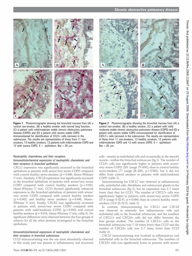
Immunohistochemical expression of neutrophilic chemokines andtheir receptors in bronchial submucosaCCL5 immunostaining was the one most abundantly observedin this study and was present in inflammatory and structural
cells—mainly in endothelial cells and occasionally in the smoothmuscle—within the bronchial submucosa (fig 1). The number ofCCL5+ cells was significantly higher in patients with severe/very severe COPD (281 (range 75–839)) than in control healthynever-smokers (77 (range 26–203), p = 0.002), but it did notdiffer from control smokers or patients with mild/moderateCOPD (table 3).
Immunostaining for CXCL7 was observed in inflammatorycells, endothelial cells, fibroblasts and submucosal glands in thebronchial submucosa (fig 2), but its expression was 2–7 timeslower than that for CCL5. The number of CXCL7+ cells wassignificantly higher in patients with severe/very severe COPD(37.6 (range 0–82.9), p = 0.004) than in control healthy never-smokers (13.8 (0–32.3), table 3).
In contrast, immunostaining for CXCL1 and CXCL6was observed in relatively few inflammatory cells andendothelial cells in the bronchial submucosa, and the numbersof CXCL1+ and CXCL6+ cells did not differ between thefour groups studied. The number of CXCL1+ cells in thebronchial submucosa was 7–10 times lower than CCL5 and thenumber of CXCL6+ cells was 2–7 times lower than CCL5(table 3).
CXCL5 immunostaining was localised in inflammatory andendothelial cells in the bronchial submucosa. The numbers ofCXCL5+ cells was significantly lower in patients with severe/
Figure 1 Photomicrographs showing the bronchial mucosa from (A) acontrol non-smoker, (B) a healthy smoker with normal lung function,(C) a patient with mild/moderate stable chronic obstructive pulmonarydisease (COPD) and (D) a patient with severe stable COPDimmunostained for identification of CCL5+ cells (arrows) in thesubmucosa. The results are representative of those from 11 non-smokers, 13 healthy smokers, 12 patients with mild/moderate COPD and13 with severe COPD. E = epithelium. Bar = 20 mm.
Figure 2 Photomicrographs showing the bronchial mucosa from (A) acontrol non-smoker, (B) a healthy smoker, (C) a patient with mild/moderate stable chronic obstructive pulmonary disease (COPD) and (D) apatient with severe stable COPD immunostained for identification ofCXCL7+ cells (arrows) in the submucosa. The results are representativeof those from 11 non-smokers, 13 healthy smokers, 12 patients withmild/moderate COPD and 13 with severe COPD. E = epithelium.Bar = 20 mm.
Chronic obstructive pulmonary disease
Thorax 2009;64:968–975. doi:10.1136/thx.2009.113647 971
on 14 February 2019 by guest. P
rotected by copyright.http://thorax.bm
j.com/
Thorax: first published as 10.1136/thx.2009.113647 on 23 A
ugust 2009. Dow
nloaded from
very severe COPD (78.3 (range 19–424), p = 0.025) than in thosewith mild/moderate COPD (167.7 (107–266), table 3).
CXCL8 immunostaining was observed in few inflammatorycells and occasionally in capillary vessels (endothelial cells) andsmooth muscle cells in the bronchial submucosa, its expressionbeing 5–15 times lower than that of CCL5. No significantdifferences were observed in the numbers of CXCL8+ cellsbetween any of the groups studied (table 3).
Immunostaining for CXCR1 was observed in inflammatorycells, endothelial cells and, to a lesser extent, in fibroblastswithin the bronchial submucosa. The number of CXCR1+ cellsin the bronchial submucosa of control healthy smokers (161.3(range 64–301), p = 0.038) was significantly higher than incontrol healthy never-smokers (69.1 (39.4–612.9)) but not thanthe other groups. Similar to CXCR1, immunostaining forCXCR2 was observed in inflammatory cells, endothelial cellsand, to a lesser extent, in fibroblasts. No significant differenceswere observed in the numbers of CXCR2+ cells counted in thebronchial submucosa of the four groups studied (table 3).
CD11b staining was observed in inflammatory cells, endothe-lial cells, fibroblasts and occasionally in the smooth muscle withno significant differences being observed between any of thegroups studied. CD44 staining was also observed in inflamma-tory cells, fibroblasts, endothelial cells and occasionally in thesmooth muscle, and again no significant differences wereobserved in the numbers of CD44+ cells counted in thebronchial submucosa between any of the groups (table 3).
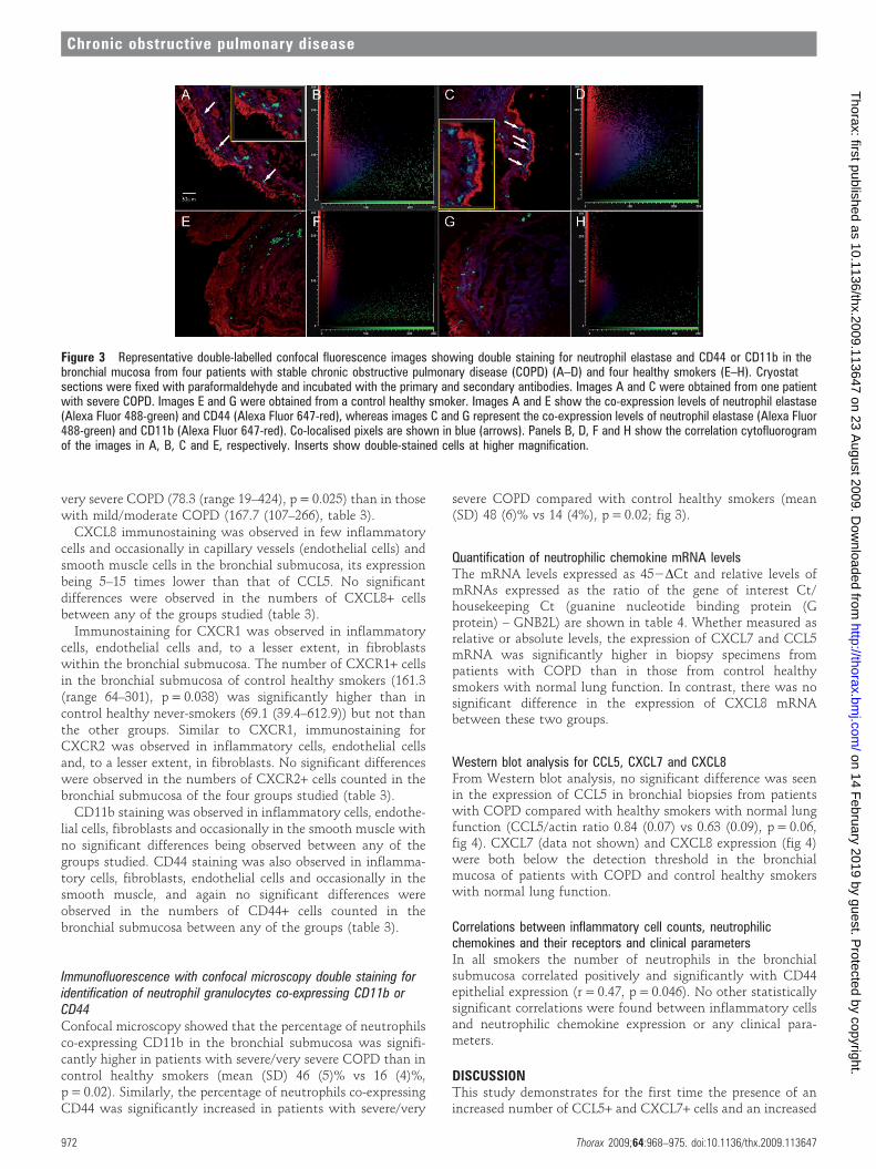
Immunofluorescence with confocal microscopy double staining foridentification of neutrophil granulocytes co-expressing CD11b orCD44Confocal microscopy showed that the percentage of neutrophilsco-expressing CD11b in the bronchial submucosa was signifi-cantly higher in patients with severe/very severe COPD than incontrol healthy smokers (mean (SD) 46 (5)% vs 16 (4)%,p = 0.02). Similarly, the percentage of neutrophils co-expressingCD44 was significantly increased in patients with severe/very
severe COPD compared with control healthy smokers (mean(SD) 48 (6)% vs 14 (4%), p = 0.02; fig 3).
Quantification of neutrophilic chemokine mRNA levelsThe mRNA levels expressed as 452DCt and relative levels ofmRNAs expressed as the ratio of the gene of interest Ct/housekeeping Ct (guanine nucleotide binding protein (Gprotein) – GNB2L) are shown in table 4. Whether measured asrelative or absolute levels, the expression of CXCL7 and CCL5mRNA was significantly higher in biopsy specimens frompatients with COPD than in those from control healthysmokers with normal lung function. In contrast, there was nosignificant difference in the expression of CXCL8 mRNAbetween these two groups.
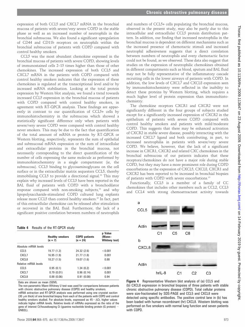
Western blot analysis for CCL5, CXCL7 and CXCL8From Western blot analysis, no significant difference was seenin the expression of CCL5 in bronchial biopsies from patientswith COPD compared with healthy smokers with normal lungfunction (CCL5/actin ratio 0.84 (0.07) vs 0.63 (0.09), p = 0.06,fig 4). CXCL7 (data not shown) and CXCL8 expression (fig 4)were both below the detection threshold in the bronchialmucosa of patients with COPD and control healthy smokerswith normal lung function.
Correlations between inflammatory cell counts, neutrophilicchemokines and their receptors and clinical parametersIn all smokers the number of neutrophils in the bronchialsubmucosa correlated positively and significantly with CD44epithelial expression (r = 0.47, p = 0.046). No other statisticallysignificant correlations were found between inflammatory cellsand neutrophilic chemokine expression or any clinical para-meters.
DISCUSSIONThis study demonstrates for the first time the presence of anincreased number of CCL5+ and CXCL7+ cells and an increased
Figure 3 Representative double-labelled confocal fluorescence images showing double staining for neutrophil elastase and CD44 or CD11b in thebronchial mucosa from four patients with stable chronic obstructive pulmonary disease (COPD) (A–D) and four healthy smokers (E–H). Cryostatsections were fixed with paraformaldehyde and incubated with the primary and secondary antibodies. Images A and C were obtained from one patientwith severe COPD. Images E and G were obtained from a control healthy smoker. Images A and E show the co-expression levels of neutrophil elastase(Alexa Fluor 488-green) and CD44 (Alexa Fluor 647-red), whereas images C and G represent the co-expression levels of neutrophil elastase (Alexa Fluor488-green) and CD11b (Alexa Fluor 647-red). Co-localised pixels are shown in blue (arrows). Panels B, D, F and H show the correlation cytofluorogramof the images in A, B, C and E, respectively. Inserts show double-stained cells at higher magnification.
Chronic obstructive pulmonary disease
972 Thorax 2009;64:968–975. doi:10.1136/thx.2009.113647
on 14 February 2019 by guest. P
rotected by copyright.http://thorax.bm
j.com/
Thorax: first published as 10.1136/thx.2009.113647 on 23 A
ugust 2009. Dow
nloaded from
expression of both CCL5 and CXCL7 mRNA in the bronchialmucosa of patients with severe/very severe COPD in the stablephase as well as an increased number of neutrophils in thebronchial submucosa. We also found a significant upregulationof CD44 and CD11b receptors on neutrophils within thebronchial submucosa of patients with COPD compared withcontrol healthy smokers.
CCL5 was the most abundant chemokine expressed in thebronchial mucosa of patients with severe COPD, showing levelsof immunostained cells 2–15 times higher than those of otherchemokines. The increased expression of both CCL5 andCXCL7 mRNA in the patients with COPD compared withcontrol healthy smokers indicates that the expression of thesechemokines is regulated at the transcriptional level and/or byincreased mRNA stabilisation. Looking at the total proteinexpression by Western blot analysis, we found a trend towardsincreased CCL5 expression in the bronchial mucosa of patientswith COPD compared with control healthy smokers, inagreement with RT-QPCR analysis. These findings are appar-ently in contrast to our quantification of CCL5+ cells byimmunohistochemistry in the submucosa which showed astatistically significant difference only when patients withsevere/very severe COPD were compared with control healthynever smokers. This may be due to the fact that quantificationof the total amount of mRNA or protein by RT-QPCR orWestern blotting, respectively, represents the sum of epithelialand submucosal mRNA expression or the sum of intracellularand extracellular proteins in the bronchial mucosa, notnecessarily corresponding to the direct quantification of thenumber of cells expressing the same molecule as performed byimmunohistochemistry in a single compartment (ie, thesubmucosa). CCL5 binding to glycosaminoglycans on the cellsurface or in the extracellular matrix sequesters CCL5, therebyimmobilising CCL5 to provide a directional signal.33 This mayexplain why increased levels of CCL5 have been reported in theBAL fluid of patients with COPD with a bronchodilatorresponse compared with non-smoking subjects,34 and whylipopolysaccharide-stimulated COPD cultured lung explantsrelease more CCL5 than control healthy smokers.25 In fact, partof this extracellular chemokine can be released after stimulationor recovered in the BAL fluid. Furthermore, the lack of asignificant positive correlation between numbers of neutrophils
and numbers of CCL5+ cells populating the bronchial mucosa,observed in the present study, may also be partly due to thisintracellular and extracellular CCL5 protein distribution pat-tern. In addition, our finding that increased neutrophilia in thebronchial mucosa is dependent on different mechanisms such asthe increased presence of chemotactic stimuli and increasedneutrophil adhesiveness suggests that a direct correlationbetween numbers of neutrophils and every chemotactic factorcould not be found, as we observed. These data also suggest thatstudies on the expression of neutrophilic chemokines obtainedin different compartments such as blood, sputum and BAL fluidmay not be fully representative of the inflammatory cascaderecruiting cells in the lower airways of patients with COPD. Inaddition, the low levels of CXCL7 and CXCL8 expression seenby immunohistochemistry were reflected in the inability todetect these proteins by Western blotting, which requires amuch higher level of protein expression than immunohisto-chemistry.
The chemokine receptors CXCR1 and CXCR2 were notsignificantly different in the four groups of subjects studiedexcept for a significantly increased expression of CXCR2 in theepithelium of patients with severe COPD compared withcontrol healthy smokers and patients with mild/moderateCOPD. This suggests that there may be enhanced activationof CXCR2 in stable severe disease, possibly interacting with theincreased CXCL7 ligand and both contributing, in part, toincreased neutrophilia in patients with severe/very severeCOPD. We believe, however, that the lack of a significantincrease in CXCR1, CXCR2 and related CXC chemokines in thebronchial submucosa of our patients indicates that thesereceptors/chemokines do not have a major role during stableCOPD, but they may have a more prominent role during COPDexacerbations as the expression of CXCL5, CXCL8, CXCR1 andCXCR2 has been reported to be increased in bronchial biopsiesof patients with COPD with severe exacerbations.15
Functionally, CCL5 is a member of a family of CCchemokines that includes other members such as CCL2, CCL3and CCL4 with strong chemoattractant activity towards
Table 4 Results of the RT-QPCR study
Healthy smokers(n = 7)
COPD patients(n = 24)
p Value(Mann–Whitney test)
Absolute mRNA levels
CCL5 19.77 (1.2) 24.32 (2.6) ,0.001
CXCL7 16.95 (1.9) 21.77 (1.8) 0.001
CXCL8 18.27 (1.5) 19.07 (1.6) 0.88
Relative mRNA levels
CCL5 0.95 (0.1) 1.34 (0.2) ,0.001
CXCL7 0.79 (0.01) 0.96 (0.14) 0.001
CXCL8 0.88 (0.06) 0.91 (0.08) 0.94
Data are shown as mean (SEM).The non-parametric Mann-Whitney U test was used for comparisons between patientswith chronic obstructive pulmonary disease (COPD) and healthy smokers.mRNA extraction and RT-QPCR analysis was performed using one cryostat section(30 mm thick) of one bronchial biopsy from each of the patients with COPD and controlhealthy smokers studied. For absolute levels, expressed as 452DCt, higher valuesindicate higher mRNA levels. Relative levels of mRNAs expressed as the ratio of thegene of interest Ct/housekeeping Ct (guanine nucleotide binding protein (G protein) 2
GNB2L).
Figure 4 Representative Western blot analysis of (a) CCL5 and(b) CXCL8 expression in bronchial biopsies of three patients with stablechronic obstructive pulmonary disease (COPD). Total cellular proteinswere size-fractionated by SDS-PAGE and CCL5 and CXCL8 weredetected using specific antibodies. The positive control lane in (b) hasbeen loaded with human recombinant (hr) CXCL8. Western blotting wasperformed on five smokers with normal lung function and seven patientswith COPD.
Chronic obstructive pulmonary disease
Thorax 2009;64:968–975. doi:10.1136/thx.2009.113647 973
on 14 February 2019 by guest. P
rotected by copyright.http://thorax.bm
j.com/
Thorax: first published as 10.1136/thx.2009.113647 on 23 A
ugust 2009. Dow
nloaded from
monocytes and natural killer (NK) cells. We previously reportedincreased levels of CCL2 and CCL4 in the BAL fluid of patientswith stable COPD compared with control smokers,35 and anupregulation of CCL3 in the bronchial epithelium and anincreased number of NK+ cells in the bronchial submucosa ofpatients with severe/very severe COPD compared with controlhealthy smokers.5 Together these findings suggest that, inpatients with stable COPD, CC chemokines may play apredominant role in sustaining tissue neutrophilia and infiltra-tion of macrophages and NK cells. In contrast, CXC chemokinesand their receptors predominate during COPD exacerba-tions.11 15 The fact that CXCL1, CXCL5 and CXCL6 were notchanged in the submucosa of patients with COPD incomparison with both control groups in the present studyfurther confirms our view of a major role for CC chemokines inthe bronchial tissue of patients with stable COPD. CCL5 is alsochemotactic for eosinophils, but an increased number ofeosinophils in the bronchial mucosa has only been demon-strated during COPD exacerbations15 and not in stable disease.4
For this reason we did not quantify the number of eosinophils inthe bronchial mucosa of patients with stable COPD.
Interestingly, we report here for the first time that expressionof the receptors CD11b and CD44 in the neutrophils is 3–4times higher in the bronchial submucosa of patients with COPDthan in control healthy smokers, suggesting a greater activationstate and higher adhesive capability of these cells to bind tosubmucosal extracellular matrix components. This furthersupports a role for these receptors in anchoring neutrophils tosome components of the extracellular matrix such as hyaluronicacid molecules and adhesion molecules such as intercellularadhesion molecule 1 (ICAM-1) and endothelial-leucocyte adhe-sion molecule 1 (ELAM-1). ELAM-1 is highly expressed inbronchial capillary vessels whereas ICAM-1 is preferentiallyexpressed in the epithelium of the bronchial mucosa.29 Thesedata, obtained from segmental and subsegmental bronchi, needto be evaluated in the smaller bronchi and bronchioles ofpatients with COPD.
Differences in the environment between tissue and sputummay account for the differences in the activation status ofneutrophils seen in the bronchial mucosa and those isolatedfrom sputum which show a hypofunctional state and chemo-tactic response.10 The fact that sputum neutrophils are usuallyin a terminal stage of life may also contribute to thesedifferences and further underlines the importance of dataobtained from analysis of the bronchial tissue of patients withCOPD.
In conclusion, the prominent expression of the chemokineCCL5 and, to a lesser extent, of CXCL7 together with higherneutrophil adhesiveness may account for the increased neu-trophilia observed in the bronchial mucosa of patients withsevere COPD. Identification of the individual contribution ofeach of these chemokines in inducing neutrophilia in patientswith severe/very severe COPD will require clinical trials usingselective antagonists or blocking antibodies to each of thesemolecules, many of which are already in development.
Funding: This work was supported by Fondazione Salvatore Maugeri, IRCCS, RicercaCorrente and Regione Piemonte, Ricerca Sanitaria Finalizzata. IMA and PJB aresupported by the Wellcome Trust.
Competing interests: None.
Ethics approval: The study conformed to the Declaration of Helsinki. Ethics consentwas obtained, bronchial biopsies were performed according to the local ethicscommittee guidelines and informed consent was obtained from each subject.
Provenance and peer review: Not commissioned; externally peer reviewed.
REFERENCES1. Global Initiative for Chronic Obstructive Lung Disease. Global strategy for the
diagnosis, management and prevention of chronic obstructive pulmonary disease. NIHPublication No 2701:1–100. NHLBI/WHO Workshop Report. Bethesda: NationalHeart, Lung and Blood Institute, April 2001 (last update 2008). http://www.goldcopd.com (accessed 24 Apr 2008).
2. Barnes PJ. Chronic obstructive pulmonary disease. N Engl J Med 2000;343:269–80.3. Hogg JC, Chu F, Utokaparch S, et al. The nature of small-airway obstruction in
chronic obstructive pulmonary disease. N Engl J Med 2004;350:2645–53.4. Di Stefano A, Caramori G, Ricciardolo FLM, et al. Cellular and molecular mechanisms
in chronic obstructive pulmonary disease: an overview. Clin Exp Allergy2004;34:1156–67.
5. Di Stefano A, Capelli A, Lusuardi M, et al. Severity of airflow limitation is associatedwith severity of airway inflammation in smokers. Am J Respir Crit Care Med1998;158:1277–85.
6. Ricciardolo FLM, Caramori G, Ito K, et al. Nitrosative stress in the bronchial mucosaof severe chronic obstructive pulmonary disease. J Allergy Clin Immunol2005;116:1028–35.
7. Hodge SJ, Hodge GL, Holmes M, et al. Flow cytometric characterization of cellpopulations in bronchoalveolar lavage and bronchial brushings from patients withchronic obstructive pulmonary disease. Cytometry B Clin Cytom 2004;61:27–34.
8. Thompson AB, Daughton D, Robbins RA, et al. Intraluminal airway inflammation inchronic bronchitis. Characterization and correlation with clinical parameters. Am RevRespir Dis 1989;140:1527–37.
9. Fujimoto K, Yasuo M, Urushibata K, et al. Airway inflammation during stable andacutely exacerbated chronic obstructive pulmonary disease. Eur Respir J2005;25:640–6.
10. Yoshikawa T, Dent G, Ward J, et al. Impaired neutrophil chemotaxis in chronicobstructive pulmonary disease. Am J Respir Crit Care Med 2007;175:473–9.
11. Viola A, Luster AD. Chemokines and their receptors: drug targets in immunity andinflammation. Annu Rev Pharmacol Toxicol 2008;48:171–97.
12. Traves SL, Culpitt S, Russell REK, et al. Elevated levels of the chemokines GRO-aand MCP-1 in sputum samples from COPD patients. Thorax 2002;57:590–5.
13. Imaizumi T, Albertine KH, Jicha DL, et al. Human endothelial cells synthesize ENA-78: relationship to IL-8 and to signaling of PMN adhesion. Am J Respir Cell Mol Biol1997;17:181–92.
14. Tanino M, Betsuyaku T, Takeyabu K, et al. Increased levels of interleukin-8 in BALfluid from smokers susceptible to pulmonary emphysema. Thorax 2002;57:405–11.
15. Qiu Y, Zhu J, Bandi V, et al. Biopsy neutrophilia, chemokine and receptor geneexpression in severe exacerbations of COPD. Am J Respir Crit Care Med2003;168:968–75.
16. Van Damme J, Wuyts A, Froyen G, et al. Granulocyte chemotactic protein-2 andrelated CXC chemokines: from gene regulation to receptor usage. J Leukoc Biol1997;62:563–9.
17. Wuyts A, Struyf S, Gijsbers K, et al. The CXC chemokine GCP-2/CXCL6 ispredominantly induced in mesenchymal cells by interleukin-1beta and is down-regulated by interferon-gamma: comparison with interleukin-8/CXCL8. Lab Invest2003;83:23–34.
18. Walz A. Generation and properties of neutrophil-activating peptide 2. Cytokines1992;4:77–95.
19. Traves SL, Smith SJ, Barnes PJ, et al. Specific CXC but not CC chemokines causeelevated monocyte migration in COPD: a role for CXCR2. J Leukoc Biol 2004;76:441–50.
20. Rossi D, Zlotnik A. The biology of chemokines and their receptors. Annu Rev Immunol2000;18:217–42.
21. Yamamoto C, Yoneda T, Yoshikawa M, et al. Airway inflammation in COPD assessedby sputum levels of interleukin-8. Chest 1997;112:505–10.
22. Gompertz S, O’Brien C, Bayley DL, et al. Changes in bronchial inflammation duringacute exacerbations of chronic bronchitis. Eur Respir J 2001;17:1112–9.
23. Costa C, Rufino R, Traves SL, et al. CXCR3 and CCR5 chemokines in induced sputumfrom patients with COPD. Chest 2008;133:26–33.
24. Fujimoto K, Yasuo M, Urushibata K, et al. Airway inflammation during stable andacutely exacerbated chronic obstructive pulmonary disease. Eur Respir J2005;25:640–6.
25. Smyth LJC, Starkey C, Gordon FS, et al. CD8 chemokine receptors in chronicobstructive pulmonary disease. Clin Exp Immunol 2008;154:56–63.
26. Noquera A, Busquets X, Sauleda J, et al. Expression of adhesion molecules and Gproteins in circulating neutrophils in chronic obstructive pulmonary disease.Am J Respir Crit Care Med 1998;158:1664–8.
27. Yamagata T, Sugiura H, Yokoyama T, et al. Overexpression of CD11b and CXCR1 oncirculating neutrophils. Its possible role in COPD. Chest 2007;132:890–9.
28. Pignatti P, Moscato G, Casarini S, et al. Downmodulation of CXCL8/IL-8receptors on neutrophils after recruitment in the airways. J Allergy Clin Immunol2005;115:88–94.
29. Di Stefano A, Maestrelli P, Roggeri A, et al. Upregulation of adhesion molecules inthe bronchial mucosa of subjects with obstructive chronic bronchitis. Am J Respir CritCare Med 1994;149:803–10.
30. Yasuda M, Nakano K, Yasumoto K, et al. CD44: functional relevance to inflammationand malignancy. Histol Histopathol 2002;17:945–50.
31. Yousefi S, Cooper PR, Mueck B, et al. cDNA representational difference analysis ofhuman neutrophils stimulated by GM-CSF. Biochem Biophys Res Commun2000;277:401–9.
Chronic obstructive pulmonary disease
974 Thorax 2009;64:968–975. doi:10.1136/thx.2009.113647
on 14 February 2019 by guest. P
rotected by copyright.http://thorax.bm
j.com/
Thorax: first published as 10.1136/thx.2009.113647 on 23 A
ugust 2009. Dow
nloaded from

32. Manders EMM, Verbeek FJ, Aten JA. Measurement of co-localization of objects indual-colour confocal images. J Microscopy 1993;169:375–438.
33. Murooka TT, Wong MM, Rahbar R, et al. CCL5-CCR5-mediated apoptosis in T cells:requirement for glycosaminoglycan binding and CCL5 aggregation. J Biol Chem2006;281:25184–94.
34. Miller M, Ramsdell J, Friedman PJ, et al. Computed tomographic scan-diagnosed chronicobstructive pulmonary disease-emphysema: eotaxin-1 is associated with bronchodilatorresponse and extent of emphysema. J Allergy Clin Immunol 2007;120:1118–25.
35. Capelli A, Di Stefano A, Gnemmi I, et al. Increased MCP-1 and MIP-1b inbronchoalveolar lavage fluid of chronic bronchitics. Eur Respir J 1999;14:160–5.
Epinephrine and dexamethasone reduce hospital admissionin children with bronchiolitisBronchiolitis is the most common acute infection of the lower respiratory tract in infants. Thismulticentre, randomised, double-blind, placebo controlled clinical trial was conducted inresponse to controversy surrounding the current treatment of bronchiolitis.
Patients were recruited at eight Canadian paediatric emergency departments from 2004through 2007. Bronchiolitis was defined as the first episode of wheezing associated with signs ofan upper respiratory tract infection during the peak respiratory syncytial virus season. A total of3556 infants (aged 6 weeks to 12 months) were assessed for eligibility; 2756 were excluded and800 were enrolled and randomised into four groups: nebulised epinephrine plus oraldexamethasone (group 1); nebulised epinephrine plus oral placebo (group 2); nebulised placeboplus oral dexamethasone (group 3); and nebulised placebo plus oral placebo (group 4).
Combining epinephrine and dexamethasone led to a reduction in the rate of hospitaladmissions within 7 days (primary outcome). There was a relative risk reduction of 35%compared with placebo. Infants in group 1 had lower respiratory rates and lower respiratorydistress assessment index scores during the first hour of the study, were discharged earlier frommedical care and appeared to return to quiet breathing and normal or almost normal feedingmore quickly (secondary outcomes) than those in the placebo group. Dexamethasone andepinephrine alone did not affect these outcomes.
Controversy still exists in the management of bronchiolitis, but this study suggests that thecombination of dexamethasone and epinephrine is beneficial and a starting point for further research.
c Plint AC, Johnson DW, Patel H, et al, for Pediatric Emergency Research Canada (PERC). Epinephrine and dexamethasone in childrenwith bronchiolitis. N Engl J Med 2009;360:2079–89.
P Terezinha Fiandeiro
Correspondence to: Dr P Terezinha Fiandeiro, ST1 CMT, North Middlesex Hospital, London, UK; [email protected]
Provenance and peer review: Not commissioned; not externally peer reviewed.
Thorax 2009;64:975. doi:10.1136/thx.2009.123273
Lung alert
Chronic obstructive pulmonary disease
Thorax November 2009 Vol 64 No 11 975
on 14 February 2019 by guest. P
rotected by copyright.http://thorax.bm
j.com/
Thorax: first published as 10.1136/thx.2009.113647 on 23 A
ugust 2009. Dow
nloaded from