associated protein
TRANSCRIPT
Intracellular Clusterin Interacts with Brain Isoforms of theBridging Integrator 1 and with the Microtubule-Associated Protein Tau in Alzheimer’s DiseaseYuan Zhou., Ikuo Hayashi., Jacky Wong, Katherine Tugusheva, John J. Renger, Celina Zerbinatti*
Department of Neuroscience, Early Development and Discovery Sciences, Merck Research Laboratories, Merck Sharp & Dohme Corp., West Point, Pennsylvania, United
States of America
Abstract
Sporadic or late-onset Alzheimer’s disease (AD) is expected to affect 50% of individuals reaching 85 years of age. The mostsignificant genetic risk factor for late-onset AD is the e4 allele of APOE gene encoding apolipoprotein E, a lipid carrier shownto modulate brain amyloid burden. Recent genome-wide association studies have uncovered additional single nucleotidepolymorphisms (SNPs) linked to AD susceptibility, including those in the CLU and BIN1 genes encoding for clusterin (CLU)and the bridging integrator 1 (BIN1) proteins, respectively. Because CLU has been implicated in brain amyloid-b (Ab)clearance in mouse models of amyloid deposition, we sought to investigate whether an AD-linked SNP in the CLU genealtered Ab42 biomarker levels in the cerebrospinal fluid (CSF). Instead, we found that the CLU rs11136000 SNP modified CSFlevels of the microtubule-associated protein Tau in AD patients. We also found that an intracellular form of CLU (iCLU) wasupregulated in the brain of Tau overexpressing Tg4510 mice, but not in Tg2576 amyloid mouse model. By overexpressingiCLU and Tau in cell culture systems we discovered that iCLU was a Tau-interacting protein and that iCLU associated withbrain-specific isoforms of BIN1, also recently identified as a Tau-binding protein. Through expression analysis of CLU andBIN1 variants, we found that CLU and BIN1 interacted via their coiled-coil motifs. In co-immunoprecipitation studies usinghuman brain tissue, we showed that iCLU and the major BIN1 isoform expressed in neurons were associated with modifiedTau species found in AD. Finally, we showed that expression of certain coding CLU variants linked to AD risk led to increasedlevels of iCLU. Together, our findings suggest that iCLU and BIN1 interaction might impact Tau function in neurons anduncover potential new mechanisms underlying the etiology of Tau pathology in AD.
Citation: Zhou Y, Hayashi I, Wong J, Tugusheva K, Renger JJ, et al. (2014) Intracellular Clusterin Interacts with Brain Isoforms of the Bridging Integrator 1 and withthe Microtubule-Associated Protein Tau in Alzheimer’s Disease. PLoS ONE 9(7): e103187. doi:10.1371/journal.pone.0103187
Editor: Jurgen Gotz, The University of Queensland, Australia
Received April 7, 2014; Accepted June 26, 2014; Published July 22, 2014
Copyright: � 2014 Zhou et al. This is an open-access article distributed under the terms of the Creative Commons Attribution License, which permitsunrestricted use, distribution, and reproduction in any medium, provided the original author and source are credited.
Data Availability: The authors confirm that all data underlying the findings are fully available without restriction. All relevant data is included within the paper.
Funding: The authors have no support or funding to report.
Competing Interests: Y. Zhou, J. Wong, J. Renger and C. Zerbinatti are full-time employees of Merck Sharp & Dohme Corp. and may hold MSD stocks and/orstock options. C. Zerbinatti also certifies here that the affiliation of manuscript authors with Merck Sharp & Dohme Corp does not alter adherence to all the PLOSONE policies on sharing data and materials, as detailed online in the PLOS ONE guide for authors.
* Email: [email protected]
. These authors contributed equally to this work.
Introduction
Multiple genome-wide association studies (GWAS) have repli-
cated a link between common single nucleotide polymorphisms in
the CLU gene (rs11136000 and rs1532278) and increased
susceptibility for late-onset Alzheimer’s disease (AD) [1–7]. In
addition, rare CLU variants revealed by next-generation sequenc-
ing have also been associated with AD risk [8]. However, the
mechanisms by which modifications in clusterin expression and/or
function alter disease risk are not yet clear [9]. Clusterin (CLU) is
synthesized as a 60–80 kD precursor protein that undergoes
internal cleavage generating a- and b-chains joined by disulfide
bonds [10]. This glycosylated heterodimeric CLU is constitutively
secreted and referred to as soluble clusterin (sCLU), or as
apolipoprotein J (apoJ), when found in association with lipopro-
teins [11]. Shorter forms of the precursor CLU have been detected
intracellularly and named cytosolic, truncated or nuclear CLU
[12–14]. Alternative splicing, internal translation initiation,
mistranslocation of sCLU, and impaired proteasomal degradation
all appear to contribute to the pool of cytosolic CLU isoforms [14].
The function of intracellular CLU (iCLU) is not completely
understood. Studies in cancer biology have linked iCLU to Bax-
mediated apoptosis [15,16]. Of relevance to AD, it has been
recently shown that iCLU levels increase quickly in cultured
primary neurons exposed to amyloid-b peptides (Ab), and that this
iCLU elevation is required for the neurotoxic downstream
signaling effects of Ab [17].
CLU expression is highest in the brain and is markedly
upregulated under situations of stress and inflammation [18,19].
Induction of CLU mRNA is observed within pyramidal neurons of
the hippocampus and the entorhinal cortex of AD patients
[20,21], and CLU immunoreactivity is found in association with
neutrophil threads, neurofibrillary tangles and amyloid plaques
[20]. Published literature suggests that CLU plays a chaperone
role for Ab, modulating both its clearance and deposition [21–23],
similar to a function proposed for apoE [24]. Seminal support for
this putative role of CLU was revealed by the double deletion of
PLOS ONE | www.plosone.org 1 July 2014 | Volume 9 | Issue 7 | e103187

CLU and apoE in a mouse model of amyloid deposition [23].
While no substantial changes in brain amyloid were detected with
the individual deletions, double knockout (KO) mice showed a
dramatic exacerbation of amyloid burden. The hypothesis is
further substantiated by findings showing direct interaction
between CLU and Ab [25,26]. However, despite these suggestive
preclinical findings, clinical data is lacking to corroborate a
significant effect of CLU on Ab burden as a major mechanism
underlying the genetic link to AD [27,28]. Healthy carriers of the
CLU rs11136000 risk allele C show decreased white matter
integrity [29], altered coupling between hippocampus and
prefrontal cortex during memory processing [30], and significant
longitudinal increases of cerebral blood flow in the hippocampus
and anterior cingulate cortex [31], indicating that CLU may also
participate in non-Ab pathways that could modulate vulnerability
to AD.
First identified as a tumor suppressor [32], the bridging
integrator 1 (BIN1) has been recently linked to AD susceptibility
by GWAS [33–37]. BIN1 is highly expressed in the brain and all
seven brain-specific BIN1 isoforms have an inserted domain that
interacts with clathrin and AP2/a-adaptin (CLAP), indicating a
key role for neuronal BIN1 in endocytosis [38]. The exact
mechanism by which polymorphisms in the BIN1 gene alter AD
risk is still unknown, but a recent study provided evidence that
BIN1 interacts with the microtubule-associated protein Tau [39].
BIN1 immunoreactivity was found in co-localization with neuro-
fibrillary tangles in the AD brain, and knockdown of the BIN1ortholog Amph partially restored the rough eye phenotype
associated with human Tau overexpression in Drosophila [39].
Here we provide evidence that the CLU rs11136000 SNP
alters CSF Tau levels in AD, and that iCLU is a Tau-interacting
protein elevated in the brain of Tau-overexpressing Tg4510 mice.
We also demonstrate that iCLU interacts with brain-specific BIN1
isoforms containing a putative coiled-coil motif. Furthermore, an
AD-risk iCLU mutant lacking the C-terminus coiled-coil motif
does not interact with BIN1, suggesting that CLU and BIN1
association is mediated via their coiled-coil domains. Lastly, we
show that iCLU and BIN1 isoform 1 are associated with modified
Tau species found in the brain of AD patients, and that certain
coding CLU mutations linked to AD risk increase the ratio
between iCLU and sCLU when transfected in cells. Together,
these genetic and functional studies establish a novel link between
two major AD-susceptibility genes, and suggest that dysfunctions
in these new Tau-interacting proteins could contribute to the
etiology of AD.
Results
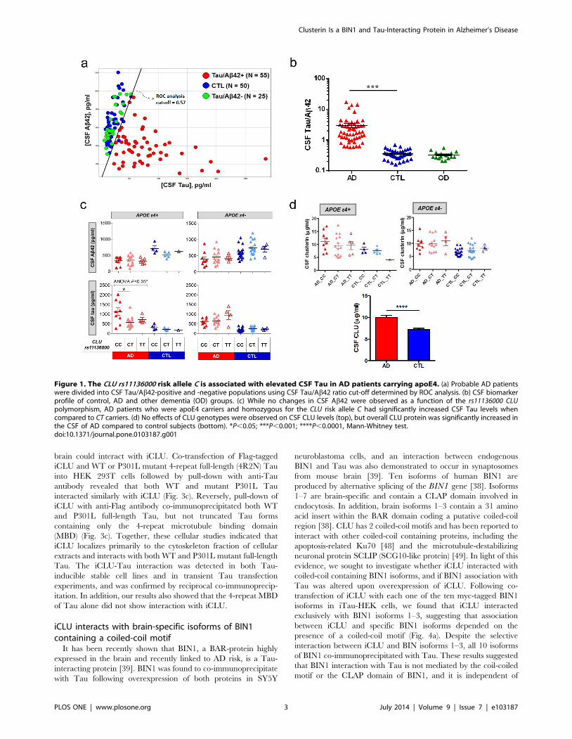
CLU rs11136000 alters CSF Tau in AD patientsTo assess potential mechanisms by which CLU confers risk for
AD, we examined effects of the CLU rs11136000 SNP on CSF
Ab42 and Tau levels, which are reliable disease biomarkers
recently used as endophenotypes in AD genetic studies [40]. For
this analysis, the AD population was enriched with patients
presenting a disease CSF Tau/Ab42 profile, which has a high
diagnostic accuracy for AD over other types of dementia [41,42].
The CSF from an initial set of 80 patients diagnosed with probable
AD and 50 control non-demented individuals was evaluated for
Ab42 and Tau content by specific ELISAs. Using receiving
operating curve (ROC) analysis of CSF Tau/Ab42 ratio to
establish a cut-off value (Fig. 1a), we identified 25 clinically
diagnosed demented individuals who lacked the projected CSF
AD profile, and were therefore subtracted from experimental
patient population (Fig. 1b). Because of previously reported effects
of apoE genotype on brain amyloid burden [24], we also
characterized subjects for their APOE variants. When stratified
by rs11136000 and APOE genotypes, we found a significant
effect of the risk allele C on CSF Tau levels in AD patients
carrying apoE4, with homozygous CC individuals showing
significantly higher CSF Tau than heterozygous CT carriers
(Fig. 1c). Consistent with previous reports [43,44], no significant
CLU genotype effects were observed on CSF Ab42 levels within
AD patients. In addition, CSF AD biomarkers Ab42 and Tau
were not affected by the CLU genotypes in either apoE4 or non-
apoE4 control subjects (Fig. 1c). While CSF CLU protein levels
were not impacted by CLU genotypes, Tau/Ab42-confirmed AD
patients had significantly higher CSF CLU than non-demented
controls (Fig. 1d), consistent with previous literature findings
[45,46]. Based on these initial results, we hypothesized a potential
link between CLU and Tau pathology in AD.
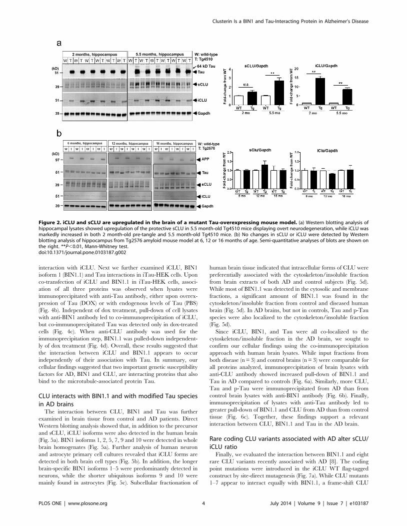
CLU is upregulated in the brain of Tg4510 Tau mousemodel
We next evaluated the expression pattern of CLU isoforms in
the hippocampus and cortex of Tg4510 mice overexpressing the
human mutant P301L Tau associated with frontotemporal
dementia [47]. While 5.5 month-old Tg4510 mice displaying
Tau pathology and neurodegeneration showed marked upregula-
tion of sCLU protein (,30–40 kD monomer) in the hippocampus,
we found that a truncated iCLU form (45–50 kD) was significantly
elevated in the hippocampus of both 2 month-old pre-tangle and
5.5 month-old tangle-bearing Tg4510 in comparison to age-
matched wild type (WT) mice (Fig. 2a). Unlike the clear changes in
brain CLU observed with Tau overexpression, Western blot
evaluation of hippocampus from Tg2576 amyloid mouse model
showed no age-dependent changes in either forms of CLU
compared to WT littermate controls (Fig. 2b). These findings
further substantiated our hypothesis for a potential physiologically
relevant connection between iCLU and Tau.
iCLU is localized to the cytoskeleton fraction andinteracts with Tau
Because iCLU had been previously detected in both cytosol and
nucleus, we first examined the subcellular fractionation of CLU
following transfection of full-length or truncated CLU cDNA
constructs in HEK 293T cells (Fig. 3a). Interestingly, we found
that most of the ,52 kD iCLU was detected in the cytoskeleton/
insoluble fraction. The precursor CLU protein (,64 kD) was
fractioned primarily to the cytosol, as predicted by ER/Golgi
localization. While the majority of sCLU (,37 kD) was localized
to the membrane fraction, likely associated with secretory Golgi
vesicles, some sCLU was also detected in the cytosolic fraction.
The primary localization of overexpressed iCLU to the cytoskel-
eton fraction led us to postulate the possibility of a direct
interaction between iCLU and the microtubule-associated protein
Tau.
To further investigate a potential interaction between CLU and
Tau, we used a doxycycline (dox)-inducible wild-type Tau HEK
293T (iTau-HEK) cell line transfected with either full-length or
truncated CLU cDNA constructs. By overexpressing the truncated
CLU construct designed to generate only the iCLU 50 kD isoform
followed by pull-down with anti-Tau antibody, we were able to co-
precipitate iCLU from iTau-HEK cell lysates (Fig. 3b). Induction
of Tau expression with dox increased Tau interaction with iCLU
(Fig. 3b). In reciprocal co-immunoprecipitation experiments, we
also evaluated whether Tau harboring the P301L mutation linked
to frontotemporal dementia and overexpressed in Tg4510 mouse
Clusterin Is a BIN1 and Tau-Interacting Protein in Alzheimer’s Disease
PLOS ONE | www.plosone.org 2 July 2014 | Volume 9 | Issue 7 | e103187
brain could interact with iCLU. Co-transfection of Flag-tagged
iCLU and WT or P301L mutant 4-repeat full-length (4R2N) Tau
into HEK 293T cells followed by pull-down with anti-Tau
antibody revealed that both WT and mutant P301L Tau
interacted similarly with iCLU (Fig. 3c). Reversely, pull-down of
iCLU with anti-Flag antibody co-immunoprecipitated both WT
and P301L full-length Tau, but not truncated Tau forms
containing only the 4-repeat microtubule binding domain
(MBD) (Fig. 3c). Together, these cellular studies indicated that
iCLU localizes primarily to the cytoskeleton fraction of cellular
extracts and interacts with both WT and P301L mutant full-length
Tau. The iCLU-Tau interaction was detected in both Tau-
inducible stable cell lines and in transient Tau transfection
experiments, and was confirmed by reciprocal co-immunoprecip-
itation. In addition, our results also showed that the 4-repeat MBD
of Tau alone did not show interaction with iCLU.
iCLU interacts with brain-specific isoforms of BIN1containing a coiled-coil motif
It has been recently shown that BIN1, a BAR-protein highly
expressed in the brain and recently linked to AD risk, is a Tau-
interacting protein [39]. BIN1 was found to co-immunoprecipitate
with Tau following overexpression of both proteins in SY5Y
neuroblastoma cells, and an interaction between endogenous
BIN1 and Tau was also demonstrated to occur in synaptosomes
from mouse brain [39]. Ten isoforms of human BIN1 are
produced by alternative splicing of the BIN1 gene [38]. Isoforms
1–7 are brain-specific and contain a CLAP domain involved in
endocytosis. In addition, brain isoforms 1–3 contain a 31 amino
acid insert within the BAR domain coding a putative coiled-coil
region [38]. CLU has 2 coiled-coil motifs and has been reported to
interact with other coiled-coil containing proteins, including the
apoptosis-related Ku70 [48] and the microtubule-destabilizing
neuronal protein SCLIP (SCG10-like protein) [49]. In light of this
evidence, we sought to investigate whether iCLU interacted with
coiled-coil containing BIN1 isoforms, and if BIN1 association with
Tau was altered upon overexpression of iCLU. Following co-
transfection of iCLU with each one of the ten myc-tagged BIN1
isoforms in iTau-HEK cells, we found that iCLU interacted
exclusively with BIN1 isoforms 1–3, suggesting that association
between iCLU and specific BIN1 isoforms depended on the
presence of a coiled-coil motif (Fig. 4a). Despite the selective
interaction between iCLU and BIN isoforms 1–3, all 10 isoforms
of BIN1 co-immunoprecipitated with Tau. These results suggested
that BIN1 interaction with Tau is not mediated by the coil-coiled
motif or the CLAP domain of BIN1, and it is independent of
Figure 1. The CLU rs11136000 risk allele C is associated with elevated CSF Tau in AD patients carrying apoE4. (a) Probable AD patientswere divided into CSF Tau/Ab42-positive and -negative populations using CSF Tau/Ab42 ratio cut-off determined by ROC analysis. (b) CSF biomarkerprofile of control, AD and other dementia (OD) groups. (c) While no changes in CSF Ab42 were observed as a function of the rs11136000 CLUpolymorphism, AD patients who were apoE4 carriers and homozygous for the CLU risk allele C had significantly increased CSF Tau levels whencompared to CT carriers. (d) No effects of CLU genotypes were observed on CSF CLU levels (top), but overall CLU protein was significantly increased inthe CSF of AD compared to control subjects (bottom). *P,0.05; ***P,0.001; ****P,0.0001, Mann-Whitney test.doi:10.1371/journal.pone.0103187.g001
Clusterin Is a BIN1 and Tau-Interacting Protein in Alzheimer’s Disease
PLOS ONE | www.plosone.org 3 July 2014 | Volume 9 | Issue 7 | e103187
interaction with iCLU. Next we further examined iCLU, BIN1
isoform 1 (BIN1.1) and Tau interactions in iTau-HEK cells. Upon
co-transfection of iCLU and BIN1.1 in iTau-HEK cells, associ-
ation of all three proteins was observed when lysates were
immunoprecipitated with anti-Tau antibody, either upon overex-
pression of Tau (DOX) or with endogenous levels of Tau (PBS)
(Fig. 4b). Independent of dox treatment, pull-down of cell lysates
with anti-BIN1 antibody led to co-immunoprecipitation of iCLU,
but co-immunoprecipitated Tau was detected only in dox-treated
cells (Fig. 4c). When anti-CLU antibody was used for the
immunoprecipitation step, BIN1.1 was pulled-down independent-
ly of dox treatment (Fig. 4d). Overall, these results suggested that
the interaction between iCLU and BIN1.1 appears to occur
independently of their association with Tau. In summary, our
cellular findings suggested that two important genetic susceptibility
factors for AD, BIN1 and CLU, are interacting proteins that also
bind to the microtubule-associated protein Tau.
CLU interacts with BIN1.1 and with modified Tau speciesin AD brains
The interaction between CLU, BIN1 and Tau was further
examined in brain tissue from control and AD patients. Direct
Western blotting analysis showed that, in addition to the precursor
and sCLU, iCLU isoforms were also detected in the human brain
(Fig. 5a). BIN1 isoforms 1, 2, 5, 7, 9 and 10 were detected in whole
brain homogenates (Fig. 5a). Further analysis of human neuron
and astrocyte primary cell cultures revealed that iCLU forms are
detected in both brain cell types (Fig. 5b). In addition, the longer
brain-specific BIN1 isoforms 1–5 were predominantly detected in
neurons, while the shorter ubiquitous isoforms 9 and 10 were
mainly found in astrocytes (Fig. 5c). Subcellular fractionation of
human brain tissue indicated that intracellular forms of CLU were
preferentially associated with the cytoskeleton/insoluble fraction
from brain extracts of both AD and control subjects (Fig. 5d).
While most of BIN1.1 was detected in the cytosolic and membrane
fractions, a significant amount of BIN1.1 was found in the
cytoskeleton/insoluble fraction from control and diseased human
brain (Fig. 5d). In AD brains, but not in controls, Tau and p-Tau
species were also localized to the cytoskeleton/insoluble fraction
(Fig. 5d).
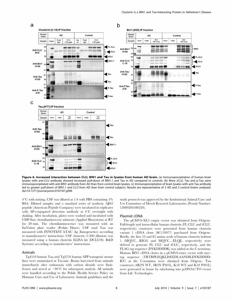
Since iCLU, BIN1, and Tau were all co-localized to the
cytoskeleton/insoluble fraction in the AD brain, we sought to
confirm our cellular findings using the co-immunoprecipitation
approach with human brain lysates. While input fractions from
both disease (n = 3) and control brains (n = 3) were comparable for
all proteins analyzed, immunoprecipitation of brain lysates with
anti-CLU antibody showed increased pull-down of BIN1.1 and
Tau in AD compared to controls (Fig. 6a). Similarly, more CLU,
Tau and p-Tau were immunoprecipitated from AD than from
control brain lysates with anti-BIN1 antibody (Fig. 6b). Finally,
immunoprecipitation of lysates with anti-Tau antibody led to
greater pull-down of BIN1.1 and CLU from AD than from control
tissue (Fig. 6c). Together, these findings support a relevant
interaction between CLU, BIN1.1 and Tau in the AD brain.
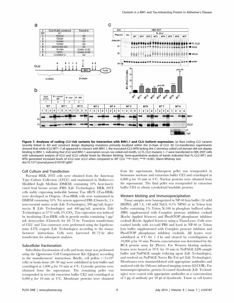
Rare coding CLU variants associated with AD alter sCLU/iCLU ratio
Finally, we evaluated the interaction between BIN1.1 and eight
rare CLU variants recently associated with AD [8]. The coding
point mutations were introduced in the iCLU WT flag-tagged
construct by site-direct mutagenesis (Fig. 7a). While CLU mutants
1–7 appear to interact equally with BIN1.1, a frame-shift CLU
Figure 2. iCLU and sCLU are upregulated in the brain of a mutant Tau-overexpressing mouse model. (a) Western blotting analysis ofhippocampal lysates showed upregulation of the protective sCLU in 5.5 month-old Tg4510 mice displaying overt neurodegeneration, while iCLU wasmarkedly increased in both 2 month-old pre-tangle and 5.5 month-old Tg4510 mice. (b) No changes in sCLU or iCLU were detected by Westernblotting analysis of hippocampus from Tg2576 amyloid mouse model at 6, 12 or 16 months of age. Semi-quantitative analyses of blots are shown onthe right. **P,0.01, Mann-Whitney test.doi:10.1371/journal.pone.0103187.g002
Clusterin Is a BIN1 and Tau-Interacting Protein in Alzheimer’s Disease
PLOS ONE | www.plosone.org 4 July 2014 | Volume 9 | Issue 7 | e103187

mutant lacking the C-terminus coiled-coil region failed to co-
immunoprecipitate with BIN1.1 (Fig. 7b) further corroborating
that iCLU and BIN1 association occurs via a coiled-coil
interaction. Moreover, overexpression of full-length CLU mutants
1–7 in HEK 293T cells showed that certain coding mutations
(MT1 and MT6) were associated with increased generation of
iCLU relative to sCLU (Fig. 7c), suggesting a potential mechanism
for pathogenicity of these variants.
Discussion
After APOE, the CLU and BIN1 genes have been identified as
the most important susceptibility loci in late-onset AD [1–7,33–
37]. It has been hypothesized that CLU is involved in amyloid
clearance, playing a protective role in AD. However, contrary to
this premise, plasma levels of CLU correlate positively with AD
severity and progression [50], and increased CLU mRNA
expression is associated with a more deteriorated disease status
[51]. It has been recently reported that elevated CSF clusterin
contributes to entorhinal atrophy in patients with mild cognitive
impairment and increased Ab42 deposition [52], suggesting that
clusterin might be involved in AD pathogenesis. In the present
study we found a link between the CLU rs11136000 SNP and
CSF Tau levels in AD patients and provided evidence that
intracellular forms of clusterin might play an important role in AD
pathology. While the secreted form of CLU (sCLU) is induced
during stress and inflammation and believed to be protective,
intracellular forms of CLU previously linked to cytotoxicity are
also upregulated under these conditions [12,14]. Mechanisms
induced by cellular stress, such as abnormal translocation of CLU
from the ER/Golgi to the cytosol [12] and impaired degradation
by the proteosome [14], as well as increased Ab levels [17] and
scrapie infection [53], all appear to drive accumulation of iCLU.
Therefore CLU mutations that alter the ratio between iCLU and
sCLU produced during stress could potentially modify risk for AD.
In support of this hypothesis, we showed that certain AD-risk
mutations in CLU significantly increased the ratio iCLU/sCLU
following expression of the full-length CLU construct in vitro.
We reported here that overexpression of human Tau in the
mouse brain was associated with a marked increase in iCLU levels
before any pathological forms of Tau were detected, suggesting
that iCLU and Tau are predicted to interact in vivo. However,
because Tg4510 mice overexpress the frontotemporal dementia
mutant P301L Tau, which has reduced ability to promote
microtubule assembly and is more prone to aggregation than
WT Tau [54] found in normal and AD patients, we performed
reciprocal co-immunoprecipitation experiments comparing the
interaction between iCLU and WT or P301L Tau in HEK 293T
cells. Our results indicated that both Tau forms co-immunopre-
cipitated with iCLU when co-expressed in a cellular system and
validated our conclusion that increased iCLU expression in
Tg4510 mouse brain is indeed suggestive of a physiologically
relevant interaction between iCLU and Tau.
Figure 3. iCLU localizes to the cytoskeleton fraction and is a Tau-interacting protein. (a) Subcellular fractionation of HEK 293T aftertransfection with full-length CLU cDNA indicated that most of iCLU was found in the cytoskeleton/insoluble fraction. (b) Transfection of sCLU or iCLUcDNA in Tau-inducible HEK293 cells (iTau-HEK) followed by pull-down with anti-Tau antibody (HT7) showed co-immunoprecipitation of iCLU. (c)Transient co-transfection of Flag-tagged iCLU construct with WT or P301L mutant full-length Tau in HEK 293T cells followed by pull-down with anti-Tau antibody (HT7) or anti-Flag antibody showed reciprocal co-immunoprecipitation of iCLU and Tau, respectively. Conversely, transient co-transfection of Flag-tagged iCLU with WT or P301L 4-repeat microtubule binding domain (MBD) of Tau alone did not result in co-immunoprecipitation of Tau by anti-Flag antibody.doi:10.1371/journal.pone.0103187.g003
Clusterin Is a BIN1 and Tau-Interacting Protein in Alzheimer’s Disease
PLOS ONE | www.plosone.org 5 July 2014 | Volume 9 | Issue 7 | e103187

Our cellular data supporting CLU as a novel Tau-interacting
protein is similar to recent findings reported for BIN1 [39]. While
iCLU interacted with both WT and P301L mutant full-length
Tau, it did not co-immunoprecipitate the 4-repeat MBD of Tau
alone, suggesting that the interaction between iCLU and Tau
occurs outside the microtubule binding region. In addition, we
showed that CLU and BIN1 co-immunoprecipitated primarily
with Tau species found in neurofibrillary tangles from AD brains.
Because initial findings from Chapuis et al. [39] suggest that BIN1
expression is detrimental, and increased expression of both CLU
and brain-specific isoforms of BIN1 has been associated with AD
status [51], we speculate that this newly discovered interaction
between CLU and BIN1 could be related to AD risk via
modulation of Tau function. CLU was previously shown to also
interact via its coil-coiled motif with SCLIP, a member of the
stathmin family proteins that modulate microtubule dynamics
through association with tubulin [49]. Therefore, BIN1 expression
levels could potentially modulate the interaction between iCLU
and proteins regulating microtubule dynamics, including Tau.
A new interaction between CLU and coiled-coil-containing
isoforms of neuronal BIN1 demonstrated here uncovers other
putative mechanisms by which CLU and BIN1 could affect AD
risk. It has been recently reported that the coiled-coil motif of
neuronal BIN1 isoforms is also required for binding to dynamin 2,
and that this interaction is essential for endocytosis [55].
Therefore, we hypothesize that increased intracellular CLU could
also impair endocytosis by interfering with the interaction between
BIN1 and dynamin 2. Endocytosis appears to be crucial in the
etiology of AD as SNPs in genes encoding endocytosis-related
proteins have been linked to the disease, including the clathrin
adaptor PICALM [1–6] and CD2AP [36]. Interestingly, both
PICALM and CD2AP have also been recently linked to Tau
pathology [56,57]. PICALM was found to co-localize and
immunoprecipitate with hyperphosphorylated and misfolded
Tau from brains of AD patients [53], while CD2AP was found
to modulate Tau toxicity in Drosophila [57]. Knockdown of cindr,
the fly ortholog of the human CD2AP, enhanced Tau-induced
retinal toxicity [57], similarly to results observed for the fly
ortholog of BIN1 [39].
In summary, our novel findings provide groundwork for future
studies that could shed light on the mechanisms by which GWAS-
identified genes CLU and BIN1 are linked to Tau pathology in
AD, and help uncover new areas for therapeutic intervention.
Materials and Methods
Reagents and AntibodiesReagents were purchased from Sigma-Aldrich unless specified
otherwise. Antibodies used in this study were as follow: anti-APP
6E10 (SIG-39320, Covance), anti-Ab42 12F4 (SIG-39142,
Covance), mouse anti-Tau Tau12 (SIG-39416, Covance), mouse
anti-Tau HT7 (MN1000, Thermo Scientific Pierce), mouse anti-
phosphorylated Tau AT8 (MN1020, Thermo Scientific Pierce),
rabbit anti-Tau H150 (SC-5587, Santa Cruz Biotechnology),
rabbit anti-Tau (A0024, Dako), rabbit anti-clusterin H330 (SC-
8354, Santa Cruz Biotechnology), goat anti-clusterin M18 (SC-
6420, Santa Cruz Biotechnology), goat anti-clusterin C18 (SC-
6419, Santa Cruz Biotechnology), mouse anti-BIN1 99D (05-449,
Figure 4. iCLU interacts with coiled-coil-containing BIN1 isoforms. (a) Co-expression of each of the ten BIN1 human isoforms with iCLU iniTau-HEK cells revealed that iCLU interacted only with brain-specific BIN1 isoforms 1–3 containing a putative coiled-coil motif within the BAR domain(black arrows); however, all isoforms of BIN1 showed interaction with Tau (white arrows). (b) Western blotting of lysates immunoprecipitated withanti-Tau HT7 antibody indicated that both iCLU and BIN1.1 were co-immunoprecipitated from both PBS and dox-treated cells. (c) Western blottingfrom BIN1 99 antibody pull-down showed co-precipitation of iCLU in both PBS and dox conditions, but Tau bands were only detected in dox-treatedcells. (d) Western blotting of anti-CLU C18 antibody immunoprecipitates showed similar bands for BIN1.1 in PBS and dox-treated cells.doi:10.1371/journal.pone.0103187.g004
Clusterin Is a BIN1 and Tau-Interacting Protein in Alzheimer’s Disease
PLOS ONE | www.plosone.org 6 July 2014 | Volume 9 | Issue 7 | e103187

Millipore), rabbit anti-Gapdh (G9545), rabbit anti-actin (A2066),
and mouse anti-Flag (F1804), mouse anti-myc 9E10 (SC-40, Santa
Cruz Biotechnology), mouse and goat IgG control (SC-2015 and
SC-2028, Santa Cruz Biotechnology), donkey anti-mouse IgG (H+L) IRDye 800 (926-32212, LI-COR Biosciences), donkey anti-
rabbit IgG (H+L) IRDye 680 (926-68073, LI-COR Biosciences),
donkey anti-rabbit IgG (H+L) IRDye 800 (926-32213, LI-COR
Biosciences), and donkey anti-goat IgG (H+L) IRDye 680 (926-
68074, LI-COR Biosciences).
Human Samples and GenotypingAll human samples were obtained from commercial sources and
analyzed anonymously. Human blood and cerebrospinal fluid
(CSF) from clinically diagnosed Alzheimer’s disease patients and
aged controls were purchased from PrecisionMed, Inc (www.
precisionmed.com). CSF samples were aliquoted and stored at 2
80uC until analysis. Genomic DNA was extracted from blood cells
using the QIAamp DNA blood midi prep (Qiagen). DNA samples
were stored at 220uC until used. APOE e2/e3/e4 and CLUrs11136000 polymorphisms were determined using polymerase
chain reaction and restriction fragment length polymorphism
(PCR-RFLP) method. All primers used for PCR were purchased
from Applied Biosystems. PCR reagents included 2.5 ml of
500 nM of each primer, 25 ng of template genomic DNA, and
25 ml of AmpliTaq Gold 360 Master Mix (Applied Biosystems).
For APOE genotyping, forward and reverse primer sequences
were: 59 ACAGAATTCGCCCCGGCCTGGTACAC 39 and 59 -
TAAGCITGGCACGGCTGTCCAAGGA 39. PCR products
were digested with HhaI (New England Biolab) at 37uC for
1 hr, and the digested products were run in 20% TBE
polyacrylamide gels. For genotyping the CLU rs11136000polymorphism, forward and reverse primer sequences were: 59
CTTTGTAATGATGTACCATCTACCC 39 and 59 AGGCTG-
CAGACTCCCTGAAT 39. PCR products were digested with
ApoI (New England Biolab) at 37uC for 1 hr, and the digested
products were run in 8% TBE polyacrylamide gels. Human brain
tissue was obtained from National Disease Research Interchange
(NDRI) and stored at 280uC until analysis.
Quantification of CSF Ab42, Tau and ClusterinCSF Ab42 was measured by a sandwich ELISA system using
6E10 as a capture antibody and alkaline phosphatase (AP)-
conjugated 12F4 as a detection antibody. A 96-well black plate
(Corning) was coated with the 6E10 monoclonal antibody at the
concentration of 2 mg/ml in 0.05 M carbonate-bicarbonate buffer,
pH 9.6, incubated overnight at 4uC with shaking, washed with
PBS containing 0.05% Tween 20 and blocked with PBS
containing 3% bovine serum albumin (BSA) for at least 24 hr at
Figure 5. Expression and subcellular localization of CLU and BIN1 isoforms in the human cortex. (a) Western blotting detection of CLUand BIN1 isoforms found in human brain lysates. (b) Western blotting analysis of human neuronal and astrocyte primary cultures revealed significantdetection of iCLU forms in both cell types. (c) Cultured human neurons express primarily brain-specific BIN1 isoform 1, and appear to lack the shorterubiquitous BIN1 isoforms 9 and 10, which are primarily represented in astrocytes. (d) Subcellular fractionation of human brain lysates showing thatBIN1.1 and CLU co-localize with Tau and p-Tau in the cytoskeleton/insoluble fraction from AD, but not control brains.doi:10.1371/journal.pone.0103187.g005
Clusterin Is a BIN1 and Tau-Interacting Protein in Alzheimer’s Disease
PLOS ONE | www.plosone.org 7 July 2014 | Volume 9 | Issue 7 | e103187
4uC with mixing. CSF was diluted at 1:4 with PBS containing 3%
BSA. Diluted samples and a standard series of synthetic Ab42
peptide (American Peptide Company) were incubated in triplicates
with AP-conjugated detection antibody at 4uC overnight with
shaking. After incubation, plates were washed and incubated with
CDP-Star chemiluminescent substrate (Applied Biosystems) at RT
for 20 min. The chemiluminescence was measured with an
EnVision plate reader (Perkin Elmer). CSF total Tau was
measured with INNOTEST hTAU Ag (Innogenetics) according
to manufacturers’ instructions. CSF clusterin (1:200 dilution) was
measured using a human clusterin ELISA kit (DCLU00, R&D
Systems) according to manufactures’ instructions.
AnimalsTg4510 human Tau and Tg2576 human APP transgenic mouse
lines were maintained at Taconic. Brains harvested from animals
immediately after euthanasia with carbon dioxide were flash-
frozen and stored at 280uC for subsequent analysis. All animals
were handled according to the Public Health Service Policy on
Humane Care and Use of Laboratory Animals guidelines and the
study protocol was approved by the Institutional Animal Care and
Use Committee of Merck Research Laboratories (Permit Number:
12089284580220).
Plasmid cDNAThe pCMV6-XL5 empty vector was obtained from Origene.
Full-length and intracellular human clusterin (FL CLU and iCLU,
respectively) constructs were generated from human clusterin
variant 1 cDNA clone (SC118977) purchased from Origene.
Briefly, the first 52 and 85 amino acids of human clusterin isoform
1 (MQVC…RIGG and MQVC…ELQE, respectively) were
deleted to generate FL CLU and iCLU, respectively, and the
FLAG tag sequence (DYKDDDDK) was added at the C-terminus.
Human BIN1 cDNA clones in a pCMV6-entry vector with myc-
tag sequence (TRTRPLEQKLISEEDLAANDILDYKDDDD-
KV) at the C-terminus were obtained from Origene. Tau
constructs (4R2N WT, 4R2N P301L, K18 WT and K18 P301L)
were generated in house by subcloning into pcDNA4/TO vector
from Life Technologies.
Figure 6. Increased interaction between CLU, BIN1 and Tau in lysates from human AD brain. (a) Immunoprecipitation of human brainlysates with anti-CLU antibody showed increased pull-down of BIN1.1 and Tau in AD compared to controls. (b) More sCLU, Tau and p-Tau wereimmunoprecipitated with anti-BIN1 antibody from AD than from control brain lysates. (c) Immunoprecipitation of brain lysates with anti-Tau antibodyled to greater pull-down of BIN1.1 and CLU from AD than from control subjects. Results are representative of 3 AD and 3 control brains analyzed.doi:10.1371/journal.pone.0103187.g006
Clusterin Is a BIN1 and Tau-Interacting Protein in Alzheimer’s Disease
PLOS ONE | www.plosone.org 8 July 2014 | Volume 9 | Issue 7 | e103187
Cell Culture and TransfectionParental HEK 293T cells were obtained from the American
Type Culture Collection (ATCC) and maintained in Dulbecco’s
Modified Eagle Medium (DMEM) containing 10% heat-inacti-
vated fetal bovine serum (FBS) (Life Technologies). HEK 293T
cells stably expressing inducible human Tau 4R2N (iTau-HEK)
were developed at Origene. iTau-HEK cells were maintained in
DMEM containing 10% Tet system approved FBS (Clontech), 16non-essential amino acids (Life Technologies), 200 mg/mL hygro-
mycin B (Life Technologies) and 400 mg/mL geneticin (Life
Technologies) at 37uC with 5% CO2. Tau expression was induced
by incubating iTau-HEK cells in growth media containing 1 mg/
mL doxycycline (Clontech) for 48–72 hr. Transient transfections
of CLU and Tau constructs were performed using the Lipofecta-
mine LTX reagent (Life Technologies) according to the manu-
facturers’ instructions. Cells were harvested 48–72 hr after
transfection for subsequent analyses.
Subcellular fractionationSubcellular fractionation of cells and brain tissue was performed
using the Qproteome Cell Compartment Kit (Qiagen) according
to the manufacturers’ instructions. Briefly, cell pellets (,56106
cells) or brain tissue (20–30 mg) were solubilized in lysis buffer and
centrifuged at 1,000 g for 10 min at 4uC. Cytosolic proteins were
obtained from the supernatant. The remaining pellet was
resuspended in ice-cold extraction buffer CE2 and centrifuged at
6,000 g for 10 min at 4uC. Membrane proteins were obtained
from the supernatant. Subsequent pellet was resuspended in
benzonase nuclease and extraction buffer CE3 and centrifuged at
6,800 g for 10 min at 4uC. Nuclear proteins were obtained from
the supernatant. The final pellet was resuspended in extraction
buffer CE4 to obtain cytoskeletal/insoluble proteins.
Western blotting and ImmunoprecipitationTissue samples were homogenized in NP-40 lysis buffer (10 mM
HEPES, pH 7.4; 140 mM NaCl; 0.5% NP40) or in Triton lysis
buffer containing 1% Triton X-100 in phosphate buffered saline
(PBS) supplemented with Complete protease inhibitor cocktail
(Roche Applied Sciences) and PhosSTOP phosphatase inhibitor
cocktail (Roche Applied Sciences) using a TissueLyzer. Cells were
washed briefly with ice-cold PBS and lysed in NP-40 or Triton
lysis buffer supplemented with Complete protease inhibitor and
PhosSTOP phosphatase inhibitor cocktails. All lysates were
solubilized at 4uC for 1–3 hr and cleared by centrifugation at
10,000 g for 10 min. Protein concentration was determined by the
BCA protein assay kit (Pierce). For Western blotting analysis,
lysates were heated at 70uC for 10 min in NuPAGE LDS sample
buffer and NuPAGE sample reducing agent (Life Technologies),
and resolved on NuPAGE Novex Bis-Tris gel (Life Technologies).
Membranes were immunoblotted with appropriate antibodies and
analyzed with the Odyssey infrared imaging system (LI-COR). For
immunoprecipitation, protein G-coated dynabeads (Life Technol-
ogies) were coated with appropriate antibodies at a concentration
of 5 mg of antibody per 50 ml of dynabeads. Tissue or cell lysates
Figure 7. Analyses of coding CLU risk variants for interaction with BIN1.1 and CLU isoform expression. (a) Rare coding CLU variantsrecently linked to AD and construct design displaying mutations primarily localized within the b-chain of CLU. (b) Co-transfection experimentsshowed that while iCLU MT1–7 all appeared to interact with BIN1.1, the truncated iCLU MT8 lacking the C-terminus coiled-coil domain did not displaybinding to BIN1.1, indicating that iCLU and BIN1.1 association occurs via coiled-coil motifs. (c) FL-CLU mutants 1–7 were transfected in HEK 293T cellswith subsequent analysis of iCLU and sCLU cellular levels by Western blotting. Semi-quantitative analysis of bands indicated that FL-CLU MT1 andMT6 generated increased levels of iCLU over sCLU when compared to WT CLU. **P,0.01; ***P,0.001, Mann-Whitney test.doi:10.1371/journal.pone.0103187.g007
Clusterin Is a BIN1 and Tau-Interacting Protein in Alzheimer’s Disease
PLOS ONE | www.plosone.org 9 July 2014 | Volume 9 | Issue 7 | e103187

were incubated with antibody-coated dynabeads for 30 min at
RT. After immunoprecipitation, samples were suspended in 16NuPAGE LDS sample buffer and NuPAGE sample reducing
agent and analyzed by Western blotting.
Primary neuronal and astrocyte culturesHuman neurons (Cat# 1520) and astrocytes (Cat #1801) were
obtained from ScienCell Research. Briefly, neurons were plated in
6-well poly-L-lysine-coated culture plates (Thermo 152035)
according to manufactures’ protocol. After 14 days in culture,
neurons were washed briefly with ice-cold PBS and lysed in RIPA
buffer (Thermo 89900) supplemented with Complete protease
inhibitor and PhosSTOP phosphatase inhibitor cocktails (Roche
Applied Sciences). For human astrocytes, cells were cultured
following manufacture instructions and 0.56106 cells were sub-
cultured in 6-well poly-L-lysine-coated culture plates for 24 hrs.
Astrocytes were then washed and lysed in the same manner as
human neurons. Lysates were solubilized at 4uC for 1 hr and
cleared by centrifugation at 16,000 g for 5 min. Protein concen-
tration was determined by the BCA protein assay kit (Pierce).
Statistical analysisReceiver operating characteristic curve (ROC) analysis was
performed using JMP 11 (SAS Institute). Group differences for
each analyte were assessed by the Student’s t test for two-group
comparisons and the One-way ANOVA test for multiple
comparisons using GraphPad Prism 5.0 (GraphPad Software
Inc.). For comparison of categorical parameters, the Pearson’s chi-
square test was used. A P value of less than or equal to 0.05 was
considered statistically significant.
Acknowledgments
The authors thank Lili Zhang, Shahriar Niroomand and Mary Savage for
sharing the iTau-HEK cell line, Tau constructs, and human brain samples,
respectively.
Author Contributions
Conceived and designed the experiments: CZ YZ IH. Performed the
experiments: YZ IH JW KT. Analyzed the data: IH YZ CZ. Contributed
reagents/materials/analysis tools: JR. Wrote the paper: CZ YZ IH.
References
1. Harold D, Abraham R, Hollingworth P, Sims R, Gerrish A, et al. (2009)
Genome-wide association study identifies variants at CLU and PICALMassociated with Alzheimer’s disease. Nat Genet 41: 1088–1093.
2. Lambert JC, Heath S, Even G, Campion D, Sleegers K, et al. (2009) Genome-
wide association study identifies variants at CLU and CR1 associated withAlzheimer’s disease. Nat Genet 41: 1094–1099.
3. Carrasquillo MM, Belbin O, Hunter TA, Ma L, Bisceglio GD, et al. (2010)Replication of CLU, CR1, and PICALM associations with Alzheimer’s disease.
Arch Neurol 67: 961–964.
4. Corneveaux JJ, Myers AJ, Allen AN, Pruzin JJ, Ramirez M, et al. (2010)Association of CR1, CLU and PICALM with Alzheimer’s disease in a cohort of
clinically characterized and neuropathologically verified individuals. Hum MolGenet 19: 3295–3301.
5. Jun G, Naj AC, Beecham GW, Wang LS, Buros J, et al. (2010) Meta-analysisconfirms CR1, CLU, and PICALM as alzheimer disease risk loci and reveals
interactions with APOE genotypes. Arch Neurol 67: 1473–1484.
6. Lee JH, Cheng R, Barral S, Reitz C, Medrano M, et al. (2011) Identification ofnovel loci for Alzheimer disease and replication of CLU, PICALM, and BIN1 in
Caribbean Hispanic individuals. Arch Neurol 68: 320–328.
7. Wijsman EM, Pankratz ND, Choi Y, Rothstein JH, Faber KM, et al. (2011)
Genome-wide association of familial late-onset Alzheimer’s disease replicates
BIN1 and CLU and nominates CUGBP2 in interaction with APOE. PLoSGenet 7: e1001308.
8. Bettens K, Brouwers N, Engelborghs S, Lambert JC, Rogaeva E, et al. (2012)Both common variations and rare non-synonymous substitutions and small
insertion/deletions in CLU are associated with increased Alzheimer risk. Mol
Neurodegener 7: 3–14.
9. Nuutinen T, Suuronen T, Kauppinen A, Salminen A (2009) Clusterin: a
forgotten player in Alzheimer’s disease. Brain Res Rev 61: 89–104.
10. de Silva HV, Harmony JA, Stuart WD, Gil CM, Robbins J (1990)
Apolipoprotein J: structure and tissue distribution. Biochemistry 5: 5380–5389.
11. Calero M, Tokuda T, Rostagno A, Kumar A, Zlokovic B, et al. (1999)
Functional and structural properties of lipid-associated apolipoprotein J
(clusterin). Biochem J 344: 375–383.
12. Nizard P, Tetley S, Le Drean Y, Watrin T, Le Goff P, et al. (2007) Stress-
induced retrotranslocation of clusterin/ApoJ into the cytosol. Traffic 8: 554–565.
13. Bettuzzi S, Rizzi F (2009) Nuclear CLU (nCLU) and the fate of the cell. Adv
Cancer Res 104: 59–88.
14. Prochnow H, Gollan R, Rohne P, Hassemer M, Koch-Brandt C, et al. (2013)
Non-secreted clusterin isoforms are translated in rare amounts from distincthuman mRNA variants and do not affect Bax-mediated apoptosis or the NF-kB
signaling pathway. PLoS One 8: e75303.
15. Zhang H, Kim JK, Edwards CA, Xu Z, Taichman R, et al. (2005) Clusterininhibits apoptosis by interacting with activated Bax. Nat Cell Biol 7: 909–915.
16. Kim N, Yoo JC, Han JY, Hwang EM, Kim YS, et al. (2011) Human nuclearclusterin mediates apoptosis by interacting with Bcl-XL through C-terminal
coiled coil domain. J Cell Physiol 227: 1157–1167.
17. Killick R, Ribe EM, Al-Shawi R, Malik B, Hooper C, et al. (2014) Clusterin
regulates b-amyloid toxicity via Dickkopf-1-driven induction of the wnt-PCP-
JNK pathway. Mol Psychiatry 19: 88–98.
18. Duguid JR, Bohmont CW, Liu NG, Tourtellotte WW (1989) Changes in brain
gene expression shared by scrapie and Alzheimer disease. Proc Natl Acad SciUSA 86: 7260–7264.
19. May PC, Lampert-Etchells M, Johnson SA, Poirier J, Masters JN (1990)
Dynamics of gene expression for a hippocampal glycoprotein elevated in
Alzheimer’s disease and in response to experimental lesions in rat. Neuron 5:
831–839.
20. McGeer PL, Kawamata T, Walker DG (1992) Distribution of clusterin in
Alzheimer brain tissue. Brain Res 579: 337–341.
21. Lidstrom AM, Bogdanovic N, Hesse C, Volkman I, Davidsson P, et al. (1998)
Clusterin (apolipoprotein J) protein levels are increased in hippocampus and in
frontal cortex in Alzheimer’s disease. Exp Neurol 154: 511–521.
22. DeMattos RB, O’dell MA, Parsadanian M, Taylor JW, Harmony JA, et al.
(2002) Clusterin promotes amyloid plaque formation and is critical for neuritic
toxicity in a mouse model of Alzheimer’s disease. Proc Natl Acad Sci USA 99:
10843–10848.
23. DeMattos RB, Cirrito JR, Parsadanian M, May PC, O’Dell MA, et al. (2004)
ApoE and clusterin cooperatively suppress Abeta levels and deposition: evidence
that ApoE regulates extracellular Abeta metabolism in vivo. Neuron 41: 193–
202.
24. Castellano JM, Kim J, Stewart FR, Jiang H, DeMattos RB, et al. (2011) Human
apoE isoforms differentially regulate brain amyloid-b peptide clearance. Sci
Transl Med 3: 89ra57.
25. Narayan P, Meehan S, Carver JA, Wilson MR, Dobson CM, et al. (2012)
Amyloid-b oligomers are sequestered by both intracellular and extracellular
chaperones. Biochemistry 51: 9270–9276.
26. Cascella R, Conti S, Tatini F, Evangelisti E, Scartabelli T, et al. (2013)
Extracellular chaperones prevent Ab42-induced toxicity in rat brains. Biochim
Biophys Acta 1832: 1217–1226.
27. Baig S, Palmer LE, Owen MJ, Williams J, Kehoe PG, et al. (2012) Clusterin
mRNA and protein in Alzheimer’s disease. J Alzheimers Dis 28: 337–344.
28. Hughes TM, Lopez OL, Evans RW, Kamboh MI, Williamson JD, et al. (2014)
Markers of cholesterol transport are associated with amyloid deposition in the
brain. Neurobiol Aging 35: 802–807.
29. Braskie MN, Jahanshad N, Stein JL, Barysheva M, McMahon KL, et al. (2011)
Common Alzheimer’s disease risk variant within the CLU gene affects white
matter microstructure in young adults. J Neurosci 31: 6764–6770.
30. Erk S, Meyer-Lindenberg A, Opitz von Boberfeld C, Esslinger C, Schnell K, et
al. (2011) Hippocampal function in healthy carriers of the CLU Alzheimer’s
disease risk variant. J Neurosci 31: 18180–18184.
31. Thambisetty M, Beason-Held LL, An Y, Kraut M, Nalls M, et al. (2013)
Alzheimer risk variant CLU and brain function during aging. Biol Psychiatry 73;
399–405.
32. Sakamuro D, Elliott KJ, Wechsler-Reya R, Prendergast GC (1996) BIN1 is a
novel MYC-interacting protein with features of a tumour suppressor. Nat Genet
14: 69–77.
33. Seshadri S, Fitzpatrick AL, Ikram MA, DeStefano AL, Gudnason V, et al. (2010)
Genome-wide analysis of genetic loci associated with Alzheimer disease. JAMA
303: 1832–1840.
34. Carrasquillo MM, Belbin O, Hunter TA, Ma L, Bisceglio GD, et al. (2011)
Replication of BIN1 association with Alzheimer’s disease and evaluation of
genetic interactions. J Alzheimers Dis 24: 751–758.
35. Hu X, Pickering E, Liu YC, Hall S, Fournier H, et al. (2011) Meta-analysis for
genome-wide association study identifies multiple variants at the BIN1 locus
associated with late-onset Alzheimer’s disease. PLoS One 6, e16616.
Clusterin Is a BIN1 and Tau-Interacting Protein in Alzheimer’s Disease
PLOS ONE | www.plosone.org 10 July 2014 | Volume 9 | Issue 7 | e103187
36. Naj AC, Jun G, Beecham GW, Wang LS, Vardarajan BN, et al. (2011)
Common variants at MS4A4/MS4A6E, CD2AP, CD33 and EPHA1 areassociated with late-onset Alzheimer’s disease. Nat Genet 43: 436–441.
37. Logue MW, Schu M, Vardarajan BN, Buros J, Green RC, et al. (2011) A
comprehensive genetic association study of Alzheimer disease in AfricanAmericans. Arch Neurol 68: 1569–1579.
38. Tan MS, Yu JT, Tan L (2013) Bridging integrator 1 (BIN1): form, function, andAlzheimer’s disease. Trends Mol Med 19: 594–603.
39. Chapuis J, Hansmannel F, Gistelinck M, Mounier A, Van Cauwenberghe C, et
al. (2013) Increased expression of BIN1 mediates Alzheimer genetic risk bymodulating Tau pathology. Mol Psychiatry 18: 1225–1234.
40. Cruchaga C, Ebbert MT, Kauwe JS (2014) Genetic discoveries in AD using CSFamyloid and tau. Curr Genet Med Rep 2: 23–29.
41. Fagan AM, Shaw LM, Xiong C, Vanderstichele H, Mintun MA, et al. (2011)Comparison of analytical platforms for cerebrospinal fluid measures of b-
amyloid 1–42, total tau, and p-tau181 for identifying Alzheimer disease amyloid
plaque pathology. Arch Neurol 68: 1137–1144.42. Schoonenboom NS, Reesink FE, Verwey NA, Kester MI, Teunissen CE, et al.
(2012) Cerebrospinal fluid markers for differential dementia diagnosis in a largememory clinic cohort. Neurology 78: 47–54.
43. Schjeide BM, Schnack C, Lambert JC, Lill CM, Kirchheiner J, et al. (2011) The
role of clusterin, complement receptor 1, and phosphatidylinositol bindingclathrin assembly protein in Alzheimer disease risk and cerebrospinal fluid
biomarker levels. Arch Gen Psychiatry 68: 207–213.44. Elias-Sonnenschein LS, Bertram L, Visser PJ (2012) Relationship between
genetic risk factors and markers for Alzheimer’s disease pathology. Biomark Med6: 477–495.
45. Nilselid AM, Davidsson P, Nagga K, Andreasen N, Fredman P, et al. (2006)
Clusterin in cerebrospinal fluid: analysis of carbohydrates and quantification ofnative and glycosylated forms. Neurochem Int 48: 718–728.
46. Sihlbom C, Davidsson P, Sjogren M, Wahlund LO, Nilsson CL (2008)Structural and quantitative comparison of cerebrospinal fluid glycoproteins in
Alzheimer’s disease patients and healthy individuals. Neurochem Res 33: 1332–
1340.
47. Ramsden M, Kotilinek L, Forster C, Paulson J, McGowan E, et al. (2005) Age-
dependent neurofibrillary tangle formation, neuron loss, and memory impair-ment in a mouse model of human tauopathy (P301L). J Neurosci 25: 10637–
10647.
48. Yang CR, Leskov K, Hosley-Eberlein K, Criswell T, Pink JJ, et al. (2000)Nuclear clusterin/XIP8, an x-ray-induced Ku70-binding protein that signals cell
death. Proc Natl Acad Sci USA 97: 5907–5912.49. Kang SW, Shin YJ, Shim YJ, Jeong SY, Park IS, et al. (2005) Clusterin interacts
with SCLIP (SCG10-like protein) and promotes neurite outgrowth of PC12 cells.
Exp Cell Res 309: 305–315.50. Schrijvers EM, Koudstaal PJ, Hofman A, Breteler MM (2011) Plasma clusterin
and the risk of Alzheimer disease. JAMA 305: 1322–1326.51. Karch CM, Jeng AT, Nowotny P, Cady J, Cruchaga C, et al. (2012) Expression
of novel Alzheimer’s disease risk genes in control and Alzheimer’s disease brains.PLoS One 7: e50976.
52. Desikan RS, Thompson WK, Holland D, Hess CP, Brewer JB, et al. (2014) The
Role of Clusterin in Amyloid-b-Associated Neurodegeneration. JAMA Neurol71: 180–187.
53. Kang SW, Yoon SY, Park JY, Kim DH (2013) Unglycosylated clusterin variantaccumulates in the endoplasmic reticulum and induces cytotoxicity. Int J Biochem
Cell Biol 45: 221–231.
54. Barghorn S, Zheng-Fischhofer Q, Ackmann M, Biernat J, von Bergen M, et al.(2000) Structure, microtubule interactions, and paired helical filament
aggregation by tau mutants of frontotemporal dementias. Biochemistry 39:11714–11721.
55. Ellis JD, Barrios-Rodiles M, Colak R, Irimia M, Kim T, et al. (2012) Tissue-specific alternative splicing remodels protein-protein interaction networks. Mol
Cell 46: 884–892.
56. Ando K, Brion JP, Stygelbout V, Suain V, Authelet M, et al. (2013) Clathrinadaptor CALM/PICALM is associated with neurofibrillary tangles and is
cleaved in Alzheimer’s brains. Acta Neuropathol 125: 861–878.57. Shulman JM, Imboywa S, Giagtzoglou N, Powers MP, Hu Y, et al. (2014)
Functional screening in Drosophila identifies Alzheimer’s disease susceptibility
genes and implicates Tau-mediated mechanisms. Hum Mol Genet 23: 870–877.
Clusterin Is a BIN1 and Tau-Interacting Protein in Alzheimer’s Disease
PLOS ONE | www.plosone.org 11 July 2014 | Volume 9 | Issue 7 | e103187