assignment ten - university of texas at...
TRANSCRIPT

Assignment TenFinal Report
Functional Genomics Research Stream • Freshman Research Initiative • by Dr. Patrick J. Killion
Your Name:
Assigned: April 14, 2009
Due: May 5, 2009
Submission: Beginning of Lecture, 2:30 PM
Format: This Printout, Report Stapled
General Introduction
IMPORTANT NOTE: Background material and explanation is purposefully omitted from this assignment text. For more background on the subject matter covered in this assignment be sure to see the lecture notes and the online course notebook, each found on the course website (course materials): http://fg.cns.utexas.edu/
Time Considerations
Please be aware of the time constraints of the various sections of this assignment and plan your attack accordingly. You should have learned by now that research cannot be procrastinated and then made up for by compressed efforts. Plan your work over the next few weeks to allow for errors, mistakes, and inexplicable experimental failure.
Assignment Format
This assignment does not take the usual form of assignments from the Functional Genomics Research Stream. By now you have become accustomed to sections dedicated to specific goals. Each of these sections contained step-by-step instructions, questions to be answered and a final sign-off section where research staff could evaluate your work. This assignment is different in that it is your final report.
It is worth 10% of your final grade.
The purpose of this assignment is to gather all that you have learned this semester about work in a molecular biology laboratory into one final document. You will be performing a few new experiments based upon methods you have been taught. You will be doing substantial scientific writing to summarize the results of these experiments and place them in context to the hierarchy of work you have learned.
Do not plagiarize online resources or other students. This entire assignment is to be performed as an individual - any copying or sharing of text or results will be considered academic dishonesty and will be dealt with accordingly.
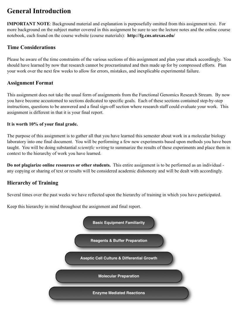
Hierarchy of Training
Several times over the past weeks we have reflected upon the hierarchy of training in which you have participated.
Keep this hierarchy in mind throughout the assignment and final report.
Basic Equipment Familiarity
Reagents & Buffer Preparation
Aseptic Cell Culture & Differential Growth
Molecular Preparation
Enzyme Mediated Reactions

Section A - RNA Preparation Images
If you do not have a very good image of your RNA preparation - make one. This may entail you running a new gel and visualizing (and saving the image) with our new gel-doc system in the laboratory. I expect that the bands would be very clear and a ladder would be present.
Important Note: If you did not get a good RNA sample during Assignment Nine (as judged by NanoDrop or gel electrophoresis) you will need to do so at this time. RNA preparation is a required skill for research in genomics. If you ran out of time during the last assignment you are not off the hook - you need to keep trying for a good preparation and final image. Please work with the research staff if you need assistance.
I expect to see at least two lanes:
‣ LadderIncrements labeled along the side, lanes labeled below.
‣ RNA sample
You will see below where this graphic required (in the report, Section III). I recommend using the USB port on the gel-doc system to save the image to a thumb-drive. Import that image into your report. Note that this RNA Preparation Images can be combined with PCR Images required in Section B (below) on one gel.
Staff Signature: ________________________________________________ Date: ________________________
Section B - PCR Images
If you do not have a very good image of your PCR - make one. Again, this may entail you running a new gel and visualizing (and saving the image) with our new gel-doc system in the laboratory. I expect that the bands would be very clear and a ladder would be present.
I expect to see at least six lanes:
‣ LadderIncrements labeled along the side, lanes labeled below.
‣ PCR: primers gene-specific a & b
‣ PCR: positive control
‣ PCR: negative control
‣ PCR: primers gene-specific a & kanB
‣ PCR: primers gene-specific a & kanC
Important Note: If you did not do the last two PCRs during Assignment Nine (gene-specific a & kanB, gene-specific a and kanC) you will need to do it now. Run the PCR and produce a new gel containing all the required bands.
You will see below where this graphic required (in the report, Section III). Ensure that all lanes are clearly labeled in your final report; a gel without labeled lanes is useless (and you will soon forget which lane contained which sample).
Staff Signature: ________________________________________________ Date: ________________________

Section C - New Experiment: Deletion Strain Growth Assays
Introduction
In Assignment Eight you learned how do study differential growth on solid media plates. In that experiment you were working with a deletion strain and you compared growth to wild type yeast on YPD plates under two different conditions: normal and heat stress temperatures. The purpose of that experiment was to determine if that specific gene (CST6) had anything to do with allowing yeast to adapt to heat stress. Most of you concluded that it did not as the two strains appeared to grow similarly under both temperature conditions.
We are now working with new deletion strains and we are interested in whether they might be involved in cell-cycle progression (or regulation thereof).
Look up the following paper (see previous material on the use of PubMed):
Niu et al. Mechanisms of cell cycle control revealed by a systematic and quantitative overexpression screen in S. cerevisiae. PLoS Genet (2008) vol. 4 (7) pp. e1000120.
You will need to look through this paper and come to understand how they used the drugs hydroxyurea and nocadozole to screen for cell cycle arrest.
Experiments
We have plates made in the lab with both of these drugs:
‣ YPD + 15 µg / mL nocodazole
‣ YPD + 50 µM hydroxyurea
You will perform the following experiments to study your deletion strain for growth defects under conditions:
‣ YPD (normal growth, 30°C - incubator on top)
‣ YPD (heat stress, 37°C - incubator on bottom)
‣ YPD + nocodazole
‣ YPD + hydroxyurea
Thus, in the end you should have four plates total.
Method Review
You can review the complete details of how this plating experiment is performed by revisiting Assignment Eight. You will need to grow overnight cultures (~ 5 mL) of both your strain and wild type yeast (S288C) in order to plate each of these experiments. Retrieve your plate from the 4°C refrigerator and inoculate a 5 mL overnight culture (do the same for S288C). The next day make 1/1, 1/10, 1/100, 1/1000 serial dilutions of both cultures and plate in the same fashion as Assignment Eight. Each plate should have both wild type yeast and your deletion strain on a separate row.
Address the questions posed in Section D of this assignment (the final report) regarding the results of your experimentation. Present your complete plates to a research staff member for evaluation and check-off.
Staff Signature: ________________________________________________ Date: ________________________

Section D - Final Report Expectations & Format
Expectations
Course Value: As mentioned, this report is worth 10% of your final grade. For this reason it is expected to be quantitatively complete and qualitatively good. I expect for it to be written completely in the voice of scientific writing as established earlier in the semester. I expect for it to be clear, well proof-read and grammatically correct.
Paragraph Formatting: The report should be spaced 1.5. This is very important. It should not be single spaced. It should not be double spaced. The former is very difficult to read while the latter looks like a second grader wrote it. Thus, 1.5 spaced or I go insane and nobody wants that to happen.
Overall Formatting: The report should contain clear section and subsection titles (sections and subsections clearly declared below in the Format Requirements). Not including items of this nature makes your report hard to read, navigate or understand - all poor choices.
Length: I am not attaching a generic length requirement. I strongly suggest you simply cover the sections denoted below. I have been very clear regarding the material I expect to see discussed and level of understanding I expect you to convey. Requirements should be clear. It should be noted I have purposefully required few final experiments for this assignment in order to give you time to author a qualitatively good document. It is my expectation that the new experiments in the assignment require no more than one week of laboratory work (~ 6 hours). Thus, you should have nearly two weeks to author this document (~ 12 hours of pseudo-laboratory time). I expect the quality and quantity of your writing to reflect that time allotted.
Your final report will contain the following subsections. Please be sure to clearly label sections with titles and subtitles.
Section I - IntroductionIn this section I wish for you to introduce the basic context of research performed by our research stream.
‣ General IntroductionThis is a short introduction in your own words - just get the ball rolling.
‣ The Eukaryotic Cell CycleReference Assignment Two and matching lecture material. For this section I recommend you borrow liberally from the text you produced for Assignment Two. You can copy and paste the entire thing - but make it fit smoothly with your other text in this final report.
‣ Saccharomyces cerevisiae as Model OrganismReference many lecture notes and online resources appropriately. You should have sufficient information from lecture notes and web references to comment on the utility of studying cell cycle progression in this eukaryotic model organism. Include genomic details about this organism and why it makes a good organism to study in a laboratory setting. Think of and comment on your experience; how easy was it for you to select a deletion strain and grow billions of cells for experimental purposes?
Section II - Hierarchy of TrainingIn this section I wish for you to discuss the hierarchy of training (as presented in a previous figure). You have creative freedom to discuss these sections in any way that you deem appropriate. What I wish for you to accomplish is to describe the important elements that were learned in each step of the hierarchy. Additionally, you have complete freedom to expand upon any of the steps - describe what you found challenging, what you learned the most from, what you were surprised by, etc.

‣ Basic Equipment FamiliarityReference Assignment Four and matching lecture material. You might especially wish to comment on the proper use of the most frequently used laboratory equipment. Additionally, concepts such as accuracy any precision are relevant and could be discussed relative to results for Assignment Four. You may recall there was an issue with this assignment in terms of believability of measurement when it came to establishing the identity of the solutions used therein. Feel free to discuss the use of any equipment you encountered during your training and call out special techniques required to use the equipment accurately and precisely.
‣ Reagent & Buffer PreparationReference Assignment Five and Six and matching lecture material. In this section you might comment on the basic tenets of single and multi-component reagent production. Additionally, this could be a good time to comment on the value of laboratory stocks and why you always need ~ 1M stocks of common reagents for dilutions and multi-component solution preparation. What are the benefits in terms of time, consistency, contamination, and reliability of experimentation? Why are buffers made at a specific pH and what role do they play in the controlling of pH during the process of experimentation (for example, what might the 10x PCR buffer be doing during PCR)?
‣ Aseptic Cell Culture & Differential GrowthReference Assignment Seven and Eight and matching lecture material. This section includes the process of plating, picking colonies, growing overnight cultures, differential growth curves and plates, spectrophotometry. There is a lot of material to reference and to talk about here. Did your results indicate that the gene CST6 had anything to do with yeast’s ability to handle heat stress? How do you know - please provide details as to the outcome of your experiment and your conclusion.
‣ Molecular PreparationReference Assignment Nine and matching lecture material. Molecular preparation is the process of harvesting from cells what you want and discarding what you do not. This covers both DNA & RNA preparations. You could discuss the process of cell culture, pelleting, lysis, phase separation, aqueous extraction, precipitation and final concentration of a nucleic acid pellet. Do you really understand what is going on during each of these steps? Prove it to me.
‣ Enzyme Mediated ReactionsReference Assignment Nine and matching lecture material. You might comment that this is a high level of molecular experimentation in that it takes all the previous steps to be successfully performed before this step becomes possible. Generally discuss what enzymes are and the role they play both in a biological setting (in the cell) and an experimental setting (in a reaction). Finally, you should discuss the specific reactions PCR and RT-PCR (discussed below) as tangible examples of laboratory-based enzyme mediated reactions.
Section III - Molecular Preparation & PCR ReflectionIn this section I wish to revisit Assignment Nine and probe specific issues in more detail. Assignment Nine was a pivotal assignment in the training component of our research stream in that it represented both the culmination and integration of all previous techniques as well utilization of new molecular methods such as PCR. It is important that these techniques are completely understood as a foundation for future research.
‣ Molecular PreparationYou likely have already discussed the process of cell culture, pelleting, lysis, phase separation, aqueous extraction, precipitation and final concentration of a nucleic acid pellet. If you have not taken the time to do so - do it here. If you have simply reference the material you already produced with a statement such as “previously discussed in Section II”. You may also choose to discuss some of the physical demands of these procedures. This might include the manner in which an aqueous layer is removed from an organic phase separation and how a Phase Lock Gel greatly helps you in this procedure.

‣ DNA PreparationDiscuss the unique parts of DNA preparation. What is the role of the glass beads? Each time ethanol is added to the preparation - what is happening at that step? Why is the pellet treated with RNase at the mid-point of the procedure? What is the purpose of this treatment in terms of DNA purification? Discuss how the quality and quantity of a DNA preparation is evaluated. Be specific - talk about what you would expect or hope to see from a NanoDrop readout of a DNA preparation.
‣ RNA PreparationDiscuss the unique parts of RNA preparation. Discuss how the quality and quantity of a RNA preparation is evaluated. Be specific - talk about what you would expect or hope to see from a NanoDrop readout of a DNA preparation. What additional information is gathered from gel-based analysis of an RNA preparation? What do the bands represent? Why does their presence indicate a high quality RNA preparation? What would lack of bands represent and why?
GRAPHIC REQUIRED:You need an image of your total RNA run on a gel here. See the RNA Preparation Images section of this assignment for more details. This should be a good quality image - run a new gel and use the new laboratory gel-doc if needed to capture a good image. Include a ladder. It is recommended that you label the gel in the Microsoft Word document using floating text boxes.
‣ Deletion Verification by Polymerase Chain ReactionIn this section I wish for you to demonstrate understanding of the PCR experiment in Assignment as a mechanism by which we proved the gene deletion in each of the deletion strains.
Introduce context - inform the reader that you are working with a deletion strain of yeast and denote which gene is putatively deleted. Why do I use the word putatively here?
Discuss the results expected. Why is there no band expected for the gene-specific primers (a, b)? Which positive control gene primers did you use? Why is the positive control PCR expected to always produce a band? Why does the negative control PCR not produce a band? If the negative control PCR produced a band - what does that imply? Is that implication good, bad or irrelevant? Finally, what is the expectation of the expectation of a PCR reaction that used the gene-specific primer a and separate primer kanB? Is the result the same as gene-specific primer d and kanC?
It is important that you fully communicate in this sub-section that you understand the PCR experiments you have performed. You did most of the work in Assignment Nine - now you are required to explain the work to me in this section of the report.
GRAPHIC REQUIRED:You need an image of your PCR reactions here. See the PCR Images section of this assignment for more details. I expect to see five lanes: gene-specific primers a and b, positive control, negative control, gene-specific primers a and generic primer kanB, as well as a ladder. It is recommended that you label the gel in the Microsoft Word document using floating text boxes.
Section IV - New Experimental Results
‣ Growth Assays - IntroductionIn Section C you looked up and studied a recent manuscript:
Niu et al. Mechanisms of cell cycle control revealed by a systematic and quantitative overexpression screen in S. cerevisiae. PLoS Genet (2008) vol. 4 (7) pp. e1000120.

This manuscript detailed the use of the drugs nocodazole and hydroxyurea as a plate-based mechanism to screen for cell-cycle related defects. Describe the mechanism by which each of these drugs work. How do they inhibit cell cycle progression? What specific effect do they have on the cellular environment when introduced? Which check-points in the eukaryotic cell cycle would be affected by each of these drugs? Any additional detail you wish to provide on the use of these drugs to screen for cell-cycle arrest is welcome and would be interesting.
‣ Growth Assays - MethodsProvide complete details as to how you performed the new growth and cell-cycle arrest assays. What were the steps involved? What materials were used? What plates were used and how were they setup. A figure or two in this section would be a good idea if you are feeling creative. You can even borrow from Assignment Eight if you wish - just label accordingly. It should be very clear to the reader the number of experiments you are performing and the purpose of each.
‣ Growth Assays - ResultsProvide complete details as to the results of the experiments. Each plate should be detailed with both results and interpretation. For each condition you should decide whether the deletion strain exhibited a growth defect relative to wild type. Additionally, you should assert conclusions as to the meaning of the result. Given each of the conditions (normal growth, heat stress, nocodazole, hydroxyurea) what is the biological interpretation of each experimental outcome?
GRAPHIC REQUESTED:It would be great if you could provide a picture of each of your plates. Try to use a phone camera or something similar. If you are not able to get pictures of your plates do not worry about this request - it is not a requirement. That said, it really adds to the strength of the report to have these pictures.
GRAPHIC REQUIRED:If you are unable to get pictures of your plates then you are required to draw a schematic of each and indicate the results on the schematic. Use lighter or darker spots to indicate growth defects (or lack thereof).

Assignment Submission
Sign Off
When you believe you have the assignment completed ask a staff member (Dr. Killion, any Functional Genomics Research Stream Undergraduate Mentor) look over your work and sign below.
What to do with your materials:‣ Discard your gels (correctly).‣ Discard all your plates (from both the incubator and the -4°C refrigerator).‣ Keep your buffers and reagents in appropriate racks.‣ Store your DNA in the -20°C freezer (completely labeled).‣ Store your RNA in the -80°C freezer in PAI 2.14 (functional genomics box).
Date: ______________________________________________
Time: ______________________________________________
Staff Name: __________________________________
Staff Signature: __________________________________
Submission
This assignment is due on May 5, 2009 - 2:30 PM.
Staple your complete report to this packet. Any time after 2:30 PM on the submission date will be considered late.
Please answer the following questions:
I completed this work as an individual:
Signature: _______________________________________ Date: ________________________________