assessing clients with bowel elimination disorders chapter 26
TRANSCRIPT
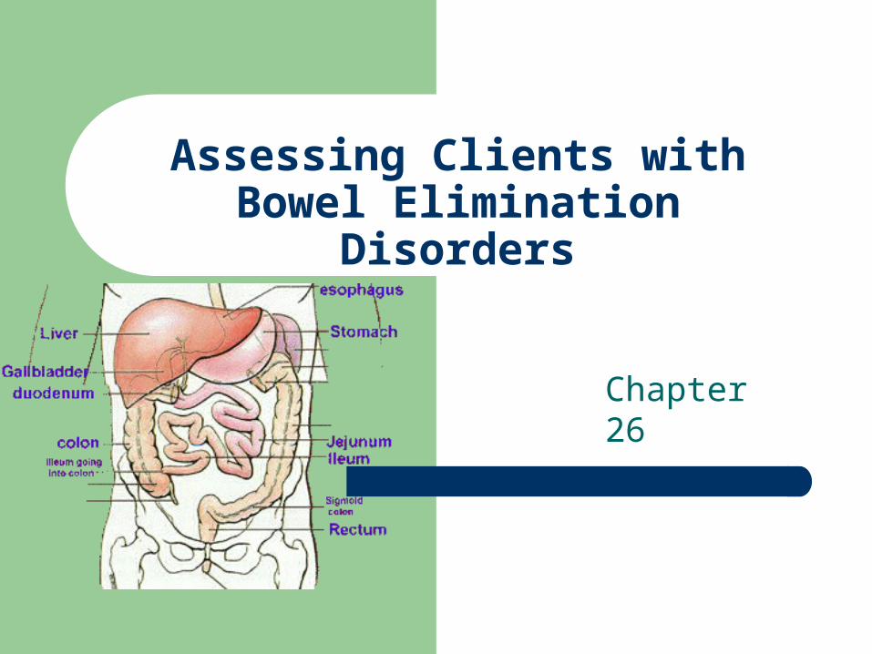
Assessing Clients with Bowel Elimination Disorders
Chapter 26
Review of Anatomy and Physiology
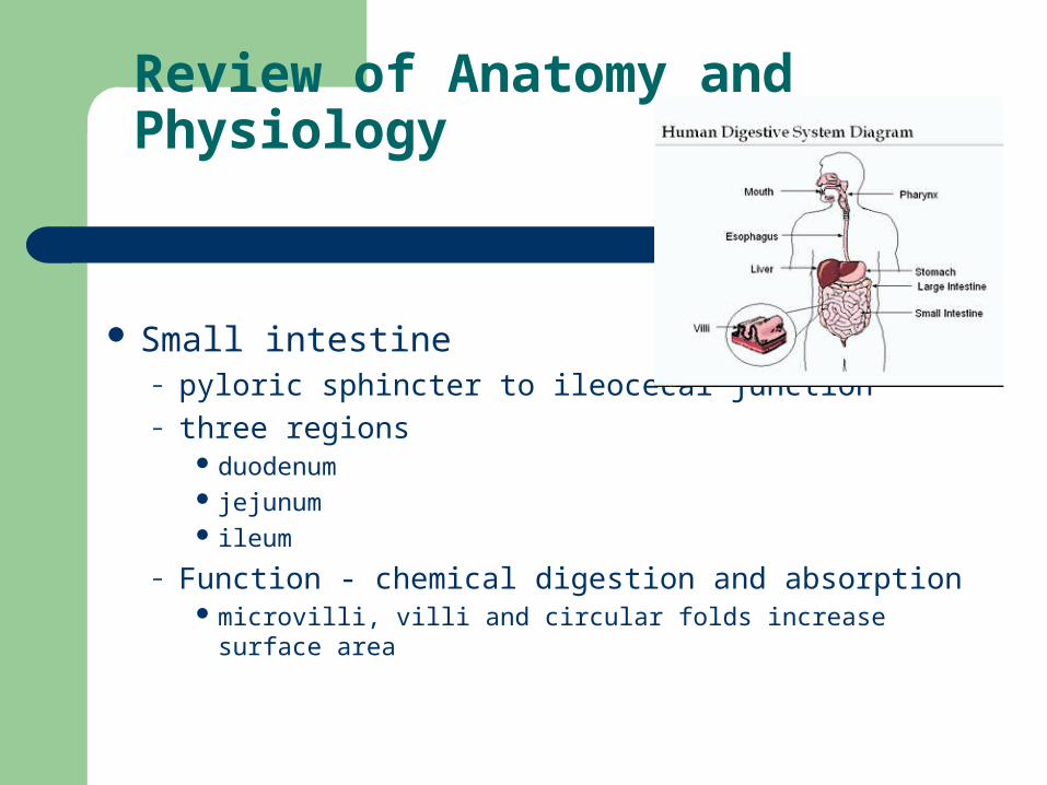
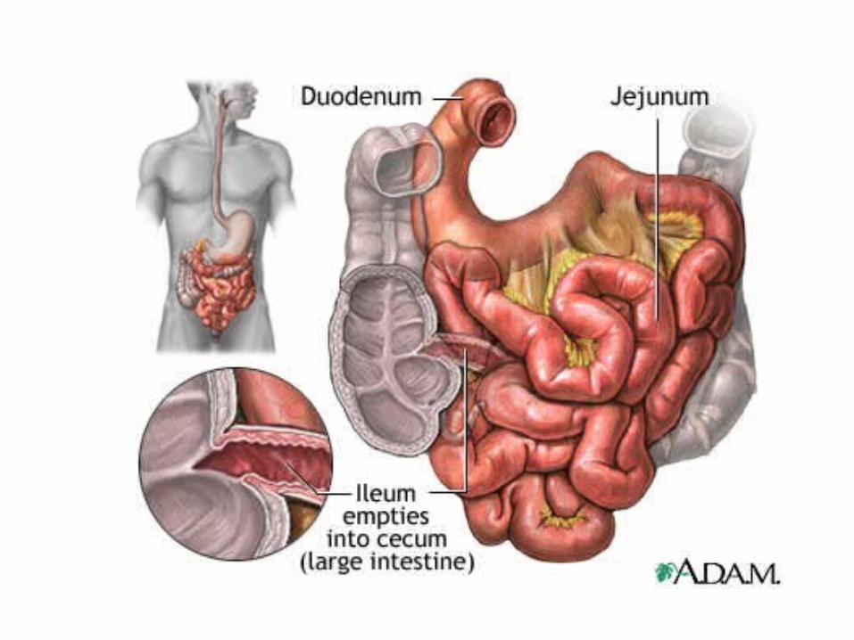
Small intestine– pyloric sphincter to ileocecal junction– three regions
duodenum jejunum ileum
– Function - chemical digestion and absorption microvilli, villi and circular folds increase surface area
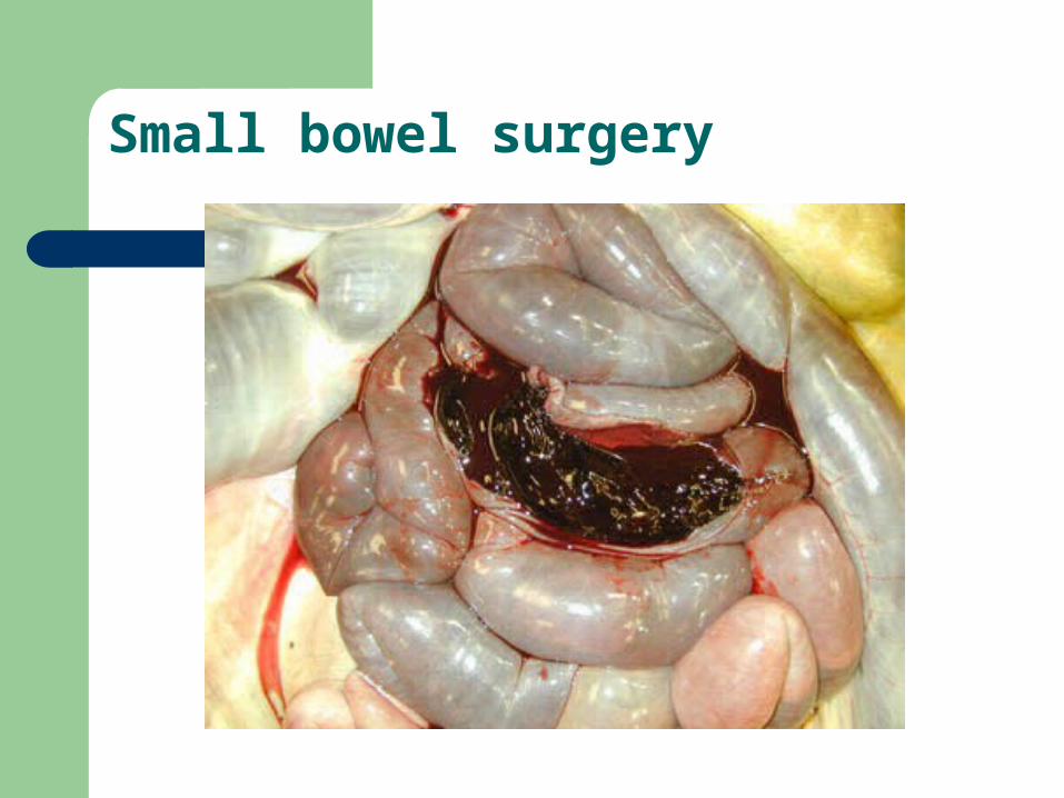
Small bowel surgery
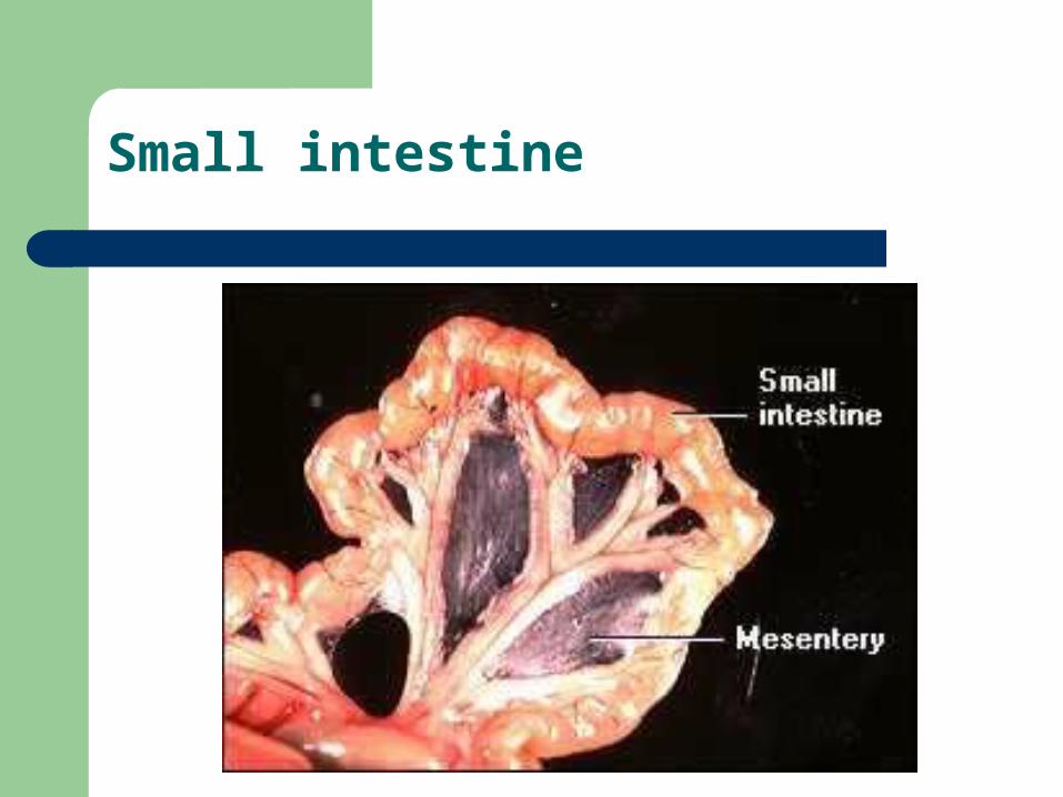
Small intestine
Review of Anatomy and Physiology
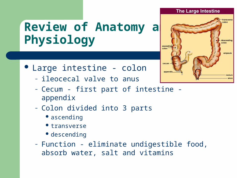
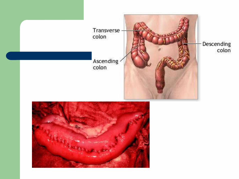
Large intestine - colon– ileocecal valve to anus– Cecum - first part of intestine - appendix– Colon divided into 3 parts
ascending transverse descending
– Function - eliminate undigestible food, absorb water, salt and vitamins
Large Intestine

Assessment of Bowel Function
Subjective– onset– characteristics– course– severity– precipitating factor– relieving factors– associated symptoms
Sample Interview Questions
Can you describe the type of cramping and abdominal pain you are having?
Have you every had bleeding from your rectum?
Have you noticed any changes in your bowel habits?
Assessing the Abdomen
Inspection, auscultation, percussion and palpation as described
Rectal exam - polyps Stool for occult blood
– + requires further testing for colon CA or GI bleeding 2nd to peptic ulcers, ulcerative colitis or diverticulosis

Blood and Stool
Melena - black tarry stool Blood on Stool - bleeding sigmoid colon, rectum Blood in Stool - colon, ulcerative colitis,
– diverticulitis, tumor, ulcer Stool black, hard = oral iron Strong odor = blood of high fat content
– steatorrhea
Nursing Care of Clients with Bowel Disorders
Chapter 26
Disorders of Intestinal Motility
Diarrhea – serious in the young and elderly– increase in the frequency, volume and fluid
content of the stool
Causes– bacteria, or parasitic infections, malaborption,
medications, diseases, allergies or pyschological
Diarrhea
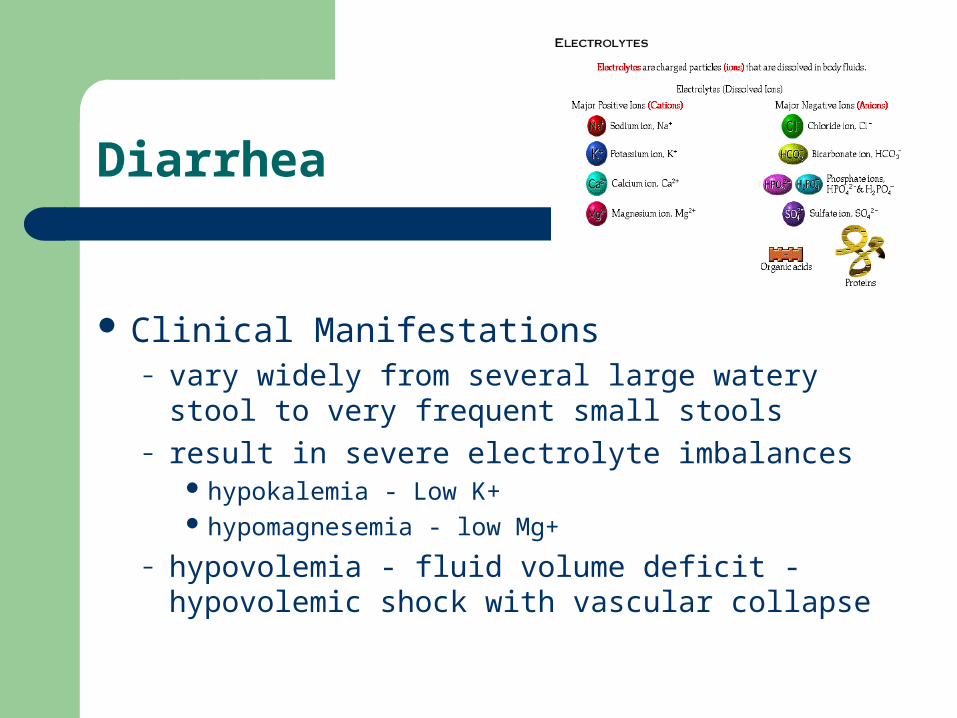
Clinical Manifestations– vary widely from several large watery stool to very
frequent small stools– result in severe electrolyte imbalances
hypokalemia - Low K+ hypomagnesemia - low Mg+
– hypovolemia - fluid volume deficit - hypovolemic shock with vascular collapse
Diarrhea
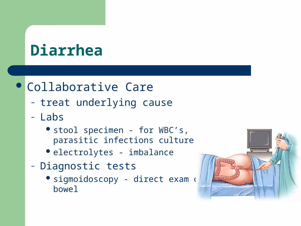
Collaborative Care– treat underlying cause– Labs
stool specimen - for WBC’s, parasitic infections culture
electrolytes - imbalance
– Diagnostic tests sigmoidoscopy - direct exam of bowel

Diarrhea
Client prep– consent, npo, enemas
Dietary management– fluid replacement - gatorade, pedialyte– bowel rest for 24 hours - add milk last
Pharmacology– absorbents, anticholinergics, antibiotics

The Client with Constipation
The infrequent or difficult passage of stool– two or less BM’s per week– affects elders - impaired health, medications,
decrease physical activity
Diagnostics– Barium enema
- tumors, diverticular disease
– colonoscopy - tumor, obstruction, take bx

Constipation
Dietary Management– high fiber - vegetable fiber– adequate fluids
Pharmacology– laxatives for short term use– bulk form agents for long term use– enemas - acute short term or as prep
Irritable Bowel Syndrome
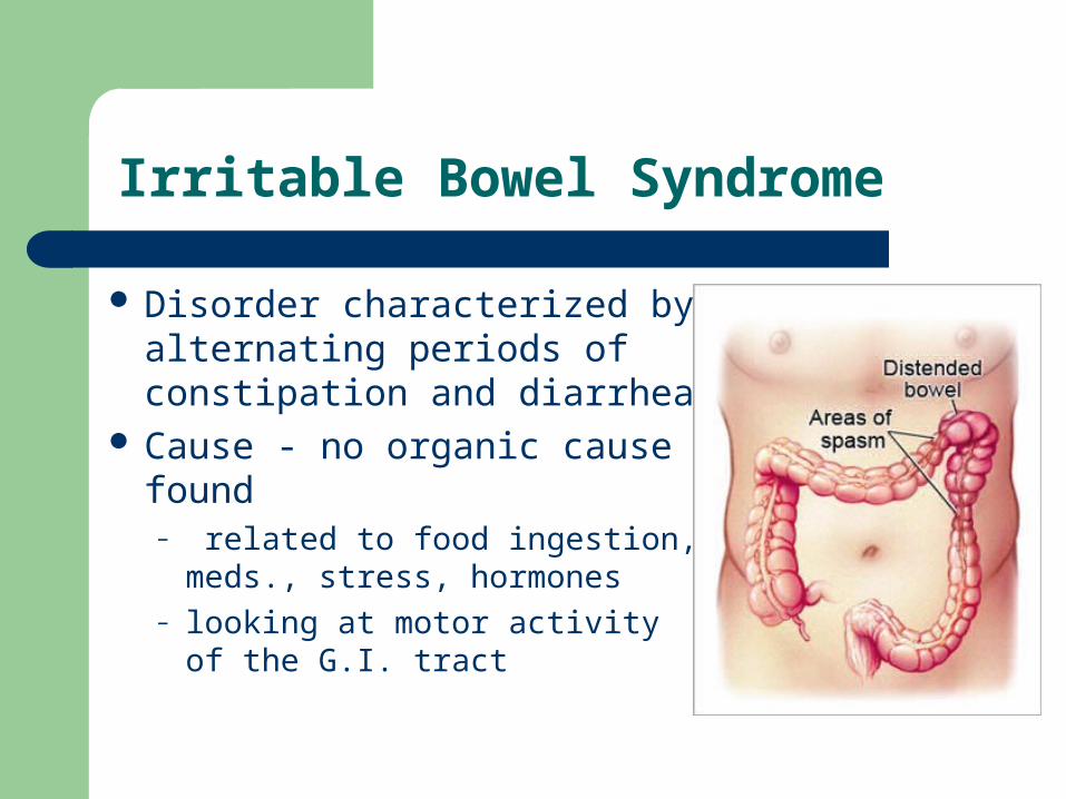
Disorder characterized by alternating periods of constipation and diarrhea
Cause - no organic cause found– related to food ingestion, meds.,
stress, hormones– looking at motor activity of the
G.I. tract
IBS
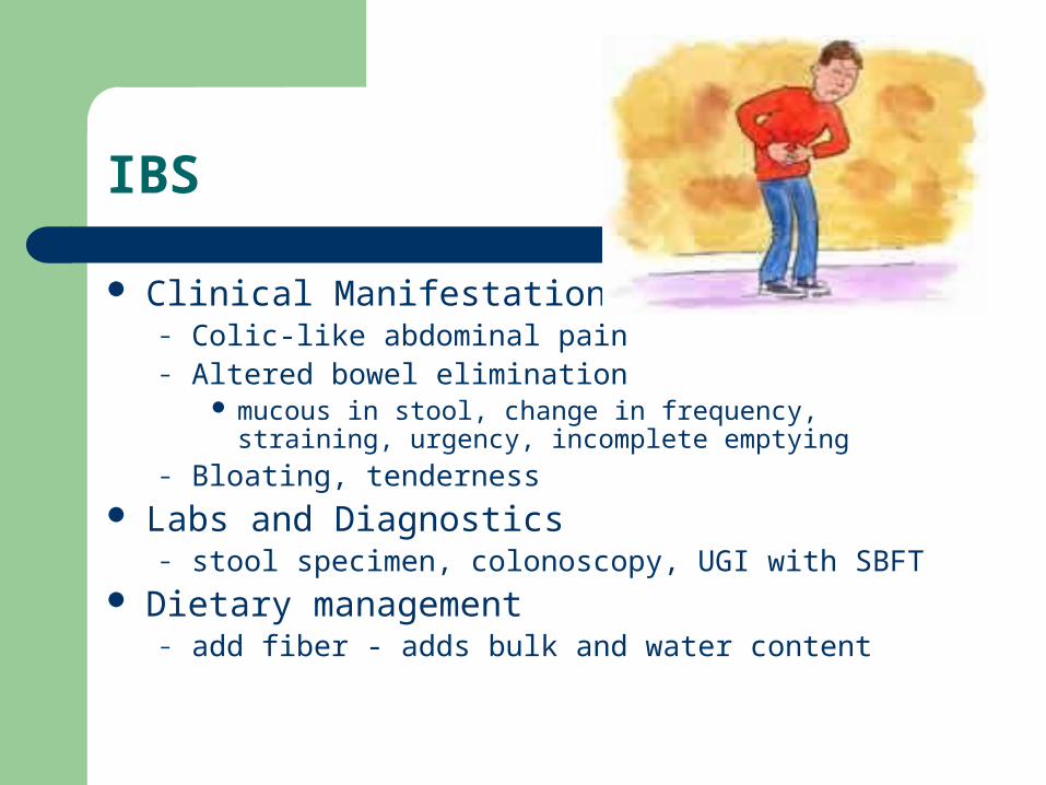
Clinical Manifestations– Colic-like abdominal pain– Altered bowel elimination
mucous in stool, change in frequency, straining, urgency, incomplete emptying
– Bloating, tenderness Labs and Diagnostics
– stool specimen, colonoscopy, UGI with SBFT Dietary management
– add fiber - adds bulk and water content
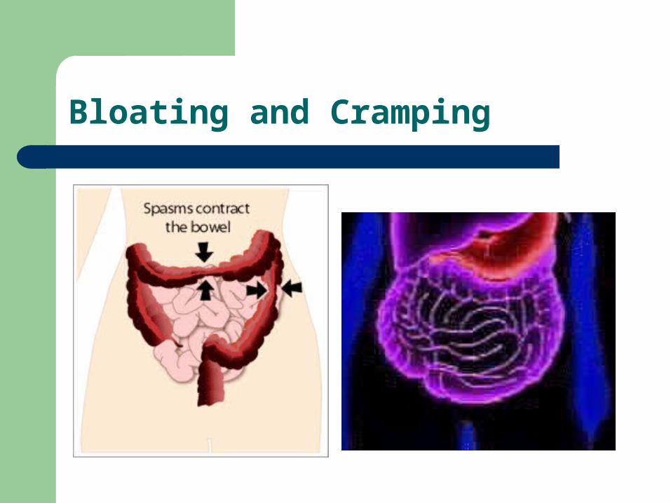
Bloating and Cramping
The Client with Fecal Incontinence
Loss of voluntary control of defecation Causes
– interfere with sensory or motor control of rectum and anal sphincters
neuro -spinal cord injury, head injury local trauma - OB tears, anal-rectal injury, surgery Other - radiation, impaction, tumors, confusion

Fecal Incontinence
Collaborative Care– dx made by history– digital exam - poor sphincter tone– treatment
bowel training program - establish regular pattern– dietary changes– stimulant - coffee, suppository, digital stimulation
surgery - colostomy
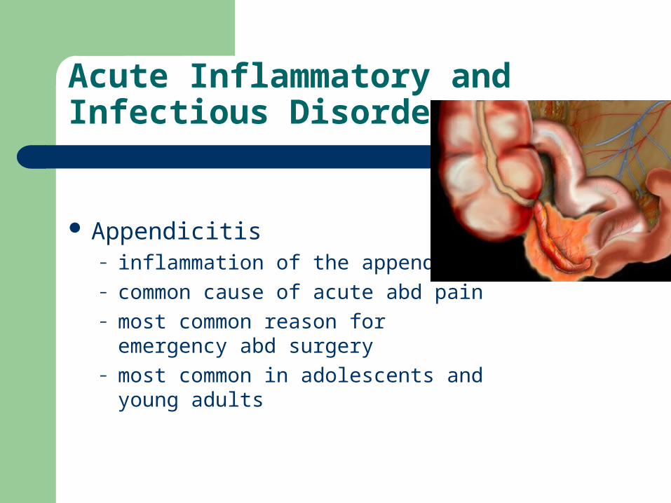
Acute Inflammatory and Infectious Disorders
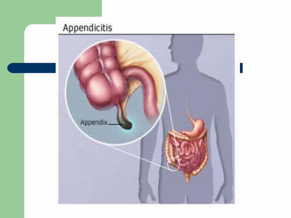
Appendicitis– inflammation of the appendix– common cause of acute abd pain– most common reason for
emergency abd surgery– most common in adolescents and
young adults
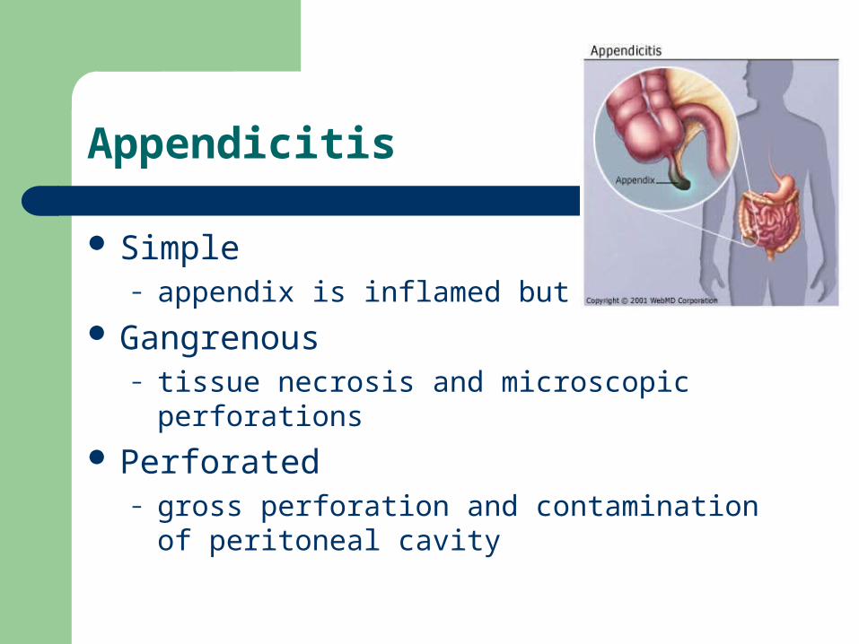
Appendicitis
Simple– appendix is inflamed but intact
Gangrenous– tissue necrosis and microscopic perforations
Perforated– gross perforation and contamination of peritoneal
cavity

Appendicitis
Clinical Manifestations
– continuous mild generalized upper abd pain– then intensifies and localizes to RLQ
rebound tenderness - tenderness on release of pressure at McBurney’s point
+ Rt heel tap pain What about pain medications?
– nausea, anorexia, vomiting, low-grade fever– perforation - increased pain, temp, abscess
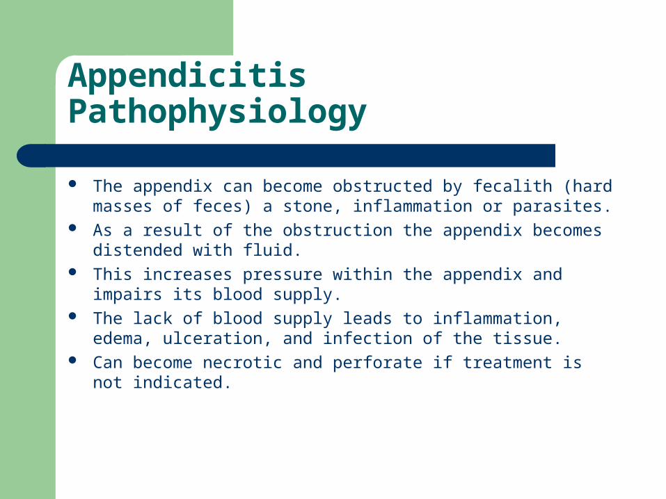
Appendicitis Pathophysiology
The appendix can become obstructed by fecalith (hard masses of feces) a stone, inflammation or parasites.
As a result of the obstruction the appendix becomes distended with fluid.
This increases pressure within the appendix and impairs its blood supply.
The lack of blood supply leads to inflammation, edema, ulceration, and infection of the tissue.
Can become necrotic and perforate if treatment is not indicated.
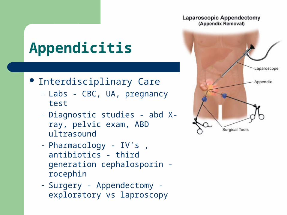
Appendicitis
Interdisciplinary Care– Labs - CBC, UA, pregnancy
test– Diagnostic studies - abd X-ray,
pelvic exam, ABD ultrasound– Pharmacology - IV’s ,
antibiotics - third generation cephalosporin - rocephin
– Surgery - Appendectomy - exploratory vs laproscopy
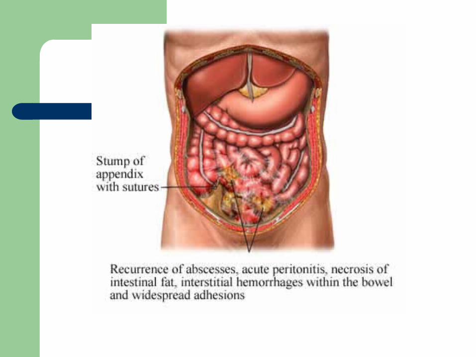

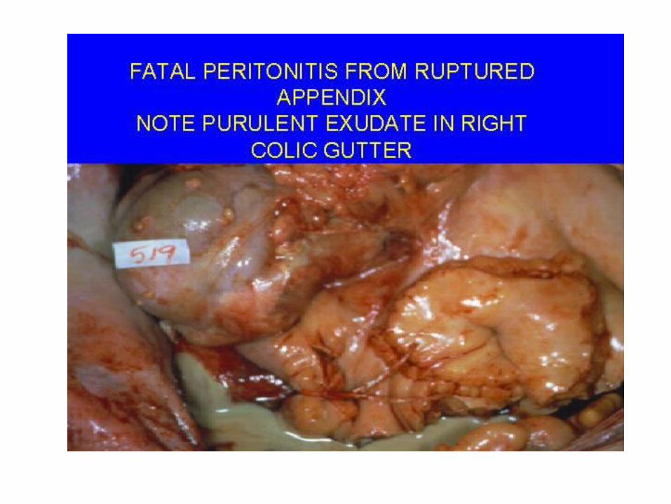
The Client with Peritonitis
Inflammation of the peritoneum - is the most significant complication of acute abdominal disorders– perforation of appendix, diverticulum, peptic ulcer,
pancreatitis or GSW– bacterial infection - E coli or klebsiella

Peritonitis
Clinical Manifestations– Abdominal Effects
Diffuse or localized pain - rebound Boardlike rigidity diminished or absent bs distention, anorexia, nausea, vomiting
– Systemic effects fever, malaise, tachycardia, restlessness shock
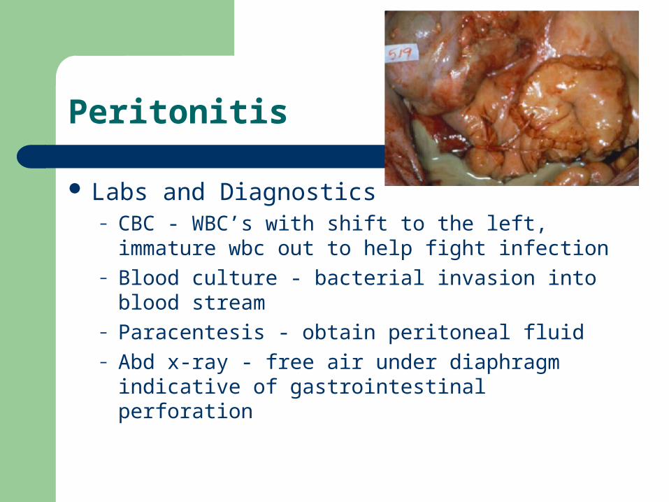
Peritonitis
Labs and Diagnostics– CBC - WBC’s with shift to the left, immature wbc
out to help fight infection– Blood culture - bacterial invasion into blood
stream– Paracentesis - obtain peritoneal fluid– Abd x-ray - free air under diaphragm indicative of
gastrointestinal perforation
Peritonitis - Interdisciplinary Care
Pharmacology– broad-spectrum antibiotics until culture report
obtained– narcotic analgesic, antipyretics
Surgery - laparotomy– peritoneal lavage
washing out cavity with copious amounts of isotonic soln drains - JP or pen rose, may be left open

Nursing Care - Peritonitis
NGT– intestinal decompression
Pain - abd distention and inflammation– assess - location, severity and type - analgesics– fowler’s - minimize stress on abd structures– alternative pain management - visualization,
medication, relaxation
Nursing Care - Peritonitis
Fluid volume deficit– I & O, vs, wt., assess for dehydration
Altered protection– monitor for sign of infection, handwashing, aseptic
technique for drsg changes
Anxiety– potential threat to life
The Client with Viral or Bacterial Infection
Gastroenteritis– describes general GI inflammation– syndrome - diarrhea, vomiting, anorexia, nausea
and pain– organisms - Staphlococcal, Salmonella,Shigella,
Botulism - life threatening, – Cholera - third world countries– dx - stool culture, tx - antibiotics, rehydration
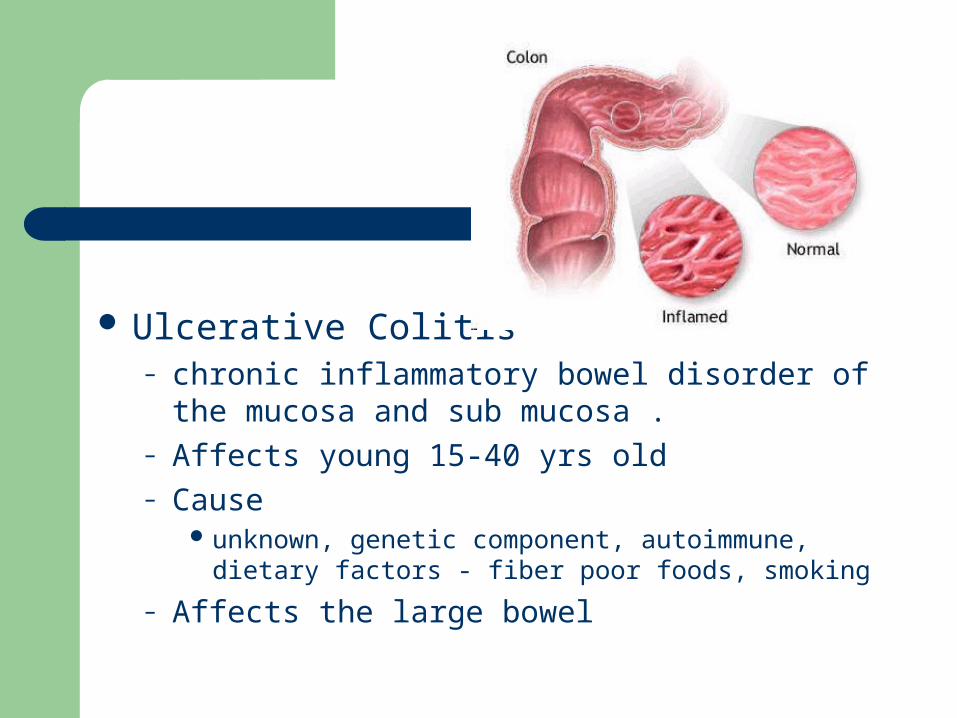
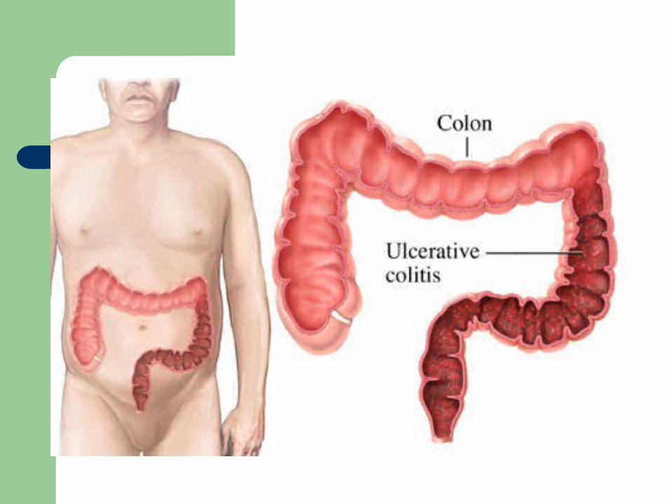
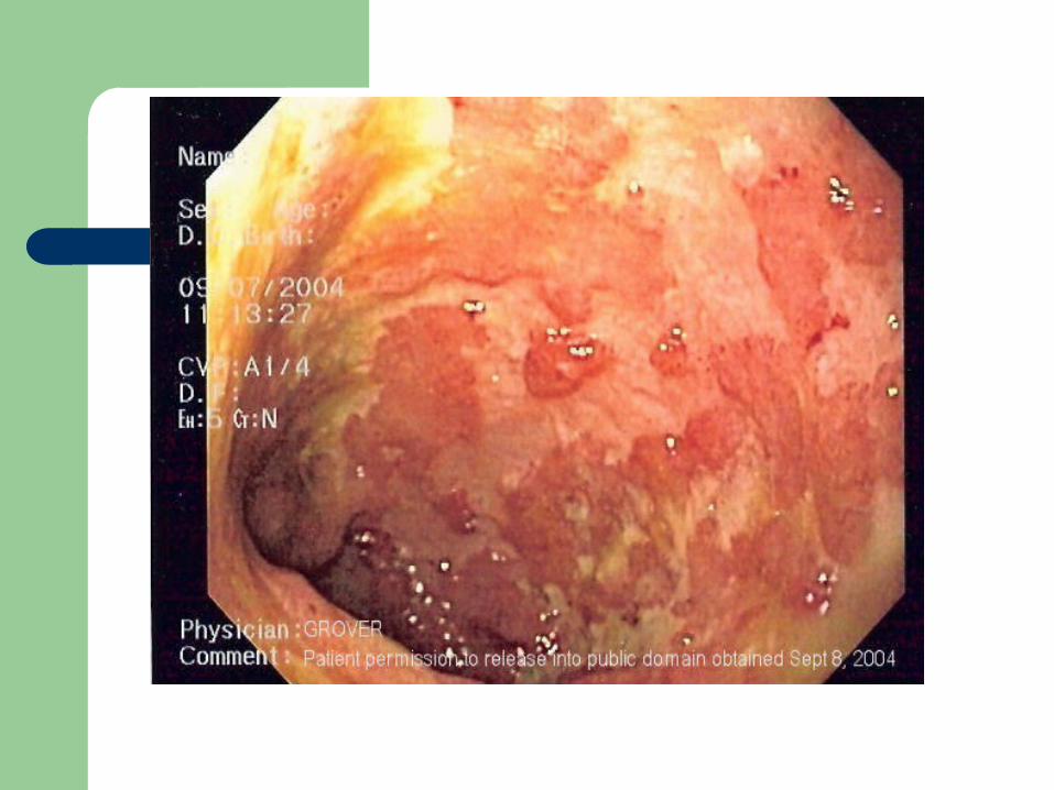
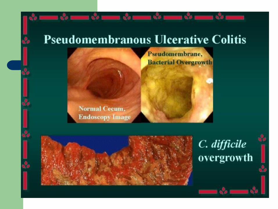
Ulcerative Colitis– chronic inflammatory bowel disorder of the
mucosa and sub mucosa .– Affects young 15-40 yrs old– Cause
unknown, genetic component, autoimmune, dietary factors - fiber poor foods, smoking
– Affects the large bowel
Ulcerative Colitis
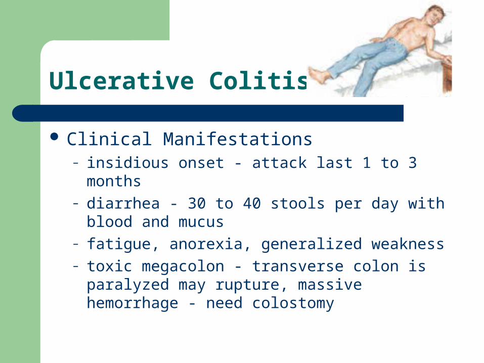
Clinical Manifestations– insidious onset - attack last 1 to 3 months– diarrhea - 30 to 40 stools per day with blood and
mucus– fatigue, anorexia, generalized weakness– toxic megacolon - transverse colon is paralyzed
may rupture, massive hemorrhage - need colostomy

Ulcerative Colitis
Interdisciplinary Care– supportive treatment– Dx - by sigmoidoscopy, edema, inflammation,
mucus and pus– Pharmacology
Azulfidine - sulfonamide antibiotic, acts topically on colonic mucosa to inhibit inflammatory process
– Dietary - npo with TPN, then low residue

Ulcerative Colitis
Surgery– not initial treatment– ileostomy
Nursing Care– relieving abd cramping– emotional support– teaching about illness and special needs– Nsg dx. - diarrhea and body image disturbance
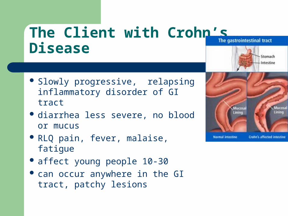
The Client with Crohn’s Disease
Slowly progressive, relapsing inflammatory disorder of GI tract
diarrhea less severe, no blood or mucus
RLQ pain, fever, malaise, fatigue affect young people 10-30 can occur anywhere in the GI
tract, patchy lesions

Crohn’s Disease
Interdisciplinary Care
– therapy is directed toward managing the symptoms and controlling the disease process
Labs and Diagnostics– Stool specimen– X-ray - UGI with SBFT - shows ulcerations, strictures and
fistulas– colonosocpy - bx

Crohn’s - Interdisciplinary Care
Pharmacology– same as ulcerative colitis - anti inflammatory– antidiarrheal - no risk of mega colon
Dietary – NPO - TPN, eliminate milk
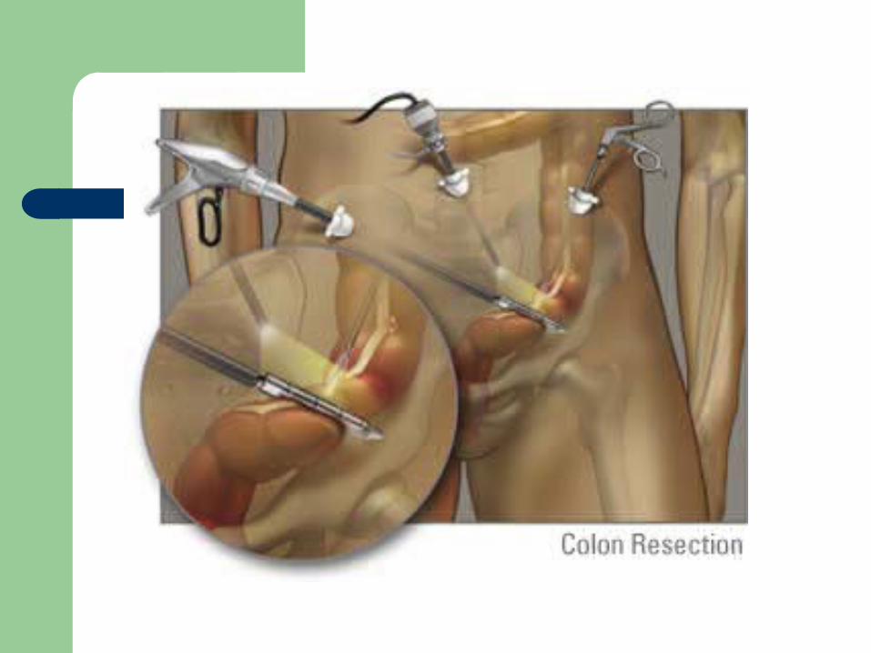
Surgery– 2nd to complications, bowel obstruction - bowel
resection


Malabsorption Syndromes
A condition in which nutrients, carbohydrates, protein, fats, water, electrolytes, minerals, and vitamins are ineffectively absorbed by the intestional mucosa– mostly disease of small intestine– surgery of small intestine
Malabsorption Syndrome
Clinical manifestations– anorexia, abd bloating, diarrhea, weight loss,
weakness, malaise, muscle cramps, anemia signs of malnutrition
Celiac Disease– hypersensitivity to gluten, protein found in cereal– Tx - gluten free diet

Malabsorption Syndrome
Lactose Intolerance– deficiency of lactase the enzymes needed for
digestion and absorbtion of lactose the primary carbohydrate in milk
– affects 90% of Asians, 75% of African Americans, high incidence among Jewish and Hispanic populations
– usually hereditary, symptoms occur in adolescence or early adulthood
Malabsorption Syndrome
Short Bowel Syndrome– from resection of significant portions of the small
intestine CA, mesenteric thrombosis with bowel infarction,
Crohn’s disease or trauma
– Treatment frequent small, high caloric and high protein meals multivitamin and mineral supplements
Neoplastic Disorders
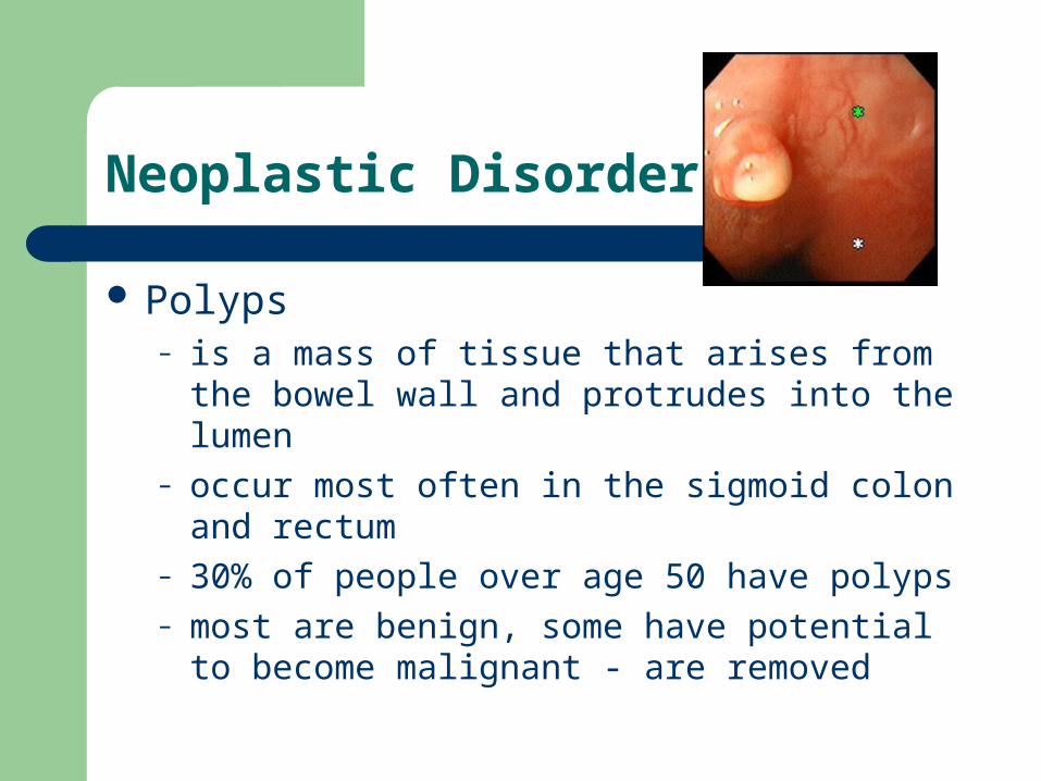
Polyps– is a mass of tissue that arises from the bowel wall
and protrudes into the lumen– occur most often in the sigmoid colon and rectum– 30% of people over age 50 have polyps– most are benign, some have potential to become
malignant - are removed
Polyps
Interdisciplinary Care– Diagnosis made by barium enema and
sigmoidoscopy or colonoscopy– Follow-up recommended because polyps tend to
recur– Consider a “silent” disease - few or no symptoms
with significant risk of CA
The Client with Colorectal Cancer
Malignant tumor arising from the epithelial tissues of the colon or rectum
2nd leading cause of cancer death in Western countries
long term survival rate is only 35% occurs more in males than females occurs after age 50
Colorectal Cancer
Risk Factors– over age 50– polyps in colon or rectum– cancer elsewhere in the body– family history– ulcerative colitis or crohn’s disease– radiation, immunodeficiency disease– dietary - high fat, high caloric, low Ca+ and fiber
Colorectal Cancer
Clinical Manifestations– no symptoms until it becomes advanced– slow growth pattern - 5-10yrs. for symptoms to
develop– bleeding– change in bowel habit - diarrhea or constipation– pain, anorexia, weight loss - advance disease
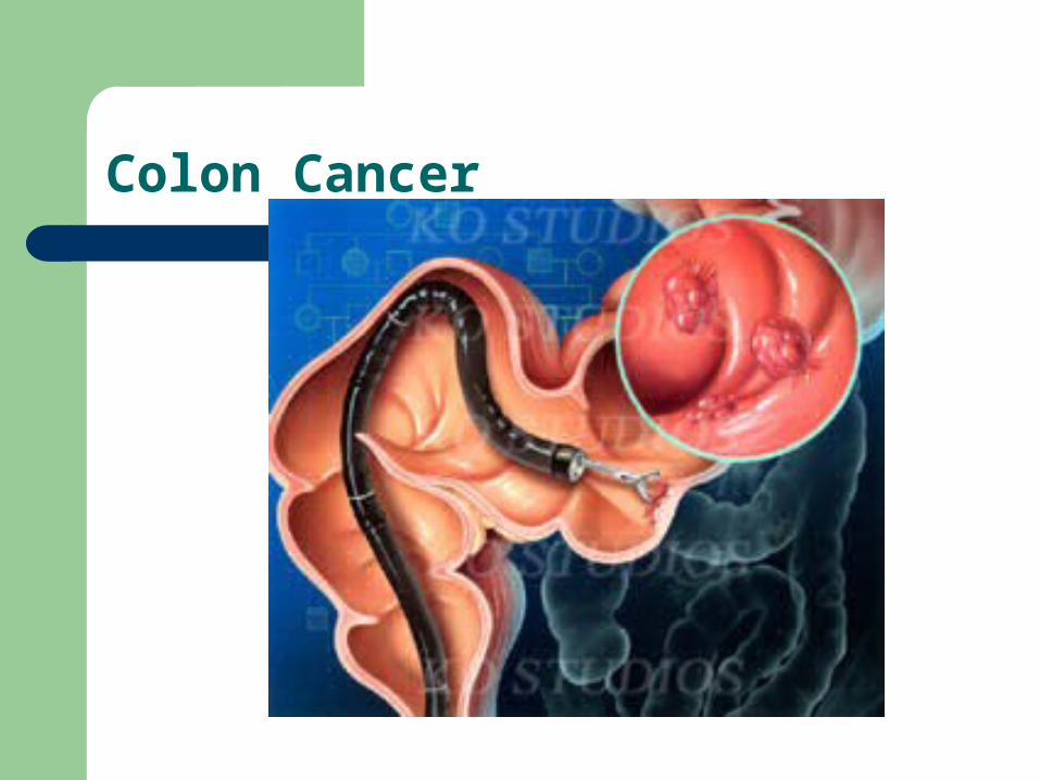
Colon Cancer

Colorectal Cancer
Interdisciplinary Care
– establish dx - colonoscopy
– surgical intervention
– adjuncts of chemotherapy and radiation
Colorectal Cancer
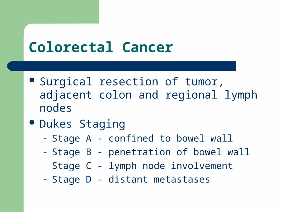
Surgical resection of tumor, adjacent colon and regional lymph nodes
Dukes Staging– Stage A - confined to bowel wall– Stage B - penetration of bowel wall– Stage C - lymph node involvement– Stage D - distant metastases

Permanent for tumors of rectum or sigmoid colon
Hartmann pouch – temporary– the distal portion of the colon is left in place and
sewn shut
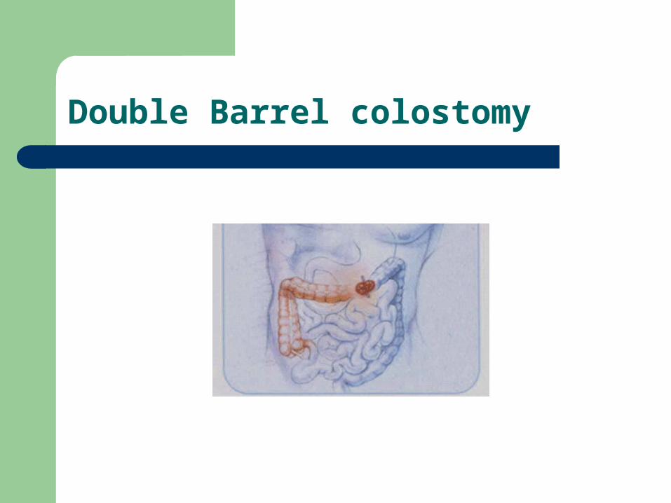
Double Barrel colostomy
Nursing Care of the Client Having Bowel Surgery
Pre-operative– consent– assess level of understanding– bowel prep
oral and parental antibiotics cathartics and enema to reduce risk of bowel
contamination
Nursing Care of the Client Having Bowel Surgery
Post-operative Nursing Care– Routine post-op care
vital signs, turn, cough, deep breath q2hrs I & O - NGT drainage, surgical drains assess for post-op hemorrhage
– Assess for bowel sounds and distention– Provide pain relief– Assess resp. status - teach to splint
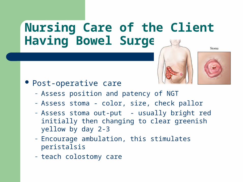
Nursing Care of the Client Having Bowel Surgery
Post-operative care– Assess position and patency of NGT– Assess stoma - color, size, check pallor– Assess stoma out-put - usually bright red initially
then changing to clear greenish yellow by day 2-3– Encourage ambulation, this stimulates peristalsis– teach colostomy care
Nursing Care of Clients Having Bowel Surgery
Effects of ostomy on Body Image– adjust to loss of body organ and dx of cancer– show acceptance of client and ostomy– concerned over the affect of cancer– develop a trusting relationship– listen actively– ostomy, cancer support groups, social services

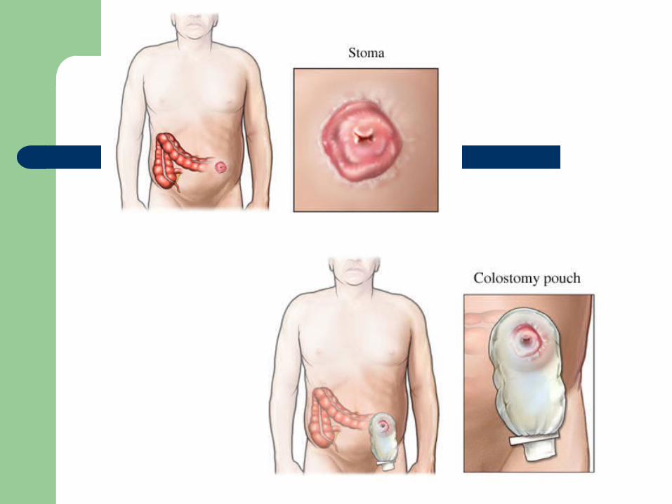
Colostomy Surgery

Case Study - Colorectal Cancer
W.C., 65yr old male, retired railroad employee, husband and father of 3 grown children. Has 3 month history of small amount of blood and mucus in stool. Has a sensation of rectal pressure and has notice his stool has changed in diameter, now is pencil thin.
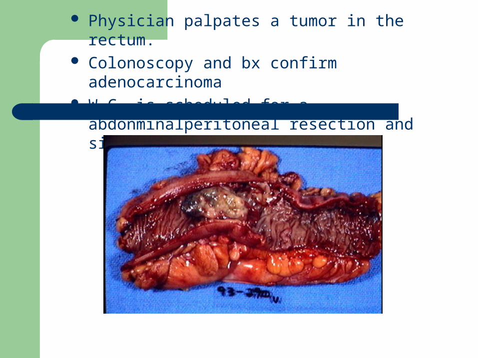
Physician palpates a tumor in the rectum. Colonoscopy and bx confirm adenocarcinoma W.C. is scheduled for a abdonminalperitoneal
resection and sigmoid colostomy

His wife has many questions and asks, why does the colostomy have to be permanent?
Why does he need erythromycin and neomycin tablets?
She then asks, is he going to be ok?

Physician Orders
Explain the rationale behind these orders– Insert NGT and connect to low intermintent
suction– Insert foley catheter– Routine post-op v.s., OOB tonight– See PCA order sheet (M.S. 1mg q 10min, up to
10mg every 4 hours)– NPO
Nursing Interventions
Explain the rationale behind these interventions– establishing a therapeutic relationship– assessing patency and position of the NGT– assessing respiratory status– assessing b.s. – assess stoma and stoma output– teaching to splinting the incision
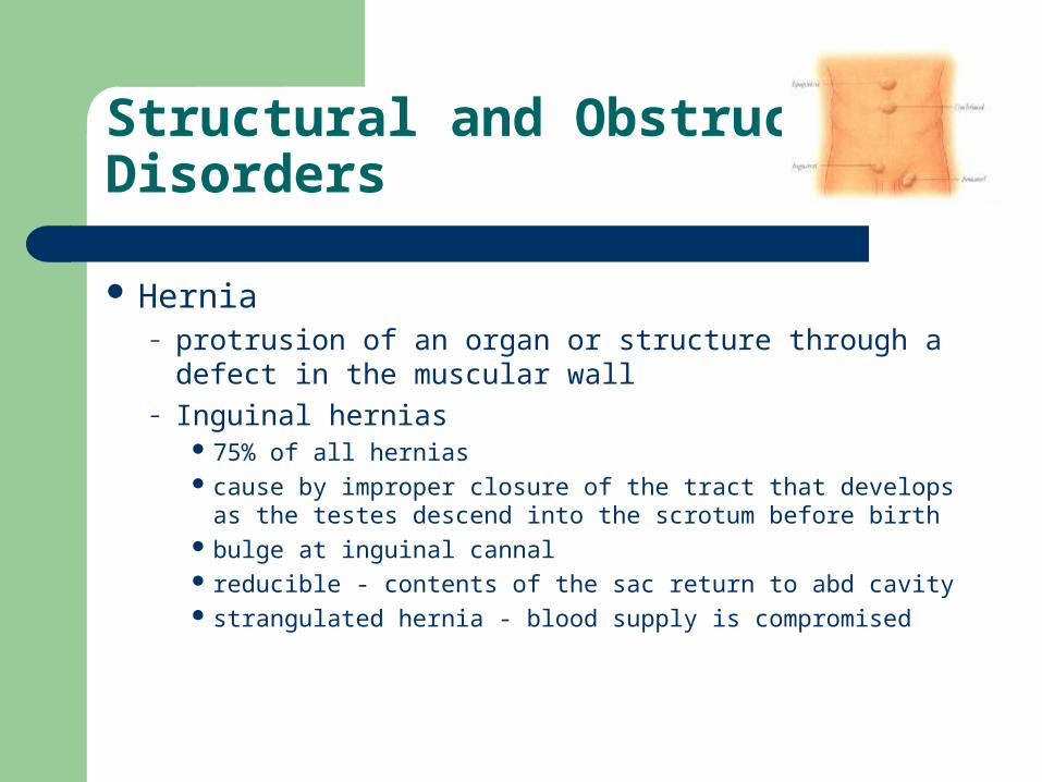
Structural and Obstructive Disorders
Hernia– protrusion of an organ or structure through a defect
in the muscular wall– Inguinal hernias
75% of all hernias cause by improper closure of the tract that develops as
the testes descend into the scrotum before birth bulge at inguinal cannal reducible - contents of the sac return to abd cavity strangulated hernia - blood supply is compromised

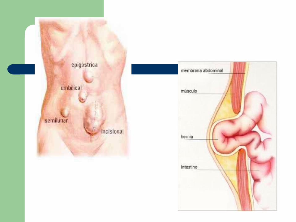
Structural and Obstructive Disorders
Umbilical hernias– occur more frequently in women– obesity, mult. pregnancies, prolonged labor– tend to enlarge steadily– strangulation is common
Incisional or Ventral hernias– occur at previous surgical incision
The Client with an Intestinal Obstruction
Occurs when intestinal contents fail to be propelled through the lumen of the bowel
Small intestine obstruction– ileum of small intestine most common site– Mechanical Obstruction
physical barrier, tumor or scar tissue
– Functional Obstruction - paralytic ileus peristalsis fails
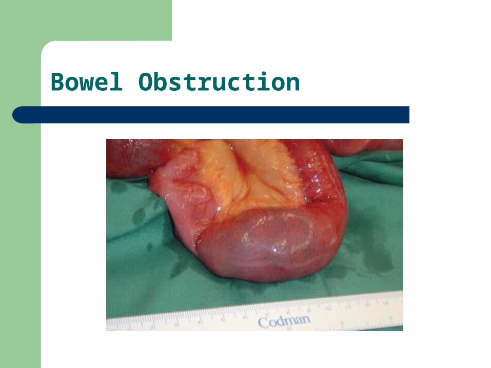
Bowel Obstruction
The Client with an Intestinal Obstruction
Clinical Manifestations– cramping, colicky abdominal pain, can be
intermittent or increase in intensity– vomiting – high-pitched tinkling bowel sounds - reflects the
bowels attempt to propel contents past the obstruction
– later stages - absent bowel sounds– electrolyte imbalance - hypovolemia - shock
The Client with an Intestinal Obstruction
Large Bowel Obstruction– usually occurs in sigmoid colon– cancer most common cause– Clinical Manifestations
abdominal pain and constipation abdomen is distended and tender to palpation
Treatment for Bowel Obstructions– NGT - functional surgery - mechanical
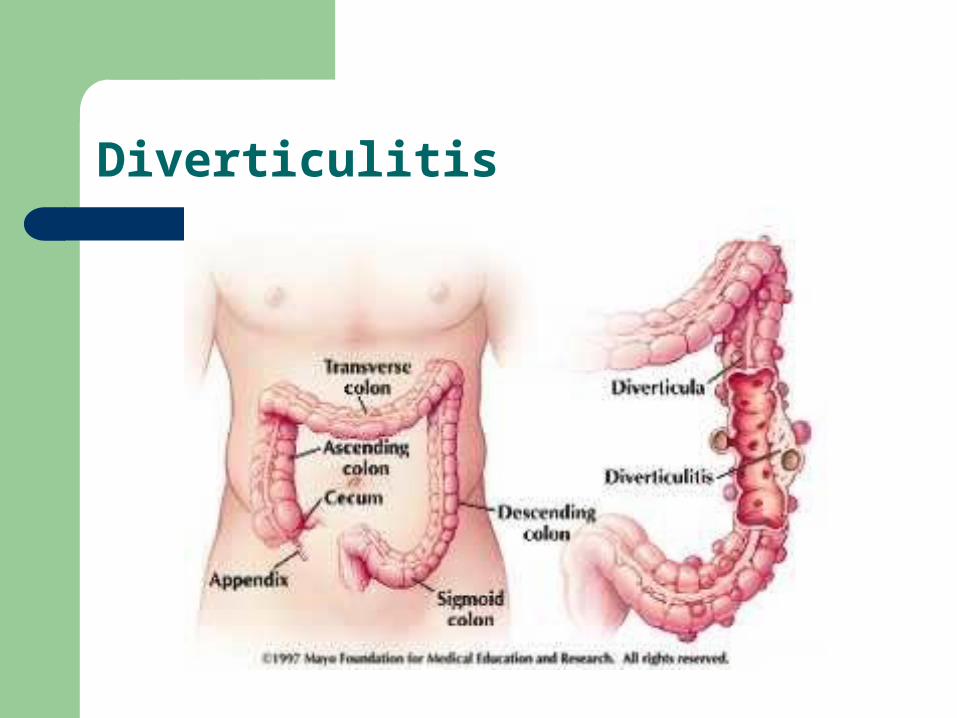
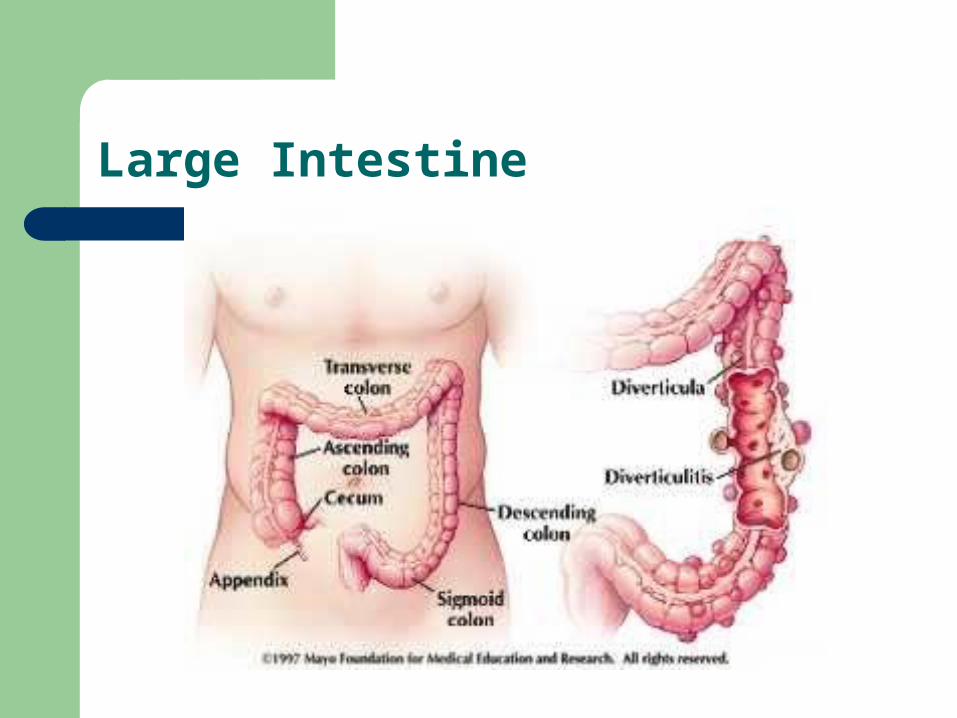
Diverticulitis
The Client with Diverticular Disease
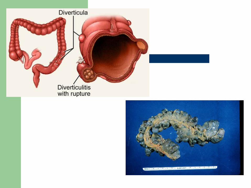
Diverticula– acquired saclike projections of mucosa through
the muscular layer of the colon– 90-95% occur in the sigmoid colon– increased incidence in US, Australia, United
Kingdom and France– related to cultural factors - diet high in refined
foods and low in fiber
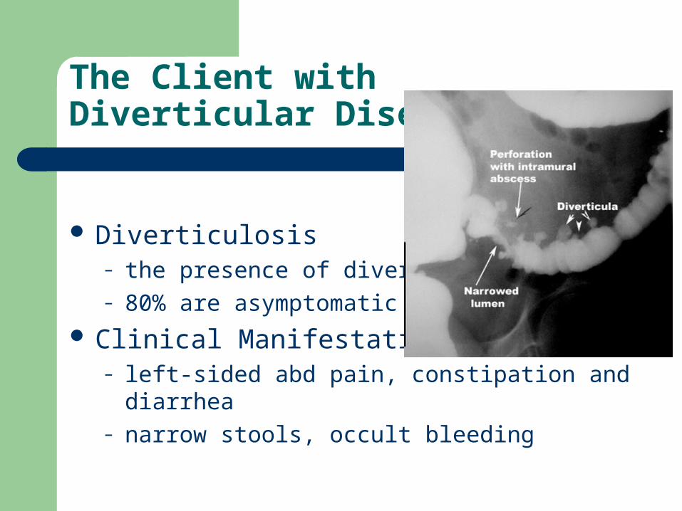
The Client with Diverticular Disease
Diverticulosis– the presence of diverticula– 80% are asymptomatic
Clinical Manifestations– left-sided abd pain, constipation and diarrhea– narrow stools, occult bleeding
The Client with Diverticular Disease
Diverticulitis– inflammation and microscopic perforation of
diverticular mucosa– undigested food becomes trapped, blood flow is
impaired - leads to abscess or peritonitis Interdisciplinary Care
– Chronic diverticular disease - dietary changes– Acute diverticulosis - bowel rest, antibiotics,
eventually surgery
Anorectal Disorders
The Client with Hemorrhoids– hemorrhoidial veins become weak, distended,
develop varices - cause is straining, pregnancy also increases intra-abdominal pressure
– internal or external bleeding, bright red, unmixed with stool pain associated with thrombosed or ulcerated
The Client with Hemorrhoids
Interdisciplinary Care– conservative therapy - diet, increase fiber, fluids,
bulk forming laxative, Preparation H– surgery
sclerotherapy - inject chemical irritant to induce inflammation - fibrosis - scarring
rubber band ligation - rubber band placed snugly around - necrosis - slough
cryosurgery - necrosed by freezing with probe
The Client with Hemorrhoids
Nursing Care - post-op– inspect rectal dressing for bleeding– pain management - position of comfort - side lying– ice pack over rectal drsg– sitz bath tid and prn bowel movement– meds - analgesics, stool softeners
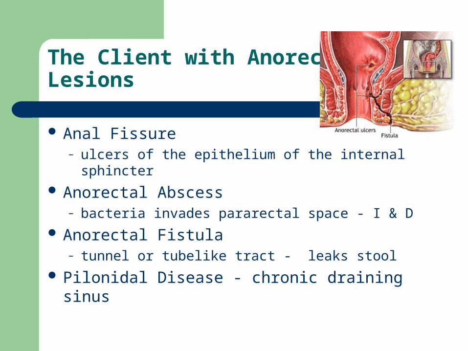
The Client with Anorectal Lesions
Anal Fissure– ulcers of the epithelium of the internal sphincter
Anorectal Abscess– bacteria invades pararectal space - I & D
Anorectal Fistula– tunnel or tubelike tract - leaks stool
Pilonidal Disease - chronic draining sinus