assessing and reducing the toxicity of 3d-printed …groverlab.org/assets/3d-tox.pdf · assessing...
TRANSCRIPT

Assessing and Reducing the Toxicity of 3D-Printed PartsShirin Mesbah Oskui,† Graciel Diamante,‡ Chunyang Liao,‡ Wei Shi,§ Jay Gan,‡ Daniel Schlenk,‡
and William H. Grover*,†
†Department of Bioengineering, University of California, Riverside, Riverside, California 92507, United States‡Department of Environmental Sciences, University of California, Riverside, Riverside, California 92507, United States§State Key Laboratory of Pollution Control and Resource Reuse, School of the Environment, Nanjing University, Nanjing, Jiangsu,China
*S Supporting Information
ABSTRACT: 3D printing is gaining popularity by providing a toolfor fast, cost-effective, and highly customizable fabrication. However,little is known about the toxicity of 3D-printed objects. In this work,we assess the toxicity of printed parts from two main classes ofcommercial 3D printers, fused deposition modeling and stereo-lithography. We assessed the toxicity of these 3D-printed parts usingzebrafish (Danio rerio), a widely used model organism in aquatictoxicology. Zebrafish embryos were exposed to 3D-printed partsand monitored for rates of survival, hatching, and developmentalabnormalities. We found that parts from both types of printers weremeasurably toxic to zebrafish embryos, with STL-printed partssignificantly more toxic than FDM-printed parts. We also developed a simple post-printing treatment (exposure to ultravioletlight) that largely mitigates the toxicity of the STL-printed parts. Our results call attention to the need for strategies for the safedisposal of 3D-printed parts and printer waste materials.
■ INTRODUCTION
Even though additive manufacturing or “3D printing” was firstintroduced in 1983,1 the technology has become widespreadonly in the past few years. The value of the 3D printing marketgrew from $288 million in 2012 to $2.5 billion in 2013 and isprojected to grow to $16.2 billion by 2018.2 Much of thisgrowth has occurred in the life sciences, where 3D printing hasfound applications in dentistry,3,4 prosthetics and implantabledevices,5,6 surgical instruments,7 and even tissue and organreplacement.8 By providing businesses, researchers, physicians,and hobbyists with custom objects and tools quickly andinexpensively, 3D printers are revolutionizing manufacturing,accelerating research, and changing how medicine is practiced.In spite of the growing popularity of 3D printers, relatively
little is known about the toxicity of 3D-printed parts. Previouswork has found that 3D-printed parts can be toxic to cancercells9 and may cause allergic or inflammatory responses5,10 andinfections11 in patients. Additionally, some 3D printers releasepotentially hazardous particles into the air during operation.12
However, the whole-organism health effects of exposure to 3D-printed parts remain largely unexplored. As 3D-printed partsfind increasing use in the medical and life science fields, theeffects of exposure to these parts need to be understood.Additionally, as consumer-grade 3D printers become morewidespread, the amount of 3D-printed parts and printer wastebeing released into the environment will also grow, and thetoxicity of these materials in the environment remains largelyunexplored.
With little known about the toxicity of 3D-printed parts,there are consequently few techniques for reducing the toxicityof these parts. Researchers have found that heating a 3D-printed part can reduce its toxicity to cancer cells, but heatingalso adversely affects the appearance of the part.9 Treating 3D-printed parts with supercritical carbon dioxide can reduce theinflammation caused when the parts are implanted in thebody,5 but this technique requires a specialized instrument thatis more expensive than many 3D printers. There is an unmetneed for simple and accessible techniques for reducing thetoxicity of 3D-printed parts in research, healthcare, andcommercial applications.In this work, we assessed the effects of 3D-printed parts on
an organism’s health and developed a simple technique forreducing the toxicity of these printed parts. We chose zebrafish(Danio rerio) as the model organism for this study. Zebrafishare widely used vertebrate model organisms that, because oftheir ability to reproduce quickly and in large numbers, makehigh-throughput screening of potential toxicants feasible andaffordable.13 There are many genetic similarities betweenhumans and zebrafish, and the relatively fast development ofsophisticated cardiovascular, nervous, and endocrine systems inthese animals makes them a very popular developmental
Received: September 11, 2015Revised: October 21, 2015Accepted: October 23, 2015Published: November 4, 2015
Letter
pubs.acs.org/journal/estlcu
© 2015 American Chemical Society 1 DOI: 10.1021/acs.estlett.5b00249Environ. Sci. Technol. Lett. 2016, 3, 1−6

model.14 As aquatic organisms, zebrafish are also a relevantmodel for understanding the bioavailability and bioaccumula-tion of chemical and biological toxicants15 and overallenvironmental toxicity. Finally, zebrafish are optically trans-parent throughout their development (embryonic and adultstage) and can be analyzed using imaging techniques to identifydeveloping pathologies and phenotypic changes in real time.
■ METHODS3D Printers. We studied the toxicity of printed parts from
the two main commercially available types of 3D printers, fuseddeposition modeling (FDM) and stereolithography (STL)printers. FDM printers feed a polymer filament into a heatednozzle that melts the polymer and deposits it layer by layeronto the growing part.16 In this study, we used the DimensionElite printer (Stratasys, Eden Prairie, MN) (Figure 1A), whichprints parts out of acrylonitrile butadiene styrene (ABS).
In contrast, STL printers use a light source to polymerize abath of photocurable liquid resin layer by layer to form afinished part.1 Because the chemical compositions of thephotocurable resins are typically not provided by printermanufacturers, little is known about the chemical and biologicalcompatibility of STL-printed parts. In this study, we used theForm 1+ printer (Figure 1B); this printer uses a 405 nm Class 1
laser to cure a resin that is a combination of methacrylatedoligomers and monomers and photoinitiators.17
3D-Printed Test Parts and Cleaning Procedures.Cylindrical test parts (40 mm diameter and 4 mm thick,shown in Figure 1C) were designed using SolidWorks(Dassault Systemes, Velizy-Villacoublay, France), exported asan .STL file, and printed using the FDM and STL printers. The3D-printed parts used in toxicity tests in Figures 2 and 3 werecleaned according to the printer manufacturers’ specifications.FDM-printed parts were submerged in a 2% (w/v) sodiumhydroxide solution for 4 h to dissolve the temporary polylacticacid supports, then rinsed with ultrapure water, and air-dried.STL-printed parts were washed in two consecutive baths ofisopropyl alcohol for 5 min each and then air-dried.
Figure 1. Commercial 3D printers and test pieces. (A) A commercialfused deposition modeling (FDM) printer (Dimension Elite printerfrom Stratasys), which deposits melted acrylonitrile butadiene styrene(ABS) layer by layer onto a stage to build a 3D-printed part. (B) Acommercial stereolithography (STL) printer (Form 1+ printer fromFormlabs, Cambridge, MA), which uses a light source to polymerize aliquid resin to form a printed part. (C) Examples of the FDM- andSTL-printed test parts used in this study (40 mm diameter and 4 mmheight). Also shown is an STL-printed part that was treated withultraviolet light (STL w UV) to reduce its toxicity. The UV treatmenthas little effect on the appearance of the printed part.
Figure 2. Survival and hatching rates of exposed zebrafish embryoscompared to control unexposed embryos. (A) Survival rates ofzebrafish embryos exposed to 3D-printed parts from a FDM printer(green), embryos exposed to parts from a STL printer (blue), embryosexposed to STL-printed parts that were treated with ultraviolet light(red), and control embryos that were not exposed to printed parts(black). Each exposure represents three replicates with 30 embryos ineach replicate. Embryos exposed to STL-printed parts had significantlylower survival rates by day 3 post-fertilization when compared to thoseof control embryos (p ≤ 0.05), with no STL-exposed embryo survivingpast day 7. However, embryos exposed to FDM- and UV-treated STL-printed parts did not have significantly decreased survival ratescompared to those of control embryos (p ≥ 0.05). (B) Hatching ratesfor the same four exposure types as in panel A. Embryos exposed toSTL-printed parts had significantly lower hatching rates by day 4 post-fertilization compared to those of control embryos (p = 0); virtuallynone of the STL-exposed embryos hatched. However, embryosexposed to FDM- and UV-treated STL-printed parts did not havesignificantly lower hatching rates in the embryos (p ≥ 0.05). Theseresults show that after STL-printed parts had been treated with UVlight, embryos exposed to the treated parts fare almost as well ascontrol embryos that were not exposed to printed parts.
Environmental Science & Technology Letters Letter
DOI: 10.1021/acs.estlett.5b00249Environ. Sci. Technol. Lett. 2016, 3, 1−6
2
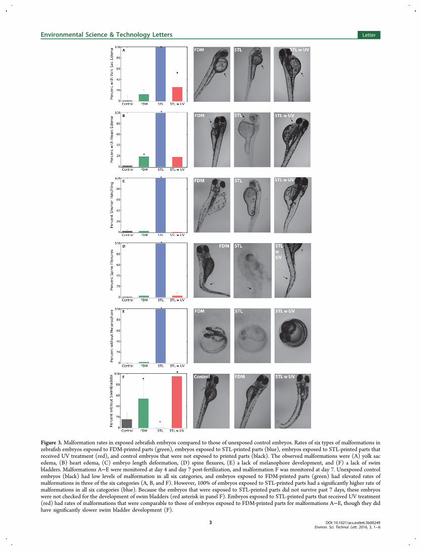
Figure 3. Malformation rates in exposed zebrafish embryos compared to those of unexposed control embryos. Rates of six types of malformations inzebrafish embryos exposed to FDM-printed parts (green), embryos exposed to STL-printed parts (blue), embryos exposed to STL-printed parts thatreceived UV treatment (red), and control embryos that were not exposed to printed parts (black). The observed malformations were (A) yolk sacedema, (B) heart edema, (C) embryo length deformation, (D) spine flexures, (E) a lack of melanophore development, and (F) a lack of swimbladders. Malformations A−E were monitored at day 4 and day 7 post-fertilization, and malformation F was monitored at day 7. Unexposed controlembryos (black) had low levels of malformation in all six categories, and embryos exposed to FDM-printed parts (green) had elevated rates ofmalformations in three of the six categories (A, B, and F). However, 100% of embryos exposed to STL-printed parts had a significantly higher rate ofmalformations in all six categories (blue). Because the embryos that were exposed to STL-printed parts did not survive past 7 days, these embryoswere not checked for the development of swim bladders (red asterisk in panel F). Embryos exposed to STL-printed parts that received UV treatment(red) had rates of malformations that were comparable to those of embryos exposed to FDM-printed parts for malformations A−E, though they didhave significantly slower swim bladder development (F).
Environmental Science & Technology Letters Letter
DOI: 10.1021/acs.estlett.5b00249Environ. Sci. Technol. Lett. 2016, 3, 1−6
3

To determine the effects of different part cleaning techniqueson the toxicity of the printed parts, additional 3D-printed partswere cleaned using alternative cleaning procedures with little orno effect on the toxicity results of the printed parts (SupportingInformation).UV Light Exposure of STL-Printed Parts. Exposure to
ultraviolet light was used to detoxify some STL-printed parts inthis study. An Intelli-Ray 400 UV light source (UvitronInternational, Inc., West Springfield, MA) with a peakirradiance of 100−120 mW/cm2 was used. Each STL-printedpart was exposed to UV light at 50% lamp power for anexposure time on each side of 30 min, for a total of 1 h ofexposure time per part.Animal Husbandry and Exposure to 3D-Printed Parts.
We assessed the toxicity of 3D-printed parts using zebrafish (D.rerio) following a specific protocol approved by the Universityof California, Riverside’s Animal Care and Use Committee(approval number 20130005). The zebrafish were wild-type ABstrain and approximately 16 months old at the time ofspawning. The fish cultures were kept in aerated aged tap water(dechlorinated) at 27 °C with a 14 h/10 h light/dark cycle.Males and females were kept separately and fed twice a day onArtemia sp. until the night before spawning, when they weretransferred to breeding aquaria. Eggs were collected the nextmorning, examined, and separated on the basis of the stage ofdevelopment. All embryos were directly exposed to theirrespective 3D-printed parts at 2 h postfertilization. Each printedpart was placed in a large sterile Petri dish (90 mm in diameterand 15 mm in height) and surrounded with approximately 45mL of ultrapure water (resistivity of 18.2 MΩ cm at 25 °C).Each printed part was exposed to 30 embryos and replicated
three times, for a total of 90 embryos used to study theeffectiveness of each cleaning technique for both printingmethods. The embryos were monitored for their survival,hatching rate, and developmental abnormalities (reducedlength, yolk sac edema, heart edema, spinal flexure, absenceof swim bladder, and discoloration) at days 4 and 7 post-fertilization by visual inspection. Dead embryos were identifiedby the loss of translucency and removed from the dish beforefurther inspection of the health of the remaining embryos.Statistical Analysis and Data Visualization. The
significance of the results was tested using the WilcoxonRank Sum nonparametric test with appropriate assumptions onR programming language. The p values were set to 0.05 to testfor the significance of treatments. The results were visualizedusing the Matplotlib package in the Python programminglanguage.
■ RESULTS AND DISCUSSION
Assessing the Toxicity of 3D-Printed Parts. Figure 2Ashows the percent survival of embryos exposed to 3D-printedparts from FDM (green) and STL (blue) printers compared tothat of unexposed control embryos (black) through 7 dayspostfertilization. While the embryos exposed to FDM-printedparts had slightly decreased average survival rates compared tothose of control embryos, the embryos exposed to STL-printedparts had significantly decreased survival rates (p ≤ 0.05), withmore than half of the embryos dead by day 3 and all dead byday 7. The percent of exposed embryos that hatched followed asimilar trend (Figure 2B): embryos exposed to FDM-printedparts had hatching rates slightly lower than those of unexposedembryos, but embryos exposed to STL-printed parts had
significantly decreased (p ≤ 0.05) hatching rates (essentiallyzero hatching).We also used six deformities as markers to assess the health
of embryos after they hatched. We monitored hatchlings foryolk sac edema (Figure 3A), heart edema (Figure 3B), reducedhatchling length (Figure 3C), the presence of spine flexures(Figure 3D), and a lack of melanophores (Figure 3E)throughout the monitoring period of 7 days, and the lack ofa swim bladder (Figure 3F) at day 7 postfertilization. Thezebrafish micrographs in Figure 3 show the most severe cases ofdeformity in each category, for embryos exposed to parts fromeach of the 3D printer types. Of the few zebrafish embryos thathatched after exposure to STL-printed parts, 100% of thehatchlings had all six malformations (blue in Figure 3). Incontrast, zebrafish embryos exposed to FDM-printed parts hadsignificantly lower rates of malformations, although FDM-exposed embryos still exhibited deformities at a rate higher thanthat of unexposed control embryos (especially for yolk sacedema) and a statistically significant increase (p ≤ 0.05) inheart edema (green in Figure 3). Embryos exposed to FDM-printed parts also exhibited significantly delayed swim bladderdevelopment (p ≤ 0.05) compared to that of the controlembryos.
Reducing the Toxicity of STL-Printed Parts. While theexact chemical compositions of the resins used in STL printersare usually trade secrets, the resins’ Material Safety Data Sheetsindicate that they usually contain acrylate and/or methacrylatemonomers:
Specific members of these classes of compounds are alreadyknown to be toxic in some situations. For example, acrylatemonomers can be acutely toxic if they are inhaled, areswallowed, or come into contact with skin.18 If the R group is ahydrogen, the resulting compounds (acrylic acid andmethacrylic acid) have been shown to have toxic effects onembryonic and fetal development in rat fetuses.19 If the Rgroup in the methacryate monomer is a methyl group, theresulting compound (methyl methacrylate) and its polymerizedform [poly(methyl methacrylate) or PMMA] have beenassociated with irreversible cardiovascular failure when theyare used as scaffolds.19 Finally, exposure to methacrylatemonomers with a variety of other R groups (ethyl, n-butyl,isobutyl, and isodecyl) has been observed to cause cytotoxicity,cardiovascular failure, gastrointestinal problems, respirationissues, and developmental malformations.19 In summary,while we do not know the exact composition of STL printerresins, ample evidence of the toxicity of the monomers in theseresins exists.On the basis of the known toxicity of acrylate and
methacrylate monomers, we hypothesized that monomers orshort-chain polymers may be leaching out of the STL-printedparts and contributing to the extreme toxicity of those parts. Totest this hypothesis, we performed gas chromatography−massspectrometry (GC−MS) analysis of water samples left incontact with STL-printed parts. The results suggest that at leastthree different chemical species are present in the leachate;these species have different retention times in GC but very
Environmental Science & Technology Letters Letter
DOI: 10.1021/acs.estlett.5b00249Environ. Sci. Technol. Lett. 2016, 3, 1−6
4
similar fragments in MS (Supporting Information). Thissupports our hypothesis that monomers or short-chainpolymers are present in the leachate from STL-printed parts,although additional analysis is necessary for a definitiveidentification.If monomers or short-chain polymers are indeed leaching out
of STL-printed parts, additional photoinduced polymerizationof the 3D-printed part might reduce the amount of thesespecies leaching out of the printed part and thus reduce thetoxicity of the part. To test this hypothesis, we exposed STL-printed parts to ultraviolet light (wavelength of 350−400 nm,peak irradiance of 100−120 mW/cm2) for 30 min on each sideof the printed part. As shown in Figure 1C, this UV exposuretreatment has a minimal effect on the appearance of the 3D-printed part. Embryos exposed to STL-printed parts that wereUV-treated fared much better than embryos exposed tountreated parts. As shown in panels A and B of Figure 2(red), the survival and hatching rates of embryos exposed totreated parts recovered to almost control levels. Embryosexposed to UV-treated STL-printed parts also showed asignificantly lower incidence of spine flexures (Figure 3D,red). All hatchlings exposed to UV-treated parts were normal inlength (Figure 3C, red) and developed normal levels ofmelanophores (Figure 3E, red). However, embryos exposed toUV-treated parts still had significantly elevated rates of yolk sacedema (p ≤ 0.05) and heart edema compared to those ofcontrol embryos (Figure 3A,B, red) and most of the embryosexposed to UV-treated parts had not developed swim bladdersby the end of day 7 (Figure 3F, red). Therefore, while our UVtreatment appears to significantly reduce the toxicity of STL-printed parts to zebrafish, it does not completely eliminate thetoxicity of these parts, and additional research intodetoxification strategies is merited.Our findings have important consequences in several
different communities. Physicians and nurses using 3D-printedparts in clinical applications need to consider the consequencesof patient exposure to these parts; researchers using 3D-printedparts in life science experiments should be on the lookout forartifacts caused by exposures of organisms to these objects, andwaste collection agencies should develop strategies for the safecollection and disposal of parts and waste materials generatedby 3D printers. The cost of 3D printers has droppeddramaticallyFDM printers are currently available for as littleas $200, and the STL printer used in this study can be boughtfor $3299and this trend is expected to continue in thecoming years. Consequently, 3D printers are spreading beyondindustry and research laboratories and into homes and smallbusinesses. The individuals using these printers may not havethe training necessary to use these printers safely and dispose oftheir wastes responsibly, and municipal waste disposal agenciesmay not have resources for collecting and treating 3D printerwaste. This situation is particularly worrisome for STL printers,which can generate liters of solvent waste contaminated withresin monomers during post-printing part cleanup. Thepotential for 3D printer toxic waste to enter waterways isalarming and deserves additional study.
■ ASSOCIATED CONTENT
*S Supporting InformationThe Supporting Information is available free of charge on theACS Publications website at DOI: 10.1021/acs.estlett.5b00249.
Results from testing the toxicity of 3D-printed partssubjected to alternative postprint cleaning techniquesand GC−MS analysis of leachates from STL-printedparts (PDF)
■ AUTHOR INFORMATION
Corresponding Author*E-mail: [email protected]. Phone: +1 (951) 827-4311.Fax: +1 (951) 827-6416.
NotesThe authors declare no competing financial interest.
■ ACKNOWLEDGMENTS
This work was supported in part by the National ScienceFoundation’s Instrument Development for Biological ResearchProgram via Grant DBI-1353974.
■ REFERENCES(1) Hull, C. W. Apparatus for production of three-dimensionalobjects by stereolithography. U.S. Patent 4,575,330, 1986.(2) Canalys Inc. 3D printing market to grow to 16.2 billion USD in2018, 2014.(3) Boyd, R. L.; Miller, R.; Vlaskalic, V. The Invisalign system in adultorthodontics: mild crowding and space closure cases. Journal of ClinicalOrthodontics 2000, 34, 203−212.(4) van Noort, R. The future of dental devices is digital. Dent. Mater.2012, 28, 3−12.(5) Popov, V.; Evseev, A.; Ivanov, A.; Roginski, V.; Volozhin, A.;Howdle, S. Laser stereolithography and supercritical fluid processingfor custom-designed implant fabrication. J. Mater. Sci.: Mater. Med.2004, 15, 123−128.(6) Matsuda, T.; Mizutani, M. Liquid acrylate-endcapped biodegrad-able poly (ε-caprolactone-co-trimethylene carbonate). II. Computer-aided stereolithographic microarchitectural surface photoconstructs. J.Biomed. Mater. Res. 2002, 62, 395−403.(7) Wong, J. Y.; Pfahnl, A. C. 3D Printing of Surgical Instruments forLong-Duration Space Missions. Aviat., Space Environ. Med. 2014, 85,758−763.(8) Murphy, K.; Dorfman, S.; Law, R. J.; Le, V. A. Devices, systems,and methods for the fabrication of tissue utilizing UV cross-linking.U.S. Patent Application 13/794,368, 2013.(9) Inoue, Y.; Ikuta, K. Detoxification of the Photocurable Polymerby Heat Treatment for Microstereolithography. Procedia CIRP 2013,5, 115−118.(10) Cassens, B. J. Inspections, Compliance, Enforcement, andCriminal Investigations, Align Technology Inc., 2010 (http://www.fda.gov/ICECI/EnforcementActions/WarningLetters/ucm234578.htm).(11) D’Urso, P. S.; Effeney, D. J.; Earwaker, W. J.; Barker, T. M.;Redmond, M. J.; Thompson, R. G.; Tomlinson, F. H. Customcranioplasty using stereolithography and acrylic. Br. J. Plast. Surg. 2000,53, 200−204.(12) Stephens, B.; Azimi, P.; El Orch, Z.; Ramos, T. Ultrafine particleemissions from desktop 3D printers. Atmos. Environ. 2013, 79, 334−339.(13) Kari, G.; Rodeck, U.; Dicker, A. P. Zebrafish: an emergingmodel system for human disease and drug discovery. Clin. Pharmacol.Ther. 2007, 82, 70−80.(14) Lieschke, G. J.; Currie, P. D. Animal models of human disease:zebrafish swim into view. Nat. Rev. Genet. 2007, 8, 353−367.(15) Carvan, M. J.; Dalton, T. P.; Stuart, G. W.; Nebert, D. W.Transgenic zebrafish as sentinels for aquatic pollution. Ann. N. Y. Acad.Sci. 2000, 919, 133−147.(16) Crump, S. S. Modeling apparatus for three-dimensional objects.U.S. Patent 5,340,433, 1994.
Environmental Science & Technology Letters Letter
DOI: 10.1021/acs.estlett.5b00249Environ. Sci. Technol. Lett. 2016, 3, 1−6
5

(17) Formlabs Inc. Materials Safety Data Sheet for Clear Photo-reactive Resin for Form 1+. 2014.(18) Yoshii, E. Cytotoxic effects of acrylates and methacrylates:relationships of monomer structures and cytotoxicity. J. Biomed. Mater.Res. 1997, 37, 517−524.(19) Autian, J. Structure-toxicity relationships of acrylic monomers.Environ. Health Perspect. 1975, 11, 141.
Environmental Science & Technology Letters Letter
DOI: 10.1021/acs.estlett.5b00249Environ. Sci. Technol. Lett. 2016, 3, 1−6
6