ase guideline update - school of medicine · ase guideline update jin kyung kim, md, phd, ... •...
TRANSCRIPT

ASE Guideline Update
Jin Kyung Kim, MD, PhD, FACC Medical Director Cardiovascular Center, Diagnostic Testing Associate Professor of Medicine UC Irvine School of Medicine October 30, 2016

2
•Guidelines released in 2016
–Guidelines for the Use of Echocardiography in the Evaluation of a Cardiac Source of Embolism
–Recommendations for the Evaluation of Left Ventricular Diastolic Function by Echocardiography: An Update
Introduction
Division of Cardiology | October 30, 2016

3
Guidelines for Use of Echo in the Evaluation of a Cardiac Source of Embolism
Division of Cardiology | October 30, 2016

4
“Echocardiography should be the primary form of cardiac imaging”
Evaluation of suspected cardiac source of embolism
Division of Cardiology | October 30, 2016

5
Appropriate Use Criteria for Echocardiography in Evaluation of Cardiac Sources of Emboli
Appropriate Use: Transthoracic Echocardiography Symptoms or conditions potentially related to suspected cardiac etiology
• Suspected cardiac mass • Suspected cardiovascular source of embolus • Initial evaluation of suspected infective endocarditis
(IE) with positive blood culture results or new murmur • Reevaluation of IE at high risk for progression,
complication, or with a change in clinical status • Known acute pulmonary embolism (PE) to guide
therapy • Reevaluation of known PE after thrombolysis or
thrombectomy for assessment of change in right ventricular (RV) function and/or pulmonary artery pressure
Division of Cardiology | October 30, 2016

6
Appropriate Use Criteria for Echocardiography in Evaluation of Cardiac Sources of Emboli
Inappropriate Use: TTE • Transient fever without evidence of bacteremia
or new murmur • Transient bacteremia with a pathogen not
typically associated with IE and/or a documented nonendovascular source of infection
• Routine surveillance of uncomplicated IE when no change in management is contemplated
• Suspected PE to establish diagnosis • Routine surveillance of prior PE with normal RV
function and pulmonary artery systolic pressure
Division of Cardiology | October 30, 2016

7
Appropriate Use: TEE
• As initial or supplemental test for evaluation for cardiovascular source of embolus with no identified noncardiac source
• As initial or supplemental test for to diagnose IE with a moderate or high pretest probability
• initial test for evaluation to facilitate clinical decision making with regard to anticoagulation, cardioversion, and/or radiofrequency ablation
Appropriate Use Criteria for Echocardiography in Evaluation of Cardiac Sources of Emboli
Division of Cardiology | October 30, 2016
8
Inappropriate Use: TEE
• Evaluation for cardiovascular source of embolus with a known cardiac source in which TEE would not change management
• Routine use of TEE when diagnostic TTE is reasonably anticipated to resolve all diagnostic and management concerns
• Surveillance of prior TEE finding for interval change when no change in therapy is anticipated
• To diagnose IE with low pretest probability (e.g., transient fever, known alternative source of infection, negative blood culture results or atypical pathogen for endocarditis)
• Evaluation when a decision has been made to anticoagulate and not to perform cardioversion
Appropriate Use Criteria for Echocardiography in Evaluation of Cardiac Sources of Emboli
Division of Cardiology | October 30, 2016

9
Echocardiography Recommended • Echocardiography should be considered in all patients with
suspected cardiac sources of embolism, especially in patients for whom clinical therapeutic decisions (such as anticoagulation or cardioversion) will depend on echocardiographic findings.
Echocardiography Not Recommended • Echocardiography is not recommended in patients for whom
the results will not guide therapeutic decisions.
TTE versus TEE • TEE is not indicated when TTE findings are diagnostic for a
cardiac source of embolism.
Recommendations for Performance of Echocardiography in Patients with Potential Cardiac Source of Embolism
Division of Cardiology | October 30, 2016
10
Thromboembolism from the left atrium and LAA
Thromboembolism from the left ventricle
Valve disease
Cardiac tumors
Embolism from the thoracic aorta
Paradoxical embolism
Cardiac and aortic embolism during cardiac surgery and percutaneous interventions
Evaluation of suspected cardiac source of embolism
Division of Cardiology | October 30, 2016

11
Evaluation of suspected cardiac source of embolism:
THROMBOEMBOLISM FROM THE LEFT ATRIUM AND LAA
Division of Cardiology | October 30, 2016
12
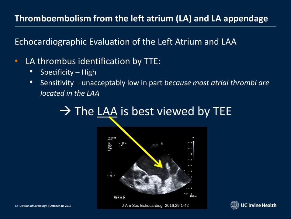
Echocardiographic Evaluation of the Left Atrium and LAA
• LA thrombus identification by TTE: • Specificity – High
• Sensitivity – unacceptably low in part because most atrial thrombi are
located in the LAA
The LAA is best viewed by TEE
Thromboembolism from the left atrium (LA) and LA appendage
Division of Cardiology | October 30, 2016 J Am Soc Echocardiogr 2016;29:1-42

13
Thromboembolism from the left atrium (LA) and LA appendage
Recommendations:
Echocardiography Recommended
• TTE is recommended in patients with suspected LA or LAA thrombus • to assess LA size and LV size and
function • to assess for underlying etiologies
of AF and additional risk factors for stroke
• TEE is superior to TTE in assessment of LAA • before cardioversion • ablation of atrial arrhythmias • percutaneous LAA closure
Division of Cardiology | October 30, 2016
J Am Soc Echocardiogr 2016;29:1-42

14
Echocardiography Potentially Useful
• Contrast echo may aid in detecting LA and LAA thrombi and may help differentiate avascular thrombi from vascular tumors.
• 3D echocardiography may provide more precise assessment of LA and LAA size and morphology.
Echocardiography Not Recommended
• Echocardiography is not recommended in patients for whom the results will not guide therapeutic decisions.
Division of Cardiology | October 30, 2016

15
Evaluation of suspected cardiac source of embolism:
THROMBOEMBOLISM FROM THE LEFT VENTRICLE
Division of Cardiology | October 30, 2016
16
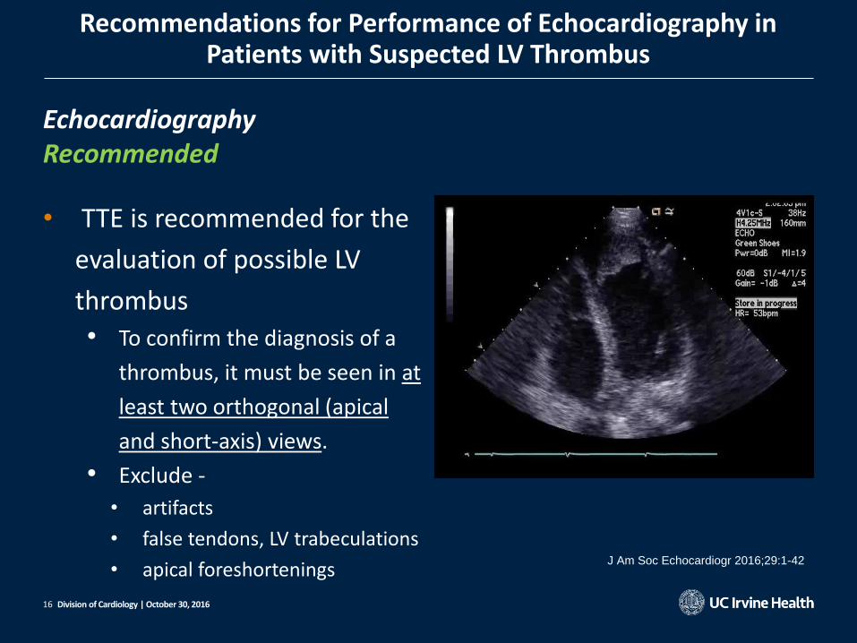
Recommendations for Performance of Echocardiography in Patients with Suspected LV Thrombus
Echocardiography Recommended
• TTE is recommended for the
evaluation of possible LV
thrombus
• To confirm the diagnosis of a
thrombus, it must be seen in at
least two orthogonal (apical
and short-axis) views.
• Exclude -
• artifacts
• false tendons, LV trabeculations
• apical foreshortenings
Division of Cardiology | October 30, 2016
J Am Soc Echocardiogr 2016;29:1-42
17
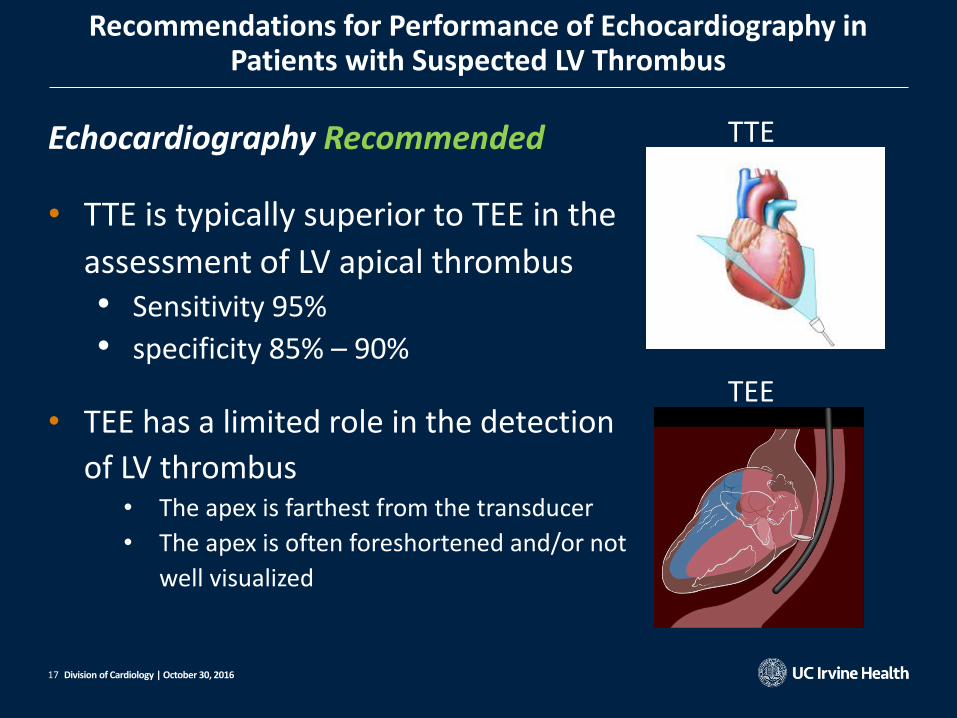
Recommendations for Performance of Echocardiography in Patients with Suspected LV Thrombus
Echocardiography Recommended
• TTE is typically superior to TEE in the
assessment of LV apical thrombus • Sensitivity 95%
• specificity 85% – 90%
• TEE has a limited role in the detection
of LV thrombus • The apex is farthest from the transducer
• The apex is often foreshortened and/or not
well visualized
Division of Cardiology | October 30, 2016
TTE
TEE

18
Recommendations for Performance of Echocardiography in Patients with Suspected LV Thrombus
Echocardiography Potentially Useful
• Contrast echocardiography may aid in detecting LV thrombi
• 3D echocardiography may provide more precise assessment of LV thrombus.
Division of Cardiology | October 30, 2016

19
Evaluation of suspected cardiac source of embolism:
VALVE DISEASE
Division of Cardiology | October 30, 2016
20
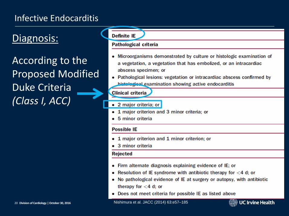
Infective Endocarditis
Diagnosis:
According to the Proposed Modified Duke Criteria (Class I, ACC)
Division of Cardiology | October 30, 2016 Nishimura et al. JACC (2014) 63:e57–185
21
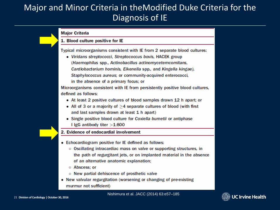
Major and Minor Criteria in theModified Duke Criteria for the Diagnosis of IE
Division of Cardiology | October 30, 2016 Nishimura et al. JACC (2014) 63:e57–185

22
Positive blood culture results + evidence of endocardial involvement constitute the definition of IE
Echocardiographic exploration for endocardial infection is mandatory
Infective Endocarditis
Division of Cardiology | October 30, 2016
Abscess Oscillating mass
on valve or on
implanted
material
New partial
dehiscence of
prosthetic valve
New valvular
regurgitation

23
Infective Endocarditis
Native valve
TTE • Specificity >90% • Sensitive 62% to 79% • Vegetations < 2 to 3 mm in size
may be missed
TEE • Sensitivity and specificity > 90%
Prosthetic valve
TTE
• Sensitivity ~ 20% to 40
TEE
• Sensitivity >80% to 90%
Division of Cardiology | October 30, 2016 J Am Soc Echocardiogr 2016;29:1-42

24
Infective Endocarditis
abscess diagnosis
TTE
• Sensitivity 28%
• Specificity 98%
TEE
• Sensitivity 87%
• Specificity 95%
Division of Cardiology | October 30, 2016
J Am Soc Echocardiogr 2016;29:1-42
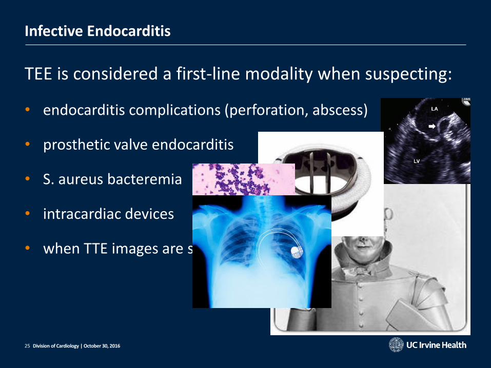
25
TEE is considered a first-line modality when suspecting:
• endocarditis complications (perforation, abscess)
• prosthetic valve endocarditis
• S. aureus bacteremia
• intracardiac devices
• when TTE images are suboptimal
Infective Endocarditis
Division of Cardiology | October 30, 2016
26
• In the setting of intermediate or high clinical suspicion for endocarditis, negative results on TTE should always be followed by TEE.
• Repeat TEE at an interval of ~ 7 days is reasonable if the clinical suspicion of IE remains high even after negative results on initial TEE
Infective Endocarditis
Division of Cardiology | October 30, 2016

27
Echocardiography Recommended
TTE is recommended for the following:
• Initial evaluation of suspected endocarditis • positive blood culture results
• new murmur
• Reevaluation for complication with a change in clinical status.
• Evaluation of hemodynamic consequences • regurgitation, shunt/fistulas, chamber enlargement, and function
• Repeat TTE at the end of antimicrobial therapy to serve as a baseline for future comparisons
.
Infective Endocarditis
Division of Cardiology | October 30, 2016

28
Echocardiography Recommended
TEE is recommended for the following:
• To diagnose IE and its complications when clinical suspicion is intermediate or high, regardless of negative results on TTE.
• As the first-line modality when complications of IE are suspected • Abscesses
• fistulas
• valve perforation
• when prosthetic valve endocarditis is suspected
Infective Endocarditis
Division of Cardiology | October 30, 2016

29
Echocardiography Not Recommended
Transient fever without bacteremia or a new murmur.
Transient bacteremia with a nontypical organism and/or documented nonintravascular infection source.
Routine surveillance of uncomplicated IE when imaging is not expected to change management.
Infective Endocarditis
Division of Cardiology | October 30, 2016
30 Division of Cardiology | October 30, 2016
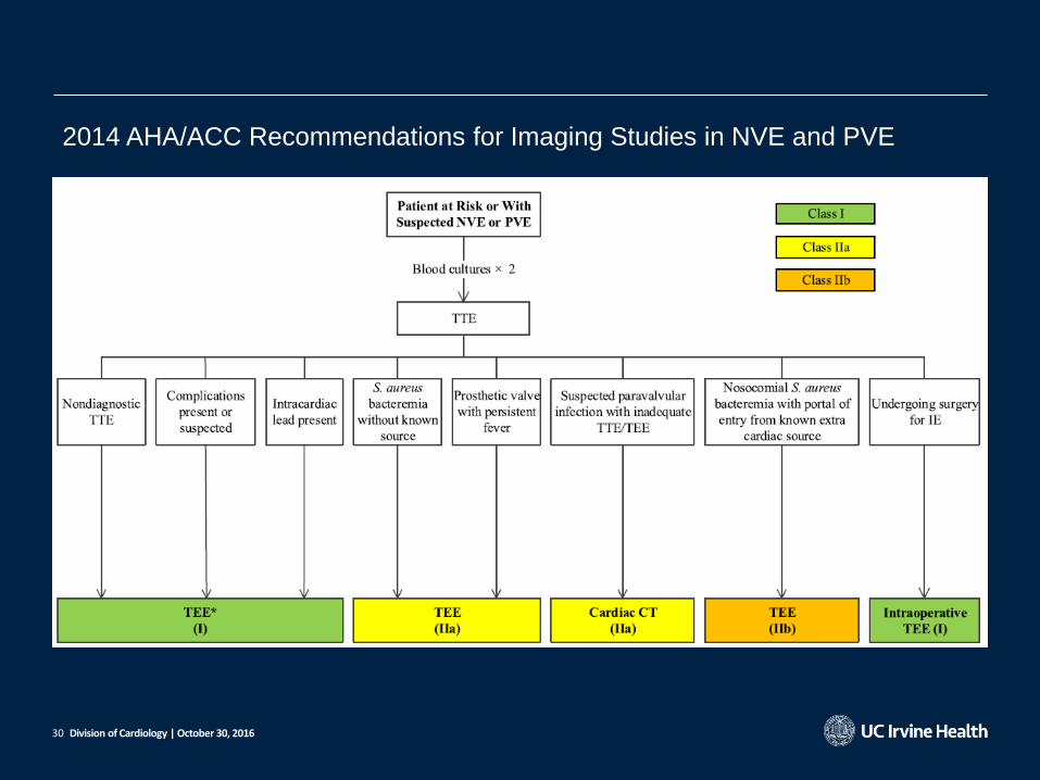
2014 AHA/ACC Recommendations for Imaging Studies in NVE and PVE

31
Nonbacterial Thrombotic Endocarditis
Verrucous Endocarditis or Libman-Sacks Endocarditis
• Composed of granular material containing immune complexes, hematoxylin bodies, and platelet thrombi, without bacteria.
• Found in up to 43% of patients with lupus
• Usually asymptomatic and not associated with valvular destruction
• Can be complicated by IE and systemic embolization.
• Affect typically the free edges of the mitral leaflets
Division of Cardiology | October 30, 2016
J Am Soc Echocardiogr 2016;29:1-42

32
Nonbacterial Thrombotic Endocarditis
Marantic Endocarditis or NBTE
• Commonly refers to noninfectious thrombotic endocarditis a/w malignancy
• Composed of platelets and fibrin
• Significant valvular dysfunction rare
• Affect the atrial side of the MV and ventricular side of the AV
• Up to 50% of patients with NBTE may incur systemic embolic events
Division of Cardiology | October 30, 2016
J Am Soc Echocardiogr 2016;29:1-42

33
Recommendations for Performance of Echocardiography in Patients with Suspected Noninfective Endocarditis
Echocardiography Recommended
• TTE surveillance in patients with • primary antiphospholipid syndrome
• SLE with secondary antiphospholipid syndrome
Nonbacterial Thrombotic Endocarditis
Division of Cardiology | October 30, 2016

34
Echocardiography Not Recommended
• Routine echocardiography is not recommended for patients with lupus in the absence of clinical signs such as fever, embolic phenomena, and new murmurs.
Nonbacterial Thrombotic Endocarditis
Division of Cardiology | October 30, 2016

35
Valvular Strands and Lambl’s Excrescences
• Echocardiographical Definition: – filiform structures, with
undulating motion – width < 2 mm – 3 - 10 mm, localized to the
line of leaflet closure
• No robust evidence that valvular strands cause systemic embolism
Division of Cardiology | October 30, 2016
J Am Soc Echocardiogr 2016;29:1-42

36
Prosthetic Valve Thrombosis
• Difficult to distinguish thrombus from vegetation echocardiographically
Patient’s clinical background and associated imaging findings become critical
• Echocardiographer must follow a systematic method of analysis and reporting • Begin with a careful echocardiographic
description of findings • End with a differential diagnosis in the
report and a most likely diagnosis
Division of Cardiology | October 30, 2016
J Am Soc Echocardiogr 2016;29:1-42

37
• The first step of the evaluation when thrombosis is suspected • Evaluate the presence and hemodynamic significance of
prosthetic dysfunction Gold standard is TTE
Prosthetic Valve Thrombosis
Division of Cardiology | October 30, 2016

38
Prosthetic Valve Thrombosis
• difficult to distinguish thrombosis from pannus formation (chronic fibrous tissue ingrowth)
• Mixed pannus-thrombus pathology is not uncommon
Division of Cardiology | October 30, 2016
J Am Soc Echocardiogr 2016;29:1-42

39
Prosthetic Valve Thrombosis
• TEE is superior in detecting
• thrombus • prosthetic leaflet motion
• TEE can reliably identify embolization risk and facilitate the decision between thrombolysis and redo surgery • Mobile • >5 to 10 mm in length • > 0.8 cm2 in area
Division of Cardiology | October 30, 2016
J Am Soc Echocardiogr 2016;29:1-42
40
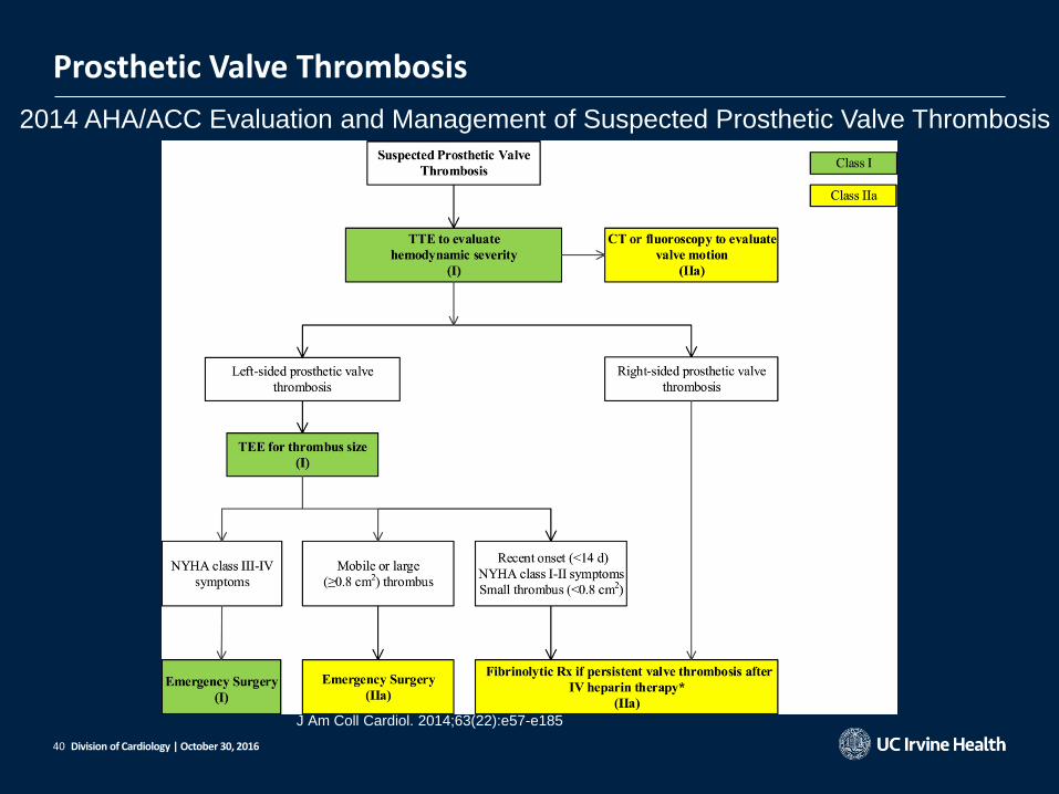
Prosthetic Valve Thrombosis
Division of Cardiology | October 30, 2016
2014 AHA/ACC Evaluation and Management of Suspected Prosthetic Valve Thrombosis
J Am Coll Cardiol. 2014;63(22):e57-e185

41
Echocardiography Recommended
• Both TTE and TEE are indicated when prosthetic valve thrombosis is suspected
• Interval studies are appropriate for re-evaluation when it would change management or guide therapy
• TTE and/or TEE are recommended for evaluation of success post thrombolysis therapy • improved valvular hemodynamics
• thrombus resolution
Prosthetic Valve Thrombosis
Division of Cardiology | October 30, 2016
42
Evaluation of suspected cardiac source of embolism:
CARDIAC TUMORS
Division of Cardiology | October 30, 2016

43
Cardiac Tumors
• Benign – 94 % – Myxoma – Lipoma – Fibroelastoma – Rhabdolyoma – Fibroma
• Malignant – 6 % – Sarcoma – Mesothelioma – Lymphoma
Division of Cardiology | October 30, 2016
• Mets to the heart 40 X more common than
primary cardiac tumors
• Primary cardiac neoplasms:

44
Cardiac Tumors
The 2 most common primary cardiac tumors in adults: myxoma & PFE
• Myxoma • Embolism in up to 1/3 of cases
• Located in LA >75%
• >90% attached with a stalk to the fossa ovalis
• Carney complex
• ~ 7% of all cardiac myxomas
• autosomal dominant
• multiple myxomas
• blue nevi
• Endocrine disorders
Division of Cardiology | October 30, 2016
J Am Soc Echocardiogr 2016;29:1-42
45
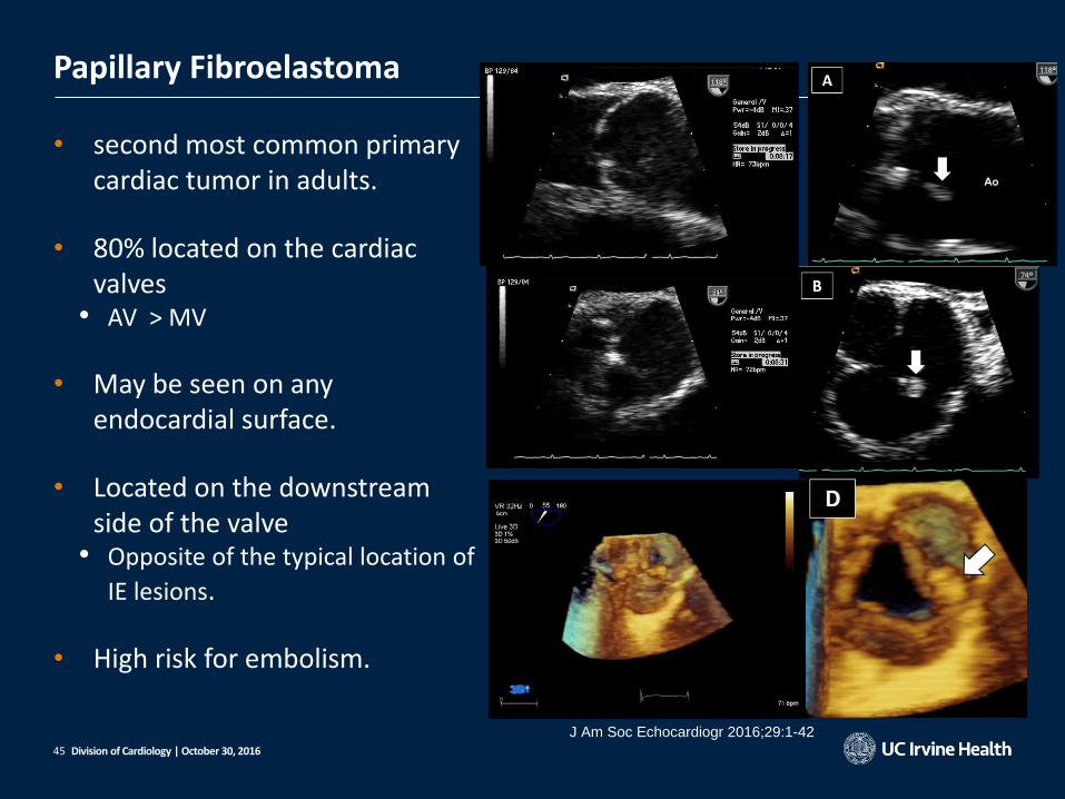
Papillary Fibroelastoma
• second most common primary cardiac tumor in adults.
• 80% located on the cardiac valves
• AV > MV
• May be seen on any endocardial surface.
• Located on the downstream side of the valve
• Opposite of the typical location of
IE lesions.
• High risk for embolism.
Division of Cardiology | October 30, 2016
J Am Soc Echocardiogr 2016;29:1-42

46
Echocardiography Recommended
• TTE in all patients suspected of having cardiac tumors
• TEE may be superior to TTE in evaluating cardiac tumors, especially myxomas and PFEs
• Surveillance echo after surgical removal of cardiac tumors with high recurrence potential (such as myxomas)
Recommendations for Echocardiographic Evaluation of Cardiac Tumors
Division of Cardiology | October 30, 2016

47
Echocardiography Not Recommended
Echocardiography is not recommended in patients for whom the results will not guide therapeutic decisions.
Recommendations for Echocardiographic Evaluation of Cardiac Tumors
Division of Cardiology | October 30, 2016

48
Evaluation of suspected cardiac source of embolism:
EMBOLISM FROM THE THORACIC AORTA
Division of Cardiology | October 30, 2016

49
TEE together with CT and MRI is the primary means of aortic plaque visualization
TEE ‘blind spot’ • A small segment of the distal ascending aorta because
of trachea between the esophagus and the aorta
Risk of embolic CVA • Plaque thickness > 4mm in the ascending aorta or arch visualized by TEE
Aortic Sources of Embolism
Division of Cardiology | October 30, 2016 J Am Soc Echocardiogr 2016;29:1-42

50
3D TEE may provide incremental diagnostic information on aortic plaques
Aortic Sources of Embolism
Division of Cardiology | October 30, 2016
J Am Soc Echocardiogr 2016;29:1-42

51
Echocardiography Recommended
• TEE is the preferred echocardiographic method for the evaluation of aortic sources of emboli.
Echocardiography Potentially Useful
• Aortic plaque may occasionally be seen on TTE. However, TTE has low sensitivity for the detection of aortic pathology, including aortic plaques,compared with TEE.
Echocardiography Not Recommended
• Echocardiography is not recommended in patients for whom the results will not guide therapeutic decisions
Recommendations for Echocardiographic Evaluation of Aortic Sources of Embolism
Division of Cardiology | October 30, 2016

52
Evaluation of suspected cardiac source of embolism:
PARADOXICAL EMBOLISM
Division of Cardiology | October 30, 2016
53
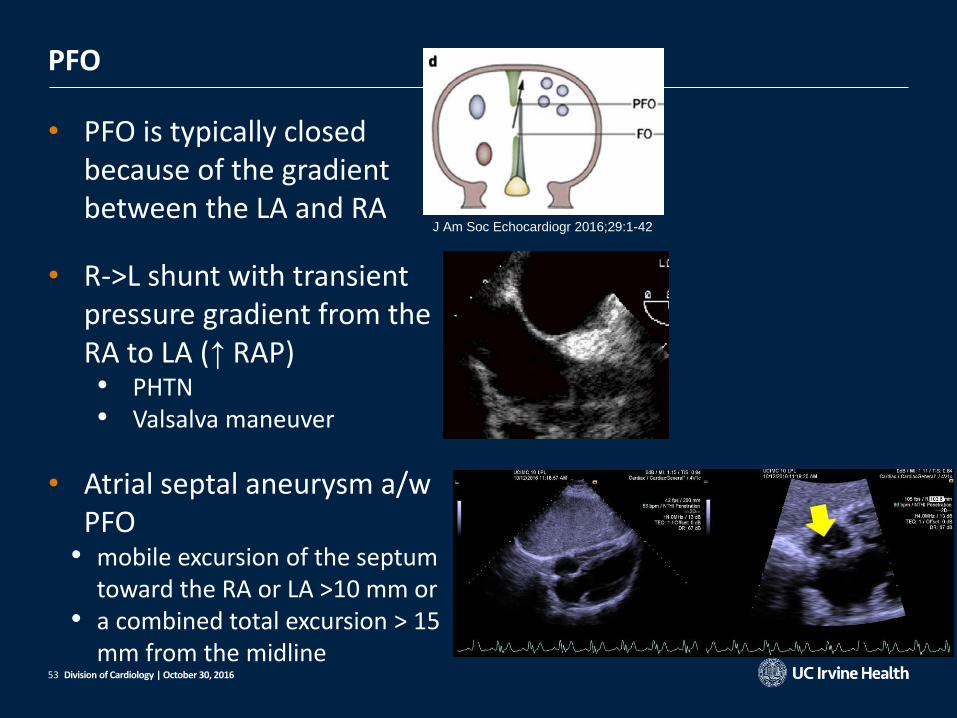
PFO
• PFO is typically closed because of the gradient between the LA and RA
• R->L shunt with transient pressure gradient from the RA to LA (↑ RAP) • PHTN • Valsalva maneuver
• Atrial septal aneurysm a/w PFO
• mobile excursion of the septum toward the RA or LA >10 mm or
• a combined total excursion > 15 mm from the midline
Division of Cardiology | October 30, 2016
J Am Soc Echocardiogr 2016;29:1-42
54
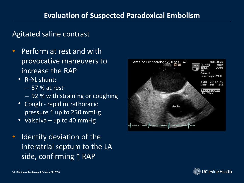
Evaluation of Suspected Paradoxical Embolism
Agitated saline contrast
• Perform at rest and with provocative maneuvers to increase the RAP
• R→L shunt: – 57 % at rest – 92 % with straining or coughing
• Cough - rapid intrathoracic pressure ↑ up to 250 mmHg
• Valsalva – up to 40 mmHg
• Identify deviation of the interatrial septum to the LA side, confirming ↑ RAP
Division of Cardiology | October 30, 2016
J Am Soc Echocardiogr 2016;29:1-42

55
PFO
• PFO is presumed when agitated saline contrast is noted in the LA within 3 cardiac cycles after complete opacification of the RA
• > 5 cardiac cycles intrapulmonary shunt or pulmonary AVM
• Timing of contrast appearance is used as a rough guide
• The best discriminator to accurately predict the location of shunting is direct visualization of the shunt.
Division of Cardiology | October 30, 2016
J Am Soc Echocardiogr 2016;29:1-42

56
+ TTE for a R-to-L shunt TEE to confirm the PFO and to exclude other shunts (e.g. secundum ASDs)
3D TEE to define the anatomy and evaluate for structural relationships
Division of Cardiology | October 30, 2016
J Am Soc Echocardiogr 2016;29:1-42

57
Echocardiography Recommended
• TTE is recommended for the evaluation of a R-to-L shunt and atrial septal anatomy in a patient with cryptogenic stroke:
• ↑ RAP with PE or DVT
• No shunt by color Doppler agitated saline injection at baseline and after coughing or Valsalva maneuver)
• TEE may be performed if TTE fails to demonstrate a R-to-L shunt
Recommendations for Echocardiographic Evaluation of Suspected Paradoxical Embolism
Division of Cardiology | October 30, 2016

58
Echocardiography Potentially Useful
3D TEE may provide incremental value in assessing atrial septal anatomy.
Echocardiography Not Recommended
Echo to establish a R-to-L shunt is not recommended in patients (typically older ones) who have other probable causes of stroke or systemic embolism.
Recommendations for Echocardiographic Evaluation of Suspected Paradoxical Embolism
Division of Cardiology | October 30, 2016
59
Evaluation of suspected cardiac source of embolism:
PULMONARY EMBOLISM
Division of Cardiology | October 30, 2016
60
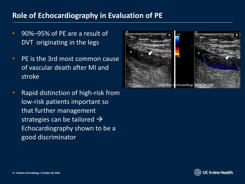
Role of Echocardiography in Evaluation of PE
• 90%–95% of PE are a result of DVT originating in the legs
• PE is the 3rd most common cause of vascular death after MI and stroke
• Rapid distinction of high-risk from low-risk patients important so that further management strategies can be tailored Echocardiography shown to be a good discriminator
Division of Cardiology | October 30, 2016
J Am Soc Echocardiogr 2016;29:1-42
61
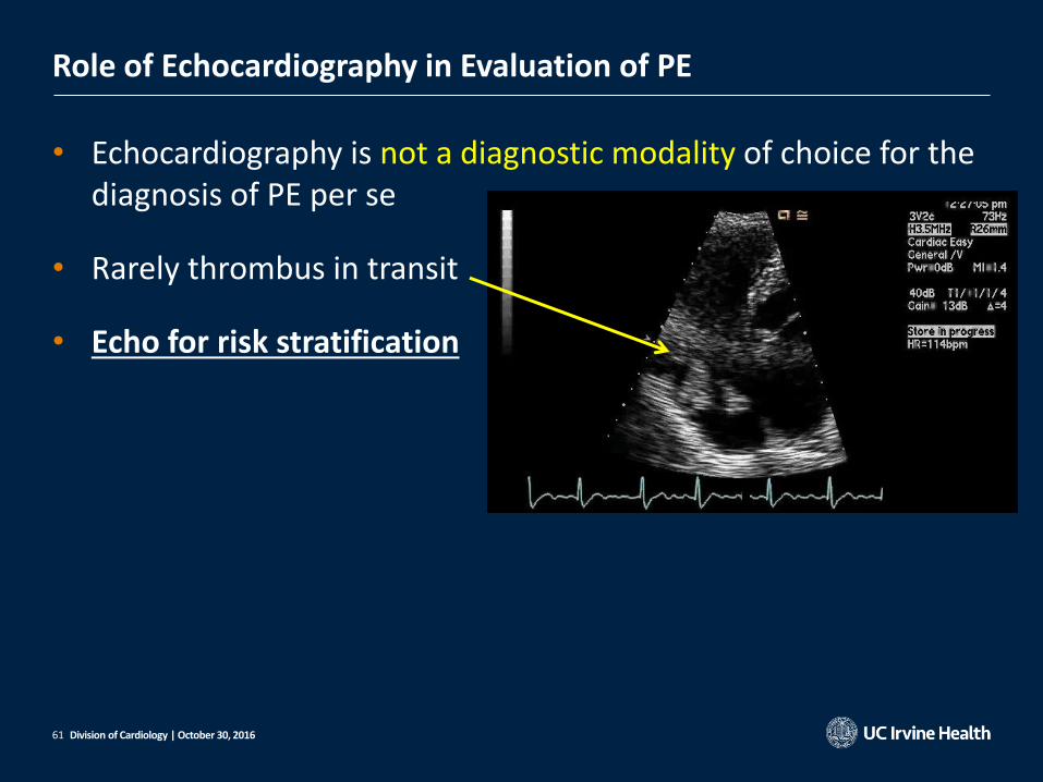
• Echocardiography is not a diagnostic modality of choice for the diagnosis of PE per se
• Rarely thrombus in transit
• Echo for risk stratification
Role of Echocardiography in Evaluation of PE
Division of Cardiology | October 30, 2016

62
The typical echocardiographic features of hemodynamically significant PE: • RV strain (RV dilatation and dysfunction) • IVS bulging into the LV • Dilated proximal PA • Elevated RVSP • Increased TR jet severity • Elevated RAP as evidenced by plethora of IVCwith no
inspiratory collapse • McConnell sign (hypokinesis of the basal and mid RV free wall,
with preserved contractility of apex) • visualization of thrombi in transit from systemic veins to
pulmonary arteries
Role of Echocardiography in Evaluation of PE
Division of Cardiology | October 30, 2016

63
Role of Echocardiography in Evaluation of PE
The typical echocardiographic features of
hemodynamically significant PE:
• RV strain (RV dilatation and dysfunction)
• IVS bulging into the LV • Dilated proximal PA • Elevated RVSP • Increased TR jet severity • Elevated RAP as evidenced by
plethora of IVCwith no inspiratory collapse
• McConnell sign (hypokinesis of the basal and mid RV free wall, with preserved contractility of apex)
• visualization of thrombi in transit from systemic veins to pulmonary arteries
Division of Cardiology | October 30, 2016
Am Soc Echocardiogr 2015;28:1-39
Am Soc Echocardiogr 2015;28:1-39 Am Soc Echocardiogr 2015;28:1-39
Am Soc Echocardiogr 2015;28:1-39

64
Role of Echocardiography in Evaluation of PE
The typical echocardiographic features of
hemodynamically significant PE:
• RV strain (RV dilatation and dysfunction)
• IVS bulging into the LV • Dilated proximal PA • Elevated RVSP • Increased TR jet severity • Elevated RAP as evidenced by
plethora of IVCwith no inspiratory collapse
• McConnell sign (hypokinesis of the basal and mid RV free wall, with preserved contractility of apex)
• visualization of thrombi in transit from systemic veins to pulmonary arteries
Division of Cardiology | October 30, 2016
FAC
3D EF
TAPSE J Am Soc Echocardiogr 2016;29:1-42

65
Role of Echocardiography in Evaluation of PE
The typical echocardiographic features of
hemodynamically significant PE:
• RV strain (RV dilatation and dysfunction)
• IVS bulging into the LV • Dilated proximal PA • Elevated RVSP • Increased TR jet severity • Elevated RAP as evidenced by
plethora of IVCwith no inspiratory collapse
• McConnell sign (hypokinesis of the basal and mid RV free wall, with preserved contractility of apex)
• visualization of thrombi in transit from systemic veins to pulmonary arteries
Division of Cardiology | October 30, 2016

66
Role of Echocardiography in Evaluation of PE
The typical echocardiographic features of
hemodynamically significant PE:
• RV strain (RV dilatation and dysfunction)
• IVS bulging into the LV • Dilated proximal PA • Elevated RVSP • Increased TR jet severity • Elevated RAP as evidenced by
plethora of IVCwith no inspiratory collapse
• McConnell sign (hypokinesis of the basal and mid RV free wall, with preserved contractility of apex)
• visualization of thrombi in transit from systemic veins to pulmonary arteries
Division of Cardiology | October 30, 2016
J Am Soc Echocardiogr 2016;29:1-42

67
Role of Echocardiography in Evaluation of PE
The typical echocardiographic features of
hemodynamically significant PE:
• RV strain (RV dilatation and dysfunction)
• IVS bulging into the LV • Dilated proximal PA • Elevated RVSP • Increased TR jet severity • Elevated RAP as evidenced by
plethora of IVCwith no inspiratory collapse
• McConnell sign (hypokinesis of the basal and mid RV free wall, with preserved contractility of apex)
• visualization of thrombi in transit from systemic veins to pulmonary arteries
Division of Cardiology | October 30, 2016
http://www.criticalecho.com/sites/default/files/images/7.11.png
http://www.cardiologyres.org/index.php/Cardiologyres/article/viewFile/187/191/
1589

68
Role of Echocardiography in Evaluation of PE
The typical echocardiographic features of
hemodynamically significant PE:
• RV strain (RV dilatation and dysfunction)
• IVS bulging into the LV • Dilated proximal PA • Elevated RVSP • Increased TR jet severity • Elevated RAP as evidenced by
plethora of IVC with no inspiratory collapse
• McConnell sign (hypokinesis of the basal and mid RV free wall, with preserved contractility of apex)
• visualization of thrombi in transit from systemic veins to pulmonary arteries
Division of Cardiology | October 30, 2016
RVSP 4(VTR)2

69
Role of Echocardiography in Evaluation of PE
The typical echocardiographic features of
hemodynamically significant PE:
• RV strain (RV dilatation and dysfunction)
• IVS bulging into the LV • Dilated proximal PA • Elevated RVSP • Increased TR jet severity • Elevated RAP as evidenced by
plethora of IVC with no inspiratory collapse
• McConnell sign (hypokinesis of the basal and mid RV free wall, with preserved contractility of apex)
• visualization of thrombi in transit from systemic veins to pulmonary arteries
Division of Cardiology | October 30, 2016

70
Role of Echocardiography in Evaluation of PE
The typical echocardiographic features of
hemodynamically significant PE:
• RV strain (RV dilatation and dysfunction)
• IVS bulging into the LV • Dilated proximal PA • Elevated RVSP • Increased TR jet severity • Elevated RAP as evidenced by
plethora of IVC with no inspiratory collapse
• McConnell sign (hypokinesis of the mid RV free wall, with preserved contractility of apex)
• visualization of thrombi in transit from systemic veins to pulmonary arteries
Division of Cardiology | October 30, 2016

71
Role of Echocardiography in Evaluation of PE
The typical echocardiographic features of
hemodynamically significant PE:
• RV strain (RV dilatation and dysfunction)
• IVS bulging into the LV • Dilated proximal PA • Elevated RVSP • Increased TR jet severity • Elevated RAP as evidenced by
plethora of IVC with no inspiratory collapse
• McConnell sign (hypokinesis of the mid RV free wall, with preserved contractility of apex)
• visualization of thrombi in transit from systemic veins to pulmonary arteries
Division of Cardiology | October 30, 2016
https://o
peni.nlm
.nih
.gov/im
gs/5
12/6
6/2
774585/P
M
C2774585_C
CR
-4-4
9_F
5.p
ng
J Am Soc Echocardiogr 2016;29:1-42

72
Echocardiography Recommended
• TTE is recommended for risk stratification in patients with PE (primarily for assessment of RV size and function).
• TEE may be considered in acutely ill, unstable patients in whom hemodynamically significant PE is suspected.
Echocardiography Not Recommended
• Echocardiography is not recommended as a primary means of diagnosing PE.
Recommendations for Echocardiography in Patients with Suspected PE
Division of Cardiology | October 30, 2016

73
Evaluation of suspected cardiac source of embolism:
CARDIAC AND AORTIC EMBOLISM DURING CARDIAC SURGERY AND PERCUTANEOUS
INTERVENTIONS
Division of Cardiology | October 30, 2016

74
Cardiac Catheterization • embolism during cardiac catheterization =1.4% - 1.9%
• the ascending aorta is likely the main source of emboli
Cardiac Surgery • The risk for embolism from the aorta during cardiac surgery is strongly
correlated with the degree of atherosclerosis in the ascending aorta
Percutaneous Interventions • Percutaneous wires, catheters, and other devices may dislodge preexisting
intracardiac and intra-aortic masses to cause systemic embolism
• Periprocedural stroke during TAVR = 1.5% - 6%
Division of Cardiology | October 30, 2016

75
Echocardiography Recommended
• TEE or ICE is recommended in all patients before intracardiac percutaneous intervention to exclude potential cardiac sources of emboli.
• The routine preoperative TEE to identify and manage aortic atheromatous disease is recommended in patients with increased risk for embolic stroke
• histories of CVA or PAD
• those with evidence of aortic atherosclerosis or calcification by MRI, CT, or
CXR.
Recommendations for Echocardiography in Patients Referred for Cardiac Surgery or Percutaneous Intervention
Division of Cardiology | October 30, 2016

76
Thank you for your attention.
Questions?
Division of Cardiology | October 30, 2016

77 Division of Cardiology | October 30, 2016

78 Division of Cardiology | October 30, 2016
http://www.pedicardiology.net/2011/03/echo
-asd-tee-evaluation-of-rims-for.html

79 Division of Cardiology | October 30, 2016
http://www.pedicardiology.net/2011/03/echo
-asd-tee-evaluation-of-rims-for.html

80 Division of Cardiology | October 30, 2016
81 Division of Cardiology | October 30, 2016

82
Mitral Annular Calcification (MAC)
Recommendations for Performance of Echocardiography in Patients with MACs.
Echocardiography Potentially Useful.
Echocardiography can establish the presence, extent, and severity of MACs.
However, MACs are typically an ncidental finding and unlikely to be an independent cause of a cardiac source of embolism.
Division of Cardiology | October 30, 2016
J Am Soc Echocardiogr 2016;29:1-42

83
1. Careful echocardiographic description • Echogenicity/echo texture: differentiate ‘‘myocardial-like’’ echogenicity vs
more echogenic patterns
• Size: length and width in millimeters
• Shape: sessile or pedunculated; amorphous, lobulated, linear, rounded,
strand-like, etc
• Location: side of valves, free edge vs base of leaflet
• Motion: dependent or independent of valvular motion; mild, moderate, or
highly mobile*
• Associations: regurgitation, stenosis, mycotic aneurysms, valvular
destruction, perivalvular abscess, perforation, prosthetic dehiscence
• A description of prosthetic valve annular position (i.e., well seated),
presence of rocking motion, and opening-closure of mechanical
mechanisms (i.e., normal disk or leaflet diastolic excursion for a mitral
prosthesis)
Recommendations for reporting valvular-associated masses
Division of Cardiology | October 30, 2016

84
2. Differential diagnosis • Always attempt to answer the clinician’s question/indication • Report 2-3 most likely explanations
– if patient’s clinical presentation is typical and echo characteristics are highly suggestive of pathology:
may use terms as suggestive of or likely represents, most consistent with, followed by terms such as less likely represents or unlikely to be.
• If clinical presentation unclear, and/or echocardiographic characteristics indistinct, report the most likely 2-3 DDx according to echocardiographic appearance, patient’s age, predisposing factors, and epidemiology
Recommendations for reporting valvular-associated masses
Division of Cardiology | October 30, 2016