ascorbic acid: metabolism and functions of a multi...
TRANSCRIPT
229
Ascorbic acid: metabolism and functions of a multi-facetted molecule Nicholas Smirnoff
Ascorbic acid (vitamin C) is the most abundant antioxidant in
plants. Its biosynthetic pathway via GDP-D-mannose and
L-galactose, which was proposed only recently, is now
supported by molecular genetic evidence from Arabidopsis
thahna and transgenic potato plants. Except for the last step
(which is located on the inner mitochondrial membrane) the
pathway is cytosolic, sharing GDP-sugar intermediates with
cell-wall polysaccharide and glycoprotein synthesis. Ascorbate
peroxidase is emerging as a key enzyme in the fine control of
H,O, concentration; its expression being controlled by redox
signals and H,O,. Convincing evidence of the involvement of
ascorbate in cell division and growth is also accumulating. Its
role as a cofactor in the synthesis of cell wall hydroxyproline-
rich glycoproteins is one mechanism for this function.
Addresses School of Biological Sciences, University of Exeter, Hatherly Laboratories, Prince of Wales Road, Exeter EX4 4PS, UK; e-mail: [email protected]
Current Opinion in Plant Biology 2000, 3:229-235
1369-5266/00/$ - see front matter 0 2000 Elsevier Science Ltd. All rights reserved.
Abbreviations A0 ascorbate oxidase APX ascorbate peroxidase GAL L-galactono-1,4-lactone GDP guanosine diphosphate GSH glutathione GUL L-gulono-1,4-lactone HL high light HRGP hydroxyproline-rich glycoprotein LL low light MDHA monodehydroascorbate PMI phosphomannose isomerase VtC vitamin C
Introduction Longevity in invertebrates and mice is associated with resistance to oxidacive stress, and with mutations that pre- vent programmed cell death in response to such stress [l]. As a result, there is great public interest in the health-pro- moting effects of antioxidants, particularly a-tocopherol (vitamin E) [Z] and ascorbic acid (vitamin C), which are required in the diet. Plants are the major sources of these vitamins and of other ‘nutriceutical’ antioxidants such as flavonoids and carotenoids. In plants, antioxidants provide protection against reactive oxygen species created both metabolically and in the environment; although the modu- lar growth pattern of plants (with the exception of seeds) makes the issue of life span less relevant for plants than for animals. It is also becoming apparent that the mosr preva- lent soluble small-molecule antioxidants, ascorbate and glutathione (GSH) [3], are multifunctional and also have
roles in photosynthesis, redox signalling, pathogen defence, metal and xenobiotic detoxification, and growth regulation.
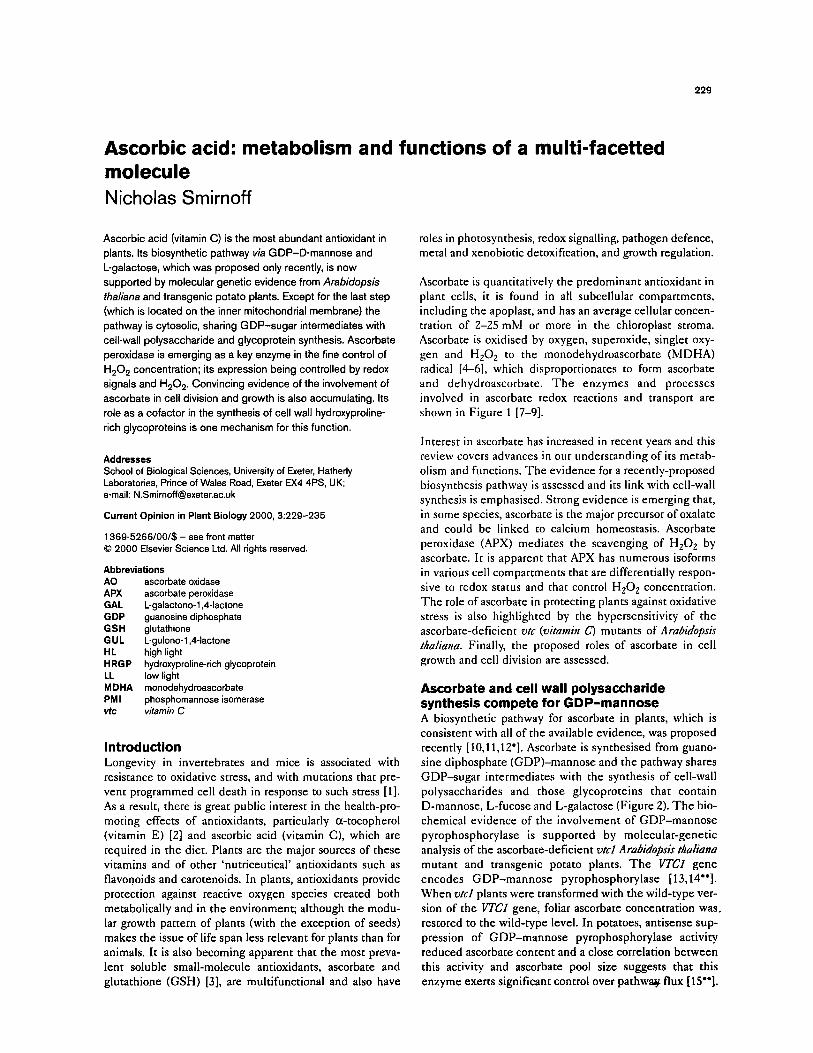
Ascorbate is quancitacively the predominant antioxidant in plant cells, it is found in aH subcellular compartments, including the apoplast, and has an average cellular concen- tration of 2-25 mM or more in the chloroplast stroma. Ascorbate is oxidised by oxygen, superoxide, singlet oxy- gen and H,O, to the monodehydroascorbate (MDHA) radical [4-61, which disproporcionates to form ascorbate and dehydroascorbate. The enzymes and processes involved in ascorbate redox reactions and transport are shown in Figure 1 [7-91.
Interest in ascorbate has increased in recent years and this review covers advances in our understanding of irs metab- olism and functions. The evidence for a recently-proposed biosynthesis pathway is assessed and its link with cell-wall synthesis is emphasised. Strong evidence is emerging that, in some species, ascorbate is the major precursor of oxalate and could be linked to calcium homeostasis. Ascorbate peroxidase (APX) mediates the scavenging of HZ02 by ascorbate. Ic is apparent that APX has numerous isoforms in various cell compartments rhac are differentially respon- sive to redox status and that control H,O, concentration. The role of ascorbace in protecting plants against oxidative stress is also highlighted by the hypersensitivity of the ascorbate-deficient vtc (vitamin c) mutants of Arabidopsis thaliatra. Finally, the proposed roles of ascorbace in cell growth and cell division are assessed.
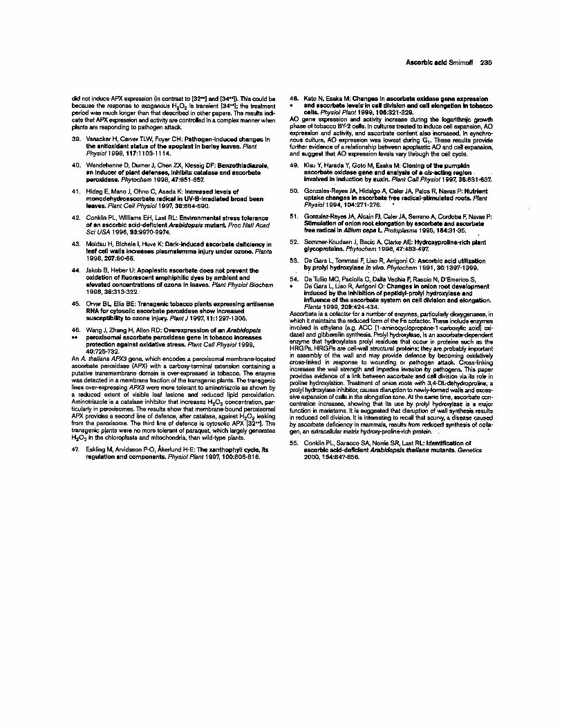
Ascorbate and cell wall polysaccharide synthesis compete for GDP-mannose A biosynthetic pathway for ascorbare in plants, which is consistent with all of the available evidence, was proposed recently [lO,l l,lZ’]. Ascorbate is synchesised from guano- sine diphosphace (GDP)-mannose and the pathway shares GDP-sugar intermediates with the synthesis of cell-wall polysaccharides and those glycoproteins that contain D-mannose, L-fucose and L-galactose (Figure 2). The bio- chemical evidence of the involvement of GDP-mannose pyrophosphorylase is supported by molecular-genetic analysis of the ascorbate-deficient vtcl Arabidopsh thaliana murant and cransgenic potato plants. The VTCI gene encodes GDP-mannose pyrophosphorylase [13,14”]. When vtcl plants were transformed with the wild-type ver- sion of the VTCI gene, foliar ascorbate concentration was, restored to the wild-type level. In potatoes, antisense sup- pression of GDP-mannose pyrophosphorylase activity reduced ascorbate content and a close correlation between this activity and ascorbate pool size suggests that this enzyme exerts significant control over pathway flux [15”].

230 Physiology and metabolism
Figure 1
hotorespiration hotosynthesis 1 lxidative burst lther oxidases 1
MDHA
I Cell wall
(d) -‘...
-----, AsA
GR s NADPH NADP Current ODinion in Plant Biohn
Redox reactions and transport of ascorbate (AsA). For clarity, only reactions in the cytosol and transport across the plasma membrane are shown. (a) Hydrogen peroxide reduction by ascorbate is catalysed by cytosolic ascorbate peroxidase (cAPX). (b) The oxidation product, monodehyroascorbate radical (MDHA), is
reduced to ascorbate by NAD(P)-dependent MDHA reductase (MDHAR). Cc) Two MDHA molecules can also disproportionate to dehydroascorbate (DHA) and ascorbate (represented non-stoichiometrically by a dotted line). Cd) DHA is reduced to ascorbate by glutathione (GSH)-dependent glutathione reductase (DHAR), which is, in
turn, regenerated from its oxidised form (GSSG) by glutathione reductase (GR). The net result is H202 removal by the ascorbate-GSH cycle at the expense of NAD(P)H [4,51. These reactions also occur in the chloroplast stroma, mitochondria and peroxisomes/glyoxysomes. APX is encoded by a gene family with distinct isoforms localised in different organelles; forms with hydrophobic tails are bound to thylakoid and peroxisomal/glyoxysomal membranes [5,281. A small proportion of APX activity is also reported in the apoplast [39]. There has been controversy over the existence of DHAR; other enzymes (e.g. glutaredoxin, thioredoxin reductase or peroxiredoxin) could reduce DHA but the evidence supports a role for a specific GSH- dependent DHAR [41. AsA and DHA occur in the cell wall. (e) AsA is transported into the wall by facilitated diffusion via a plasma membrane transporter in exchange for DHA [El. (f) Ascorbate oxidase (AO) is a secreted glycoprotein that catalyses ascorbate oxidation in the wall. (g) The resulting MDHA is probably reduced by a plasma-membrane cytochrome b system [7]. It is likely that cytosolic ascorbate is the electron donor and that (h) a plasma- membrane-bound MDHAR on the cytosolic side [91 regenerates ascorbate. A0 expression correlates with cell expansion although a causal relationship although its mechanism of action has not been established.
In some cases, the antisense suppression resulted in a on a non-phosphorylated sugar. The pathway prior to GAL reduction of mannose and galactose, but not of fucose, is probably cytosolic but the last step, the oxidation of GAL residues in wall polysaccharides. Incorporation of these to ascorbate by GAL dehydrogenase, is mitochondrial. sugar residues into glycoproteins was, however, unaffected GAL dehydrogenase has been purified and cloned from ([15”]; GL Wheeler, PL Conklin, N Smirnoff, unpub- cauliflower and sweet potato; the genes that encode it in lished data). It has been suggested that most plant species each of these two plant species are nearly identical and lack phosphomannose mutase (PMI) activity and this include a putative mitochondrial targeting sequence accounts for the toxicity of exogenous mannose, which [18,19]. GAL dehydrogenase is bound to the inner mem- accumulates as mannose-6-phosphate [16,17]. Lack of brane of the mitochondria [ZO’]. GAL donates electrons to PM1 would necessitate an alternative route to GDP-man- cytochrome c between complexes III and IV, and the active nose such as a GDP-glucose-Z-epimerase. Nevertheless, site probably faces the intermembrane space (CH Foyer, the wtcl mutation and antisense potato results argue for a personal communication) so that GAL does not need to be role for PMI. transported into the mitochondrial matrix.
GDP-L-galactose is produced by a double epimerisation of GDP-D-mannose [7]. The enzymes that break GDP-L- galactose down to free L-galactose have been detected ([7]; GL Wheeler, N Smirnoff, unpublished data) but have not yet been characterised in detail. A newly-discovered NAD+-dependent L-galactose dehydrogenase oxidises L-galactose to L-galactono-1,4-lactone (GAL), the immedi- ate ascorbate precursor [lO,ll]. This enzyme, which we have purified and cloned (GL Wheeler, S Gatzek, N Smirnoff, unpublished data) is specific for L-galactose and is, as far as we know, the only plant dehydrogenase acting
The current evidence suggests that the mannose pathway predominates in the biosynthesis of ascorbate in plants, but we cannot rule out contributions from other pathways. Conversion of radiolabelled (methyl)-D-galacturonate and D-glucuronolactone to ascorbate was noted by Loewus and colleagues some time ago [12’], and the involvement of these compounds has been confirmed by the increased ascorbate pool found after feeding methyl-D-galacturonate, D-glucuronolactone and methyl-D-glucuronate to A. thaliana cell cultures (21’1. The physiological significance of these conversions requires further investigation.

Ascorbic acid Smimoff 231
L-gulono-1,4-lactone (GUL), the precursor used by mam- mals in place of GAL, is slowly converted to ascorbate by plants. A separate GUL-oxidising enzyme could exist as purified GAL dehydrogenase is completely specific for GAL. It is now thought that the osone pathway [12’] of ascorbate biosynthesis is not physiologically important [Z?‘].
The recent advances in identifying the ascorbate biosyn- thetic pathway will allow investigation of its control, most importantly in relation to light intensity. Ascorbate accu- mulation is increased at high light intensity [23-E], a response which presumably reflects its use in H,Oz detox- ification (described below), regeneration of oxidised a-tocopherol [6] and as a cofactor for violaxanthin de-epox- idase [ZS]. Ascorbate concentration decreases in darkened leaves of some species, and this is partially reversed by sucrose or glucose feeding [23]. Synthesis of ascorbate is repressed or feedback-inhibited in pea seedling embryon- ic axes after their ascorbate content has been increased by feeding with ascorbate [26’].
Oxalate and tartrate are synthesised from ascorbate Ascorbate is cleaved at CZ/C3 to form oxalate and at either CZ/C3 or C4/C.5, depending on species, to produce L-tar- trate [12’]. Oxalate can also be formed from glycolate. In Pistia stratiotes, however, labelling studies show that oxalate is predominantly derived from ascorbate rather than glycolate [27”], and calcium oxalate crystals are accu- mulated in specialised cells (i.e. idioblasts). Calcium oxalate synthesis could therefore be involved in the regu- lation of calcium concentration. The enzyme catalysing CZ/C3 cleavage has not been identified but idioblast cells would be a good place to start searching for it.
Ascorbate peroxidase is a key enzyme for controlling H,O, concentration APX catalyses the reduction of HzOz to water and has high specificity and affinity for ascorbate as reductant [S]. Its sequence is distinct from other peroxidases, and different forms of APX occur in the chloroplasts, cytosol, mitochon- dria, peroxisomes and glyoxysomes. Membrane-bound APXs occur on the peroxisome and thylakoid membranes [5,28]. Hydrogen peroxide is formed by oxygen reduction by the chloroplast and mitochondrial electron transport chains; by certain oxidases, notably peroxisomal glycolate oxidase, dur- ing photorespiration; and during the oxidative burst associated with the hypersensitive response to pathogens. Oxygen reduction by Photosystem I, coupled with removal of the resulting HzOz by APX, is termed the Mehler-peroxi- dase reaction and contributes to the regulation of the redox state of photosynthetic electron carriers [5,29]. Recent work on the response of APX expression to intense light and pathogen attack has highlighted the importance of APX activity in controlling HzOz concentration in signalling.
Photoinhibition and photo-oxidation are caused when low light (LL) acclimated leaves are exposed to high light
Figure 2
- tndo mplasmic
,Cuculum P-4:
cell w ‘all polysaccharides and gl ycoproteins
4-T
1 (W
Mannose-6-P -e + Mannitol
4 Irz 1
(c)
Mannqse-l -P GTP
x (d) Ivrcr)
PPi
GDP-mannose+-+ GDP-glucose .
Glucose-6-P
1 (a)
Fructose-6-P
I \ Fe’ GDP-L-fucose t GDP-L-galactose
t 0 L-Galactose
NAD
(g) NADH
1 L-Galactono-,I ,Glactone Ascorbate
A
The proposed biosynthetic pathway of ascorbate via GDP-mannose and L-galactose [lo]. The diagram illustrates links with other pathways, including polysaccharide and glycoprotein synthesis from GDP-sugars. Mannitol is a major carbon translocation compound in some species. GDP-mannose could be formed from GDP-glucose, although lower ascorbate concentration in plants with reduced GDP-mannose pyrophosphorylase activity [14”,15”], suggests that this is not a major route. Reduced GDP-mannose pyrophosphorylase activity also lowers the mannose and L-galactose content of cell-wall polysaccharides. The final oxidation of L-galactono-1,4-lactone to ascorbate on the inner mitochondrial membrane [201 uses cytochrome c as an electron acceptor and is therefore coupled to mitochondrial electron transport. Ascorbate and L-galactono-I ,4- lactone presumably enter and leave the intermembrane space freely through porins in the outer membrane. Ascorbate biosynthesis is one of many biosynthetic functions that takes place in plant, but not in animal mitochondria In contrast, the final step of ascorbate biosynthesis in mammals is catalysed by a microsomal oxidase. Alternative pathways via D-galacturonate and D-glucuronate, or their lactones or esters [21 *I, are not illustrated and their physiological significance has not yet been established. The enzymes are: (a) glucose phosphate isomerase; (b) phosphomannose isomerase; (c) phosphomannose mutase; Cd) GDP-mannose pyrophosphorylase; (e) GDP-mannose-3,5-epimerase; (0 uncharacterised enzymes; (g) L-galactose dehydrogenase; and (h) L-galactono-1,4-lactone dehydrogenase.
(HL) intensity. Acclimation involves the induction of antioxidant and photoprotective (e.g. zeaxanthin-depen- dent non-photochemical quenching) systems [5,24,29,30]. HL causes increased HzOz formation in chloroplasts, either from the Mehler reaction or Photosystem II, and in

232 Physiology and metabolism
peroxisomes as a result of faster photorespiration. Exposure of A. thaliana leaves to HL causes a rapid (i.e. within 30 min) increase in the transcription of APXI and APXZ, which encode two isoforms of cytosolic APX [31]. The promotors for APXl and APXZ have been isolat- ed and fusions with a luciferase reporter gene have been made. The transformants were then used to investigate the pattern of APXZ expression and the signalling process- es that control it [32”]. APX induction is correlated to increased reduction-state of quinone B and plastoquinone. Thus, APXexpression is regulated in same way as has been proposed for light-intensity-dependent expression of other photosynthetic genes [33]. The expression of both antiox- idant and photosynthetic genes is therefore co-ordinated through a common redox-signalling system.
Hydrogen peroxide itself is also involved in APX induc- tion: exogenous application of HzOz increases APX expression in LL, whereas catalase infiltration inhibits the expression of this gene in HL [32”]. Hydrogen peroxide also induces cytosolic APX expression in non-photosyn- thetic tissue [34”]. Induction of APX by its own substrate may provide a sensitive means of controlling its concentra- tion (assuming that the enzyme is active). This system has also provided evidence of a systemic plant response to HL, possibly mediated by HzOz, because leaves remote from the one exposed to HL showed an increase in APXZ::LUCIFERASE expression and a small increase in HzOz concentration. Because the APXZ promoter has an extremely sensitive response, it will prove to be a valuable tool for identifying the promotor DNA sequences and DNA binding proteins involved in responses to HzOz or other redox signals. Further progress towards this goal might be made with the use of a recessive A. thaliana mutant that has increased tolerance to salinity-induced photo-oxidation [35”]. The mutant has APX and superox- ide dismutase activities that are above wild-type levels, particularly after exposure to salt. It is possible that the mutation affects a gene that regulates the expression of antioxidant genes.
Hydrogen peroxide is produced during pathogen attack, and it could provide a direct defence against the pathogen and contribute to programmed cell death in the hypersen- sitive. response. These responses are localised and therefore more difficult to investigate than the leaf responses to HL described above. Antisense suppression of catalase in tobacco results in the expression of patho- genesis-related genes [36]. I f HzOz is the key to this mechanism, local activity of catalase and APX should be restricted during pathogen attack. Cytosolic APX tran- scripts increase in virus-infected tobacco but APX protein concentrations actually decrease as a result of the inhibi- tion of polypeptide elongation on the ribosomes [37,38”]. These findings illustrate the danger of equating increased transcript levels to increased enzyme activity, and suggest that suppression of peroxide scavenging by APX is impor- tant for defence against pathogens [39]. The inhibition of
catalase and APX by salicylic acid, which is produced in response to infection, might also contribute to local sup- pression of HzOz scavenging 1401.
WC mutants and transgenic plants provide evidence of the role of ascorbate in stress resistance Various stresses, including UVB, increase the prevalence of MDHA radicals [41]. The A. thaliana wtcl mutant has only 30% of wild-type ascorbate [42], which is sufficient for normal growth under non-stressful conditions. A similar reduction in ascorbate concentrations in potatoes express- ing antisense GDP-mannose pyrophosphorylase, however, causes visible lesions on leaves and stems, and premature die-back of the shoot [15”]. It is not known if these symp- toms are caused by ascorbate deficiency or by defects in the cell wall. Vtcf is hypersensitive to ozone, ultraviolet-B light and SO,, but its ozone sensitivity is reversed by exogenous ascorbate [42]. Plants kept in the dark for a pro- longed period are known to be more ozone sensitive, and it has been suggested that this is a result of reduced apoplastic ascorbate [43]. Use of an apoplast-localised dye, whose fluorescence is abolished by oxidation, however, suggests that apoplastic ascorbate is not the major reduc- tant of ozone in the apoplast [44]. Nevertheless, cytosolic ascorbate may be important because antisense reduction of cytosolic APX activity increases the ozone sensitivity of tobacco [45]. Presumably the induction of cytosolic APX (described above) [31,32”] improves the scavenging of HzOz that escapes from the chloroplasts and peroxisomes after evading chloroplast APX and peroxisomal catalase. Furthermore, over-expression of peroxisomal APX3 increases HzOz tolerance [46”]. Ascorbate is probably a limiting factor in the violaxanthin de-epoxidase reaction in the thylakoid lumen [47]; it will therefore be of interest to determine if the reduced ascorbate content of wtc mutants affects their zeaxanthin-mediated photoprotection [30].
Ascorbate oxidase and prolyl hydroxylase: roles for ascorbate in growth? Ascorbate oxidase (AO) is a cell wall localised glycoprotein belonging to the family of blue copper oxidase enzymes. Its role in plants has not been defined but its activity and expres- sion are closely correlated to rapid cell expansion [48’] and it is induced by auxin [49]. A causal relationship that might explain this correlation has not yet been demonstrated and no mechanism of action has yet been established [6]. One possi- bility is that A0 generates MDHA radicals. These radicals are then reduced by transmembrane electron transport (Figure 1) thereby depolarising the plasma membrane and stimulating H+-ATPase activity. In this model, cell expansion is stimulat- ed by enhanced wall loosening or ion uptake: exogenous MDHA radicals stimulate onion root growth and ion uptake [50,51]. It is likely that transgenic plants with altered A0 expression will soon be used to clarify the function of AO. Another role for ascorbate in growth is an involvement in the synthesis of hydroxyproline-rich glycoproteins (HRGPs). Ascorbate is a cofactor for prolyl hydroxylase (as it is for a

Ascorbic acid Smimoff 233
range of other oxygenases [6]), which post-translationally hydroxylates proline residues. The HRGPs are cell wall struc- tural proteins and are involved in wall synthesis in dividing cells [SZ]. Prolyl hydroxylase is inhibited by 3,CDL-dehy- droproline causing increased ascorbate concentration and reduced cell division in onion roots [53,54’]. These results show that HRGP synthesis in meristems is a function of ascorbate and could provide an explanation for the reported effects of ascorbate on cell division.
Conclusions and future developments Within the past few years knowledge of ascorbate metabo- lism and function in plants has greatly increased. The identification of the ascorbate biosynthetic pathway will allow us to manipulate the ascorbate content of plants, as has already been achieved with its fellow antioxidants GSH [3,4] and a-tocopherol [Z]. This should provide fur- ther information on the functions of ascorbate, including those involved in photosynthesis, stress resistance, growth, development and oxidative-stress signalling, that have been identified in my review. Metabolic engineering of ascorbate biosynthesis to produce plants with increased ascorbate concentration will not only provide crops with improved nutritional value, but may well improve their growth and stress resistance. Ascorbate does not act in iso- lation, so it will be exciting to see the effect of engineering ascorbate biosynthesis along with GSH, a-tocopherol, carotenoids and other antioxidant enzymes; this goal is now within reach.
Update A recent paper by Conklin et al. [SS] describes the identi- fication and mapping of three ascorbate-deficient mutants (UC& 3 and 4) in addition to the previously characterised wtcl [13,14”]. These mutants contain between ZO-30% of wild-type ascorbate. Not all of the mutant alleles produce phenotypes that are hypersensitive to ozone, suggesting that the relationship between ascorbate content and ozone resistance [42-45] is not predictable. These mutants should prove useful for identifying further genes involved in ascorbate metabolism and exploring the role of ascor- bate in resistance to oxidative stress.
Acknowledgements I am grateful fo 0 Arrigoni, PL Conklin, MW Davcy, CH Foyer, FA Locwus and GL Wheeler for discussions and access to rcsuks in press. Funding for my laboratory is provided by the Biotechnology and Biological Sciences Research Council (UK) and Bio-Technical Resources (Manirowoc, Wisconsin, USA).
References and recommended reading Papers of particular interest, published within the annual period of review, have been highlighted as:
l of special interest “of outstanding interest
1. Migliaccio E, Giorgio M, Mele S, Pellici G, Reboldi P, Pandolfi PP, Lanfrancone L, Pelicci PG: The p55’hS adaptor protein controls oxldatlve stress response and life span In mammals. Nature 1999, 402:309-313.
2. Shintani D, DellaPenna D: Elevating the vitamin E content of plants through metabolic englnwring. Science 1 QQ6,252:2095-2100.
3.
4.
5.
5.
7.
3.
Q.
10.
11.
12. .
Cobbett CB: Phytochelatin biosynthesis and function In heavy metal detoxification. Curr Opin Plant Ho/ 2000,3:21 l-21 5. .
Noctor G, Foyer CH: Ascorbate and glutathione: keeping actlve oxygen under control. Ann Rev Plant Physiol Plant MO/ Bioll QQ5, 49:249-279.
Asada K: The water-water cycle In chloroplastp: scavenging of actlve oxygens and dlsslpatlon of excess photons. Ann Rev Plant Physiol Plant Mol Bioll QQQ, 50:501-539.
Smimoff N: The function and metabolism of ascorbic acid in plants. Ann Botany 1 QQ5,75:651-559.
Horemans N, Asard H, Csubergs RI: The role of ascorbate free radical as an electron acceptor to cytochmme bmediated trans- plasma membrane electron trangport in higher plants. Plant PhysiollQQ4. 104:1455-l 455.
Horemans N, Asard H, Caubargs RJ: Carrier mediated uptake of dehydmascorbate Into higher plant plasma membrane vesicles shows trans-stimulation. FEBS Leti 1 QQ5,421:41-44.
Berczi A, Mollsr IM: NADH-monodehydmpascorbate oxldoreductase is one of the redox enzymes In spinach leaf plasma membranes. Plant Physio/19Q8,116:102Q-1035.
Wheeler GL, Jones MA, Smimoff N: The biosynthetic pathway of vitamin C In higher plants. Nafure 1 QQ5,393:355-359.
Smimoff N, Wheeler GL: Ascorbic acid metabolism In planta. In Plant Carbohydrale Biochemistry. Edited by Bryant JA, Surrell MM, Kruger NJ. Oxford: Bios Scientific Publishers; 1 QQQ:215-229.
Loewus FA: Biosynthesis and metabolism of ascorbic acid In plants and of analogs of ascorbic add In fungi. Phytochem 1 Q9Q, 52:1Q3-210. _
A Concise but comprehensnre review ot ascorbate blosynthesls and catabo- lism providing an assessment of the current ideas about biosynthesis, alter- native pathways and a historical perspective.
13. Conklin PL, Pallanca JE, Last RL, Smimoff N: L-Ascorbic acid metabolism In the ascorbate-deficient Ar&dopsis mutant vtcl. Plant PhysiollQQ7,115:1277-1255.
14. Conklin PL, Norris SR, Wheeler GL, Williams EH, Smimoff N, . . Last RL: Genetic evidence for the role of GDP-mannose In plant
ascorbic acid (vitamin C) biosynthesis. Proc Nat/ Acad SW’ USA 1 QQQ, 95:4195-4203.
This paper, along with [15’*1, provides genetic evidence of the proposed [lOI involvement of GDP-mannose in ascorbate biosynthesis. The enzyme catal- yses the fonation of GDP-mannose from mannose-l-phosphate and GTP. The gene encoding the vtcl mutation of A. thaliana, which causes ascorbate deficiency, was cloned and found to have homology with GDP-mannose pyrophosphotylase. l&cl leaves had lower GDP-mannose pyrophosphory- lase a&vii than the wild-type, and expression of wild-type UC1 in the mutant plants restored ascorbate concentration. Reduced GDP-mannosa pymphosphorylase activity is caused by a point mutation that reduces enzyme-specific activity; transcript levels are unaffected in the mutant.
15. Keller R, Springer F, Ranz A, Kossmann 1: Antisense Inhibition of the l . GDP-mannose pymphosphorylase reduces the ascorbate
content In transgenic plants leading to developmental changes during senescence. Plant J 1 QQQ, 1 %131-l 41.
This paper, along with [14”], provides genetic evidence of the involvement of GDP-mannoss in ascorbate biosynthesis. GDP-mannose pyrophospha rylase was cloned from potato using an A. thaliana expressed sequence tag. Antisense expression of this gene in potato resulted in plants with reduced ascorbate concentration. GDP-mannose pyrophosphorylase appears to control ascotbate biosynthesis pathway flux because there is a close cor~e- lation between reduction in activity of this enzyme and ascorbate content. The mannose and galactose content of cell wall polysacdrarides was also reduced in leaves, as would be expected if GDP-mannose and GDP-L- galactose are precursors of hsmicelluloses. The antisense plants also showed necmtic leaf and stem lesions, and accelerated leaf senescence. It is not known if these symptoms are caused by ascorbate deficiency 0~ altered cell wall composition.
15. Pego JV, Weisbeck PJ, Smeekens SCM: Mannose inhibits Arabidopsis germination vla a hexoklnase-mediated step. Plant Physiol 1 QQQ, 119:1017-l 023.
li! Stein JC, Hansen G: Mannose Induces an endonudease wponslble for DNA laddering in plants P/ant physiol 1 QQQ, 121:71-79.
15. Ostergaard J, Peniau G, Cavey MW, Bauw G, Van Montagu M: Isolation and cDNA cloning for L-galactonoylactone dehydmgenase, en enzyme Involved In the biosynthesis of ascorbic add In plants. J Ho/ Chem 1 QQ7,272:30009-30015.
234 Physiology and metabolism
19. lmai T, Katita S, Shatori G, Hattori M, Nunome T, Oba K, Hirai M: L-Galactobo+actone dehydrogenase from sweet potato: purification and cDNA sequence analysis. Plant Cell P/rysio/ 1996, 39:1350-l 356.
20. Siendones E, Gonxalsz-Reyes JA, Santos-Ocafta C, Navas P, .- C4rdoba F: Biosynthesis of ascorbic acid in kidney bean.
L-galactono-y-la&one dehydmgenase is an lntrlnsic protein located at the mltochondrial inner membrane. Plant Phvsiol1999, 120:907-912.
Lgafactono-1,4-factone dehydmgenass is the fast enxyme in the ascorbata biceynthesis pathway and its association with the mitochondrfa has been recog- nised for some time. lhii oacer uses purified Phaseolus vulaariis mitochondria to show that the enzyme is &ated in’ the inner mitochondn~ membrane. This finding raises the possibifii that ascorbate biosynthesis could be controlled through a link to mkochondriaf, and therefore chloroplastic, metabolism.
21. Davey MW, Gilot C, Persiau G, Ostergaard J, Han Y, Bauw GC, Van . Montaau MC: Ascorbate biosvnthesis in Arsfridoosis cell
suspekion culture. P/ant Ph~sio/l999,121:535~543. This paper confirms that L-gafactose and L-galactono-1 ,Clactone, the pro- posed immediate precursors of ascorbate [lo], are readily converted to ascorbate by A. thdiana cell suspensions. In addition, a number of uranic acid derivatives (i.e. methyl-D-galacturonate, D-glucuronolactone and methyl-D-glucuronate) that have previously been shown to be incorporated into ascorbate at radiotracer levels 112.1 cause small but detectable increas- es in ascorbate. The results suggest that alternative pathways involving uron- ic acid intermediates could make a small contribution to ascorbate synthesis.
22. Paflanca JE Smimoff N: Ascorbic acid metabolism in pea . seedlings. A comparison of D-glucosone, L-sorbosone and
L-galactono-1,4-lactone as ascorbate precursors. P/ant Physiol 1999,120:453-461.
This paper assssses the importance of the osone pathway of ascorbate syn- thesis [12*1 in which D-glucosone and L-sorbosone are proposed as ascor- bate precursors. Isotope-dilution experiments show that sorbosone is not involved in ascorbate synthesis. No evidence for glucosone synthesis was found and exogenous glucosone was toxic to pea seedlings. There was a close relationship between the appearance of ascorbate and ascorbate oxi- dase activity; both are almost absent until 20 h after imbibing the seeds, and the start of embryonic axis growth. These results are further circumstantial evidence of the role of ascorbate and ascorbate oxidase in cell growth.
23. Smimoff N, Pallanca JE: Ascorbate metabolism in relation to oxldatlve stress. Biochem Sot lkns.l.996,24:472-476.
24. Grace SC, Logan BA: Acclimation of follar antioxldant systems to arowth lrradisnce in three broadleaved evemreen soecies. P/ant ~hysio/1996,112:1631-1640.
- .
25. Eskfing M, Akerkmd HE: Changes in the quantftfes of violaxanthln de-epoxidase, xanthophylls and ascorbate in spinach upon shift from low to high light Photosynth Res 1996,57:41-50.
26. Pallanca JE, Smimoff N: The control of ascorbic acid synthesis and turnover In pea seedlings. J Exp Botany 2000,51 :in press.
;he authors show that ascorbate synthesis from ‘4Gglucose is strongly inhib ited by pm-loading pea seedlings with ascorbate. This effect is Seen within 3 h of ascotbate loading and provides evidence that ascorbate synthesis is con- trolled by feedback inhibition. The enzymes subject to feedback regulation must now beidentified; this will contribute towards optimising the metabolic engineering of ascorbate biosynthesis Ascorbate loading increased the rate of ascorbate turnover, determined by metabolism of ‘4Casoorbate. The rats of ascotbate turnover is direotfy proportional to its pool size.
27. Keates SE, Tarfyn NM, Loswus FA, Franceschi VR: L-galactose: . . source of oxalic acid in calcium oxalate deposition in pi&a
stratiotes. Phytochem 2000, in press. Pulse-chase experiments with 1% ascorbata, L-gafactose and glycolate show that ascorbate, not glycolate, is the major source of oxalate in Pistia sfrefiofes. This floating aquatic plant stores calcium oxalats in specialised cells ffdioblasts) as a result of stimulation of ascorbate and oxafate synthe- sis by calcium. Also, contrary to the usual supposition, there is appreciable turnover of f”coxafate, showing it is not a dead-end product.
26. lshikawa T, Yoshimura K, Sakai K, Tamoi M, Takeda T, Shigeoka S: Molecular characterixatlon and ohvsloloolcal role of a glyoxysome-bound ascorbate Rer&ldasa from spinach. Plant Cell Physioll996,39:23-34.
. . 29. Foyer CH, Noctor G: Leaves in the dark see In the light Science
1999,284:699-601.
30. Niyogi KK: Photoprotectlon revisltsd: genetic and molecular approaches. Ann Rev Plant Physiol Plant MO/ Biol1999, 50333-359.
31. Karpinsfri S, Eacobar C, Karpinska B, Creissen G, Mullineaux PM: Pjt0twynthdo elachn transport regplates the. expressjon of
32. . .
cytosolic aswrbate peroxidesa genes in Arebidopsk during excess light Plant Cell 1997,9:627-640.
Karpinski S, Reynolds H, Karpinska B, Wingsle G, Crfessen G, Mullineaux P: Systemic signalling and acclimation in response to excess excitation energy in Arebidopsis. Science 1999, 264:654-657.
Leaves exposed to high light intensity show rapid increase in expression of cytosolic ascorbate peroxidase as part of a defence against HaO,, which is generated by the Mehler reaction and photorespiration 129,311. The tran- scripts of one ascorbate peroxidase, APX2, are not expressed in low light but increase within 30 min of exposure to high light. A. thaliana was transformed with a fusion of the APX2 promoter and lucifsrase. This fusion was expressed in the same way as the native gene, allowing lucifsrase activity to report APX2 expression in intact leaves exposed to high light. It also provides a sensitive assay for APX2 exoression in leaf extracts. This svstem was used to show that mani’pulation of the redox state of the photo&tern II electron acceptors quinone B and plastoquinone, using the electron transport inhibitors 3-(3,4- dichlorophenol)-1 ,l -dimethylurea and 2,5-dibmmo-6-isopropyl-3-methyl-1,4- benxoquinone (DBMIB), alters APX2 expression. Increased reduction, as occurs in high light or in the presence of DBMIB, increases APX2 expression. Antioxidant defsnces are therefore coupled to photosynthesis and use a sim- ilar rsdox-sensing system as photosynthetic enzymes [33]. APX2 expression is also increased by its own substrate, ascorbate; the high light response is reduced by infiltrating leaves with catalase. This paper demonstrates a novel systemic response in which exposure of one leaf to high light induces APX2 expression and reduces photoinhibition in untreated leaves. This phenorne- non was named ‘systemic acquired acclimation’ and H,Oa is suggested as a component of the signalling system.
33. Pfannschmidt T, Nilsson A, Allen JF: Photosynthetic control of chlomplast gene expression. Nature 1999,397:625-626.
34. Morfta S, Kaminaka H, Masumura T, Tanaka K: Induction of rice l * cytosolic ascorbate pemxidase mRNA by oxidatfve stress: the
involvement of hydrogen peroxide In oxidatlve stress signalling. Plant Cell Physioll999,40:417-422.
Cytosolic APX expression in cultured rice embryos is controlled by HaO,. Both HaOa and paraquat are shown to elevate endogenous peroxide and increase APX transcripts within 2 h in a dose-dependent manner. Paraquat generates peroxide via superoxide; inhibition of superoxide dismutase by N,N-diethyldithiocarbamate prevented APX induction showing that superox- ide is not involved. Catalase and APX inhibitors (i.e. aminotriaxole and hydroxyurea) also induced APX transcripts and increased endogenous HaO,. This paper, along with f32.1, now provides convincing evidence that cytosolic APX gene expression is controlled by H,O,. It should now be pos- sible to uncover the signal transduction systems involved in H,O, signalling. As a cautionary note, this paper and [32”1 do not show that increased APX transcript level is associated with more protein or enzyme activity; in some circumstances [371 this is not the case.
35. Tsugane K, Kobayashi K, Niwa Y, Ohba Y, Wada K, Kobayshi H: A l 0 recessive Ambidopsis mutant that grows photoautotrophlcally
under salt stress shows enhanced active oxygen detoxification. Plant Cell 1999,11:1195-l 206.
An A. thaliana mutant that could be affected in a signalling pathway for oxidative stress is identified. The recessive mutant @stl) is more resistant to salt stress in high light. This treatment causes photo-oxidation, eventually resulting in bleaching of the cotyledons. APX and superoxide dismutase activity are induced by salt treatment. These enzymes have higher activity in the mutant and, notably in the case of APX, the salt-induced increase in activity is greater in the mutant than in wild-type plants. The results, although preliminary, indicate that the gene encoding this mutation could be involved in signalling oxidative stress.
36.
37.
36. . .
Takahashi H, Chen 2, Du H, Liu Y, Klessig DF: Development of necrosis and activation of disease resistance in tobacco plants with severely reduced catalase levels. P/ant J 1997,11:993-l 005.
Mittler R, Feng XG, Cohen M: Post-transcriptional suppression of cytosollc aswrbate peroxldase expression during pathogen- Induced oroorammed cell death in tobacco. P/ant Cell 1996. lo:461 -473.-
Miiler R, Lam E, Shulaev V, Cohen M: Signals controlling the expression of cytosolic ascorbate peroxldase during pathogen- Induced programmed cell death In tobacco. P/ant MO/ No/ 1999, 39:1025-i 035.
pr&rammed wll death response to tobacco mosaic virus. i p&ious paper [37l had shown that this did not lead to increased APX protein, indeed trans- lation was suppressed, suggesting that HaOa, which is required for pathogen defenw, is pmtected. This paper investigates the induction of APX transcripts in detail, and shows that EGTA with a calcium ionophore, paraquat, salicylic acid (see also 1401) and gkrtathione all induce APX expression. Saficylic acid is not essential to the pathogen response because tobacco expressing salicy- fate, hydroxy@-basn,ormal APX expression. Hydrpgen.wr@de application
Ascorbic acid Smimoff 235
did not induce APX expression (in contrast to j32”l and j34”j). This could be because the response to exogenous HsO, is transient [34-l; the treatment period was much longer than that described in other papers. The results indi- cate that APX expression and activity are controlled in a complex manner when plants are responding to pathogen attack
39.
40.
41.
42.
43.
44.
45.
46. . .
Vanacker H, Carver TLW, Foyer CH: Pathogen-induced changes in the antioxidant status of the apoplast In barley leaves. Plan? Physio/1996,117:1103-1114.
Wendehenne D, Dumer J, Chen ZX, Klessig DF: Benzothladiexole, an Inducer of plant defenses, inhibits catalase and ascorbate peroxidase. Phytochem 1996,47:651-657.
Hideg E, Mano J, Ohno C, Asada K: Increased levels of monodehydroascorbate radical in UV-B-irradiated broad bean leaves. Plant Cell PhysiollQQ7, 39:664-690.
Conklin PL, Williams EH, Last RL: Envlmnmental stress tolerance of en ascorbic acid-deficient Arabidopsis mutant Proc Nat/ Acad Sci USA 1996,93:9970-9974.
Moldau H, Bichele I, Huve K: Dark-induced ascorbate deftciency in leaf cell walls lncraasas plasmalemma injury under ozone. Plants 1996,207:60-66.
Jakob B, Hebsr U: Apopleettc ascorbate does not prevent the oxidation of fluorescent emphiphilic dyes by ambient and elevated concentrations of ozone in leaves. Plant Physiol Biochem 1998,36:313-322.
Orvar BL, Ellis BE: Transgenic tobacco plants expressing antisense RNA for cytosolic ascorbete pemxidase show increased susceptibility to ozone injury. P/an/J 1997,l Id 297-l 305.
Wang J, Zhang H, Allen RD: Overexpression of en Ambidopsis pemxisomal ascorbate pemxidese gene in tobacco increases protection against oxidative stress. P/ant Cell Physiol 1999, 40~725-732.
An A. fhaliana APX3 gene, which encodas a peroxisomal membrane-located ascorbate peroxidase (APX) with a carboxy-terminal extension containing a putative transmembrane domain is over-expressed in tobacco. The enzyme was detected in a membrane fraction of the transgenic plants. The transgenic lines over-expressing APX3 wars more tolerant to aminotriaxole as shown by a reduced extent of visible leaf lesions and reduced lipid peroxidation. Aminotriazole is a catalase inhibitor that increases H&e concentration, par- ticularly in peroxisomss. The results show that membrane-bound peroxisomal APX provides a second line of defence, after catalase, against HxO, leaking from the peroxisome. The third line of defence is cytosolic APX 132’1. The transgenic plants wsre no more tolerant of paraquat, which largely generates H,O, in the chloroplasts and mitochondria, than wild-type plants.
47. Eskling M, Arvidsson P-O, Akerlund H-E: The xanthophyll cycle, its ragulatlon and components. Physiol Plant 1997,l Q&606-61 6.
46. Kato N, Esaka M: Changes In ascorbate oxfdase gene expression . and ascorbate levels In cell divfslon and cell elongation In tobacco
cells. Physiol Plant 1999,105:321-329. A0 gene expression and activity increase during the logarithrntc growth phase of tobacco BY-2 cells. In cultures treated to induce cell expansion, A0 expression and activity, and ascorbate content also increased. In synchro- nous culture, A0 expression was lowest during Gt. These results provide further evidence of a relationship between apoplastic A0 and cell expansion, and suggest that A0 expression levels vary thrbugh the cell cycle.
49.
50.
51.
52.
53.
54. .
Kisu Y, Harada Y, Goto M, Esaka M: Cloning of the pumptdn ascorbate oxidase gene and enelysis of a c/s-acting region involved In Induction by auxin. Plant Cell Physio/lQQ7,39:631-637’
Gonzales-Reyas JA, Hidalgo A, Caler JA, Pales R, Navas P: Nutrient uptake changes In ascorbate free radical-stimulated roots. Plant Physio/1994,104:271-276. *
Gonzalez-Reyes JA, Alcain FJ, Caler JA, Senano A, Cordoba F, Navas P: StImulatfon of onion root elongation by ascorbate and ascorbate free radical In A//ium cepe L Protoplasma 1995,184:31-35.
Sommer-Knudsen J, Bacic A, Clarke AE: Hydmxypmllne-rich plant glycopmteins. Phytochem 1996,47:463-497
De Gara L, Tommasi F, Liso R, Anigoni 0: Ascorbic acid utlllzatton by pmlyl hydmxylase in t&o. Phytochem 1991,30:1397-1399.
Da Tullio MC, Paciolla C, Daka Vechia F, Rascio N, D’EmericoS, De Gara L, Liso R, Anigoni 0: Changes in onion root development induced by the Inhibition of peptidyl-pmlyl hydmxylese and influence of the ascorbate system on cell division and elongation. Planta 1999,209:424-434.
Ascorbate is a cofactor for a number of enzymes, particulariy dioxygenases, in which it maintains the reduced form of the Fe cofactor. These include enxymes involved in ethylene (e.g. ACC [I -aminocyclopropanel oarboxylic acid] oxi- dase) and gibbsrallin synthesis. Prolyl hydroxylase, is an ascorbata-dependent enzyme that hydroxylatas protyl residues that occur in proteins such as the HRGPs. HRGPs are cell-wall structural proteins; they are pmbably important in assembly of the wall and may provide defence by becoming oxidativsly cross-linked in response to wounding or pathogen atta& Cross-linking increases the wall strength and impedes invasion by pathogens. This paper provides evidence of a link between ascorbate and cell division via its role in proline hydroxylation. Treatment of onion roots with 3,4-DL-dehydropmline, a pmlyl hydroxylese inhibitor, causes disruption to newly-formed walls and axwe sive expansion of calls in the elongation zone. At the same time, ascorbate con- centration increases, showing that its use by prolyl hydroxylase is a major function in me&ems. It is suggested that disruption of wall synthesis results in reduced cell division. lt is interesting to recall that scurvy, a disease caused by ascorbate deficiency in mammals, results from reduced synthesis ot col!a- gen, an extracellular matrix hydmxy-pmlinerich protein. .
55. Conklin PL, Saracco SA, Norris SR, Last RL: lden9ficetion of ascorbic acid-deficient Arabidopsis theliene mutants. Genetics 2000,154:647-656.