ascertainment of silent myocardial infarction in patients
TRANSCRIPT
aDepartme
Rotterdam, the
Medicine, Taip
National Yang
dam, the Neth
dam, Amsterd
Medical Unive
tional CardiologZentrum f€ur
DZHK Stando
tle upon Tyne
castle Univers
Medical Unive
Marta Hospital
versity, Quebe
Singapore; mInnDepartment o
land; oDepartm
lands; pD�epa
0002-9149/© 2
https://doi.org/
ARTICLE IN PRESS
Ascertainment of Silent Myocardial Infarction inPatients Undergoing Percutaneous Coronary
Intervention (from the GLOBAL LEADERS Trial)
Chun Chin Chang, MDa,b, Ernest Spitzer, MDa,c, Ply Chichareon, MDd, Kuniaki Takahashi, MDd,Rodrigo Modolo, MDd, Norihiro Kogame, MDd, Mariusz Tomaniak, MDa,e, Hidenori Komiyama, MDd,
Sing-Chien Yap, MD, PhDa, Stephen P. Hoole, MDf, Tommaso Gori, MDg, Azfar Zaman, MDh,Bernhard Frey, MDi, Rui Cruz Ferreira, MDj, Olivier F. Bertrand, MD, PhDk, Tian Hai Koh, MDl,
Amanda Sousa, MDm, Aris Moschovitis, MDn, Robert-Jan van Geuns, MD, PhDa,o,Philippe Gabriel Steg, MDp, Christian Hamm, MDq,r, Peter J€uni, DMs,t, Pascal Vranckx, MD, PhDu,
Marco Valgimigli, MD, PhDn, Stephan Windecker, MDn, Patrick W. Serruys, MD, PhDv,*,Osama Soliman, MD, PhDa,c, and Yoshinobu Onuma, MD, PhDa,c
nt o
Net
ei V
Mi
erla
am
rsity
gy,
Kar
rt Rh
NHS
ity,
rsity
, Li
c C
stit
f Ca
ent
rtem
019
10.
Q-wave myocardial infarction (QWMI) comprises 2 entities. First, a clinically evident MI,which can occur spontaneously or be related to a coronary procedure. Second, silent MIwhich is incidentally detected on serial electrocardiographic (ECG) assessment. The preva-lence of silent MI after percutaneous coronary intervention (PCI) in the drug-eluting stentera has not been fully investigated. The GLOBAL LEADERS is an all-comers multicentertrial which randomized 15,991 patients who underwent PCI to 2 antiplatelet treatment strate-gies. The primary end point was a composite of all-cause death or nonfatal new QWMI at2-years follow-up. ECGs were collected at discharge, 3-month and 2-year visits, and analyzedby an independent ECG core laboratory following the Minnesota code. All new QWMI werefurther reviewed by a blinded independent cardiologist to identify a potential clinical corre-late by reviewing clinical information. Of 15,968 participants, ECG information was completein 14,829 (92.9%) at 2 years. A new QWMI was confirmed in 186 (1.16%) patients. Transientnew Q-waves were observed in 28.5% (53 of 186) of them during the follow-up. The majorityof new QWMI (78%, 146 of 186) were classified as silent MI due to the absence of a clinicalcorrelate. Silent MI accounted for 22.1% (146 of 660) of all MI events. The prevalence ofsilent MI did not differ significantly between treatment strategies (experimental vs reference:0.88% vs 0.98%, p = 0.5027). In conclusion, we document the prevalence of silent MI in anall-comers population undergoing PCI in this large-scale randomized trial. © 2019 ElsevierInc. All rights reserved. (Am J Cardiol 2019;00:1−8)
f Cardiology, Thoraxcenter, Erasmus Medical Center,
herlands; bDivision of Cardiology, Department of Internal
eterans General Hospital, Institute of Clinical Medicine,
ng University, Taipei, Taiwan; cCardialysis B.V., Rotter-
nds; dAcademic Medical Center, University of Amster-
, the Netherlands; eFirst Department of Cardiology,
of Warsaw, Warsaw, Poland; fDepartment of Interven-
Royal Papworth Hospital, Cambridge, United Kingdom;
diologie, Kardiologie I, University Medical Center, and
ein-Main, Mainz, Germany; hFreeman Hospital, Newcas-
Hospitals Trust and Institute of Cellular Medicine, New-
United Kingdom; iDepartment of Internal Medicine II,
Vienna, Vienna, Austria; jCardiology Department, Santa
sbon, Portugal; kQuebec Heart-Lung Institute, Laval Uni-
ity, Quebec, Canada; lNational Heart Center Singapore,
uto Dante Pazzanese de Cardiologia, Sao Paulo, Brazil;
rdiology, University of Bern, Inselspital, Bern, Switzer-
of Cardiology, Radboud UMC, Nijmegen, the Nether-
ent de Cardiologie, Hopital Bichat, Hopitaux
Universitaires Paris Nord Val de Seine, Assistance Publique - Hopitaux de
Paris, Paris, France; qKerckhoff Clinic, Bad Nauheim, Germany; rDZHK
(German Centre for Cardiovascular Research), Frankfurt, Germany; sAp-
plied Health Research Centre, Li Ka Shing Knowledge Institute of St
Michael’s Hospital, Toronto, Ontario, Canada; tDepartment of Medicine,
Institute of Health Policy, Management and Evaluation University of Tor-
onto, Toronto, Ontario, Canada; uJessa Ziekenhuis, Faculty of Medicine and
Life Sciences at the Hasselt University, Hasselt, Belgium; and vNHLI, Impe-
rial College London, London, United Kingdom. Manuscript received May 6,
2019; revised manuscript received and accepted August 30, 2019.
Funding: This study was sponsored by the European Cardiovascular
Research Institute (ECRI, Rotterdam, the Netherlands) that received funding
from one device (Biosensors International Ltd, Europe) and two drug manu-
facturers (Astra Zeneca, Cambridge United Kingdom; The Medicines Com-
pany, Parsippany; United States of America).
See page 7 for disclosure information.
*Corresponding author: Tel: +31-10-4635260; fax: +31-10-4369154.
E-mail address: [email protected] (P.W. Serruys).
www.ajconline.orgElsevier Inc. All rights reserved.
1016/j.amjcard.2019.08.049
Silent or unrecognized myocardial infarction (MI) wasidentified as a clinical entity several decades before the eraof percutaneous coronary intervention (PCI).1 Currently,silent MI is defined by the appearance of new pathological
Q-waves on an electrocardiogram (ECG) or cardiac imag-ing evidence of infarction without an identifiable clinicalcorrelate (e.g., acute coronary syndrome or coronary revas-cularization) during follow-up.2 The frequency of silent MI
ARTICLE IN PRESS
2 The American Journal of Cardiology (www.ajconline.org)
has been reported as comprising around 1/3 of all MIs fromseveral large cohort studies.3,4 A mounting body of evi-dence has demonstrated that silent MI is associated with anincreased risk of all-cause death, heart failure, and reinfarc-tion.5−7 Despite its prognostic impact, silent MI is rarelyaddressed in contemporary coronary intervention trials. Theprevalence of silent MI after PCI with drug-eluting stentshas not yet been fully investigated. Timing of surveillance,analysis methodology, and appropriate use of this end pointare current research interests. In this context, we report onthe assessment of new Q-wave MI (QWMI) in theGLOBAL LEADERS trial and summarize findings withrespect to silent and clinically evident events.
Methods
The details of the GLOBAL LEADERS trial have beenreported previously.8,9 In the investigator-initiated, multi-center, prospective randomized GLOBAL LEADERS trial,15,991 patients who underwent PCI with Biolimus A9 elut-ing stents were randomly assigned to 2 antiplatelet treat-ment strategies. In the experimental treatment strategy,patients received aspirin 75 to 100 mg once daily in combi-nation with ticagrelor 90 mg twice daily for 1 month; fol-lowed by ticagrelor 90 mg twice daily monotherapy for23 months (irrespective of the clinical presentation). In thereference treatment strategy, patients received aspirin 75 to100 mg daily in combination with either clopidogrel 75 mgonce daily in patients with stable coronary artery disease orticagrelor 90 mg twice daily in patients with acute coronarysyndromes for 1 year; followed by aspirin 75 to 100 mgonce daily monotherapy between 1 and 2 years. The pri-mary end point was a composite of all-cause death or nonfa-tal new QWMI at 2 years of follow-up.
Resting 12-lead ECGs at hospital discharge, 3-monthsfollow-up, and the 2-year end-of-trial visit and anyavailable intercurrent ECGs, related to suspected ischemicevents, were inspected for quality and technical errors andanalyzed by an independent ECG-core laboratory (Cardial-ysis, Rotterdam, The Netherlands). The ECG acquisitionguidelines are provided in the appendix. All of the missingECGs or noninterpretable ECGs were queried, and theinvestigator were requested to forward a new recording.
Serial comparison of sequential ECGs was performed toidentify patients with new appearance of Q waves (majorQ-QS wave abnormalities 1-1-1 to 1-2-7 according to theMinnesota Code 2009).10 All core laboratory detected newQWMIs were reviewed by a blinded independent medicalreviewer (BIMR). Where new Q waves, with respect to theimmediately preceding ECGs (first reference ECG was atdischarge), were identified, the BIMR confirmed or rejectedthe event as a new QWMI and if confirmed also assigned adate, based on review of the reported clinical events to thenew QWMI. Where no clinical correlate was identified, thedate of the new QWMI was arbitrarily assigned to the dateof the qualifying ECG. When the BIMR disagreed with thenew Q waves, he requested a reassessment by the core lab.The initial or subsequent assessment by the core lab wasthe final decision. When the other co-primary end point(all-cause death) occurred or when there was no clinicalevent up to 2 years occurred and there was no 2-year ECG
it was assumed that no new QWMI had occurred. When anew QWMI followed by death in a short time (<28 days), itwas considered to be fatal and did not count for the endpoint of new QWMI.
The BIMR was also responsible for reviewing the site-reported new QWMI that had not been reported by the corelab. This review was performed after the 2-year ECG hadbeen received or was confirmed to be permanently missing.When the BIMR identified a likely new QWMI betweendischarge and 2-years follow-up, the core lab was requestedto reassess the ECGs. The ECG-core lab also identified newoccurrences of left bundle branch block (LBBB) on serialECGs. When a new LBBB was identified, the BIMR deter-mined whether a likely ischemic event (prolonged ischemicchest pain, significant rise in cardiac biomarkers or imagingevidence of loss of viable myocardium) occurred accordingto the electronic clinical record form when necessary withadditional source documents. A new occurrence of LBBBcounted as a new QWMI equivalent only when a qualifyingischemic event was identified. The new QWMI equivalentwas assigned to the date of the qualifying ischemicevent. ECG analysts and the BIMR were unaware of thestudy-group assignments. With respect to clinically evidentMI (periprocedural or spontaneous), it was site-reportedaccording to the Third Universal Myocardial Infarctiondefinition as study specific MI criteria.11
Categorical variables were presented as percentages andnumbers and compared with the use of the Chi-square testor Fisher’s exact test. Continuous variables were comparedwith Student’s t test or analysis of variance test. Kaplan-Meier method was used to estimate the cumulative rates oftime to event end points and log-rank test was performed toexamine the differences between groups. A 2-sided p valueof <0.05 was considered to indicate statistical significance.Data were analyzed using SPSS software (version 25,SPSS, Chicago, Illinois).
Results
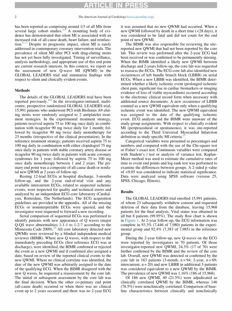
The GLOBAL LEADERS trial enrolled 15,991 patients,of whom 23 subsequently withdrew consent and requesteddeletion of their data from the database, leaving 15,968patients for the final analysis. Vital status was obtained inall but 8 patients (99.95%). The study flow chart is shownin Figure 1. At 2-year follow-up, the ECG information wascomplete in 93.3% (7,446 of 7,980) patients in the experi-mental group and 92.4% (7,383 of 7,985) in the referencegroup.
During the 2-year follow-up, new Q-waves on the ECGwere reported by investigators in 70 patients. Of thoseinvestigator-reported new QWMI, 24.3% (17 of 70) werefurther confirmed by the BIMR and the review of the corelab. Overall, new QWMI was detected or confirmed by thecore lab in 183 patients (3-month, n = 94; 2-year, n = 69;intercurrent, n = 20) and new LBBB in additional 3 patientswas considered equivalent to a new QWMI by the BIMR.The prevalence of new QWMI was 1.16% (186 of 15,968).
Of 186 new QWMI, 40 (21.5%) were adjudicated asclinically correlated QWMI by the BIMR, whereas 146(78.5%) were nonclinically correlated. Comparison of base-line characteristics between clinically and nonclinically
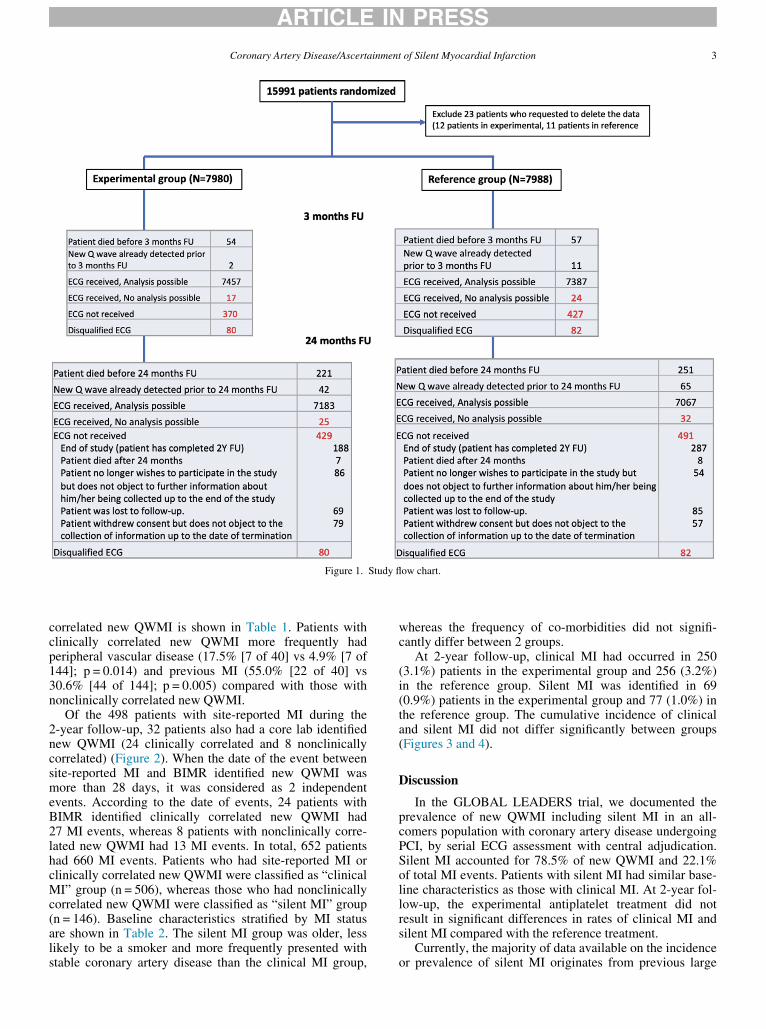
Figure 1. Study flow chart.
ARTICLE IN PRESS
Coronary Artery Disease/Ascertainment of Silent Myocardial Infarction 3
correlated new QWMI is shown in Table 1. Patients withclinically correlated new QWMI more frequently hadperipheral vascular disease (17.5% [7 of 40] vs 4.9% [7 of144]; p = 0.014) and previous MI (55.0% [22 of 40] vs30.6% [44 of 144]; p = 0.005) compared with those withnonclinically correlated new QWMI.
Of the 498 patients with site-reported MI during the2-year follow-up, 32 patients also had a core lab identifiednew QWMI (24 clinically correlated and 8 nonclinicallycorrelated) (Figure 2). When the date of the event betweensite-reported MI and BIMR identified new QWMI wasmore than 28 days, it was considered as 2 independentevents. According to the date of events, 24 patients withBIMR identified clinically correlated new QWMI had27 MI events, whereas 8 patients with nonclinically corre-lated new QWMI had 13 MI events. In total, 652 patientshad 660 MI events. Patients who had site-reported MI orclinically correlated new QWMI were classified as “clinicalMI” group (n = 506), whereas those who had nonclinicallycorrelated new QWMI were classified as “silent MI” group(n = 146). Baseline characteristics stratified by MI statusare shown in Table 2. The silent MI group was older, lesslikely to be a smoker and more frequently presented withstable coronary artery disease than the clinical MI group,
whereas the frequency of co-morbidities did not signifi-cantly differ between 2 groups.
At 2-year follow-up, clinical MI had occurred in 250(3.1%) patients in the experimental group and 256 (3.2%)in the reference group. Silent MI was identified in 69(0.9%) patients in the experimental group and 77 (1.0%) inthe reference group. The cumulative incidence of clinicaland silent MI did not differ significantly between groups(Figures 3 and 4).
Discussion
In the GLOBAL LEADERS trial, we documented theprevalence of new QWMI including silent MI in an all-comers population with coronary artery disease undergoingPCI, by serial ECG assessment with central adjudication.Silent MI accounted for 78.5% of new QWMI and 22.1%of total MI events. Patients with silent MI had similar base-line characteristics as those with clinical MI. At 2-year fol-low-up, the experimental antiplatelet treatment did notresult in significant differences in rates of clinical MI andsilent MI compared with the reference treatment.
Currently, the majority of data available on the incidenceor prevalence of silent MI originates from previous large
Table 1
Blinded independent medical reviewer-identified clinically correlated versus nonclinically correlated new Q-wave myocardial infarction
Clinically correlated
new QWMI
(n = 40)
Nonclinically correlated
new QWMI
(n = 146)
p Value
Age (years) 65.4 § 10.6 67.0 § 10.6 0.414
Sex
Male 34 /40 (85.0%) 105/146 (71.9%) 0.104
Female 6 /40 (15.0%) 41/146 (28.1%)
Body mass index (kg/m2) 27.6 § 4.8 28.3% § 4.2 0.322
Medical history
Diabetes mellitus 15/40 (37.5%) 44/146 (30.1%) 0.444
Insulin-dependent diabetes mellitus 4/40 (10.0%) 17/146 (11.6%) 1.000
Hypertension 30/40 (75.0%) 108/145 (74.5%) 1.000
Hypercholesterolemia 29/39 (74.4%) 91/141 (64.5%) 0.337
Current smoker 11/40 (27.5%) 28/146 (19.2%) 0.276
Peripheral vascular disease 7/40 (17.5%) 7/144 (4.9%) 0.014
Chronic obstructive pulmonary disease 5/40 (12.5%) 8/145 (5.5%) 0.159
Impaired renal function* 7/40 (17.5%) 25/146 (17.1%) 1.000
Previous stroke 3/40 (7.5%) 4/145 (2.8%) 0.173
Previous myocardial infarction 22/40 (55.0%) 44/144 (30.6%) 0.005
Previous percutaneous coronary intervention 21/40 (52.5%) 52/146 (35.6%) 0.067
Previous coronary artery bypass grafting 6/40 (15.0%) 17/1462 (11.6%) 0.590
Clinical presentation
Stable coronary artery disease 26/40 (65.0%) 86/146 (58.9%) 0.585
Acute coronary syndrome 14/40 (35.0%) 60/146 (41.1%)
Antiplatelet treatment
Experimental strategy 14/40 (35.0%) 69/146 (47.3%) 0.209
Reference strategy 26/40 (65.0%) 77/146 (52.7%)
Data are shown as mean § standard deviation or number of patients (%).
*Defined as an estimated glomerular filtration rate of creatinine clearance of <60 ml/min per 1.73 m2 based on the Modification of Diet in Renal Disease
formula. QWMI = Q-wave myocardial infarction.
ARTICLE IN PRESS
4 The American Journal of Cardiology (www.ajconline.org)
cohorts of patients without PCI.4 In our study, the preva-lence of silent MI (0.91%) was in the range of previouslyreported data (0.37% to 3.37%).4 The Bypass AngioplastyRevascularization Investigation 2 Diabetes study12 in 2,368diabetic patients with coronary artery disease assigned toeither prompt revascularization or medical therapy alone
Figure 2. Classification of site-reported and blinded independent medical review
correlated and 8 nonclinically correlated) patients had both blinded independe
infarction and site-reported myocardial infarction. When the date of event betwee
events. * 8 patients had 13 MI events; ** 24 patients had 27 MI events, MI = myoc
showed that nonfatal silent MI occurred in 0.97% (23 of2,368) of patients and accounted for 8% of all MI during anaverage of 5.3-year follow-up.13 Recently, Zhang et alshowed that silent MI occurred in 3.3% of participants whowere free of cardiovascular disease at baseline in the Ath-erosclerosis Risk In Communities study during a median
er-identified new Q-wave myocardial infarction. Thirty-two (24 clinically
nt medical reviewer (BIMR)-identified nonfatal new Q-wave myocardial
n 2 types of MI was more than 28 days, it was considered as 2 independent
ardial infarction.

Figure 3. Kaplan−Meier curve for clinical myocardial infarction at 2 years.
Table 2
Baseline characteristics of patients stratified by myocardial infarction status
No myocardial
infarction
(n = 15,316)
Silent myocardial
infarctiony
(n = 146)
Clinical myocardial
infarctionz
(n = 506)
p Value
Age (years) 64.5 §10.2x 67.0 § 10.6{ 64.4 § 11.2 0.013
Sex 0.382
Male 11,760/15,316 (76.8%) 105/146 (76.9%) 389/506 (76.2%)
Female 3,556/15,316 (23.2%) 41/146 (23.1%) 117/506 (23.8%)
Body mass index (kg/m2) 28.1 § 4.5 28.2 § 4.6 28.3 § 4.2 0.834
Medical history
Diabetes mellitus 3,817/15,305 (24.9%){ 44/146 (30.1%) 177/506 (35.0%) <0.001Insulin-dependent diabetes mellitus 1,146/15,270 (7.5%){ 17/146 (11.6%) 60/505 (11.9%) <0.001Hypertension 11,241/15,263 (73.6%) 108/145 (74.5%) 366/506 (72.3%) 0.781
Hypercholesterolemia 10,322/14,835 (69.6%) 91/141 (64.5%) 355/489 (72.4%) 0.151
Current smoker 3,992/15,316 (26.1%) 28/146 (19.2%){ 149/506 (29.4%) 0.037
Peripheral vascular disease 945/14,231 (6.2%){ 7/144 (4.9%) 53/502 (10.6%) <0.001Chronic obstructive pulmonary disease 776/14,470 (5.1%) 8/145 (5.5%) 37/505 (7.3%) 0.081
Impaired renal function* 2037/15,232 (13.4%){ 25/146 (17.1%) 109/505 (21.6%) <0.001Previous stroke 397/15,294 (2.6%) 4/145 (2.8%) 20/506 (4.0%) 0.172
Previous myocardial infarction 3,482/15,275 (22.8%){ 44/144 (30.6%) 184/503 (36.6%) <0.001Previous percutaneous coronary intervention 4,950/10,352 (32.3%){ 52/146 (35.6%) 219/506 (43.3%) <0.001Previous coronary artery bypass grafting 862/14,441 (5.6%)x,{ 17/146 (11.6%) 64/506 (12.6%) <0.001
Clinical presentation 0.017
Stable angina 8154/15316 (53.2%){ 86/146 (58.9%){ 241/506 (47.6%)
Acute coronary syndrome 7162/15316 (46.8%){ 60/146 (41.1%){ 265/506 (52.4%)
Data are shown as mean § standard deviation or number of patients (%).
*Defined as an estimated glomerular filtration rate of creatinine clearance of <60 ml/min per 1.73 m2 based on the Modification of Diet in Renal Disease
formula.yEqual to BIMR-identified nonclinically correlated Q-wave MI.z Patients had either site-reported MI or BIMR-identified clinically correlated new QWMI or both. Patients (n = 8) who had concomitant site-reported MI
and BIMR-identified nonclinically correlated MI were not included. ANOVA test was used to compare continuous variables.x p <0.05 (compare to silent MI group).{ p <0.05 (compare to clinical MI group).
ARTICLE IN PRESS
Coronary Artery Disease/Ascertainment of Silent Myocardial Infarction 5
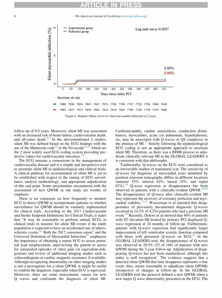
Figure 4. Kaplan−Meier curve for silent myocardial infarction at 2 years.
ARTICLE IN PRESS
6 The American Journal of Cardiology (www.ajconline.org)
follow-up of 8.9 years. Moreover, silent MI was associatedwith an increased risk of heart failure, cardiovascular death,and all-cause death.6,7 In the abovementioned 2 studies,silent MI was defined based on the ECG findings with theuse of the Minnesota code10 or the Novacode14,15 which arethe 2 most widely used ECG coding system providing pre-dictive values for cardiovascular outcomes.16
The ECG remains a cornerstone in the management ofcardiovascular disease and is a simple and inexpensive toolto ascertain silent MI in epidemiological and clinical trials.A clinical pathway for ascertainment of silent MI is yet tobe established with respect to the timing of ECG surveil-lance, analysis methodology, and appropriate adjudicationof this end point. Some uncertainties encountered with theassessment of new QWMI in our study are worthy ofemphasis.
There is no consensus on how frequently to monitorECG to detect QWMI in asymptomatic patients or whethersurveillance for QWMI should be routinely implementedfor clinical trials. According to the 2017 Cardiovascularand Stroke Endpoint Definitions for Clinical Trials, it statesthat “It may be reasonable to perform annual ECGs inclinical trials to monitor for silent MI events if the studypopulation is expected to have an accelerated rate of athero-sclerotic events.”2 Both the 2017 consensus report2 and theUniversal Definition of Myocardial Infarction11 emphasizethe importance of obtaining a repeat ECG to assess poten-tial lead misplacement, interviewing the patient to assessfor unreported episodes of chest pain (or equivalent-indi-gestion) and review of additional imaging studies such asechocardiogram or cardiac magnetic resonance if available.Although recognizing abnormality on other imaging studiesis not a prerequisite for a silent QWMI, it might be usefulto confirm the diagnosis, especially when ECG is equivocal.Moreover, there are some nonischemic causes for newQ waves and confounds the diagnosis of silent MI.
Cardiomyopathy, cardiac amyloidosis, conduction distur-bances, myocarditis, acute cor pulmonale, hyperkaliemia,etc, may be associated with Q waves or QS complexes inthe absence of MI.17 Strictly following the epidemiologicalECG coding is not an appropriate approach to ascertainsilent MI. Therefore, as there was a BIMR process to adju-dicate clinically relevant MI in the GLOBAL LEADERS itis consistent with that philosophy.
Traditionally, Q-waves on the ECG were considered asan irreversible marker of transmural scar. The sensitivity ofQ-waves for diagnosis of myocardial scars identified bypositron emission tomography differs in different locations(anterior 53%, inferior 62%, lateral 25%, and septal67%).18 Q-wave regression or disappearance has beenobserved in patients with a clinically-evident QWMI.19,20
The disappearance of Q-wave after clinically-evident MImay represent the recovery of coronary perfusion and myo-cardial viability.21,22 Wasserman et al reported that disap-pearance of previously documented diagnostic Q-wavesoccurred in 14.2% of 4,254 patients who had a previous MIevent.20 Recently, Delewi et al showed that 40% of patientswith ST elevation MI treated by primary PCI displayed Q-wave regression at 24 months of follow-up. Furthermore,patients with Q-wave regression had significantly largerimprovement of left ventricular systolic function comparedwith those with persistent Q-waves on ECG.23 In theGLOBAL LEADERS trial, the disappearance of Q-waveswas observed in 28.5% (53 of 186) of patients with newQWMI during the 2-year follow-up. Adjudication of disap-pearing Q-waves has not been standardized, although thisentity is well recognized.2 The evidence suggests that adetected silent QWMI that later disappears represents a trueevent, thus, seems reasonable to adjudicate silent QWMIsirrespective of changes at follow-up. In the GLOBALLEADERS trial the protocol defined a new QWMI when anew major Q wave abnormality presented on the ECG. The
ARTICLE IN PRESS
Coronary Artery Disease/Ascertainment of Silent Myocardial Infarction 7
Steering Committee decided to include all new QWMIregardless of the disappearance or persistence of Q-wavesduring follow-up. The clinical relevance of Q-wave disap-pearance in patients with silent MI warrants further investi-gation.
With respect to the correlation between site-reportednew QWMI and core lab-identified new QWMI, there wereonly 24.3% of site-reported new QWMI confirmed as non-fatal new QWMI by the BIMR and the core lab. This dis-crepancy might be attributed to different definition of newQ-waves or we may overlook some true site-reported newQWMI when Q-waves regressed and there was no eventECG to confirm they had ever been there. In addition, site-reported new QWMI followed by a death event within28 days were also disregarded since these were defined asfatal events.
There are some limitations to be acknowledged in ourstudy. First of all, the ECG information was incomplete in7.1% of patients (ECG not received 5.7% and unanalyzable1.4%). The prevalence of new QWMI and silent MI mightbe slightly underestimated in this context. Secondly, policyof a confirmatory ECG to exclude subtle lead misplacementand lack of other imaging studies to confirm the diagnosisof silent MI had to be acknowledged. Lastly, limitedresource of this investigator-initiated study precluded for-mal event adjudication and monitoring in 15,991 study par-ticipants.
In conclusion, in this large-scale randomized trial, weshowed that silent MI comprised 1/5 of all MI in an all-comers population with coronary artery disease undergoingPCI. One third of new Q-waves disappeared over time inour study. Given the frequency of silent MI and the needfor precise predefined rules for its ascertainment, the valid-ity of silent MI as part of a composite end point requiresfurther scrutiny.
Disclosures
Dr. Chang received grants from Taipei Veterans GeneralHospital-National Yang-Ming University Excellent Physi-cian Scientists Cultivation Program (No.107-V-A-002).
Dr. Spitzer received institutional grants from EuropeanCardiovascular Research Institute, during the conduct ofthe study.
Dr. Gori received speaker�s fees from Abbott Vascular.Dr. Zaman received lecture and consulting fees from
sanofi, astra, daiichi-sankyo.Dr. Steg received grants and personal fees from Bayer/
Janssen, grants and personal fees from Merck, grants andpersonal fees from Sanofi, grants and personal fees fromAmarin, personal fees from Amgen, personal fees fromBristol Myers Squibb, personal fees from Boehringer-Ingel-heim, personal fees from Pfizer, personal fees from Novar-tis, personal fees from Regeneron, personal fees from Lilly,personal fees from AstraZeneca, grants, personal fees andnon-financial support from Servier, outside the submittedwork.
Dr. J€uni received grants from Canadian Institutes ofHealth Research (CIHR), during the conduct of the study;grants from Astra Zeneca, grants from Biotronik, grantsfrom Biosensors International, grants from Eli Lilly, grants
from The Medicines Company, outside the submitted work;and Peter J€uni serves as unpaid member of the steeringgroup of trials funded by Astra Zeneca, Biotronik, Biosen-sors, St. Jude Medical and The Medicines Company.
Dr. Hamm received personal fees from AstraZeneca,outside the submitted work.
Dr. Vranckx received personal fees from Astra Zeneca,personal fees from Bayer Health Care, personal fees fromDaiichi Sankio, personal fees from Terumo, personal feesfrom CLS Behring, outside the submitted work.
Dr. Valgimigli received grants and personal fees fromAbbott, personal fees from Chiesi, personal fees fromBayer, personal fees from Daiichi Sankyo, personal feesfrom Amgen, grants and personal fees from Terumo, grantsfrom Medicure, personal fees from Alvimedica, grants andpersonal fees from Astrazeneca, personal fees from Biosen-sors, outside the submitted work.
Dr. Windecker received research and educational grantsfrom Abbott, Amgen, Bayer, BMS, Boston Scientific, Bio-tronik, CSL Behring, Edwards Lifescience, Medtronic,Polares and Sinomed.
Dr. Serruys received personal fees from Cardialysis, per-sonal fees from Medtronic, personal fees from Sino MedicalSciences Technology, personal fees from Soci�et�e EuropaDigital Publishing, personal fees from Stentys France, per-sonal fees from Philips/Volcano, personal fees from St.Jude Medical, outside the submitted work.
Dr. Onuma reports being a member of advisory board ofAbbott vascular.
Acknowledgment
The authors would like to acknowledge the major contri-bution of Eugene McFadden to this scientific document andspecifically acknowledge for his intellectual input in manu-script editing.
Supplementary materials
Supplementary material associated with this article canbe found in the online version at https://doi.org/10.1016/j.amjcard.2019.08.049.
1. Kannel WB, Abbott RD. Incidence and prognosis of unrecognizedmyocardial infarction. An update on the Framingham study. N Engl JMed 1984;311:1144–1147.
2. Hicks KA, Mahaffey KW, Mehran R, Nissen SE, Wiviott SD, Dunn B,Solomon SD, Marler JR, Teerlink JR, Farb A, Morrow DA, TargumSL, Sila CA, Hai MTT, Jaff MR, Joffe HV, Cutlip DE, Desai AS,Lewis EF, Gibson CM, Landray MJ, Lincoff AM, White CJ, BrooksSS, Rosenfield K, Domanski MJ, Lansky AJ, McMurray JJV, TchengJE, Steinhubl SR, Burton P, Mauri L, O’Connor CM, Pfeffer MA,Hung HMJ, Stockbridge NL, Chaitman BR, Temple RJ, StandardizedData Collection for Cardiovascular Trials I. 2017 Cardiovascular andstroke endpoint definitions for clinical trials. Circulation 2018;137:961–972.
3. Sheifer SE, Manolio TA, Gersh BJ. Unrecognized myocardial infarc-tion. Ann Intern Med 2001;135:801–811.
4. Pride YB, Piccirillo BJ, Gibson CM. Prevalence, consequences, andimplications for clinical trials of unrecognized myocardial infarction.Am J Cardiol 2013;111:914–918.
5. Burgess DC, Hunt D, Li L, Zannino D, Williamson E, Davis TM,Laakso M, Kesaniemi YA, Zhang J, Sy RW, Lehto S, Mann S, KeechAC. Incidence and predictors of silent myocardial infarction in type 2
ARTICLE IN PRESS
8 The American Journal of Cardiology (www.ajconline.org)
diabetes and the effect of fenofibrate: an analysis from the FenofibrateIntervention and Event Lowering in Diabetes (FIELD) study. EurHeart J 2010;31:92–99.
6. Zhang ZM, Rautaharju PM, Prineas RJ, Rodriguez CJ, Loehr L, Rosa-mond WD, Kitzman D, Couper D, Soliman EZ. Race and sex differen-ces in the incidence and prognostic significance of silent myocardialinfarction in the Atherosclerosis Risk in Communities (ARIC) Study.Circulation 2016;133:2141–2148.
7. Qureshi WT, Zhang ZM, Chang PP, Rosamond WD, Kitzman DW,Wagenknecht LE, Soliman EZ. Silent myocardial infarction and long-term risk of heart failure: The ARIC Study. J Am Coll Cardiol2018;71:1–8.
8. Vranckx P, Valgimigli M, Windecker S, Steg PG, Hamm C, Juni P,Garcia-Garcia HM, van Es GA, Serruys PW. Long-term ticagrelormonotherapy versus standard dual antiplatelet therapy followed byaspirin monotherapy in patients undergoing biolimus-eluting stentimplantation: rationale and design of the GLOBAL LEADERS trial.EuroIntervention 2016;12:1239–1245.
9. Vranckx P, Valgimigli M, Juni P, Hamm C, Steg PG, Heg D, van EsGA, McFadden EP, Onuma Y, van Meijeren C, Chichareon P, BenitE, Mollmann H, Janssens L, Ferrario M, Moschovitis A, ZurakowskiA, Dominici M, Van Geuns RJ, Huber K, Slagboom T, Serruys PW,Windecker S, Investigators GL. Ticagrelor plus aspirin for 1 month,followed by ticagrelor monotherapy for 23 months vs aspirin plus clo-pidogrel or ticagrelor for 12 months, followed by aspirin monotherapyfor 12 months after implantation of a drug-eluting stent: a multicentre,open-label, randomised superiority trial. Lancet 2018;392:940–949.
10. Prineas RJ CR, Zhang Z-M. The Minnesota Code Manual of Electro-cardiographic Findings. London: Springer Science & Business Media;2009.
11. Thygesen K, Alpert JS, Jaffe AS, Simoons ML, Chaitman BR, WhiteHD, Joint ESCAAHAWHFTFftUDoMI, Katus HA, Lindahl B, Mor-row DA, Clemmensen PM, Johanson P, Hod H, Underwood R, Bax JJ,Bonow RO, Pinto F, Gibbons RJ, Fox KA, Atar D, Newby LK, Gal-vani M, Hamm CW, Uretsky BF, Steg PG, Wijns W, Bassand JP,Menasche P, Ravkilde J, Ohman EM, Antman EM, Wallentin LC,Armstrong PW, Simoons ML, Januzzi JL, Nieminen MS, GheorghiadeM, Filippatos G, Luepker RV, Fortmann SP, Rosamond WD, Levy D,Wood D, Smith SC, Hu D, Lopez-Sendon JL, Robertson RM, WeaverD, Tendera M, Bove AA, Parkhomenko AN, Vasilieva EJ, Mendis S.Third universal definition of myocardial infarction. Circulation2012;126:2020–2035.
12. Group BDS, Frye RL, August P, Brooks MM, Hardison RM, KelseySF, MacGregor JM, Orchard TJ, Chaitman BR, Genuth SM, GoldbergSH, Hlatky MA, Jones TL, Molitch ME, Nesto RW, Sako EY, SobelBE. A randomized trial of therapies for type 2 diabetes and coronaryartery disease. N Engl J Med 2009;360:2503–2515.
13. Chaitman BR, Hardison RM, Adler D, Gebhart S, Grogan M, Ocampo S,Sopko G, Ramires JA, Schneider D, Frye RL, Bypass Angioplasty Revas-cularization Investigation 2 Diabetes Study G. The Bypass AngioplastyRevascularization Investigation 2 diabetes randomized trial of differenttreatment strategies in type 2 diabetes mellitus with stable ischemic heartdisease: impact of treatment strategy on cardiac mortality and myocardialinfarction. Circulation 2009;120:2529–2540.
14. Chaitman BR, Zhou SH, Tamesis B, Rosen A, Terry AB, ZumbehlKM, Stocke K, Takase B, Gussak I, Rautaharju PM. Methodology ofserial ECG classification using an adaptation of the NOVACODE forQ wave myocardial infarction in the Bypass Angioplasty Revasculari-zation Investigation (BARI). J Electrocardiol 1996;29:265–277.
15. Rautaharju PM, Park LP, Chaitman BR, Rautaharju F, Zhang ZM. TheNovacode criteria for classification of ECG abnormalities and theirclinically significant progression and regression. J Electrocardiol1998;31:157–187.
16. Zhang ZM, Prineas RJ, Eaton CB. Evaluation and comparison of theMinnesota Code and Novacode for electrocardiographic Q-ST waveabnormalities for the independent prediction of incident coronary heartdisease and total mortality (from the Women’s Health Initiative). Am JCardiol 2010;106:18−25.e12.
17. Thygesen K, Alpert JS, Jaffe AS, Chaitman BR, Bax JJ, Morrow DA,White HD, Group ESCSD. Fourth universal definition of myocardialinfarction (2018). Eur Heart J 2019;40:237–269.
18. Yang H, Pu M, Rodriguez D, Underwood D, Griffin BP, Kalahasti V,Thomas JD, Brunken RC. Ischemic and viable myocardium in patientswith non-Q-wave or Q-wave myocardial infarction and left ventriculardysfunction: a clinical study using positron emission tomography, echo-cardiography, and electrocardiography. J Am Coll Cardiol 2004;43:592–598.
19. Karnegis JN, Matts J, Tuna N. Development and evolution of electro-cardiographic Minnesota Q-QS codes in patients with acute myocar-dial infarction. Am Heart J 1985;110:452–459.
20. Wasserman AG, Bren GB, Ross AM, Richardson DW, HutchinsonRG, Rios JC. Prognostic implications of diagnostic Q waves aftermyocardial infarction. Circulation 1982;65:1451–1455.
21. Barold SS, Falkoff MD, Ong LS, Heinle RA. Significance of transientelectrocardiographic Q waves in coronary artery disease. Cardiol Clin1987;5:367–380.
22. Nagase K, Tamura A, Mikuriya Y, Nasu M. Significance of Q-waveregression after anterior wall acute myocardial infarction. Eur Heart J1998;19:742–746.
23. Delewi R, Ijff G, van de Hoef TP, Hirsch A, Robbers LF, Nijveldt R,van der Laan AM, van der Vleuten PA, Lucas C, Tijssen JG, van Ros-sum AC, Zijlstra F, Piek JJ. Pathological Q waves in myocardialinfarction in patients treated by primary PCI. JACC Cardiovasc Imag-ing 2013;6:324–331.