arterial blood gas sampling - society for acute...
TRANSCRIPT

Arterial Blood Gas Sampling
Simon Giles
Consultant Nurse
Critical Care

Aims and Objectives
Identify the indications for blood gas
sampling
Discuss the process of arterial puncture
Highlight complications of blood gas
sampling
Discuss arterial blood gas values and their
implications to patient management

Forewarning
Arterial Blood Gases are a diagnostic adjunct and should not blinker clinical judgement
You may not be able to interpret some results – don’t get too concerned!
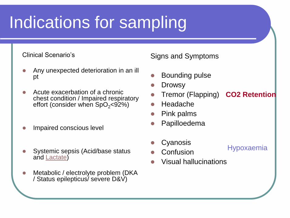
Indications for sampling
Clinical Scenario’s
Any unexpected deterioration in an ill pt
Acute exacerbation of a chronic chest condition / Impaired respiratory effort (consider when SpO2<92%)
Impaired conscious level
Systemic sepsis (Acid/base status and Lactate)
Metabolic / electrolyte problem (DKA / Status epilepticus/ severe D&V)
Signs and Symptoms
Bounding pulse
Drowsy
Tremor (Flapping)
Headache
Pink palms
Papilloedema
Cyanosis
Confusion
Visual hallucinations
CO2 Retention
Hypoxaemia

Indications for sampling
Monitoring of the Critically ill patient
Monitoring treatment of known
respiratory failure
Anyone ventilated invasively or
non-invasively
Peri-arrest / post-arrest
assessment
Any major trauma
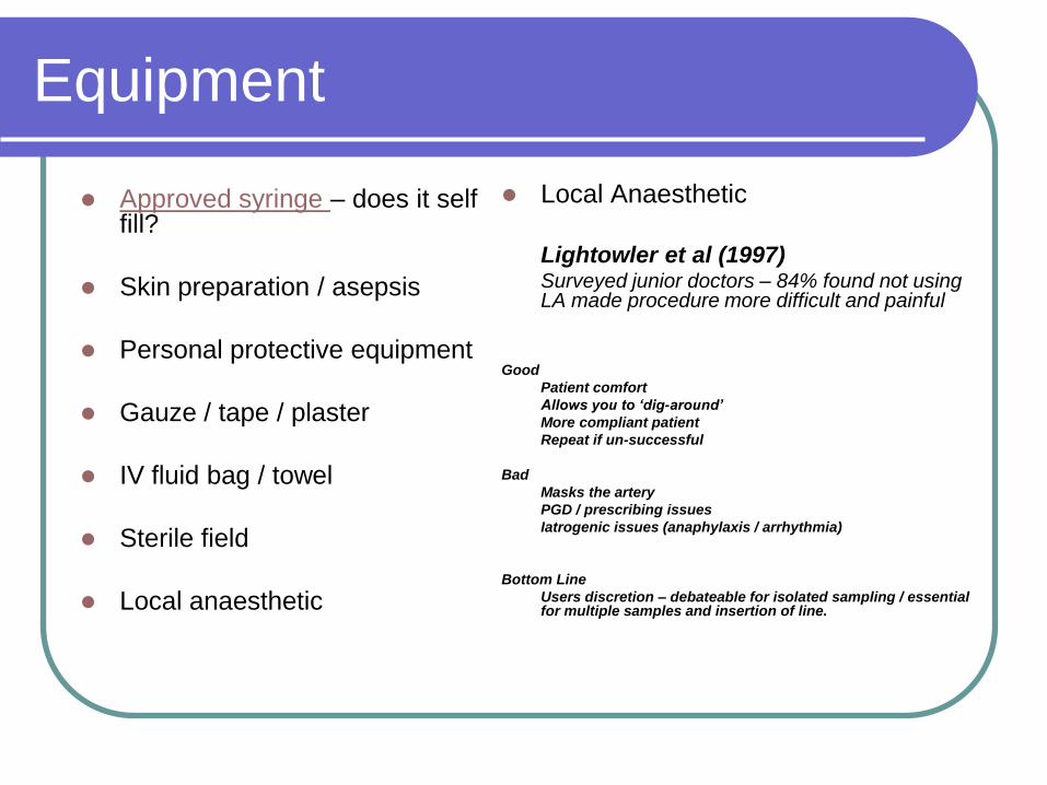
Equipment
Approved syringe – does it self fill?
Skin preparation / asepsis
Personal protective equipment
Gauze / tape / plaster
IV fluid bag / towel
Sterile field
Local anaesthetic
Local Anaesthetic
Lightowler et al (1997)Surveyed junior doctors – 84% found not using LA made procedure more difficult and painful
Good
Patient comfort
Allows you to ‘dig-around’
More compliant patient
Repeat if un-successful
Bad
Masks the artery
PGD / prescribing issues
Iatrogenic issues (anaphylaxis / arrhythmia)
Bottom Line
Users discretion – debateable for isolated sampling / essential for multiple samples and insertion of line.

Procedure
Identification of patient -
is there a clinical need?
Obtain consent
written / verbal & document
Access to sampler
Correct medium to transit
sample
Identify site
Aseptic procedure
Oxygen therapy
(Consider removal NOT
if pt in extremis!)

Puncture Sites

Preferred Sites
Radial
Easily accessible
Easily compressible
Post sampling can be observed easily

Preferred Sites
Femoral
Artery
(NAVY)
Femoral
Larger artery - collapsed
patient looses radial pulse
Disadvantages
More structures
‘Dirty area’
Accessibility

Femoral Artery
Clean area with a
suitable antiseptic
Needle at 90 degrees
to skin
NAVY
Pressure for 5 mins
minimum
N A V Y

Radial - Modified Allen’s Test
1) Elevate hand and make a tight fist for 30 seconds.
2) Apply firm pressure to ulnar and radial arteries to occlude them.
3) Still elevated, open the hand It should appear blanched (pallor can be observed at the finger nails).
4) Ulnar pressure is released and the colour should return in 7 seconds.
Inference: Ulnar artery supply to the hand is sufficient and it is safe to cannulate/prick the radial
If colour does not return or returns after 7–10 seconds, then the ulnar artery supply to the hand is not sufficient and the radial artery therefore cannot be safely stabbed
Anatomical basis
The hand is normally supplied by blood from the ulnar and radial arteries. The arteries undergo anastamosis in the hand. Thus, if the blood supply from one of the arteries is cut off, the other artery can supply adequate blood to the hand. A minority of people lack this dual blood supply.


Procedure
Position patient Radial, slight wrist
extension (IV bag / towel)
Femoral, abduct legs (genitals and abdomen)
Palpate artery
Palpate and clean skin
Prepare syringe - small amount of air

Procedure
Position fingers distally and proximally to needle insertion site / roll finger over artery
Advance needle Radial - 45 degrees to
skin
Femoral - 90 degrees to skin
When in artery blood should pulsate into syringe / may need to aspirate
Remove and apply immediate pressure for 5 mins

Sample Transit
Room temperature samples should be analysed within 10 – 15 mins
Store in iced water - samples should be analysed within one hour
Storing directly on ice can haemolyse sample (lower pH and PaO2 / increase PaCO2 and K+)
Possible elevation of potassium levels by prolonged chilling
Don’t shake the samples – affects potassium calibration and causes haemolysis
Gentle rolling of the syringe will reduce chance of clot formation
Remove air from the sample
Use approved syringes in machine – under-calculated heparin volumes can affect accuracy

Post procedure
Record in notes and ensure therapy is
adjusted as required
Recheck patient
further bleeding
Sensory defecit

Caution!
Negative Allen’s test (collateral circulation test)
Should not be taken through or distally to a surgical shunt (Fistula) or through a bypass graft (groin)
Should not be taken through areas of infection or via a limb with evidence of peripheral vascular disease (Raynaud’s Disease)
Caution with groin if evidence of thrush
Bleeding diathesis / coagulopathy (relative contraindication post thrombolysis)
When you haven’t done the basics!

Hazards / Complications
Haematoma
Arteriospasm
Emboli
Infection
Haemorrhage
Vessel trauma
Arterial occlusion
Nerve damage
Pain
Anaphylaxis (L.A.)
Needlestick injury to
clinician
Vaso-vagal episode

Arteriospasm / Embolism

Haematoma

Pathological Allen’s Test

Competency / Training
Trust Policy for non-medical practitioners to sample?
Appropriate training and assessment criteria
Need to maintain competence
Record of competency trust audit
Individual audit – record / case records

Blood Gas Analysis
The 5 step approach……


5-Step approach to arterial blood gas interpretation
1. Assess oxygenation
Is the patient hypoxic?
Is there a significant alveolar-arterial gradient?
2. Determine status of the pH or H+ concentration
pH > 7.45 (H+ < 35 nmol l-1) – alkalaemia
< 7.35 (H+ > 45 nmol l-1) – acidaemia
3. Determine respiratory component
PaCO2 > 6.0 kPa (45 mmHg) – respiratory acidosis
< 4.7 kPa (35 mmHg) – respiratory alkalosis
4. Determine metabolic component
HCO3- < 22 mmol l-1 – metabolic acidosis
> 26 mmol l-1 – metabolic alkalosis
0.1333kPa is 1mmHg
mmHg /7.5 approximates to kPa
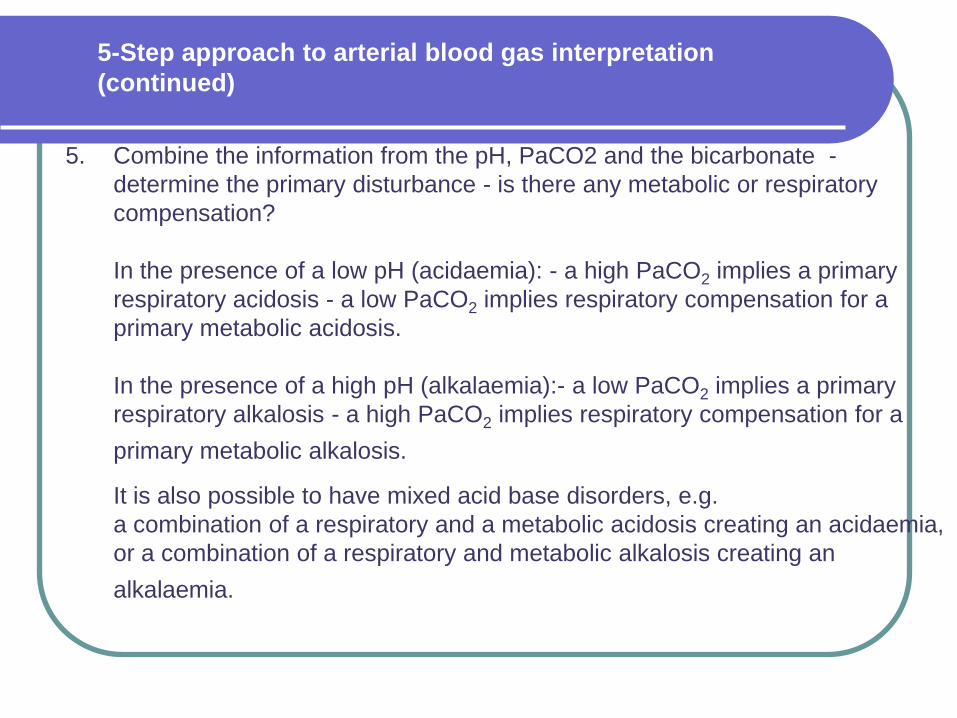
5. Combine the information from the pH, PaCO2 and the bicarbonate -
determine the primary disturbance - is there any metabolic or respiratory
compensation?
In the presence of a low pH (acidaemia): - a high PaCO2 implies a primary
respiratory acidosis - a low PaCO2 implies respiratory compensation for a
primary metabolic acidosis.
In the presence of a high pH (alkalaemia):- a low PaCO2 implies a primary
respiratory alkalosis - a high PaCO2 implies respiratory compensation for a
primary metabolic alkalosis.
It is also possible to have mixed acid base disorders, e.g.
a combination of a respiratory and a metabolic acidosis creating an acidaemia,
or a combination of a respiratory and metabolic alkalosis creating an
alkalaemia.
5-Step approach to arterial blood gas interpretation
(continued)

Physiological Response
For optimal cellular function / homeostasis, the body will naturally buffer itself in order to maintain an equilibrium.
Will buffer quickly by utilising the lungs to ‘blow-off’ or maintain CO2.
(CO2 is an acid)
The kidneys also act as a buffer by removing or keeping bicarbonate.
(Bicarbonate is an alkaline)

5-Step approach to arterial blood gas interpretation
Mixed Picture
Adjustments in both the respiratory and
metabolic components
HCO3- or base excess HCO3
- or base excess Metabolic
CO2 CO2 Respiratory
AlkalosisAcidosis

Respiratory Failure
Type 1 Low O2Normal / Low PaCO2
Asthmatics / LVF
Type 2 Low O2
Elevated PaCO2
Elevated HCO3
COPD

* If the compensation is virtually complete the pH may be in the normal range –
over compensation does not occur.
Those marked in bold are particularly common after cardiac arrest.
Summary of changes in pH, PaCO2 and HCO3-
in acid-base disorders
Acid-base disorder pH PaCO2 HCO3-
Respiratory acidosis (Asthma/COPD) N
Metabolic acidosis (DKA) N
Respiratory alkalosis (PE / hypervent.) N
Metabolic alkalosis (Vomiting) N
Respiratory acidosis with renal
compensation
*
Metabolic acidosis with respiratory
compensation (DKA – Kussmauls)
*

* If the compensation is virtually complete the pH may be in the normal range –
over compensation does not occur.
Those marked in bold are particularly common after cardiac arrest.
Summary of changes in pH, PaCO2 and HCO3-
in acid-base disorders (continued)
Acid-base disorder pH PaCO2 HCO3-
Respiratory alkalosis with renal
compensation
*
Metabolic alkalosis with respiratory
compensation
*
Mixed metabolic and respiratory
acidosis
Mixed metabolic and respiratory
alkalosis

Scenario 1
Initial Information
A 69 year old lady is brought to the emergency
department after a witnessed out-of-hospital VF cardiac
arrest.
The paramedics arrived approximately 8 minutes post
collapse, poor CPR had been given. The paramedics
had successfully restored spontaneous circulation after
3 shocks.
On arrival to ED:
(GCS 3/15)
ETT in-situ ventilated with 60% oxygen via ventilator
HR 118 min BP 140/100 mmHg.

Scenario 1 (continued)
Arterial blood gas analysis reveals:
FiO2 0.6 (60%) Normal Values
pH 7.10 7.35 – 7.45
PaCO2 6.2 kPa 4.7 – 6.0 kPa
PaO2 7.5 kPa > 10 kPa on air
HCO3- 14 mmol l-1 22 – 26 mmol l-1
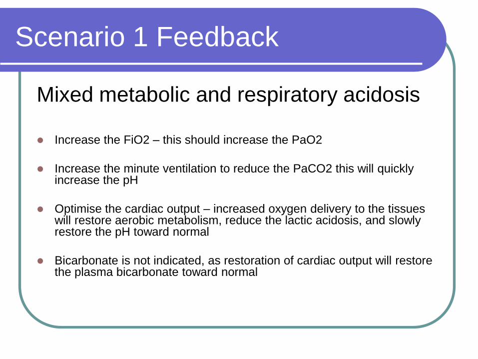
Scenario 1 Feedback
Mixed metabolic and respiratory acidosis
Increase the FiO2 – this should increase the PaO2
Increase the minute ventilation to reduce the PaCO2 this will quickly increase the pH
Optimise the cardiac output – increased oxygen delivery to the tissues will restore aerobic metabolism, reduce the lactic acidosis, and slowly restore the pH toward normal
Bicarbonate is not indicated, as restoration of cardiac output will restore the plasma bicarbonate toward normal

Scenario 2
Initial Information
A 65 year old man with severe COPD has just collapsed
in the waiting room.
On initial assessment by the ED nurse he is apnoeic but
has an easily palpable carotid pulse at 90 min-1.
The nurse is attempting to ventilate his lungs with a bag
-mask and supplemental oxygen (with reservoir) as the
ED team are summoned.

Scenario 2 (continued)
Arterial blood gas analysis reveals:
FiO2 0.85 (85%) estimated Normal Values
pH 7.10 7.35 – 7.45
PaCO2 18.0 kPa 4.7 – 6.0 kPa
PaO2 19.5 kPa > 10 kPa on air
HCO3- 36 mmol l-1 22 – 26 mmol l-1

Scenario 2 Feedback
Severe respiratory acidosis
The patient’s arterial blood is well oxygenated
There is a compensatory increase in bicarbonate, which reflects a chronically raised PaCO2 – this is consistent with severe COPD
The significant acidaemia (low pH) and very high PaCO2 indicate an additional acute respiratory acidosis (occurring around the time of the respiratory arrest) – in the presence of a fully compensated chronic respiratory acidosis the pH would be near normal.
If considered appropriate, the active treatment of this patient would include:
- Tracheal intubation and positive pressure ventilation

Scenario 3
A 23yr old female is brought to the ED by her parents she has experienced some emotional upheaval (‘boyfriend dumped her’). She is upset, hyperventilating and complains of her fingers feeling tingly.
A junior doctor has asked you to comment on the results……
RR 28 HR 116 BP 128/60

Scenario 3
Arterial blood gas analysis reveals:
FiO2 0.21 (21%) Normal Values
pH 7.51 7.35 – 7.45
PaCO2 2.65 kPa 4.7 – 6.0 kPa
PaO2 13.4 kPa > 10 kPa on air
HCO3- 23 mmol l-1 22 – 26 mmol l-1

Scenario 3 Feedback
Respiratory Alkalosis
Was an ABG indicated?
Paper Bag
Close curtains and lecture!!
Encourage deep slow breaths
Observe

Scenario 4
Initial Information
An 23 year old insulin dependent diabetic is brought to
the Emergency Department.
He has been unwell with the ‘man-flu’ for 3/7, has
vomited several times and not taken his insulin.
On arrival:
HR 130 min-1 BP 90/65 mmHg.
Spontaneous breathing, RR 35 min-1
Oxygen 4 l min-1 via Hudson mask
GCS 14 (E3, M6, V5)

Scenario 4 (continued)
Arterial blood gas analysis reveals:
FiO2 0.28 (28%) venturi Normal Values
pH 7.15 7.35 – 7.45
PaCO2 2.48 kPa 4.7 – 6.0 kPa
PaO2 22.0 kPa > 10 kPa on air
HCO3- 6.1 mmol l-1 22 – 26 mmol l-1
The blood glucose is 30 mmol l-1 and there are ketones+++
in the urine

Scenario 4 Feedback
Metabolic acidosis with partial compensation provided by a respiratory alkalosis
These blood gas values are consistent with severe diabetic ketoacidosis.
The treatment would include:
- Fluid resuscitation – initially, with normal saline
- Insulin
- The use of bicarbonate is controversial but many clinicians would give it in the presence of an acidaemia of this severity, particularly if it did not improve quickly after starting insulin and fluid resuscitation

Scenario 5 Initial Information
A 75 year old man is on the surgical ward 2 days after a laparotomy
for a perforated sigmoid colon secondary to diverticular disease.
He has become hypotensive over the last 6 hours. His vital signs
are:
Heart rate 120 min-1 – sinus tachycardia
warm peripheries
Blood pressure 70/40 mmHg
Respiratory rate 35 breaths min-1
SpO2 on oxygen 92%
Urine output 50 ml in the last 6 hours
GCS 13 (E3, M6, V4)

Scenario 5 (continued)
Arterial blood gas analysis reveals:
FiO2 0.4 (40%) approx Normal Values
pH 7.12 7.35 – 7.45
PaCO2 4.5 kPa 4.7 – 6.0 kPa (35–45 mmHg)
PaO2 8.2 kPa > 10 kPa (75 mmHg) on air
HCO3- 12 mmol l-1 22 – 26 mmol l-1
BE - 15 mmol l-1 +/- 2 mmol l-1

Simon Giles Consultant Nurse/ Critical
Care

References
www.the-abg-site.com
www.en.wikipedia.org/wiki/Main_Page
Beaumont T (1997) How to guides: arterial blood gas sampling. Care of the Critically Ill.
13, 1,
British Thoracic Society (2002) Non-invasive ventilation in acute respiratory
failure. Thorax. 57, 3, 192-211.
British Thoracic Society (1997) Guidelines for the management of chronic
obstructive pulmonary disease. Thorax. 52, Supplement 5.
Clutton-Brock T (1997) The assessment and monitoring of respiratory function. In Goldhill D, Withington P (Eds) Textbook of Intensive Care. London,Chapman & Hall.
Coombs M (2001) Making sense of arterial blood gases. Nursing Times.97, 27, 36-38.

References
Dellinger, R.P. et al (2008) Surviving Sepsis Campaign: International guidelines for management of severe sepsis and septic shock. Critical Care Medicine; 36: 1, 296–327. Giner J, Casan P, Belda J, et al. Pain during arterial puncture Chest 1996;110:1443-5.
Lightowler JV, Elliott MW. Local anaesthetic infiltration prior to arterial puncture for blood gas analysis: a survey of current practice and a randomised double blind placebo controlled trial. J R Coll Physicians Lond 1997;31:645-6.
Jarvis MA, Jarvis CL, Jones PR, Spyt TJ (October 2000). "Reliability of Allen's test in selection of patients for radial artery harvest". Ann. Thorac. Surg. 70 (4): 1362–5.
NICE (2007) Acutely Ill Patients in Hospital: Recognition of and Response to Acute Illness in Adults in Hospital. www.nice.org.uk
Pagana, K.D., Pagana T.J. (2006) In: Ruholl, L. (2006) Arterial blood gases: analysis and nursing responses. Medsurg Nursing: Official Journal of the Academy of Medical-surgical Nurses; 15: 6, 343–349.
Simpson, H. (2004) Interpretation of arterial blood gases: a clinical guide for nurses. British Journal of Nursing; 13: 9, 522–527.
Woodrow, P. (2004) Arterial blood gas analysis. Nursing Standard; 18: 21, 45–52.

Oxygenation
Normal Range 10 - 14 kPa
Is the PaO2 less than 8kpa (hypoxic)?
Is the patient on O2?
Ensure the FiO2 / delivered O2 is
documented on the gas sampler.
Is there a significant alveolar-arterial gradient?
1% 02 is approximately 1kPa.
Normal drop inspired to alveolar is approximately 10.0 kPa
Thus on an inspired oxygen concentration of 50% should give an approximate PaO2 of 40 kPa
FiO2 0.21 (Air 21%)
0.4 (40%)
5 Step
92%

pH
1 2 3 4 5 6 7 8 9 10 11 12 13 14
Acid AlkalineNeutral
The normal Range is 7.35 – 7.45
What is the pH doing ?
A range less than 7.35 is acidaemia (acidosis)
A range greater than 7.45 is alkaleamia (alkalosis)> 7.45 (H+ < 35 nmol l-1) alkalaemia
< 7.35 (H+ > 45 nmol l-1) acidaemia
5 Step

PaCO2
Carbon dioxide is an acidic gas and is responsible for the respiratory component of the blood gas. PaCO2 levels can fluctuate quickly by increasing / decreasing respiratory rates
Normal Range is 4.7 - 6.0 kPa
Does the pH reflect changes associated with PaCO2 levels?
PaCO2 >6.0 kPa Respiratory acidosis (Hypercapnia)
PaCO2 <4.7 kPa Respiratory alkalosis (Hypocapnia)
5 Step

Bicarbonate / Metabolic
Determine metabolic component
HCO3 < 22 mmol l-1 – metabolic acidosis
> 26 mmol l-1 – metabolic alkalosis
Bicarbonate is an alkaline and acts as a buffer,
Bicarbonate takes several hours to adjust
therefore elevated or lowered levels normally
indicate sub-acute / chronic changes.
Back

Venous versus arterial results
What parameters stay the same?
PH
Hb / electrolytes / COHb
Lactate – slightly higher on venous sample Normal range less than 1mmol/l
Back