aromatic-turmerone induces neural stem cell proliferation ... · aromatic-turmerone induces neural...
TRANSCRIPT
Hucklenbroich et al. Stem Cell Research & Therapy 2014, 5:100http://stemcellres.com/content/5/4/100
RESEARCH Open Access
Aromatic-turmerone induces neural stem cellproliferation in vitro and in vivoJoerg Hucklenbroich1,2, Rebecca Klein2,3, Bernd Neumaier3, Rudolf Graf3, Gereon Rudolf Fink1,2,Michael Schroeter1,2,3 and Maria Adele Rueger1,2,3*
Abstract
Introduction: Aromatic (ar-) turmerone is a major bioactive compound of the herb Curcuma longa. It has beensuggested that ar-turmerone inhibits microglia activation, a property that may be useful in treating neurodegenerativedisease. Furthermore, the effects of ar-turmerone on neural stem cells (NSCs) remain to be investigated.
Methods: We exposed primary fetal rat NSCs to various concentrations of ar-turmerone. Thereafter, cell proliferationand differentiation potential were assessed. In vivo, naïve rats were treated with a single intracerebroventricular (i.c.v.)injection of ar-turmerone. Proliferative activity of endogenous NSCs was assessed in vivo, by using noninvasive positronemission tomography (PET) imaging and the tracer [18F]-fluoro-L-thymidine ([18F]FLT), as well as ex vivo.
Results: In vitro, ar-turmerone increased dose-dependently the number of cultured NSCs, because of an increase inNSC proliferation (P < 0.01). Proliferation data were supported by qPCR-data for Ki-67 mRNA. In vitro as well as in vivo,ar-turmerone promoted neuronal differentiation of NSCs. In vivo, after i.c.v. injection of ar-turmerone, proliferating NSCswere mobilized from the subventricular zone (SVZ) and the hippocampus of adult rats, as demonstrated by both [18F]FLT-PET and histology (P < 0.05).
Conclusions: Both in vitro and in vivo data suggest that ar-turmerone induces NSC proliferation. Ar-turmerone thusconstitutes a promising candidate to support regeneration in neurologic disease.
IntroductionCurcumin and ar-turmerone are the major bioactive com-pounds of the herb Curcuma longa. Although many studieshave demonstrated curcumin to possess antiinflammatoryand neuroprotective properties (reviewed by [1]), to date,the effects of ar-turmerone remain to be elucidated. Forexample, antitumor properties, exerted via the induc-tion of apoptosis [2] and inhibition of tumor cell invasion[3], have been attributed to ar-turmerone. Park et al. [4,5]recently suggested that ar-turmerone also possessesantiinflammatory properties resulting from the blockadeof key signaling pathways in microglia. Because micro-glia activation is a hallmark of neuroinflammation andis associated with various neurologic disorders, inclu-ding neurodegenerative diseases [6,7] and stroke [8,9],
* Correspondence: [email protected] Neuroscience, Institute of Neuroscience and Medicine (INM-3),Research Centre Juelich, Leo-Brandt-Straße 52425, Jülich, Germany2Department of Neurology, University Hospital of Cologne, Cologne, GermanyFull list of author information is available at the end of the article
© Hucklenbroich et al.; licensee BioMedCreative Commons Attribution License (http:/distribution, and reproduction in any mediumDomain Dedication waiver (http://creativecomarticle, unless otherwise stated.
2014
ar-turmerone constitutes a promising therapeutic agentfor various neurologic disorders.The regenerative potential of endogenous neural
stem cells (NSCs) plays an important role in neuro-degenerative disease and stroke. Endogenous NSCs aremobilized by cerebral ischemia [10] as well as byvarious neurodegenerative diseases [11,12], although theirintrinsic regenerative response is insufficient to enablefunctional recovery. The targeted (that is, pharmacologic)activation of endogenous NSCs has been shown to enhanceself-repair and recovery of function in the adult brain inboth stroke [13,14] and neurodegeneration [15]. Impor-tantly, NSCs and microglia relevantly interact with eachother, thereby affecting their respective functions [16,17].Thus, with the perspective of ar-turmerone as a thera-
peutic option in mind, we investigated the effects ofar-turmerone on NSCs in vitro and in vivo.
Central Ltd. This is an Open Access article distributed under the terms of the/creativecommons.org/licenses/by/4.0), which permits unrestricted use,, provided the original work is properly credited. The Creative Commons Publicmons.org/publicdomain/zero/1.0/) applies to the data made available in this

Hucklenbroich et al. Stem Cell Research & Therapy Page 2 of 92014, 5:100http://stemcellres.com/content/5/4/100
Material and methodsCell cultureNSCs were cultured from fetal rat cortex at embryonic day14.5, as described previously [18]. Cells were expanded asmonolayer cultures in serum-free DMEM/F12 medium (LifeTechnologies, Darmstadt, Germany) with N2 supplement(Gibco, Karlsruhe, Germany) and fibroblast growth factor(FGF2; 10 ng/ml; Invitrogen, Karlsruhe, Germany) for 5 daysand were replated in a 24-well plate at 10,000 cells per cm2.FGF2 was included throughout the experiments.Ar-turmerone (Fluka, Munich, Germany) was added to
cultures at replating at concentrations of 0, 1.56, 3.125,6.25, 12.5, and 25 μg/ml. All experiments were performedin triplicate. After 72 hours, representative pictures weretaken by using an inverted fluorescence phase-contrastmicroscope (Keyence BZ-9000E). Three images weretaken per well, and cells were counted by using the soft-ware ImageJ with a threshold of 20 px (National Institutesof Health, Bethesda, MD, USA, Version 1.47 k).To determine the ratio of proliferating cells, 10 μM bro-
modeoxyuridine (BrdU; Fluka, Munich, Germany) wasadded to cultures for 6 hours, before cells were fixed with4% PFA. Again, all experiments were performed in tripli-cate. Cells were stained with mAb against BrdU to identifyproliferating cells (clone BU-33, dilution 1:100; Sigma-Aldrich, Munich, Germany). For antigen-retrieval beforestaining, sections were incubated in 2 N HCl for 30 mi-nutes. For visualization, FITC-labeled anti-mouse IgG wasused (Invitrogen); all cells were additionally counter-stained with Hoechst 33342 (Life Technologies). To calcu-late the ratio of proliferating cells, BrdU-positive cellswere divided by the total cell number in each sample, andmean values were established among equally treated cells.To establish its effect on cell survival, ar-turmerone
was added to NSC cultures for 24 hours. To discrimi-nate between live and dead cells, the live/dead cell-mediated cytotoxicity kit (Life Technologies, cat. no.L7010) was used according to the manufacturer’sinstructions. Both viable and dead NSCs were countedin n = 6 samples per condition, and a ratio of survivingcells was calculated for each field of view; mean valueswere calculated for each concentration tested.To assess the differentiation potential of NSCs treated
with ar-turmerone, mitogen was withdrawn during theexpansion phase, followed by a differentiation phase of10 days, in the absence (control) or presence of 6.25 μg/mlar-turmerone. Immunocytochemistry with markers foryoung neurons (TuJ1), astrocytes (GFAP), and oligodendro-cytes (CNPase) was used to verify all three differentiated fatesof NSCs, whereas SOX2 marked undifferentiated NSCs.
Real-time quantitative PCR (RT-qPCR)RNA from cells was isolated by using the RNeasy Mini Kit(Qiagen, Hilden, Germany). Total RNA concentration and
purity were evaluated photometrically. Total RNA wasconverted to c-DNA by reverse transcription with theQuantitect reverse transcription kit (Qiagen). The primerused for Ki67 was obtained from Biolegio (Nijmegen,The Netherlands). The sequences of the primers wereas follows: (a) forward: TCTTGGCACTCACAGTCCAG,and (b) reverse: GCTGGAAGCAAGTGAAGTCC. Theq-PCR reaction was carried out by using 10 ng total RNAin a 20-μl reaction (Quantitect Reagents, Qiagen) accor-ding to the manufacturer’s instructions. The samples wereamplified and quantified on a Rotorgene 2000 (Corbett,Sydney, Australia) by using the following thermal cyclerconditions: activation: 95°C 10 minutes; cycling: 50 cycles,step 1: 92°C, 15 seconds, step 2: 52°C, 15 seconds, andstep 3: 72°C, 40 seconds. PCR product integrity was evalu-ated by melting-point analysis and agarose gel electro-phoresis. Each sample and gene was normalized toRPL13a as reference gene [19]. Ki67 mRNA levels werenormalized to endogenous RPL13a expression (ΔCT);normalized values were then expressed as 2-ΔCt. Meanvalues were calculated for treated and untreated cells.
Animals and surgeryAll animal procedures were in accordance with theGerman Laws for Animal Protection and were approvedby the local animal care committee (Buero derTierschutzbeauftragten, MPIfNF, Cologne, Germany), aswell as local governmental authorities (LANUV NRW84–02.04.2012.A116). Spontaneously breathing maleWistar rats weighing 290 to 330 g were anesthetizedwith 5% isoflurane and maintained with 2.5% isofluranein 65%:35% nitrous oxide/oxygen. Throughout surgicalprocedures, the body temperature was maintained at37.0°C with a thermostatically controlled heating pad.
Intracerebroventricular injectionsOne group of animals (n = 3) underwent a single intra-cerebroventricular (i.c.v.) injection of 3 mg ar-turmeroneat a concentration of 1 mg/μl. For control, n = 6 rats werevehicle-injected with the identical volume of normalsaline. Under anesthesia with 1.5% isoflurane, each rat’sskull was fixated in a stereotaxic frame in plane orienta-tion. After incision of the skin, the bregma was exposed,and a burr hole was drilled over the right lateral ventricleby using the following stereotaxic coordinates: bregma,AP −0.9 mm; ML, −1.4 mm; and VD, +3.8 mm. Ar-turmerone dissolved in normal saline, or respectively, puresaline as control, was injected at 1 μl/min. After injection,the needle was left in place for another 5 minutes to allowa distribution of the solution within the ventricles. Theneedle was thereafter withdrawn slowly, and the skinsutured with nonabsorbing silk.After each procedure, all animals were allowed to
recover from anesthesia and were put back into their

Hucklenbroich et al. Stem Cell Research & Therapy Page 3 of 92014, 5:100http://stemcellres.com/content/5/4/100
home cages, where they were given access to food andwater ad libitum.
BrdU injectionsIn all animals, the tracer bromodeoxyuridine (BrdU) wasinjected intraperitoneally for 5 days, starting on the day ofi.c.v. injection, at a concentration of 50 mg/kg per injec-tion, as described previously [18]. This regimen resultedin a cumulative dose of 250 mg/kg BrdU per animal.
Positron emission tomography (PET)[18F]-fluoro-L-thymidine ([18F]FLT) was synthesized asdescribed previously [20]. Seven days after i.c.v. injectionof ar-turmerone or placebo, respectively, PET imagingwas performed on a microPET Focus 220 scanner(Concorde Microsystems, Inc., Knoxville, TN, USA;63 image planes; 1.5-mm full width at the half ma-ximum). Animals were anesthetized with 5% isoflurane,maintained with 2% isoflurane in a 65%:35% nitrousoxide/oxygen atmosphere, and placed in the scanner.Temperature was monitored by using a rectal probe andmaintained at 37°C ± 0.5°C by a thermostatically con-trolled water-flow system (Medres, Cologne, Germany).After a 10-minute transmission scan for attenuationcorrection, rats received an intravenous bolus injectionof [18F]FLT (1.0 to 2.2 mCi/rat), and emission data wereacquired for 60 minutes. PET data were reconstructed intwo time frames of 1,800 seconds. The last frame (thatis, minutes 31 to 60 after tracer injection) was used forimage analysis.
Image analysisPET images were co-registered to anatomic data of a 3Drat-brain atlas constructed from the brain slices pre-sented by Swanson [21]. Based on the 3D anatomic data,ellipsoid volumes of interest (VOIs) measuring 4 mm3
were placed to cover the subventricular zone (SVZ) as wellas the dentate gyrus region of the hippocampus. A stan-dard uptake value (SUV) was calculated for each VOI,dividing maximal VOI activity by the decay-correctedinjected radioactive dose per body weight. SUVs wereindividually determined and then averaged betweenanimals within each group.
ImmunohistochemistryAfter PET imaging, or 7 days after ar-turmerone treat-ment, rats were deeply anesthetized and decapitated.The brains were rapidly removed, frozen in isopentane,and stored at −80°C before further histologic and immu-nohistochemical processing. Ten-μm-thick adjacent ser-ial coronal brain sections were cut at 500-μm intervalsand stained with anti-BrdU to identify proliferating cells(mAb clone BU-33, dilution 1:200; Sigma-Aldrich), orwith anti-doublecortin (DCX) to identify neuroblasts
(rabbit polyclonal, dilution 1:1,000, Sigma-Aldrich). Forantigen-retrieval before BrdU staining, sections weremicrowave-heated in 0.01M citrate buffer, pH 6.0, for5 minutes, followed by 2 N HCl at 37°C for 30 minutes.For visualization, the ABC Elite kit (Vector Laboratorieswith diaminobenzidine (Sigma-Aldrich) as the finalreaction product was used.To quantify the width of the SVZ and of the dentate
gyrus of the hippocampus, it was measured on threeconsecutive BrdU-stained slices per animal, and an ave-rage was calculated per animal. To quantify the numberof neuroblasts in the SVZ, their number was counted onthree consecutive DCX-stained slices within a standar-dized field-of-view for each animal. For both schemes ofquantification, mean values were calculated for eachgroup of animals.
Statistical analysisDescriptive statistics were performed with MicrosoftExcel 2003 (Microsoft Corp., Redmond, WA, USA).One-way ANOVA tests (followed by Holm-Sidak posthoc test) were performed with SigmaPlot 11.0 forWindows (Systat Software Inc., San Jose, CA, USA).Statistical significance was set at P < 0.05.
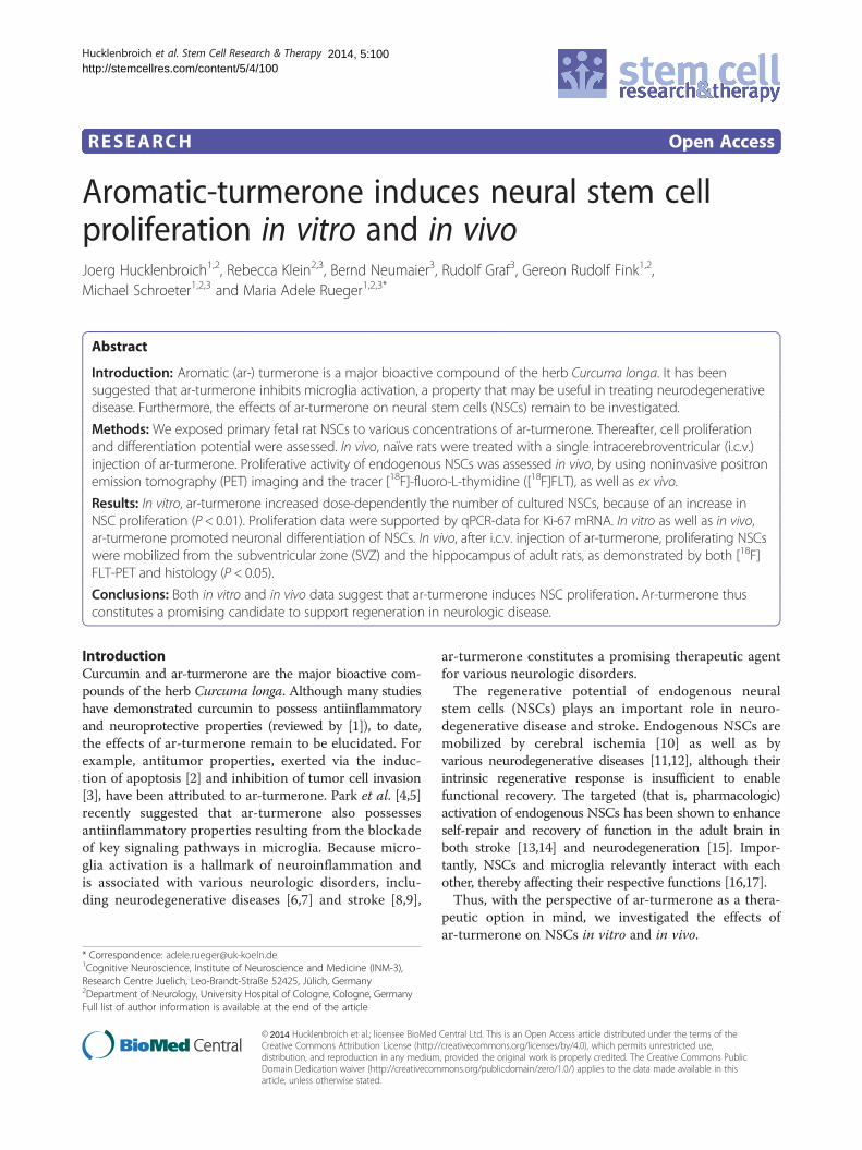
ResultsEffects on NSC proliferation in vitroTo assess the effects of ar-turmerone on NSC in primaryculture, rat fetal NSC were grown in the presence ofvarious concentrations of ar-turmerone for 72 hours.Cell numbers significantly increased when NSCs weretreated with 3.125 to 25 μg/ml ar-turmerone (P < 0.05),with a maximum increase of NSC numbers by ~80% at6.25 μg/ml (Figure 1A; P < 0.01).With the BrdU-incorporation assay, we next investi-
gated whether this increase in NSC number was causedby an increase in NSC proliferation. Indeed, treatmentwith certain concentrations of ar-turmerone significantlyincreased the percentage of proliferating NSCs from ~50%to ~80% (Figure 1B; P < 0.01). This result was verified onthe mRNA level by using qPCR for the proliferationmarker Ki67. In line with the BrdU data, treatment with6.25 μg/ml ar-turmerone led to a significant increase inKi67 mRNA (Figure 1C; P < 0.05).To assess whether ar-turmerone affected NSC survival,
viable and dead cells were determined after 24 hours,and the proportion of surviving cells was quantified foreach concentration of ar-turmerone. Concentrationsbetween 1.56 and 6.25 μg/ml that had yielded the ma-ximum effect on NSC proliferation did not affect cellsurvival. Higher concentrations of 12.5 and 25 μg/ml ledto a significant decrease in the number of viable NSCs(Figure 1D; P < 0.05).
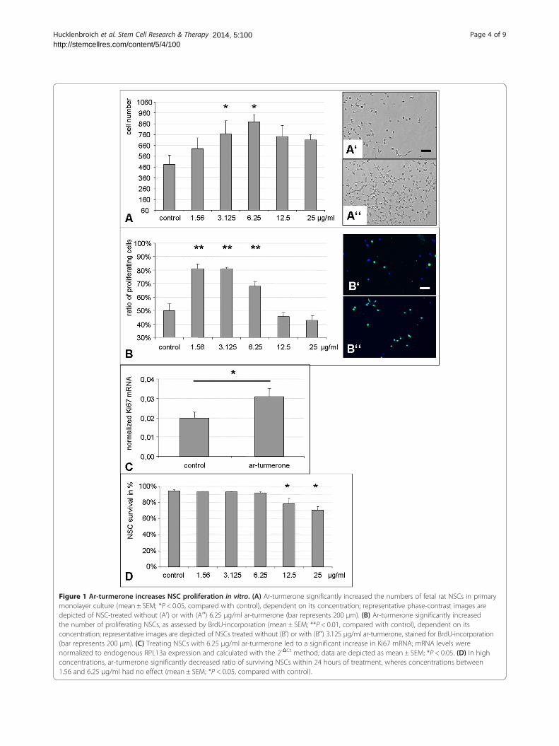
Figure 1 Ar-turmerone increases NSC proliferation in vitro. (A) Ar-turmerone significantly increased the numbers of fetal rat NSCs in primarymonolayer culture (mean ± SEM; *P < 0.05, compared with control), dependent on its concentration; representative phase-contrast images aredepicted of NSC-treated without (Aʹ) or with (Aʹʹ) 6.25 μg/ml ar-turmerone (bar represents 200 μm). (B) Ar-turmerone significantly increasedthe number of proliferating NSCs, as assessed by BrdU-incorporation (mean ± SEM; **P < 0.01, compared with control), dependent on itsconcentration; representative images are depicted of NSCs treated without (Bʹ) or with (Bʹʹ) 3.125 μg/ml ar-turmerone, stained for BrdU-incorporation(bar represents 200 μm). (C) Treating NSCs with 6.25 μg/ml ar-turmerone led to a significant increase in Ki67 mRNA; mRNA levels werenormalized to endogenous RPL13a expression and calculated with the 2-ΔCt method; data are depicted as mean ± SEM; *P < 0.05. (D) In highconcentrations, ar-turmerone significantly decreased ratio of surviving NSCs within 24 hours of treatment, wheres concentrations between1.56 and 6.25 μg/ml had no effect (mean ± SEM; *P < 0.05, compared with control).
Hucklenbroich et al. Stem Cell Research & Therapy Page 4 of 92014, 5:100http://stemcellres.com/content/5/4/100

Hucklenbroich et al. Stem Cell Research & Therapy Page 5 of 92014, 5:100http://stemcellres.com/content/5/4/100
Differentiation potential of NSCsTo assess the effect of ar-turmerone on the differentiationpotential of NSCs in vitro, cells in the expansion phasewere treated with or without 6.25 μg/ml ar-turmeroneand allowed to differentiate for 10 days by withdrawalof FGF2. Compared with that in untreated controlcells, the differentiation process was significantly
Figure 2 Ar-turmerone induces neurogenesis in vitro and in vivo. (A)of 6.25 μg/ml ar-turmerone. Immunocytochemistry 10 days after growth-fathe turmerone-treated group, but more young neurons. The generation of(mean ± SEM; **P < 0.01, compared with control). (B) Representative image(left), TuJ1-positive young neurons (middle), and GFAP-positive astrocytes (ar-turmerone, significantly more DCX-positive neuroblasts were observed inSEM; **P < 0.01). (D) Representative staining of DCX-positive neuroblasts in
accelerated in ar-turmerone-treated NSCs, with fewerundifferentiated (SOX2-positive) cells 10 days afterFGF2-withdrawal (Figure 2A; P < 0.01). Moreover,ar-turmerone-treated NSCs preferentially differenti-ated into young neurons, as assessed by TuJ1 staining,compared with untreated control cells (Figure 2A, B;P < 0.01). The generation of GFAP-positive astrocytes
NSCs were allowed to differentiate in the absence (control) or presencector discontinuation revealed fewer undifferentiated (SOX2+) NSCs inastrocytes and oligodendrocytes was not affected by ar-turmerones of differentiated cells include CNPase-positive oligodendrocytesright); bar represents 50 μm. (C) After i.c.v. injection of 3 mg (1 mg/μl)the SVZ compared with placebo-injected control animals (mean ±the SVZ (bar represents 50 μm).
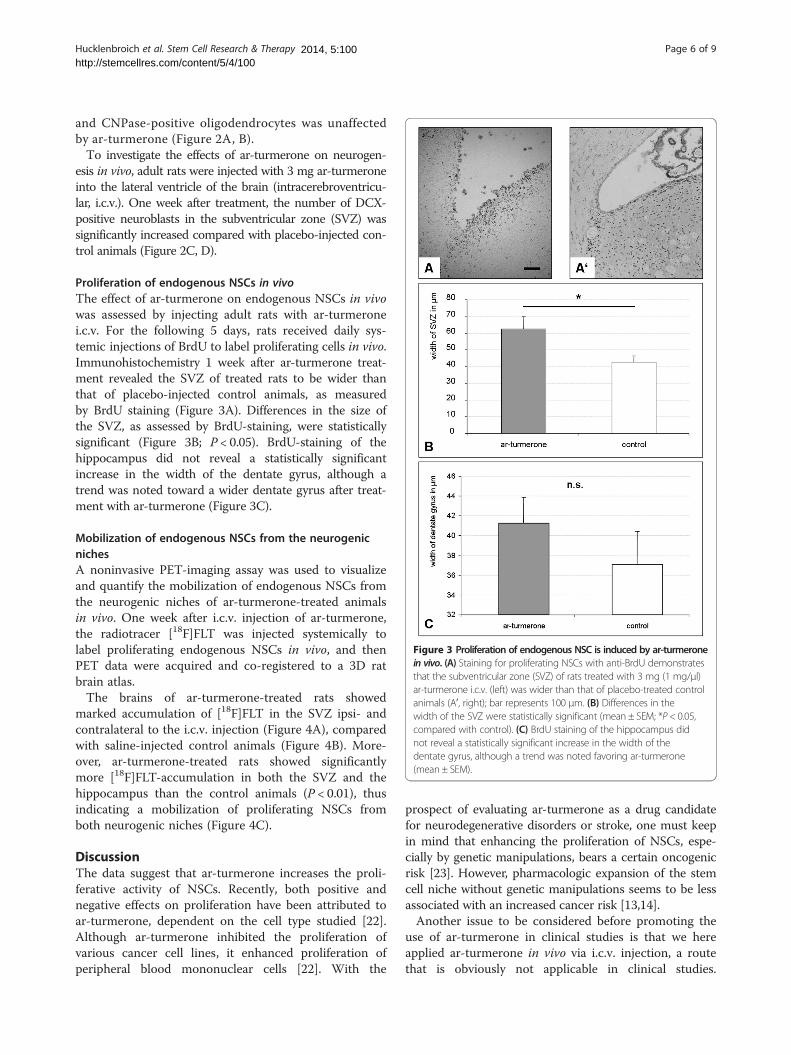
Figure 3 Proliferation of endogenous NSC is induced by ar-turmeronein vivo. (A) Staining for proliferating NSCs with anti-BrdU demonstratesthat the subventricular zone (SVZ) of rats treated with 3 mg (1 mg/μl)ar-turmerone i.c.v. (left) was wider than that of placebo-treated controlanimals (Aʹ, right); bar represents 100 μm. (B) Differences in thewidth of the SVZ were statistically significant (mean ± SEM; *P < 0.05,compared with control). (C) BrdU staining of the hippocampus didnot reveal a statistically significant increase in the width of thedentate gyrus, although a trend was noted favoring ar-turmerone(mean ± SEM).
Hucklenbroich et al. Stem Cell Research & Therapy Page 6 of 92014, 5:100http://stemcellres.com/content/5/4/100
and CNPase-positive oligodendrocytes was unaffectedby ar-turmerone (Figure 2A, B).To investigate the effects of ar-turmerone on neurogen-
esis in vivo, adult rats were injected with 3 mg ar-turmeroneinto the lateral ventricle of the brain (intracerebroventricu-lar, i.c.v.). One week after treatment, the number of DCX-positive neuroblasts in the subventricular zone (SVZ) wassignificantly increased compared with placebo-injected con-trol animals (Figure 2C, D).
Proliferation of endogenous NSCs in vivoThe effect of ar-turmerone on endogenous NSCs in vivowas assessed by injecting adult rats with ar-turmeronei.c.v. For the following 5 days, rats received daily sys-temic injections of BrdU to label proliferating cells in vivo.Immunohistochemistry 1 week after ar-turmerone treat-ment revealed the SVZ of treated rats to be wider thanthat of placebo-injected control animals, as measuredby BrdU staining (Figure 3A). Differences in the size ofthe SVZ, as assessed by BrdU-staining, were statisticallysignificant (Figure 3B; P < 0.05). BrdU-staining of thehippocampus did not reveal a statistically significantincrease in the width of the dentate gyrus, although atrend was noted toward a wider dentate gyrus after treat-ment with ar-turmerone (Figure 3C).
Mobilization of endogenous NSCs from the neurogenicnichesA noninvasive PET-imaging assay was used to visualizeand quantify the mobilization of endogenous NSCs fromthe neurogenic niches of ar-turmerone-treated animalsin vivo. One week after i.c.v. injection of ar-turmerone,the radiotracer [18F]FLT was injected systemically tolabel proliferating endogenous NSCs in vivo, and thenPET data were acquired and co-registered to a 3D ratbrain atlas.The brains of ar-turmerone-treated rats showed
marked accumulation of [18F]FLT in the SVZ ipsi- andcontralateral to the i.c.v. injection (Figure 4A), comparedwith saline-injected control animals (Figure 4B). More-over, ar-turmerone-treated rats showed significantlymore [18F]FLT-accumulation in both the SVZ and thehippocampus than the control animals (P < 0.01), thusindicating a mobilization of proliferating NSCs fromboth neurogenic niches (Figure 4C).
DiscussionThe data suggest that ar-turmerone increases the proli-ferative activity of NSCs. Recently, both positive andnegative effects on proliferation have been attributed toar-turmerone, dependent on the cell type studied [22].Although ar-turmerone inhibited the proliferation ofvarious cancer cell lines, it enhanced proliferation ofperipheral blood mononuclear cells [22]. With the
prospect of evaluating ar-turmerone as a drug candidatefor neurodegenerative disorders or stroke, one must keepin mind that enhancing the proliferation of NSCs, espe-cially by genetic manipulations, bears a certain oncogenicrisk [23]. However, pharmacologic expansion of the stemcell niche without genetic manipulations seems to be lessassociated with an increased cancer risk [13,14].Another issue to be considered before promoting the
use of ar-turmerone in clinical studies is that we hereapplied ar-turmerone in vivo via i.c.v. injection, a routethat is obviously not applicable in clinical studies.
Figure 4 Endogenous NSCs in the neurogenic niches of the rat brain are mobilized by ar-turmerone in vivo. (A) [18F]FLT-PET of a ratbrain 1 week after intracerebroventricular injection of ar-turmerone shows enhanced accumulation of [18F]FLT in the subventricular zone com-pared with (B) Saline-injected control brain, indicating an increase of proliferating endogenous NSCs caused by ar-turmerone. (C) Ar-turmerone-treated rats showed significantly more [18F]FLT accumulation in the SVZ and the hippocampus than did control animals (mean ± SEM; **P < 0.01).
Hucklenbroich et al. Stem Cell Research & Therapy Page 7 of 92014, 5:100http://stemcellres.com/content/5/4/100
However, another recent study found good bioavailabi-lity of ar-turmerone after both intravenous or intra-peritoneal injection in the mouse [24].In vivo, ar-turmerone expanded the width of the SVZ
by ~45%. An expansion of this NSC niche has also beendemonstrated for other pharmacologic agents suchas growth factors [13,14]. In a similar experimentalsetting, we previously observed that FGF2 expanded theSVZ by ~350%, whereas a combination of the Notchligand Delta-like 4 and insulin led to an increase in thewidth of the SVZ of ~66% [18]. Our data thereforesuggest that the effect of ar-turmerone on the NSCniche in vivo is somewhat smaller than that of ”classic”NSC-activation pathways. Nevertheless, the pleiotropiceffects of ar-turmerone render it a promising drug forfurther studies.Ar-turmerone was recently described to inhibit the
LPS- or Aß-induced activation of microglia through in-hibition of NF-κB, JNK-, and p38-MAPK pathways [4,5].Microglia activation as the hallmark of an innate inflam-matory response of the central nervous system (CNS)has been found in many neurologic disorders that areconsidered to be primarily nonimmunogenic, such asstroke [25,26], traumatic brain injury (TBI [27], Parkinsondisease [6], or Alzheimer disease [7]. NSC and immunecells interact extensively [16,17,28-30]. Therefore, thera-peutically regulating one entity’s fate is likely to influencethe other.
Yet, our knowledge about the interaction of NSCsand inflammatory responses in the CNS with regard toregeneration and functional recovery to date remainsscarce. On attraction by proinflammatory cytokines,endogenous NSCs considerably affect this regenerativeresponse [31,32], for example, through inducing remyeli-nization [33] and neuroprotection [15]. As ar-turmeroneboth limits microglia activation and induces NSC prolife-ration, it constitutes a promising future drug candidate tosupport regeneration in neurologic disorders.In the presence of mitogen in cell culture, as well as
under physiological conditions in vivo, we found ar-turmerone to promote neurogenesis. However, after FGF2discontinuation in vitro, treatment with ar-turmerone ledto an accelerated decrease of undifferentiated NSC, indi-cating an early exit from the cell cycle. This effect suggeststhat ar-turmerone may act as a weak antagonist on theFGF-receptor only in the absence of the ligand.Further studies are needed to clarify such a putative
relationship. In support of this notion, a recent reportsuggests that ar-turmerone acts as an antagonist on therelated epidermal growth factor (EGF) receptor [34].Noninvasive in vivo imaging is a crucial tool for transla-
tion from bench to bedside (that is, from experimentalanimal to human studies). We used PET imaging and theradiotracer [18F]FLT that enables imaging and measuringof proliferation, thereby allowing noninvasive detectionand quantification of endogenous NSC mobilization in the
Hucklenbroich et al. Stem Cell Research & Therapy Page 8 of 92014, 5:100http://stemcellres.com/content/5/4/100
adult rat brain in vivo [18]. This imaging assay is capableof monitoring the effects of drugs aimed at expanding theNSC niche [35]. By using [18F]FLT-PET, we here found ar-turmerone to mobilize NSCs from both neurogenicniches, the SVZ and the dentate gyrus of the hippo-campus, in vivo. Thus, this study provides further evi-dence for NSC activation by ar-turmerone, spanning fromcell-culture findings to in vivo imaging.
ConclusionsIn this study, we investigated the effects of ar-turmeroneon NSCs in vitro and in vivo. Ar-turmerone increased thenumber of NSCs both in cell culture and in the adult ratbrain in vivo. This increase resulted from enhanced NSCproliferation and led to promoted neurogenesis duringdifferentiation. In vivo, ar-turmerone mobilized endoge-nous NSCs from both neurogenic niches, the SVZ and thehippocampus. We propose that ar-turmerone constitutesa promising future drug candidate to support regenerationin neurologic disorders.
Abbreviations[18F]FLT: [18F]-fluoro-L-thymidine; ar-turmerone: aromatic turmerone;BrdU: bromodeoxyuridine; CNPase: 2´,3´-cyclic nucleotide 3´-phosphodiesterase; DCX: doublecortin; DMEM: Dulbecco Modified EagleMedium; FGF2: fibroblast growth factor 2; GFAP: glial fibrillary acidic protein;HCl: hydrogen chloride; i.c.v.: intracerebroventricular; NSC: neural stem cell;PCR: polymerase chain reaction; PET: positron-emission tomography;RNA: ribonucleic acid; SUV: standard uptake value; SVZ: subventricular zone;TuJ1: neuron-specific class III beta-tubulin; VOI: volume of interest.
Competing interestsThe authors declare that they have no competing interests.
Authors’ contributionsJH carried out all cell-culture experiments and PCR analyses, and performedsome of the statistical analyses. RK carried out the in vivo experiments,including surgery and imaging, and performed some of the statisticalanalyses. BN produced the radiotracer for PET-imaging. RG, GRF, and MSparticipated in the design and coordination of the study and helped to draftthe manuscript. MAR conceived of, designed, and coordinated the study,helped with the statistical analyses, and drafted the manuscript. All authorsread and approved the final manuscript.
AcknowledgementsThis work was supported by the Koeln Fortune Program/Faculty of Medicine,University of Cologne, Germany (106/2012), and the EU FP7 project“NeuroFGL.” We thank Mrs. Claudia Drapatz and Mrs. Katrin Eckstein forexcellent technical assistance.
Author details1Cognitive Neuroscience, Institute of Neuroscience and Medicine (INM-3),Research Centre Juelich, Leo-Brandt-Straße 52425, Jülich, Germany.2Department of Neurology, University Hospital of Cologne, Cologne, Germany.3Max Planck Institute for Neurological Research, Cologne, Germany.
Received: 27 May 2014 Revised: 12 August 2014Accepted: 12 August 2014 Published:
References1. Mythri RB, Bharath MM: Curcumin: a potential neuroprotective agent in
Parkinson’s disease. Curr Pharm Des 2012, 18:91–99.2. Lee Y: Activation of apoptotic protein in U937 cells by a component of
turmeric oil. BMB Rep 2009, 42:96–100.
26 Sep 2014
3. Park SY, Kim YH, Kim Y, Lee SJ: Aromatic-turmerone attenuates invasionand expression of MMP-9 and COX-2 through inhibition of NF-kappaBactivation in TPA-induced breast cancer cells. J Cell Biochem 2012,113:3653–3662.
4. Park SY, Jin ML, Kim YH, Kim Y, Lee SJ: Anti-inflammatory effects ofaromatic-turmerone through blocking of NF-kappaB, JNK, and p38MAPK signaling pathways in amyloid beta-stimulated microglia.Int Immunopharmacol 2012, 14:13–20.
5. Park SY, Kim YH, Kim Y, Lee SJ: Aromatic-turmerone’s anti-inflammatoryeffects in microglial cells are mediated by protein kinase A and hemeoxygenase-1 signaling. Neurochem Int 2012, 61:767–777.
6. Hirsch EC, Hunot S: Neuroinflammation in Parkinson’s disease: a targetfor neuroprotection? Lancet Neurol 2009, 8:382–397.
7. Perrin RJ, Fagan AM, Holtzman DM: Multimodal techniques for diagnosisand prognosis of Alzheimer’s disease. Nature 2009, 461:916–922.
8. Iadecola C, Anrather J: The immunology of stroke: from mechanisms totranslation. Nat Med 2011, 17:796–808.
9. Schroeter M, Dennin MA, Walberer M, Backes H, Neumaier B, Fink GR,Graf R: Neuroinflammation extends brain tissue at risk to vital peri-infarcttissue: a double tracer [(11)C]PK11195- and [(18)F]FDG-PET study.J Cereb Blood Flow Metab 2009, 29:1216–1225.
10. Liu J, Solway K, Messing RO, Sharp FR: Increased neurogenesis in thedentate gyrus after transient global ischemia in gerbils. J Neurosci 1998,18:7768–7778.
11. Curtis MA, Penney EB, Pearson AG, Roon-Mom WM, Butterworth NJ, Dragunow M,Connor B, Faull RL: Increased cell proliferation and neurogenesis in theadult human Huntington’s disease brain. Proc Natl Acad Sci U S A 2003,100:9023–9027.
12. Jin K, Peel AL, Mao XO, Xie L, Cottrell BA, Henshall DC, Greenberg DA:Increased hippocampal neurogenesis in Alzheimer’s disease. Proc NatlAcad Sci U S A 2004, 101:343–347.
13. Androutsellis-Theotokis A, Leker RR, Soldner F, Hoeppner DJ, Ravin R, PoserSW, Rueger MA, Bae SK, Kittappa R, McKay RD: Notch signalling regulatesstem cell numbers in vitro and in vivo. Nature 2006, 442:823–826.
14. Nakatomi H, Kuriu T, Okabe S, Yamamoto S, Hatano O, Kawahara N, TamuraA, Kirino T, Nakafuku M: Regeneration of hippocampal pyramidal neuronsafter ischemic brain injury by recruitment of endogenous neuralprogenitors. Cell 2002, 110:429–441.
15. Androutsellis-Theotokis A, Rueger MA, Park DM, Mkhikian H, Korb E, PoserSW, Walbridge S, Munasinghe J, Koretsky AP, Lonser RR, McKay RD:Targeting neural precursors in the adult brain rescues injured dopamineneurons. Proc Natl Acad Sci U S A 2009, 106:13570–13575.
16. Mosher KI, Andres RH, Fukuhara T, Bieri G, Hasegawa-Moriyama M, He Y,Guzman R, Wyss-Coray T: Neural progenitor cells regulate microgliafunctions and activity. Nat Neurosci 2012, 15:1485–1487.
17. Vukovic J, Colditz MJ, Blackmore DG, Ruitenberg MJ, Bartlett PF:Microglia modulate hippocampal neural precursor activity in responseto exercise and aging. J Neurosci 2012, 32:6435–6443.
18. Rueger MA, Backes H, Walberer M, Neumaier B, Ullrich R, Simard ML,Emig B, Fink GR, Hoehn M, Graf R, Schroeter M: Noninvasive imaging ofendogenous neural stem cell mobilization in vivo using positronemission tomography. J Neurosci 2010, 30:6454–6460.
19. Yao L, Chen X, Tian Y, Lu H, Zhang P, Shi Q, Zhang J, Liu Y: Selection ofhousekeeping genes for normalization of RT-PCR in hypoxic neural stemcells of rat in vitro. Mol Biol Rep 2012, 39:569–576.
20. Jacobs AH, Rueger MA, Winkeler A, Li H, Vollmar S, Waerzeggers Y,Rueckriem B, Kummer C, Dittmar C, Klein M, Heneka MT, Herrlinger U,Fraefel C, Graf R, Wienhard K, Heiss WD: Imaging-guided gene therapyof experimental gliomas. Cancer Res 2007, 67:1–10.
21. Swanson L: Brain Maps: Structure of the Rat Brain (Vol. 3). London:Academic Press; 2003.
22. Yue GG, Chan BC, Hon PM, Lee MY, Fung KP, Leung PC, Lau CB: Evaluation ofin vitro anti-proliferative and immunomodulatory activities of compoundsisolated from Curcuma longa. Food Chem Toxicol 2010, 48:2011–2020.
23. Holland EC, Celestino J, Dai C, Schaefer L, Sawaya RE, Fuller GN: Combinedactivation of Ras and Akt in neural progenitors induces glioblastomaformation in mice. Nat Genet 2000, 25:55–57.
24. Orellana-Paucar AM, Afrikanova T, Thomas J, Aibuldinov YK, Dehaen W,de Witte PA, Esguerra CV: Insights from zebrafish and mouse models onthe activity and safety of ar-turmerone as a potential drug candidate forthe treatment of epilepsy. PLoS One 2013, 8:e81634.
Hucklenbroich et al. Stem Cell Research & Therapy Page 9 of 92014, 5:100http://stemcellres.com/content/5/4/100
25. Schroeter M, Jander S, Huitinga I, Witte OW, Stoll G: Phagocytic response inphotochemically induced infarction of rat cerebral cortex: the role ofresident microglia. Stroke 1997, 28:382–386.
26. Schroeter M, Jander S, Witte OW, Stoll G: Heterogeneity of the microglialresponse in photochemically induced focal ischemia of the rat cerebralcortex. Neuroscience 1999, 89:1367–1377.
27. Giulian D, Chen J, Ingeman JE, George JK, Noponen M: The role ofmononuclear phagocytes in wound healing after traumatic injury toadult mammalian brain. J Neurosci 1989, 9:4416–4429.
28. Butovsky O, Ziv Y, Schwartz A, Landa G, Talpalar AE, Pluchino S, Martino G,Schwartz M: Microglia activated by IL-4 or IFN-gamma differentiallyinduce neurogenesis and oligodendrogenesis from adult stem/progenitor cells. Mol Cell Neurosci 2006, 31:149–160.
29. Rolls A, Shechter R, London A, Ziv Y, Ronen A, Levy R, Schwartz M:Toll-like receptors modulate adult hippocampal neurogenesis.Nat Cell Biol 2007, 9:1081–1088.
30. Ziv Y, Ron N, Butovsky O, Landa G, Sudai E, Greenberg N, Cohen H, Kipnis J,Schwartz M: Immune cells contribute to the maintenance ofneurogenesis and spatial learning abilities in adulthood. Nat Neurosci2006, 9:268–275.
31. Belmadani A, Tran PB, Ren D, Miller RJ: Chemokines regulate the migrationof neural progenitors to sites of neuroinflammation. J Neurosci 2006,26:3182–3191.
32. Widera D, Mikenberg I, Elvers M, Kaltschmidt C, Kaltschmidt B:Tumor necrosis factor alpha triggers proliferation of adult neural stemcells via IKK/NF-kappaB signaling. BMC Neurosci 2006, 7:64.
33. Arnett HA, Mason J, Marino M, Suzuki K, Matsushima GK, Ting JP:TNF alpha promotes proliferation of oligodendrocyte progenitors andremyelination. Nat Neurosci 2001, 4:1116–1122.
34. Sun M, Ma WN, Guo Y, Hu ZG, He LC: Simultaneous screening of fourepidermal growth factor receptor antagonists from Curcuma longa viacell membrane chromatography online coupled with HPLC-MS. J Sep Sci2013, 36:2096–2103.
35. Rueger MA, Muesken S, Walberer M, Jantzen SU, Schnakenburg K, Backes H,Graf R, Neumaier B, Hoehn M, Fink GR, Schroeter M: Effects of minocyclineon endogenous neural stem cells after experimental stroke. Neuroscience2012, 215:174–183.
Cite this article as: Hucklenbroich et al.: Aromatic-turmerone inducesneural stem cell proliferation in vitro and in vivo. Stem Cell Research &Therapy
10.1186/scrt500
2014, 5:100
Submit your next manuscript to BioMed Centraland take full advantage of:
• Convenient online submission
• Thorough peer review
• No space constraints or color figure charges
• Immediate publication on acceptance
• Inclusion in PubMed, CAS, Scopus and Google Scholar
• Research which is freely available for redistribution
Submit your manuscript at www.biomedcentral.com/submit