apredictivescoreforthrombosisassociatedwithbreast...
TRANSCRIPT
A Predictive Score for Thrombosis Associated with Breast, Colorectal,
Lung, or Ovarian Cancer: The Prospective COMPASS–Cancer-
Associated Thrombosis Study
GRIGORIS T. GEROTZIAFAS,a,b ALI TAHER,c HIKMAT ABDEL-RAZEQ,d ESSAM ABOELNAZAR,e ALEX C. SPYROPOULOS,f SALEM EL SHEMMARI,g
ANNETTE K. LARSEN,a ISMAIL ELALAMY,a,b on behalf of the COMPASS–CAT Working GroupaCancer Biology and Therapeutics, INSERM U938, Institut Universitaire de Canc�erologie (IUC), Facult�e de M�edecine Pierre et Marie Curie,Universit�e Pierre et Marie Curie (UPMC), Sorbonne Universit�es, Paris, France; bService d’H�ematologie Biologique Hopital Tenon, HopitauxUniversitaires Est Parisien, Assistance Publique Hopitaux de Paris, Paris, France; cDepartment of Internal Medicine, Division of Hematology/Oncology, American University of Beirut, Lebanon; dDepartment of Internal Medicine, King Hussein Cancer Center, Amman, Jordan;eSurgery Department, Umm Al-Qura University, Mecca, Saudi Arabia; fDepartment of Medicine, Anticoagulation and Clinical ThrombosisServices, Hofstra Northwell School of Medicine, Northwell Health System, Manhasset, New York, USA; gDepartment of Medical Oncology,Kuwait Cancer Control Center, Kuwait City, KuwaitDisclosures of potential conflicts of interest may be found at the end of this article.
Key Words. Risk assessment model • Cancer-associated thrombosis • Breast cancer • Lung cancer • Ovarian cancer • Colon cancer
ABSTRACT
Background. The stratification of outpatients on chemotherapyfor breast, colorectal, lung, and ovarian cancers at risk of venousthromboembolism (VTE) remains an unmet clinical need. Thederivation of a risk assessment model (RAM) for VTE in thesepatients was the aim of the study “Prospective Comparison ofMethods for thromboembolic risk assessment with clinical Per-ceptions and AwareneSS in real life patients–Cancer AssociatedThrombosis” (COMPASS–CAT).Patients and Methods. The derivation cohort consisted of1,023 outpatients. Patients on low molecular weight heparin(LMWH) thromboprophylaxis were excluded. Documentedsymptomatic VTEwas the endpoint of the study.Results. Patients had breast (61%), colorectal (17%), lung(13%), or ovarian cancer (8.6%) at localized (30%) or advancedstage (70%). In 64% of patients, cancer was diagnosed withinthe last 6 months prior to inclusion. Most of them were onchemotherapy when assessed. Symptomatic VTE occurred in8.5% of patients. The COMPASS–CAT RAM includes the
following variables: (a) anthracycline or anti-hormonal therapy,(b) time since cancer diagnosis, (c) central venous catheter, (d)stage of cancer, (e) presence of cardiovascular risk factors, (f)recent hospitalization for acute medical illness, (g) personal his-tory of VTE, and (h) platelet count. At 6 months, patients strati-fied at low/intermediate and high-risk groups had VTE rates of1.7% and 13.3%, respectively. The area under the curve ofreceiver operating characteristics analysis was 0.85. The sensi-tivity and specificity of the RAM were 88% and 52%, respec-tively. The negative and positive predictive values of the RAMwere 98% and 13%, respectively.Conclusion. The COMPASS–CAT RAM includes reliable andeasily collected VTE risk predictors and, in contrast to theKhorana score, it is applicable after the initiation of anti-cancer treatment in patients with common solid tumors. Itsrobustness for stratification of patients at high and low/inter-mediate VTE risk needs to be externally validated. The Oncol-
ogist 2017;22:1222–1231
Implications for Practice: The Prospective Comparison of Methods for thromboembolic risk assessment with clinical Perceptionsand AwareneSS in real life patients–Cancer Associated Thrombosis (COMPASS–CAT) study provides a new risk assessment model(RAM) for venous thromboembolism (VTE) applicable in outpatients with breast, colorectal, lung or ovarian cancer. The COMPASS–CAT RAM is robust, applicable during chemotherapy and determines the need for VTE pr�evention by including reliable and easilycollected VTE predictors associated with cancer status, its treatment as well as with patients’ characteristics and comorbidities. Anindependent external validation of the RAM is indicated before its use in clinical practice.
This article was published online on 26 May 2017. The copyright line for this article was changed on 24 April 2018 after original onlinepublication.
Correspondence: Grigoris T. Gerotziafas, M.D., Ph.D., Service d’H�ematologie Biologique, Hopital Tenon, 4 Rue de la Chine, INSERM U938 UPMC,Paris, 75020, France. Telephone: 133156016197; e-mail: [email protected] Received October 19, 2016; accepted for publicationMarch 8, 2017; published Online First on May 26, 2017. http://dx.doi.org/10.1634/theoncologist.2016-0414
The Oncologist 2017;22:1222–1231 www.TheOncologist.com Oc AlphaMed Press 2017
Symptom Management and Supportive Care
by guest on March 28, 2019
http://theoncologist.alphamedpress.org/
Dow
nloaded from

INTRODUCTION
Venous thromboembolism (VTE) significantly increases themortality and deteriorates the quality of life for cancer patients[1–3]. The overall incidence of symptomatic VTE in ambulatorypatients with breast, colon, lung, or ovarian cancer is approxi-mately 3% [4–7]. However, the risk of VTE increases by sixfoldin outpatients on chemotherapy and in patients with advanceddisease [8–10]. A risk of this magnitude, as well as the hetero-geneity of ambulatory cancer patients, does not justify univer-sal administration of thromboprophylaxis [11]. Thus, a routineassessment to identify patients at high risk for VTE is recom-mended [12–16]. However, to date, a reliable risk assessmenttool for ambulatory patients on anticancer treatment for com-mon solid tumors remains an unmet medical need.
The only currently available risk assessment model (RAM),presented by Khorana et al., was constructed by a post hocanalysis of a database from the “Awareness of Neutropenia inChemotherapy Study Group” Registry [17]. The Khorana scoreis applicable in patients with solid tumors at the initiation ofchemotherapy and among clinical predictors includes pre-chemotherapy levels of hemoglobin, platelets, and white bloodcells count [17–19]. The accuracy of the Khorana score is lowwhen applied to patients with lung, colon, or ovarian cancer.For example, this score was unable to predict cancer-associatedthrombosis (CAT) in approximately 70% of a cohort of 3,212patients enrolled in the SAVE-ONCO study, which assessed theefficacy and safety of semuloparin in primary prophylaxis of VTE[20, 21]. Furthermore, a recent study showed that the Khoranascore was unable to predict VTE risk in patients with lung cancer[22]. Therefore, an accurate risk assessment tool applicable foroutpatients with breast, colorectal, lung, or ovarian cancerremains an unmet medical need. Taking into consideration thatthe awareness for VTE prevention among oncologists is not yetoptimal, the availability of a RAM applicable to ambulatorypatients on anticancer treatment could have an additional edu-cational value by increasing the attention among clinicians aboutthe prevention of CAT.
The multicenter, prospective, longitudinal, non-interventionalCOMPASS–CAT study (Prospective Comparison of Methods forthromboembolic risk assessment with clinical Perceptions andAwareneSS in real life patients-Cancer Associated Thrombosis)was undertaken in outpatients suffering from breast, colon, lung,or ovarian cancer. The aim of the study was to identify the mostrelevant risk factors for symptomatic VTE and to develop a RAMapplicable to patients after the initiation of anticancer treatment.We here describe the derivation of the COMPASS–CAT RAM.
MATERIALS AND METHODS
Study Design and ParticipantsThe study was an investigator-initiated multinational, prospec-tive, and non-interventional trial. Ambulatory cancer patients,with histologically confirmed cancer of the breast, lung, colon,or ovaries, were recruited and followed from November 2013to November 2015. Patients at assessment for inclusion in thestudy were receiving or were planned to receive the recom-mended anticancer treatments according to the institutionalpractices.The exclusion criteria were as follows: (a) age youngerthan 18 years, (b) life expectancy less than 3 months, (c)ongoing pregnancy, (d) major psychiatric disorders, (e) recent
(<6 months) episode of VTE or acute coronary syndrome, (f)active anticoagulant treatment (for any indication), (g) sched-uled open elective curative surgery under general anesthesiafor abdominal, pelvic, or lung cancer, and (h) hospitalizationdue to stroke, acute coronary syndrome, congestive heart fail-ure, or acute respiratory failure. Consecutive patients from theambulatory anticancer clinics were assessed for eligibility. Atthe follow-up visits, at 3, 6, and 12 months after inclusion,patients were interviewed and clinical records were analyzedregarding the occurrence of symptomatic VTE, bleeding epi-sodes, disease evolution, and anticancer treatments. Afterinclusion in the study, investigators were free to decide whetherto apply thromboprophylaxis according to local clinical practiceand individual perception of the risk. As per protocol, patientswho received any kind of thromboprophylaxis after inclusionwere not included in the derivation cohort for the RAM.
All patients enrolled in the study provided written informedconsent. The study protocol was approved by the institutionalreview boards or ethics committees of all participatinginstitutions.
OutcomesThe primary endpoint was symptomatic and objectively con-firmed VTE including deep vein thrombosis (DVT), pulmonaryembolism (PE), or both (DVT and PE); central venous catheter(CVC) thrombosis or upper limb vein thrombosis (not related tothe CVC); or vein thrombosis of rare localization (i.e., splanchnicvein or cerebral vein thrombosis). Symptomatic VTE had to bedocumented by at least one of the following methods: colorEcho-Doppler, computerized tomography, magnetic resonanceimaging, angiography, or scintigraphy. The investigators con-firmed the occurrence of VTE by analysis of the patients’ medi-cal files, taking into consideration the results of the imagingmethods and the administration of therapeutic doses of antico-agulant by the treating physician. Patients with incidental VTEwere not included in the analysis for the RAM derivationbecause research for this form of thrombosis has not reacheddefinitive conclusions regarding the need to treat with anticoa-gulant therapy. Occurrence of VTE and evolution of the diseasewere registered during the follow-up visits and cross-checkedby analysis of the medical records.
Definitions for Key Predictors for VTEEligible patients were interviewed at the inclusion visit using astandardized clinical research form (CRF) that included VTE riskfactors described in the literature [23–25]. The CRF alsoassessed the status of the disease, the ongoing treatments, thedevices, and the values of hemogram and laboratory parame-ters of liver and renal function measured within 1 week prior toenrollment. The comorbidities and VTE risk factors non-relatedto the cancer were defined as follows: renal function was con-sidered as normal if the estimated creatinine clearance rateusing Cockcroft-Gault formula was �60 mL/min per 1.73 m2.Liver impairment was defined as transaminase increase twofoldhigher than the upper normal level. The body mass index (BMI)at the day of the assessment was stratified into three groups:normal weight (BMI less than 25), overweight (BMI greaterthan or equal to 25 and less than 30), or obese (BMI greaterthan or equal to 30).
Gerotziafas, Taher, Abdel-Razeq et al. 1223
www.TheOncologist.com Oc AlphaMed Press 2017
by guest on March 28, 2019
http://theoncologist.alphamedpress.org/
Dow
nloaded from

The predictors “hyperlipidemia,” “hypertension,” “diabetes,”“personal history of acute coronary syndrome,” “stroke,” and“peripheral artery disease” appeared individually in the CRF,were assessed at the inclusion, and refer to objectively diagnosedconditions according to the respective diagnostic criteria. Sepa-rate variables were created according to the number of risk fac-tors coexisting in a patient (one, two, three, or four risk factorstogether) and their relative risk for CAT was evaluated in themultivariate analysis.
Total bed rest with bathroom privileges for >3 days wasevaluated if occurring within one month prior to inclusion inthe study.
Pulmonary disease includes any active pulmonary disease(except cancer) requiring treatment and present to the patientat least one month prior to inclusion in the study.
The “hospitalization” was defined as hospitalization for anynon-surgical reason occurring within the last 3 months beforeassessment.
The “stage” of cancer was dichotomized into two catego-ries: “local stage” and “advanced stage.” The latter was com-posed of “locally advanced and metastatic disease.”
The “time since cancer diagnosis” refers to the timebetween the day of the assessment and the objective firstdiagnosis of the cancer or the recurrence of the cancer (if thepatient was in complete remission).
The “anti-hormonal therapy” refers exclusively to the treat-ments recommended for women with hormone receptor-positive breast cancer.
Statistical AnalysisThe number of patients included in the study was calculatedaccording to the following assumptions: (a) the model had tobe constructed according to the rule of thumb, the so-called
events per variable (EPV) 1–10, (b) less than 10 variables shouldbe included in the model in order for it to be easy to use, and(c) it should include the most clinically relevant risk factors forVTE [26–28]. According to the above conditions, the number ofindependent VTE risk factors that were expected to provide suf-ficient accuracy of the model was about 5–10 VTE events perrisk factor [28]. Thus 50–100 symptomatic VTE events wererequired to respond to the above conditions. In addition, thetotal number of patients included in the study was based onthe estimation that the mortality during the first 6 monthsfrom inclusion would be about 10% and that the fraction ofpatients lost during follow-up or patients with missing datawould be approximately 15%. Continuous variables aredescribed by mean and standard deviation and categorical vari-ables by frequency and percentage. Descriptive statistics for rel-evant baseline characteristics are provided with correspondingfrequency and standard deviation or interquartile range(depending on a Gaussian or a skewed distribution). The chi-square test was used to identify baseline differences in qualita-tive variables between patients who presented symptomaticVTE and patients who did not. A comparison of quantitativevariables between two groups was performed using the Stu-dent’s t test, depending on the distribution of the data. Patientswho, after the inclusion, received pharmacological thrombo-prophylaxis were excluded. Patients who had no missing dataat 6 months from inclusion were used for the derivation of themodel.
The model development started by defining the sympto-matic documented index VTE event as the dependent variable.The first step consisted of the univariate analysis to identify thevariables associated with VTE risk. The selection of independentvariables was done at the level of 5% using the stepwiseprocedure.
Figure 1. Flow chart of the patients enrolled in COMPASS–CAT study.Abbreviations: COMPASS–CAT RAM, COMPASS–CAT risk assessment model; LMWH, low molecular weight heparin.
1224 Predicting VTE Risk in Ambulatory Cancer Patients
Oc AlphaMed Press 2017
by guest on March 28, 2019
http://theoncologist.alphamedpress.org/
Dow
nloaded from

The multivariable logistic regression model was used toexplore the effect of independent variables on VTE risk. The var-iables found to be significant in the univariate analysis (p< .05)and the variables known to be relevant risk factors for VTE (i.e.,personal history of VTE) were included in the multivariate anal-ysis. In each step of multivariate analysis, the variable with thehighest p value was excluded from the model. To prevent erro-neous inclusion of predictors into the model, the rule of thumb,EPV 1–10, was applied: one candidate predictor per 10 out-come events was included in the data set [28, 29]. Calibrationof the model was controlled with the Hosmer-Lemeshow test.To further evaluate the calibration of the model, patients withVTE were stratified into 10 groups and the number of expectedVTE events in each group was plotted against those withobserved VTE events. Similarly, the observed number ofpatients without VTE was plotted against the expected patientswithout any VTE. The discrimination capacity of the model wastested with receiver operating characteristics (ROC) analysisand the area under the curve (AUC) was calculated. An addi-tional criterion for the selection of predictors included in themodel was based on the individual ability to improve the AUCof the ROC analysis. Model discrimination performance wasevaluated by calculating sensitivity, specificity, positive predic-tive value (PPV), and negative predictive value (NPV) for bothcohorts.
RESULTS
Study Population
Derivation Cohort
A total of 1,355 patients were enrolled in the COMPASS–CATstudy. Four hundred eighty-nine patients were recruited in Paris(36%), 348 patients in Beyrouth (26%), 214 patients in Amman(16%), 200 patients in Djeddha (15%), 54 patients in Kuwait(4%), and 50 patients in Damas (4%). Among the 1,355patients, 154 patients (11%) received thromboprophylaxis withthe low molecular weight heparin enoxaparin (4,000 anti-Xa IUs.c. daily) for a period ranging from 7–90 days after inclusion.
Table 1. Demographic data, cancer characteristics andassociated treatments, comorbidities, and risk factors forVTE non-related to the cancer in the derivation cohort ofevaluable patients at 6 months follow-up (n 5 1,023)
CharacteristicsDerivation cohort(n 5 1,023), n (%)
Age (years)
Mean6 sd 556 12
Range 23–89
Gender
Male 191 (18.7)
Female 832 (81.3)
BMI
Normal 427 (41.7)
Overweight 339 (33.1)
Obesity 258 (25.2)
Type of cancer
Breast cancer 629 (61.5)
Colon cancer 170 (16.6)
Lung cancer 136 (13.3)
Ovarian cancer 88 (8.6)
Stage of cancer
Localized 307 (30.0)
Locally advanced 311 (30.4)
Metastatic 405 (39.6)
Time since cancer diagnosis
0–3 months 444 (43.4)
4–6 months 209 (20.4)
7–12 months 101 (9.9)
13–24 months 122 (11.9)
Relapsed cancer 147 (14.4)
Anticancer treatment and devices
On active treatment when assessed 911 (89.1)
Presence of CVC 326 (31.9)
Type of anticancer treatments
Anthracycline containing 356 (34.8)
Anti-hormonal therapy 265 (25.9)
Platinum containing 261 (25.5)
Antiangiogenic 197 (19.3)
Radiotherapy 335 (32.7)
Performance status, ECOG
I or II 920 (90)
III or IV 103 (10)
Comorbidities and VTE risk factorsnon-related with the cancer
Hypertension 280 (27.4)
Hyperlipidemia 229 (22.4)
Diabetes 123 (12.0)
Infection 68 (6.6)
Total bed rest with bathroomprivileges for >3 days
62 (6.1)
Coronary artery disease 52 (5.1)
Table 1. (continued)
CharacteristicsDerivation cohort(n 5 1,023), n (%)
Pulmonary disease 49 (4.8)
Chronic obstructivepulmonary disease
49 (4.8)
Liver impairment 34 (3.3)
Renal impairment 26 (2.5)
Peripheral artery disease 25 (2.4)
Ischemic Stroke 15 (1.5)
Heart failure NYHA class I or II 12 (1.13)
Heart failure NYHA class III or IV 2 (0.15)
Varicose veins 137 (13.4)
Hospitalization during the last3 months prior inclusion
83 (8.1)
Personal history of VTE 59 (5.8)
Abbreviations: BMI, body mass index; CVC, central venous catheter;ECOG, Eastern Cooperative Oncology Group; NYHA, New York HeartAssociation; VTE, venous thromboembolism.
(continued)
Gerotziafas, Taher, Abdel-Razeq et al. 1225
www.TheOncologist.com Oc AlphaMed Press 2017
by guest on March 28, 2019
http://theoncologist.alphamedpress.org/
Dow
nloaded from
At the end of the follow-up period, 40 patients (3%) were elimi-nated because of missing data, mainly from the variables of thehemogram, 81 patients (6%) were lost during follow-upbecause they moved to another location, and 128 patientsdied. Most of the patients (89.1%) were on anticancer treat-ment when enrolled. In 43.4% of the patients, cancer was diag-nosed within 3 months prior to the enrollment and they wereon anticancer treatment for a median of 33 days (minimum 11days, maximum 90 days; 95% confidence interval [CI], 39–43).In 20% of patients, cancer was diagnosed within 4–6 monthsbefore the inclusion into the study and they were on activeanticancer treatment for a median period of 98 days (minimum91 days, maximum 160 days; 95% CI, 109–114). The one-yearmortality rate was 9.4%. The flow chart of the patients enrolledin the study is depicted in Figure 1. All centers had comparablerates of mortality, missing data, and lost patients. The demo-graphic and clinical characteristics of the enrolled patients aresummarized in Table 1.
Follow-Up and VTEAt 3-months follow-up, 68 patients presented with a sympto-matic VTE. At 6-months follow-up, 10 additional patients hadsymptomatic VTE. At 12-months follow-up, 10 new patients
developed VTE, raising the annual incidence of VTE to 8.6%.Analytical data on VTE localization and distribution according tothe type of cancer are shown in Table 2. In the subgroup ofpatients who received thromboprophylaxis with enoxaparinafter enrollment in the study, two patients manifested VTE(1.3%) during the 6-months follow-up.
Risk Factors for VTEIn the univariate analysis, the following predictors were foundto be significantly associated with the occurrence of sympto-matic VTE: overweight or obesity (odds ratio [OR]5 1.80 vs.normal weight; 95% CI, 0.98–2.55; p 5 .04) and hospitalizationwithin 3 months prior to assessment (OR5 3.64, 95% CI,1.62–6.82; p< .001).
The presence of at least one cardiovascular risk factor wasassociated with a significant increase in the risk of VTE(OR5 3.02, 95% CI, 1.41–7.22; p 5 .007). The risk of VTEincreased with the number of the cardiovascular risk factors asfollows: for two cardiovascular risk factors, the OR was 2.80(95% CI, 1.22–6.72; p 5 .001); for three cardiovascular risk fac-tors, the OR was 3.50 (95% CI, 1.22–10.15; p 5 .001); for fourcardiovascular risk factors, the OR was 4.20 (95% CI, 1.14–11.23;p 5 .002).
Table 2. Localization and distribution of VTE according to the type of cancer. Values are number of events and percentageof VTE per type of cancer
Localizationof VTE
Breast cancer(n 5 629), n (%)
Colorectal cancer(n 5 170), n (%)
Lung cancer(n 5 136), n (%)
Ovarian cancer(n 5 88), n (%)
Total cohort(n 5 1,023), n (%)
PE 6 (0.95%) 5 (2.94%) 2 (1.47%) 4 (4.55%) 17 (1.66%)
Iliofemoral DVT 4 (0.64%) 1 (0.59%) 2 (1.47%) 1 (1.14%) 8 (0.78%)
Distal DVT 33 (5.25%) 1 (0,59%) 1 (0.74%) 0 35 (3.4%)
ULVT 4 (0.64%) 6 (3.53%) 0 0 10 (0.98%)
CVC thrombosis 7 (1.11%) 3 (1.76%) 0 1 (1.14%) 11 (1,08%)
Other 4 (0.64%) 2 (1.18%) 0 1 (1.14%) 7 (0,68%)
Total 58 (9.22%) 18 (10.59%) 5 (3.68%) 7 (7.95%) 88 (8.6%)
Abbreviations: CVC, central venous catheter; DVT, deep vein thrombosis; PE, pulmonary embolism; ULVT, upper limb vein thrombosis; VTE, venousthromboembolism.
Table 3. Relative risk and 95% confidence intervals of variables, which according to the multivariate regression were signif-icantly associated with the risk of VTE
Predictors of VTERelative risk95% confidence interval, n (range) p value
Anthracycline-containing chemotherapy 2.33 (1.02–5.33) .04
Anti-hormonal therapy in women withbreast cancer
6.40 (3.16–12.96) .0001
Hospitalization 5.41 (2.90–10.08) .0001
Cardiovascular risk factors andcomorbidities (composed by at least two ofthe following predictors: personal history ofperipheral artery disease, ischemic stroke,coronary artery disease, hypertension,hyperlipidemia, diabetes, obesity)
5.18 (1.10–13.40) .0007
Time since cancer diagnosis� 6 months 4.10 (2.10–7.98) .0001
CVC 3.24 (1.56–6.72) .0015
Platelets count� 350 3 109/L 2.53 1.35–4.74 .0038
Advanced stage of cancer 1.93 (0.92–2.64) .0048
Abbreviations: CVC, central venous catheter; VTE, venous thromboembolism.
1226 Predicting VTE Risk in Ambulatory Cancer Patients
Oc AlphaMed Press 2017
by guest on March 28, 2019
http://theoncologist.alphamedpress.org/
Dow
nloaded from

Time since cancer diagnosis of less than 6 months(OR5 2.54, 95% CI, 0.84–2.43; p 5 .0001) and personal his-tory of VTE (OR5 2.37, 95% CI, 0.93–2.73; p 5 .001) werealso significant risk factors for VTE. The risk of VTE was signifi-cantly higher in patients with advanced or metastatic diseasecompared to those with localized disease (OR5 1.71, 95% CI,0.92–1.83; p 5 .04).
Patients on anti-hormonal therapy for breast cancer had sig-nificantly higher VTE risk compared with those without(OR5 2.64, 95% CI, 0.82–2.23; p< .001). Anthracycline-containing chemotherapy was also an independent risk factorfor VTE (OR5 5.33, 95% CI, 2.73–10.41; p 5 .0001)
The VTE risk was significantly higher in patients with partialremission or no response to treatment compared with thosewith complete remission (OR5 5.82, 95% CI, 1.22–11.24;p 5 .001).
Among the biomarkers, only platelet count higher than 3503 109/L was significantly associated with the risk of sympto-matic VTE (OR5 2.94, 95% CI, 0.93–3.01; p 5 .001).
Multivariate AnalysisThe multivariate analysis showed that the following variableswere significantly associated with the risk of VTE: anti-hormonal therapy (OR5 6.4, 95% CI, 3.16–12.96; p 5 .0001),hospitalization (OR5 5.41, 95% CI, 2.90–10.08; p 5 .0001), car-diovascular risk factors and comorbidities (OR5 5.18, 95% CI,1.10–13.40; p 5 .0007), and time since cancer diagnosis �6months (OR5 4.1, 95% CI, 2.10–7.98; p 5 .0001).
Hospitalization combined with anticancer treatment signifi-cantly increased the risk of VTE (OR5 8.4, 95% CI, 2.90–10.08;p 5 .013). Other significant VTE predictors were the presence
of CVC (OR5 3.24, 95% CI, 1.56–6.72; p 5 .0015), plateletcount� 350 3 109/L (OR5 2.53, 95% CI, 0.92–2.64; p 5
.0038), anthracycline-containing chemotherapy (OR5 2.33,95% CI, 1.02–5.33; p 5 .04), and an advanced stage of cancer(OR5 1.93, 95% CI, 0.92–2.64; p 5 .0048). Cancer evolution wasstrongly and significantly associated with the stage.
Table 3 shows the relative risk and the 95% CIs of variables,which, according to the multivariate regression, were signifi-cantly associated with the risk of VTE.
Derivation of the Risk Assessment ModelThe entire population evaluable after 6-months follow-up was1,023 patients. The dependent variable is the VTE risk and allpredictors are binary: 1 (Yes) or 0 (No). Multivariate logisticanalysis led to the following equation:
VTE risk 5 25.4876 1 (1.6876*Hospitalization) 1 (1.6441*
At least two cardiovascular risk factors or comorbidities) 2
(0.6963*Advanced stage of cancer) 1 (1.8565* Anti-hormonal
therapy or anthracycline-containing therapy) 1 (1.1768*CVC)
1 (1.4108*Time since cancer diagnosis�6 months) 1 (0.9274*
Platelet count� 350 3 109/L) 1 (0.3726*Personal history of
VTE)
To simplify the model, a score was formulated by calculat-ing an integral numeric value for each predictor according tothe degree of its significance stemming from the multipleregression coefficients (Table 4).
The model stratified patients into high and low/intermediaterisk for VTE. Accordingly, the cut-off values for the stratificationof patients using the COMPASS–CAT RAM and the simplifiedCOMPASS–CAT score are shown in Figure 2. For each separatetype of cancer, the distribution of VTE events in the highand low/intermediate risk groups was similar to that in the totalpopulation (Fig. 2).
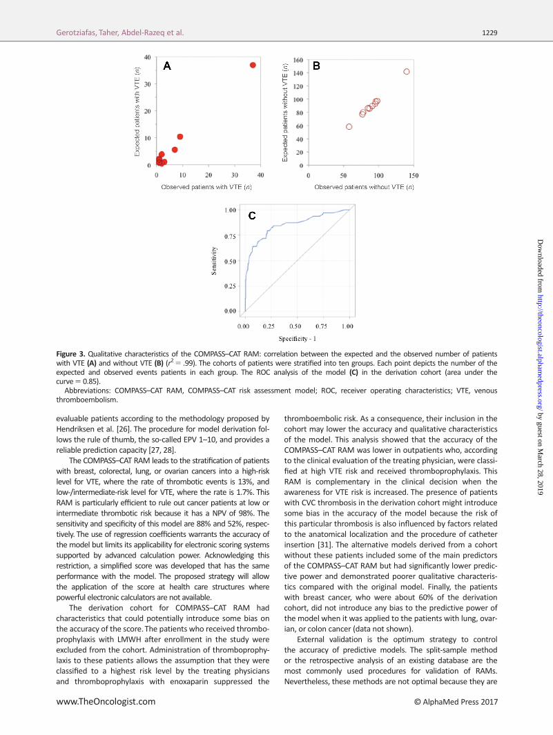
Qualitative Characteristics of Risk Assessment ModelThe model and the score at the cut-off value for high risk level(>24.7 and �7, respectively), had a NPV of 98% and a PPV of13%. The sensitivity and the specificity of the COMPASS–CATRAM was 88% and 52%, respectively. According to the Hosmer-Lemeshow test, a p 5 .23 showed that the model was well cali-brated. Plotting the expected VTE events, according to themodel, against the observed VTE events, as well as theexpected against the observed number of patients without anyVTE event, confirmed the good calibration of the model(r25 .99; Fig. 3). The ROC curve was plotted to evaluate the dis-crimination power of the model between the high-risk and thelow-/intermediate-risk population for VTE. The AUC was 0.85,indicating very good discrimination capacity.
An alternative model derived after the elimination of breastcancer patients from the derivation cohort had the samepredictors and similar qualitative characteristics as the initialCOMPASS–CAT RAM. The inclusion of patients with CVC throm-bosis into the derivation cohort as well as the elimination ofthe patients who received prophylaxis with enoxaparin afterenrollment in the study did not bias the accuracy of the modelbecause the alternative models developed according to thesescenarios were less accurate than the COMPASS–CAT RAM(data not shown).
Table 4. Simplified COMPASS–CAT Score for VTE predictionin ambulatory patients with common cancers on anti-cancer therapy
Predictors for VTE Scorea
Cancer-related risk factors
Anti-hormonal therapy for womenwith hormone receptor-positivebreast cancer or on anthracyclinetreatment
6
Time since cancer diagnosis� 6 months 4
CVC 3
Advanced stage of cancer 2
Predisposing risk factors
Cardiovascular risk factors (composedby at least two of the followingpredictors: personal history ofperipheral artery disease, ischemicstroke, coronary artery disease,hypertension, hyperlipidemia,diabetes, obesity)
5
Recent hospitalization foracute medical illness
5
Personal history of VTE 1
Biomarkers
Platelets count� 350 3 109/L 2aLow/Intermediate risk: 0–6; high risk: �7.Abbreviations: CVC, central venous catheter; VTE, venousthromboembolism.
Gerotziafas, Taher, Abdel-Razeq et al. 1227
www.TheOncologist.com Oc AlphaMed Press 2017
by guest on March 28, 2019
http://theoncologist.alphamedpress.org/
Dow
nloaded from

DISCUSSION
A new RAM for VTE applicable to outpatients after the initiationof anticancer treatment for common solid tumors was derivedfrom the prospective COMPASS–CAT study. The COMPASS–CATRAM includes VTE risk factors related to patient characteristicsand comorbidities as well as variables related to the cancer andits treatments. The COMPASS–CAT RAM is composed of well-defined and easily collected predictors that provide a globalevaluation of VTE risk. The COMPASS–CAT RAM can be appliedto outpatients at any time after treatment initiation during thepatient’s anticancer therapy. The predictors used in the modelare as follows: (a) recent hospitalization (<3 months), (b) cardi-ovascular risk factors, (c) stage of cancer, (d) anti-hormonal ther-apy for women with breast cancer or anthracycline-containingchemotherapy, (e) presence of a CVC, (f) time since cancer diag-nosis, (g) platelet count� 350 3 109/L, and (h) personal historyof VTE. The model stratifies patients into high and low/interme-diate levels of VTE risk. The multinational design of the study,which is one of its strengths, allowed the identification of theimpact of both cancer-related and patient-related risk factorsand, therefore, this simple RAM responds to the generalizabilitycriteria for risk assessment tools [26, 27].
In the first part of the study, the most clinically relevant riskfactors of VTE were identified, and subsequently the RAM wasdeveloped using data from the first 6 months of follow-upbecause the vast majority of thromboembolic events occurredwithin this interval.We demonstrate herein that hospitalizationwithin the last 3 months prior to assessment is an independentrisk factor for VTE in outpatients who are on therapy for one ofthe studied cancers. This finding is in agreement with the datareported by a recently published population-based case-control
study [30]. We also show that after initiation of anticancertreatment, patient-related risk factors are major determinantsfor the risk of CAT. Indeed, cardiovascular risk factors and/orcardiovascular comorbidities were associated with a fivefoldincrease of VTE risk. Noteworthy, VTE risk further increaseswhen multiple cardiovascular risk factors and comorbidities arepresent. Overweight or obesity and the personal history ofthrombosis are also independent VTE risk factors. Amongcancer-related variables, the univariate analysis showed thatthe predictor “time since cancer diagnosis,” which refers to thetime elapsed between assessment and cancer diagnosis, figuresamong the major risk factors for VTE. Indeed, patients withcancer diagnosed within 6 months prior to assessment had2.5-fold higher VTE risk as compared with those for whom thisinterval period was longer than 6 months. The risk of VTE wasabout twofold higher in patients with advanced cancer diseasecompared with those with localized stage, and it was independ-ent of the therapeutic strategy. Anti-hormonal treatment (towomen with breast cancer) or anthracycline-containing chemo-therapy were independent risk factors for VTE. Interestingly,the presence of CVC was found to be an independent risk fac-tor for VTE. However, the design of the present study does notallow a precise evaluation of the impact of the CVC on the riskfor DVTand/or PE. The concept that the risk imparted by CVC isalso systemic is supported by the data reported by Ashraniet al. [30]. Patients with partial remission or refractory diseasehad almost sixfold higher VTE risk compared with those withcomplete remission. Lastly, a platelet count higher than 350 3
109/L was associated with a significant increase of VTE risk inagreement with previous reports [17].
Following this analysis, a new RAM was constructed. Thederivation of the model was carried out for the entire cohort of
Figure 2. Incidence of VTE according to the stratification of patients to risk levels using the COMPASS–CAT RAM and the simplified score.The number of VTE events per type of cancer in each level of risk is shown.Abbreviations: COMPASS–CAT RAM, COMPASS–CAT risk assessment model; VTE, venous thromboembolism.
1228 Predicting VTE Risk in Ambulatory Cancer Patients
Oc AlphaMed Press 2017
by guest on March 28, 2019
http://theoncologist.alphamedpress.org/
Dow
nloaded from
evaluable patients according to the methodology proposed byHendriksen et al. [26]. The procedure for model derivation fol-lows the rule of thumb, the so-called EPV 1–10, and provides areliable prediction capacity [27, 28].
The COMPASS–CAT RAM leads to the stratification of patientswith breast, colorectal, lung, or ovarian cancers into a high-risklevel for VTE, where the rate of thrombotic events is 13%, andlow-/intermediate-risk level for VTE, where the rate is 1.7%. ThisRAM is particularly efficient to rule out cancer patients at low orintermediate thrombotic risk because it has a NPV of 98%. Thesensitivity and specificity of this model are 88% and 52%, respec-tively. The use of regression coefficients warrants the accuracy ofthe model but limits its applicability for electronic scoring systemssupported by advanced calculation power. Acknowledging thisrestriction, a simplified score was developed that has the sameperformance with the model. The proposed strategy will allowthe application of the score at health care structures wherepowerful electronic calculators are not available.
The derivation cohort for COMPASS–CAT RAM hadcharacteristics that could potentially introduce some bias onthe accuracy of the score.The patients who received thrombo-prophylaxis with LMWH after enrollment in the study wereexcluded from the cohort. Administration of thromboprophy-laxis to these patients allows the assumption that they wereclassified to a highest risk level by the treating physiciansand thromboprophylaxis with enoxaparin suppressed the
thromboembolic risk. As a consequence, their inclusion in thecohort may lower the accuracy and qualitative characteristicsof the model. This analysis showed that the accuracy of theCOMPASS–CAT RAM was lower in outpatients who, accordingto the clinical evaluation of the treating physician, were classi-fied at high VTE risk and received thromboprophylaxis. ThisRAM is complementary in the clinical decision when theawareness for VTE risk is increased. The presence of patientswith CVC thrombosis in the derivation cohort might introducesome bias in the accuracy of the model because the risk ofthis particular thrombosis is also influenced by factors relatedto the anatomical localization and the procedure of catheterinsertion [31]. The alternative models derived from a cohortwithout these patients included some of the main predictorsof the COMPASS–CAT RAM but had significantly lower predic-tive power and demonstrated poorer qualitative characteris-tics compared with the original model. Finally, the patientswith breast cancer, who were about 60% of the derivationcohort, did not introduce any bias to the predictive power ofthe model when it was applied to the patients with lung, ovar-ian, or colon cancer (data not shown).
External validation is the optimum strategy to controlthe accuracy of predictive models. The split-sample methodor the retrospective analysis of an existing database are themost commonly used procedures for validation of RAMs.Nevertheless, these methods are not optimal because they are
Figure 3. Qualitative characteristics of the COMPASS–CAT RAM: correlation between the expected and the observed number of patientswith VTE (A) and without VTE (B) (r25 .99). The cohorts of patients were stratified into ten groups. Each point depicts the number of theexpected and observed events patients in each group. The ROC analysis of the model (C) in the derivation cohort (area under thecurve5 0.85).
Abbreviations: COMPASS–CAT RAM, COMPASS–CAT risk assessment model; ROC, receiver operating characteristics; VTE, venousthromboembolism.
Gerotziafas, Taher, Abdel-Razeq et al. 1229
www.TheOncologist.com Oc AlphaMed Press 2017
by guest on March 28, 2019
http://theoncologist.alphamedpress.org/
Dow
nloaded from

vulnerable to hazardous effects of the cohort composition, andfor this reason, they were not used in the present study. How-ever, we applied the split sample method for internal validationof the RAM and we confirmed its validity (data not shown). Theabsence of a validation cohort is an evident limitation of ourstudy that imposes the need for external validation as a prereq-uisite for its routine use in clinical practice. However, the pro-spective design of the study is a strength for the derivation ofthis new RAM. Incidental VTE was not systematically assessedin patients enrolled in the study; therefore, this variable wasnot included in the analysis. This might represent a limitationon the accuracy of the COMPASS–CAT RAM. An additional limi-tation of the present study is that the number of VTE events inlung cancer patients was unusually low, allowing for thehypothesis that some thromboembolic events were missed atdiagnosis or that others could be associated with fatal PE andthus contributed to the mortality.
The COMPASS–CAT RAM targets patients with commonsolid cancer who are receiving anticancer treatment, whereasthe Khorana score is applicable in patients at the initiation ofthe chemotherapy. Patients with breast cancer represent about60% of the whole study population, reflecting the real-life situa-tion that this is the most frequent type of cancer in the commu-nity. Almost all patients enrolled in the derivation cohort werealready on chemotherapy, allowing for the application of theRAM after the initiation of the anticancer treatment. This is anadvantage of the COMPASS–CAT RAM considering that theawareness for VTE risk is rather low among oncologists, andtherefore, the probability of missing an evaluation of VTE riskbefore treatment initiation is high [32, 33]. Many patients in thederivation cohort who experienced VTE had symptomatic distalDVT. Although some authors have questioned the clinical rele-vance of distal DVT, and the proximal DVT is preferred as theendpoint in clinical studies, we should underline that cancerpatients who experience isolated symptomatic distal DVT are athigh risk of recurrence [34–36]. Moreover, according to theinternational recommendations, the therapeutic strategy forcancer-associated distal DVT is not different when comparedwith proximal DVT [12–14]. As a consequence, the ensemble ofthe clinical endpoints defined in the study represents commonfeatures of CATand allows a wider RAM applicability.
CONCLUSIONThe prospective COMPASS–CAT study provides a new, accurateRAM for VTE in outpatients on anticancer treatment for com-mon solid tumors that allows stratification of patients at highand low/intermediate risk for VTE. The originality of this RAM isthat it includes reliable and easily collected VTE predictors asso-ciated with cancer evolution and its treatments as well as withpatient characteristics and comorbidities. It is applicable forpatients suffering the most frequent types of solid tumors,which have major impact on VTE burden, and it can be appliedwhile the patient is on chemotherapy, thus permitting re-evaluation of VTE risk during the patient’s journey. It has beenderived from a cohort of patients who were prospectivelyrecruited and followed, and this provides its robustness. TheCOMPASS–CAT RAM can easily identify cancer patients on anti-cancer treatment at low or intermediate risk of VTE and ruleout the need for an antithrombotic primary prevention
strategy. An independent validation of the COMPASS–CAT RAMshould allow its routine use in clinical practice.
ACKNOWLEDGMENTS
COMPASS–CAT Working Group:Joseph Gligorov, Breast Cancer Expert Centre, Medical Oncol-
ogy Service, Hopital Tenon, Hopitaux Universitaires Est Parisien,Assistance Publique Hopitaux de Paris, Cancer Biology and Ther-apeutics, INSERM U938, Institut Universitaire de Canc�erologie(IUC), Universit�e Pierre et Marie Curie (UPMC), Sorbonne Uni-versit�es, Paris. Facult�e deM�edecine Pierre et Marie Curie, Paris,France.
Jean Pierre Lotz, Medical Oncology and Cellular TherapyDepartment, APREC (Alliance Pour la Recherche En Canc�erolo-gie), Hopital Tenon, Hopitaux Universitaires Est Parisien, Assis-tance Publique Hopitaux de Paris, Universit�e Pierre et MarieCurie (UPMC), Sorbonne Universit�es, Paris. Facult�e deM�edecine Pierre et Marie Curie, Paris, France.
Isabelle Mah�e, Internal Medicine Department, Louis MourierHospital, APHP, Paris 7 University, Colombes, France.
Marwan Bachour, Medical Oncology Department, Al Bayr-ouni University Hospital of Damascus, Syria.
The authors would like to acknowledge Dr. Antonis Voyatzisand Dr. Hisham Mahmoud from Sanofi Middle East for theirprecious support of the COMPASS–CAT project. The authorswould also like to thank Prof. Ander Cohen for his advice ondata analysis and the encouragement for developing the RAM,and Mme Rabiatou Sangare for the statistical analysis and thesubstantial contributions made for interpretation of the results.The study was presented in 2016 at the ASCO Annual Meeting(J Clin Oncol 2016;34:e21662a).
The study was supported financially by Sanofi Middle East(DIREG_L_05534). Protocol development, construction of thedatabase, data collection, statistical analysis, data interpreta-tion, and manuscript writing were all done by the investigatorswith no involvement from the funding sources.
AUTHOR CONTRIBUTIONSConception/design: Grigoris T. Gerotziafas, Ali Taher, Hikmat Abdel-Razeq,Essam AboElnazar
Provision of study material or patients: Grigoris T. Gerotziafas, Ali Taher, HikmatAbdel-Razeq, Essam AboElnazar, Salem El Shemmari
Collection and/or assembly of data: Grigoris T. Gerotziafas, Ali Taher, HikmatAbdel-Razeq, Essam AboElnazar, Salem El Shemmari
Data analysis and interpretation: Grigoris T. Gerotziafas, Ali Taher, HikmatAbdel-Razeq, Essam AboElnazar, Alex C. Spyropoulos, Salem El Shemmari,Annette K. Larsen
Manuscript writing: Grigoris T. Gerotziafas, Annette K. LarsenFinal approval of manuscript: Grigoris T. Gerotziafas, Ali Taher, Hikmat Abdel-Razeq, Essam AboElnazar, Alex C. Spyropoulos, Salem El Shemmari, AnnetteK. Larsen
DISCLOSURES
Grigoris T. Gerotziafas: Sanofi, Leo, Aspen, Bayer, BoehringerIngelheim (C/A, H), SanofiMiddle East (DIREG_L_05) (RF); Ali Taher:Novartis Pharmaceuticals (H), Novartis Pharmaceuticals, CelegeneCorporation (RF). Alex C. Spyropoulos: Janssen, Boehringer Ingelheim,Daichi Sankyo, Bristol-Myers Squibb, Pfizer, Portol (C/A), Janssen,Boehringer Ingelheim (RF). The other authors indicated no financialrelationships.(C/A) Consulting/advisory relationship; (RF) Research funding; (E) Employment; (ET) Expert
testimony; (H) Honoraria received; (OI) Ownership interests; (IP) Intellectual property rights/
inventor/patent holder; (SAB) Scientific advisory board
1230 Predicting VTE Risk in Ambulatory Cancer Patients
Oc AlphaMed Press 2017
by guest on March 28, 2019
http://theoncologist.alphamedpress.org/
Dow
nloaded from

REFERENCES
1. Lyman GH, Khorana AA, Falanga A. Thrombosisand cancer: Emerging data for the practicing oncolo-gist. Am Soc Clin Oncol Educ Book, 2013.
2. Gary T, Belaj K, Steidl K et al. Asymptomatic deepvein thrombosis and superficial vein thrombosis inambulatory cancer patients: Impact on short-termsurvival. Br J Cancer 2012;107:1244–1248.
3. Chew HK, Wun T, Harvey DJ et al. Incidence ofvenous thromboembolism and the impact on sur-vival in breast cancer patients. J Clin Oncol 2007;25:70–76.
4. Walker AJ,West J, Card TR et al.When are breastcancer patients at highest risk of venous throm-boembolism? A cohort study using English healthcare data. Blood 2016;127:849–857.
5. Ahern TP, Horv�ath-Puh�o E, Spindler KG et al.Colorectal cancer, comorbidity, and risk of venousthromboembolism: Assessment of biological interac-tions in a Danish nationwide cohort. Br J Cancer2016;114:96–102.
6. Salla E, Dimakakos EP, Tsagkouli S et al. Venousthromboembolism in patients diagnosed with lungcancer. Angiology 2016;67:709–724.
7. Chen EC, Papa N, Lawrentschuk N et al. Inci-dence and risk factors of venous thromboembolismafter pelvic uro-oncologic surgery—A single centerexperience. BJU Int 2016;117(suppl 4):50–53.
8. Tran BH, Nguyen TJ, Hwang BH et al. Risk factorsassociated with venous thromboembolism in 49,028mastectomy patients. Breast 2013;22:444–448.
9. Moore RA, Adel N, Riedel E et al. High incidenceof thromboembolic events in patients treated withcisplatin-based chemotherapy: A large retrospectiveanalysis. J Clin Oncol 2011;29:3466–3473.
10. Wun T, White RH. Epidemiology of cancer-related venous thromboembolism. Best Pract ResClin Haematol 2009;22:9–23.
11. Akl EA, Kahale LA, Ballout RA et al. Parenteralanticoagulation in ambulatory patients with cancer.Cochrane Database Syst Rev 2014:CD006652.
12. Kahn SR, Lim W, Dunn AS et al. Prevention ofVTE in nonsurgical patients: Antithrombotic Therapyand Prevention of Thrombosis, 9th ed: AmericanCollege of Chest Physicians Evidence-Based ClinicalPractice Guidelines. Chest 2012;141(suppl 2):e195S–e226S.
13. Nicolaides AN, Fareed J, Kakkar AK et al. Pre-vention and treatment of venous thromboembo-lism—International Consensus Statement. Int Angiol2013;32:111–260.
14. Debourdeau P, Farge D, Beckers M et al. Inter-national clinical practice guidelines for the treatmentand prophylaxis of thrombosis associated with cen-tral venous catheters in patients with cancer.J Thromb Haemost 2013;11:71–80.
15. Di Nisio M, Porreca E, Otten HM et al. Primaryprophylaxis for venous thromboembolism in ambu-latory cancer patients receiving chemotherapy.Cochrane Database Syst Rev 2014:CD008500.
16. Lyman GH, Bohlke K, Khorana AA et al. Venousthromboembolism prophylaxis and treatment inpatients with cancer: American Society of ClinicalOncology clinical practice guideline update 2014.J Clin Oncol 2015 20;33:654–656.
17. Khorana AA, Kuderer NM, Culakova E et al.Development and validation of a predictive modelfor chemotherapy-associated thrombosis. Blood2008;111:4902–4907.
18. Verso M, Agnelli G, Barni S et al. A modifiedKhorana risk assessment score for venous throm-boembolism in cancer patients receiving chemother-apy: The Protecht score. Intern Emerg Med 2012;7:291–292.
19. Ay C, Dunkler D, Marosi C et al. Prediction ofvenous thromboembolism in cancer patients. Blood2010;116:5377–5382.
20. Agnelli G, George DJ, Kakkar AK et al. Semulo-parin for thromboprophylaxis in patients receivingchemotherapy for cancer. N Engl J Med 2012;366:601–609.
21. George D, Agnelli G, Fisher W et al. Venousthromboembolism (VTE) prevention with semulo-parin in cancer patients initiating chemotherapy:Benefit-risk assessment by VTE risk in SAVE-ONCO.Paper presented at: American Society of Hematol-ogy 53rd Annual meeting; December 10–13, 2011;San Diego, California.
22. Mansfield A, Tafur AJ,Wang CE et al. Predictorsof active cancer thromboembolic outcomes: Valida-tion of the Khorana score among patients with lungcancer. J Thromb Haemost 2016;14:1773–1778.
23. Alikhan R, Cohen AT, Combe S et al. Risk factorsfor venous thromboembolism in hospitalizedpatients with acute medical illness: Analysis of theMEDENOX Study. Arch Intern Med 2004;164:963–968.
24. Caprini JA, Arcelus JI, Reyna JJ. Effective riskstratification of surgical and nonsurgical patients forvenous thromboembolic disease. Semin Hematol2001;38:12–19.
25. Mokhtari M, Salameh P, Kouchek M et al. TheAVAIL ME Extension: A multinational Middle East-ern survey of venous thromboembolism risk andprophylaxis. J Thromb Haemost 2011;9:1340–1349.
26. Hendriksen JM, Geersing GJ, Moons KGet al. Diagnostic and prognostic prediction mod-els. J Thromb Haemost 2013;11(suppl 1):129–141.
27. Harrell FE Jr, Lee KL, Mark DB et al. Multivari-able prognostic models: Issues in developing mod-els, evaluating assumptions and adequacy, andmeasuring and reducing errors. Stat Med 1996;15:361–387.
28. Peduzzi P, Concato J, Kemper E et al. A simula-tion study of the number of events per variable inlogistic regression analysis. J Clin Epidemiol 1996;49:1373–1379.
29. Wasson JH, Sox HC, Neff RK et al. Clinical pre-diction rules: Applications and methodologicalstandards. N Engl J Med 1985;313:793–799.
30. Ashrani AA, Gullerud RE, Petterson TM et al.Risk factors for incident venous thromboembolismin active cancer patients: A population based case-control study.Thromb Res 2016;139:29–37.
31. Parienti JJ, Mongardon N, M�egarbane B et al.Intravascular complications of central venous cathe-terization by insertion site. N Engl J Med 2015;373:1220–1229.
32. Sevestre MA, Belizna C, Durant C et al. Compli-ance with recommendations of clinical practice inthe management of venous thromboembolism incancer: The CARMEN study. J Mal Vasc 2014;39:161–168.
33. Aggarwal A, Fullam L, Brownstein AP et al.Deep vein thrombosis (DVT) and pulmonary embo-lism (PE): Awareness and prophylaxis practicesreported by patients with cancer. Cancer Invest2015;33:405–410.
34. Dentali F, Pegoraro S, Barco S et al. Clinical his-tory of cancer patients with isolated distal deep veinthrombosis: A multicenter cohort study. Thromb Res2016;(140 suppl 1):S168
35. Ho P, Lim HY, Chua CC et al. Retrospectivereview on isolated distal deep vein thrombosis(IDDVT) - A benign entity or not? Thromb Res 2016;142:11–16.
36. Sartori M, Migliaccio L, Favaretto E et al. Twoyears outcome of isolated distal deep vein thrombo-sis.Thromb Res 2014;134:36–40.
Gerotziafas, Taher, Abdel-Razeq et al. 1231
www.TheOncologist.com Oc AlphaMed Press 2017
by guest on March 28, 2019
http://theoncologist.alphamedpress.org/
Dow
nloaded from