approach to the unfolding and folding dynamics of add a … · the journal of chemical physics 145,...
TRANSCRIPT
THE JOURNAL OF CHEMICAL PHYSICS 145, 014104 (2016)
Approach to the unfolding and folding dynamics of add A-riboswitch uponadenine dissociation using a coarse-grained elastic network model
Chunhua Li,
1,2
Dashuai Lv,
1
Lei Zhang,
1
Feng Yang,
1
Cunxin Wang,
1
Jiguo Su,
3,a)
and Yang Zhang
2,a)
1College of Life Science and Bioengineering, Beijing University of Technology, Beijing 100124, China2Department of Computational Medicine and Bioinformatics, University of Michigan, Ann Arbor,Michigan 45108, USA3College of Science, Yanshan University, Qinhuangdao 066004, China
(Received 13 April 2016; accepted 17 June 2016; published online 6 July 2016)
Riboswitches are noncoding mRNA segments that can regulate the gene expression via alteringtheir structures in response to specific metabolite binding. We proposed a coarse-grained Gaussiannetwork model (GNM) to examine the unfolding and folding dynamics of adenosine deaminase (add)A-riboswitch upon the adenine dissociation, in which the RNA is modeled by a nucleotide chainwith interaction networks formed by connecting adjoining atomic contacts. It was shown that theadenine binding is critical to the folding of the add A-riboswitch while the removal of the ligand canresult in drastic increase of the thermodynamic fluctuations especially in the junction regions betweenhelix domains. Under the assumption that the native contacts with the highest thermodynamicfluctuations break first, the iterative GNM simulations showed that the unfolding process of theadenine-free add A-riboswitch starts with the denature of the terminal helix stem, followed by theloops and junctions involving ligand binding pocket, and then the central helix domains. Despitethe simplified coarse-grained modeling, the unfolding dynamics and pathways are shown in closeagreement with the results from atomic-level MD simulations and the NMR and single-moleculeforce spectroscopy experiments. Overall, the study demonstrates a new avenue to investigate thebinding and folding dynamics of add A-riboswitch molecule which can be readily extended for otherRNA molecules. Published by AIP Publishing. [http://dx.doi.org/10.1063/1.4954992]
I. INTRODUCTION
In recent years, the studies of functional noncodingRNAs have received unprecedented attention.1–3 Riboswitchesare the natural genetic control segments found in the50 untranslated regions (UTRs) of many mRNAs of bacteriaand fungi. They can regulate gene expression at the levelof transcription or translation that is highly sensitive tothe cellular concentrations of specific metabolites.2–4 Sofar, various riboswitches have been discovered which canspecifically recognize a large variety of ligands, such asadenine, glycine, lysine, and glucosamine-6-phosphate.5–8
Riboswitches are generally composed of two domains: theaptamer domain and the downstream expression platform.The former can fold into an intricate three-dimensionalstructure upon binding to a specific metabolite when theconcentration of the metabolite exceeds a threshold. The latterswitches a gene on/o↵ through its structural changes triggeredby metabolite-induced large-scale conformational changesin the aptamer domain.8 The adenosine deaminase (add)A-riboswitch is one of the structurally simplest riboswitchesand has been widely studied.9–12 The folded aptamer domainof the add A-riboswitch bound with the ligand adeninewas structurally characterized by X-ray crystallography with
a)Authors to whom correspondence should be addressed. Electronic ad-dresses: [email protected] and [email protected]
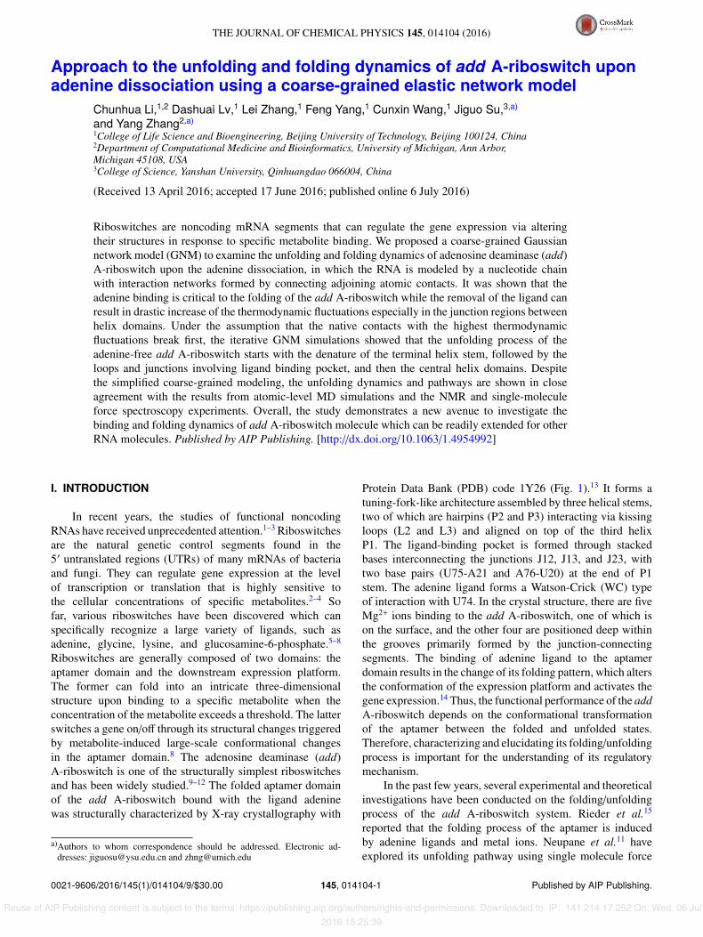
Protein Data Bank (PDB) code 1Y26 (Fig. 1).13 It forms atuning-fork-like architecture assembled by three helical stems,two of which are hairpins (P2 and P3) interacting via kissingloops (L2 and L3) and aligned on top of the third helixP1. The ligand-binding pocket is formed through stackedbases interconnecting the junctions J12, J13, and J23, withtwo base pairs (U75-A21 and A76-U20) at the end of P1stem. The adenine ligand forms a Watson-Crick (WC) typeof interaction with U74. In the crystal structure, there are fiveMg2+ ions binding to the add A-riboswitch, one of which ison the surface, and the other four are positioned deep withinthe grooves primarily formed by the junction-connectingsegments. The binding of adenine ligand to the aptamerdomain results in the change of its folding pattern, which altersthe conformation of the expression platform and activates thegene expression.14 Thus, the functional performance of the addA-riboswitch depends on the conformational transformationof the aptamer between the folded and unfolded states.Therefore, characterizing and elucidating its folding/unfoldingprocess is important for the understanding of its regulatorymechanism.
In the past few years, several experimental and theoreticalinvestigations have been conducted on the folding/unfoldingprocess of the add A-riboswitch system. Rieder et al.15
reported that the folding process of the aptamer is inducedby adenine ligands and metal ions. Neupane et al.11 haveexplored its unfolding pathway using single molecule force
0021-9606/2016/145(1)/014104/9/$30.00 145, 014104-1 Published by AIP Publishing.
Reuse of AIP Publishing content is subject to the terms: https://publishing.aip.org/authors/rights-and-permissions. Downloaded to IP: 141.214.17.252 On: Wed, 06 Jul2016 15:25:39
014104-2 Li et al. J. Chem. Phys. 145, 014104 (2016)
FIG. 1. Three-dimensional structure of add A-riboswitch with five Mg2+ ionsand a ligand adenine (PDB code 1Y26). The ligand and Mg2+ ions are coloredwith red and blue, respectively. The secondary structures of the system arelabelled correspondingly with P1, P2, and P3 for stems, L2 and L3 for loops,and J12, J13, and J23 for junctions.
measurements and observed a series of intermediates duringits unfolding process. Using a self-organized polymer modelwith the Langevin dynamics, Lin and Thirumalai14 describedforce-triggered unfolding of this system and obtained thesame unfolding sequence as the experimental observation.Priyakumar and MacKerell16 performed a 40 ns all-atommolecular dynamics (MD) simulation to investigate theunfolding behavior in both presence and absence of ligand,but the complete unfolding process was not produced and onlystructural deviations were observed in the J23 junction and P1stem. In addition, some investigations have concentrated onthe roles of adenine ligand on the structural stability of addA-riboswitch. Researchers found that the ligand binding playsa critical role in stabilizing the binding pocket, especially forthe folded P1 stem.11,16,17 While the all-atom MD simulationcan provide useful information on the fluctuations andconformational changes of biomacromolecules, it is generallydi�cult to obtain a complete folding/unfolding picture throughMD due to the prohibitive computational costs.
The coarse-grained method has proved to be ane�cient tool to explore the large-scale functional motionsof biomolecular systems. Recently, we proposed a Gaussiannetwork model (GNM) method, which was successfullyapplied to protein folding/unfolding studies.18,19 In thismethod, the unfolding process of proteins is modeled throughgradually breaking the non-covalent native contacts accordingto the distance fluctuations of residue pairs. This process canbe considered to be composed of a series of quasi-equilibrium
states corresponding to a slowly increasing temperature,during which the molecule has su�cient time to search forthe state with a maximum entropy increase at each unfoldingstep.
In this work, we extended the iterative GNM approachto explore the unfolding process of the add A-riboswitch,with the changes of cross-correlations between nucleotidefluctuations and the fluctuations in the fast modes analyzedin detail. Additionally, we tried to interpret the roles ofthe adenine ligand in stabilizing the folded aptamer from atopological point of view.
II. METHODS
A. Gaussian network model
In the Gaussian network model, the three-dimensionalstructure of a molecule is described as an elastic networkof nodes that are connected by harmonic springs with acertain cuto↵ distance.20,21 In this work, the RNA chainis represented by a set of linked nodes, with each nodestanding for a nucleotide, where the interactions of backbone-backbone, base-base, and backbone-base, and the e↵ects ofMg2+ ions and adenine are considered in the construction ofthe nucleotide networks. In addition to the backbone links, thenucleotide nodes are connected in the model when one of thefollowing requirements is satisfied:
I. The distance between P atoms 8.5 Å.II. The distance between mark atoms 8.0 Å.
III. The distance between the P and mark atoms from di↵erentnucleotides 8.0 Å.
IV. Both distances of two P atoms from the same Mg2+ ion8.5 Å.
V. Both distances of two mark atoms from the mark atom ofthe ligand adenine 8.0 Å.
The mark atoms refer to the four atoms (N6 foradenine, O6 for guanine, N4 for cytosine and O4 for uracil),representing the four kinds of bases, respectively, all of whichparticipate in the formation of hydrogen bonds in canonicalbase pairs.22 The requirements I, II, and III are designed totake into account the interactions of backbone-backbone, base-base, and backbone-base, respectively, and V for consideringthe mediating role of the ligand.
It should be pointed out that as Mg2+ ions are importantfor structure stabilization and folding of the system, all thefive Mg2+ ions are considered in the network construction.Mg2+ ions mainly play a role in mediating the interactionsamong the negative charges of the highly acidic phosphatebackbone, permitting the system to form and retain the foldedstructures. Thus, in the network construction we consider themediating roles of Mg2+ ions (not treated as separated nodes)by connecting the nucleotide nodes when both distances oftheir P atoms from the same Mg2+ ion 8.5 Å, as shown inrequirement IV.
Di↵erent from the conventional GNM in which theforce constants are identical for the springs of covalentand noncovalent pairs, the spring constants of non-bondednucleotides are set as � while those of the covalently bonded
Reuse of AIP Publishing content is subject to the terms: https://publishing.aip.org/authors/rights-and-permissions. Downloaded to IP: 141.214.17.252 On: Wed, 06 Jul2016 15:25:39
014104-3 Li et al. J. Chem. Phys. 145, 014104 (2016)
pairs along the chain backbone are chosen as c�. The valuesof c and � are determined by fitting the predicted fluctuationsagainst the crystallographic B-factors. This setting allows usto consider the heterogeneity of RNA chain interactions, sincethe covalent bonding is generally much stronger than thenon-covalent ones.
The internal Hamiltonian of the RNA molecule systemcan be written as23
H =12�⇥�RT (� ⌦ E)�R
⇤, (1)
where �R = {�R1,�R2, . . .�RN} represents the 3N-dimensional column vectors of fluctuations of the nodesin the network, where N is the number of nucleotides; thesuperscript T denotes the transpose; E is the third-orderidentity matrix; ⌦ is the direct product; and � is the N ⇥ Nsymmetric matrix in which the elements are given by
�i j =
8>>>>>>>><>>>>>>>>:
�c if |i � j | = 1�1 if |i � j | > 1 and nucleotides i and j are connected0 if |i � j | > 1 and nucleotides i and j are not connected�X
i, j,i
�i j if i = j
. (2)
The mean-square fluctuation (MSF) of each nucleotidenode and the cross-correlation fluctuations between di↵erentnucleotide nodes are in proportion to the diagonal and o↵-diagonal elements of the pseudo-inverse of the � matrix. Theinverse of the matrix can be decomposed as
��1 = U⇤�1UT, (3)
where U is an orthogonal matrix whose columns ui (1 < i N) are the eigenvectors of �, and ⇤ is a diagonal matrixof eigenvalues �i of �. The cross-correlation fluctuationsbetween the ith and jth nucleotides are written as
⌦�Ri · �Rj
↵=
3kBT�
⇥��1⇤
i j, (4)
where kB is the Boltzmann constant and T is the absolutetemperature. When i = j, the MSF of the ith nucleotide can beobtained. The B-factor, also called temperature factor, whichis related to the MSF, can be computed with the expression
Bi =8⇡2
3h�Ri · �Rii =
8⇡2kBT�
⇥��1⇤
ii. (5)
The MSF of the ith nucleotide associating with the kth modeis obtained by
h�Ri · �Riik =3kBT�
��1k [uk]i[uk]i. (6)
The MSF in the distance vector Ri j between the nucleotides iand j can be calculated with the expression24
h��Ri j
�2i = ⌧⇣Ri j � R0i j
⌘2�= h��Ri � �Rj
�2i= h�Ri · �Rii +
⌦�Rj · �Rj
↵� 2
⌦�Ri · �Rj
↵=
3kBT�
⇣⇥��1⇤
ii +⇥��1⇤
j j � 2⇥��1⇤
i j
⌘, (7)
where Ri j and R0i j are the instantaneous and equilibrium
separation vectors between nucleotides i and j. In the GNM,
the cross-correlation is normalized as
Ci j =
⌦�Ri · �Rj
↵fh�R2
i i ·D�R2
j
Eg1/2 . (8)
B. Iterative Gaussian network model
The unfolding process of the riboswitch moleculesis modeled based on the thermodynamic fluctuations.Generally, with a higher fluctuation the native contacts ofriboswitches between nucleotides are expected to break ina higher possibility, which results in unfolded structures.The fluctuations in the distance between all nucleotides arecalculated by the GNM. The nonlinear elasticity during RNAunfolding is considered via iterative normal mode calculations,i.e.,18
I. The MSFs of the distances in all nucleotide pairs arecalculated according to the native structure topology withEq. (7).
II. The contact in the nucleotide pair with the largest distancefluctuation is broken. Then, a new matrix � is constructed,which represents a new topology during RNA unfolding.
III. The MSFs of the distances in all nucleotide pairs arerecalculated based on the new matrix � with Eq. (7).
IV. The above steps II and III are repeated until all thenoncovalent contacts are broken.
V. All the topologies of di↵erent conformations during RNAunfolding are obtained and the unfolding pathway can bederived from the above data.
It should be noted that many previous studies havebeen conducted on examining the roles of Mg2+ ions onthe add A-riboswitch, indicating that they mainly a↵ect thepre-organization and stabilization of the folded conformationrather than the intervention of the unfolding process.12,15
Therefore, we did not specifically examine the unfoldingbehavior induced by Mg2+ release. Here, the contacts mediated
Reuse of AIP Publishing content is subject to the terms: https://publishing.aip.org/authors/rights-and-permissions. Downloaded to IP: 141.214.17.252 On: Wed, 06 Jul2016 15:25:39
014104-4 Li et al. J. Chem. Phys. 145, 014104 (2016)
by Mg2+ are not treated specially and the unfolding process issimulated purely in a fluctuation-dependent manner.
III. RESULTS AND DISCUSSION
A. Force constants for the covalentand noncovalent interactions
The add A-riboswitch is modeled by the Gaussiannetwork model system with each node representing anucleotide and the interaction network formed by the adjoiningatomic contacts (see Sec. II). The spring constant valuesof covalent and noncovalent interactions, i.e., c� and �,are determined by matching the calculated and experimentalB-factor values. As the absolute value of � does not a↵ectthe relative size of nucleotide fluctuations, it has no influenceon the correlation between the calculated and experimentalB-factors. Therefore, we first set � = 1 to determine thevalue of c via maximizing the correlation between thecomputed and experimental B-factors. The value of � was thendetermined through normalizing the calculated fluctuationwith the experimental B-factors. According to this method, weobtained c = 8.0 and � = 0.125 kBT /Å2, where the correlationcoe�cient between the theoretical and experimental B-factorsis 0.728.
Compared to the traditional GNM with uniform springcontacts, the introduction of heterogeneity in the covalentand noncovalent spring contacts in our model improves thecorrelation coe�cient of B-factors from 0.606 to 0.728. Part ofthe e↵ect is due to the consideration of the mediating roles ofMg2+ ions in our network construction. Without consideringthe Mg2+ ions, the correlation coe�cient of B-factors willreduce to 0.717, which partly highlights the roles of Mg2+
ions in the stabilization and folding of the molecule.We also computed this correlation coe�cient using the
conventional RNA single-point GNM, in which one noderepresents one nucleotide and the connections between themare determined only based on the distances between P atoms;25
this gives an even lower correlation coe�cient 0.587. Theresults indicate that the new iterative GNM approach, whichconsiders the RNA backbone-backbone, backbone-base, base-base interactions, and Mg2+ ion binding, and meanwhiledistinguishes the covalent from noncovalent interactions,can help generate a better accordance with experimentalfluctuation data.
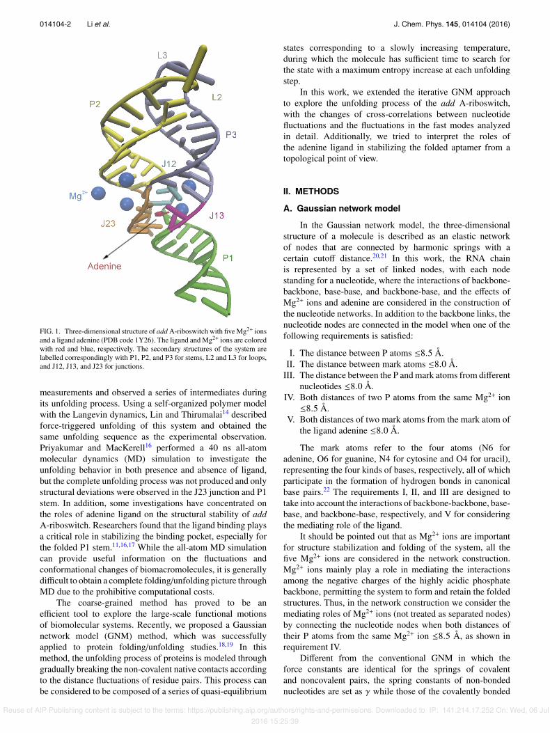
B. Roles of ligand adenine and Mg2+ ionson the dynamics of add A-riboswitch
The ligand adenine was found to be important for thestructure and function of add A-riboswitch.26 To examinethe specific role of the ligand adenine on the dynamics of thesystem, we computed the MSF of each nucleotide in the GNMwith and without adenine. Fig. 2 shows that the nucleotideshave a relatively higher MSF in the adenine-free state than inthe adenine-binding state, although the di↵erence in MSFsvaries along the sequence. The most significant increasein MSFs occurs in the three junctions (J12, J13, and J23)associated with the binding pockets as well as the nucleotides
FIG. 2. Fluctuations of nucleotides of add A-riboswitch in ligand-boundstate (solid line) and ligand-free one (shaded line), respectively.
at the terminal P1 stem that are near the binding pocket. Inparticular, the region from U74-A76 pairing with the adeninehas the highest MSF increase. Similar MSF changes havebeen observed in the former atomic MD and experimentalstudies,12,16,17,27 suggesting that the adenine ligand-bindingindeed plays an important role in stabilizing the bindingpocket, especially for the folded P1 stem and J23 junctionregions. Functionally, binding with the adenine ligand leads toa change in the stability of P1 stem, which in e↵ect, constitutesa structural switch in the expression platform, since the30 strand of P1 stem is involved in alternative base pairing withthe Shine-Dalgarno sequence of the expression platform in theabsence of adenine binding, which masks the Shine-Dalgarnosequence and causes the stop of translation.8,28,29
The ligand dissociation leads to a marked rise in MSFat J23 U51 (base pairing with ligand), which causes furtherinstability of its neighbors C50 and U49 that are involved inthe formation of base triplets with the top base pairs of P1stem: U75-A21 and A76-U20, respectively.12 This explainsthe causal relationship between the fluctuations of P1 stemand J23 junction, observed by Sharma et al.12 Additionally,the data imply that the transmission pathway of structuralchanges caused by ligand dissociation may start from ligand-bound U51, and then to its neighbors, finally to the P1 stem.Also, the high flexibility of J23 in the absence of the adenineligand suggests that the ligand should associate and dissociatefrom the riboswitch in the vicinity of J23.
Recently, the experimental and theoretical researcheshave reported that Mg2+ ions appear to be important inpreorganizing and stabilizing the aptamer fold.12,15 In order todetect the roles of Mg2+ ions on the dynamics of the system,we calculated the MSF of each nucleotide in the GNM ofthe system with and without Mg2+ ions (see Figure S1 ofthe supplementary material38). Overall, the nucleotides haverelatively higher MSFs in the Mg2+ ion free state than in theMg2+ ion binding state, especially for the regions around theligand-binding pocket: J12, J23, and the junction proximalends of three stems P1 (U20-A21), P2 (G42-A45), and P3(C69-U71), which was also observed in the previous MDsimulation study.12
Reuse of AIP Publishing content is subject to the terms: https://publishing.aip.org/authors/rights-and-permissions. Downloaded to IP: 141.214.17.252 On: Wed, 06 Jul2016 15:25:39
014104-5 Li et al. J. Chem. Phys. 145, 014104 (2016)
In conclusion, both ligand adenine and Mg2+ ions showtheir importance in stabilizing the folded conformation ofthe riboswitch. Ligand adenine mainly contributes to thestabilization of P1 and J23, and the e↵ect of Mg2+ ions ismainly reflected in stabilizing the ligand binding pocket.
C. Pathway of add A-riboswitch unfolding
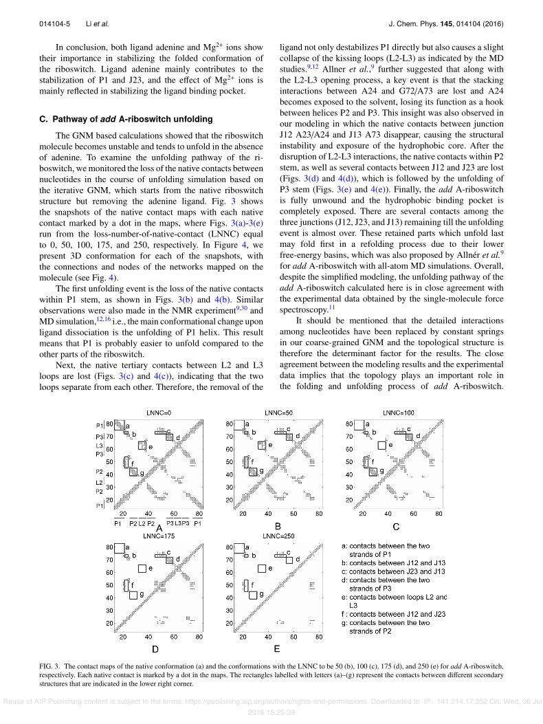
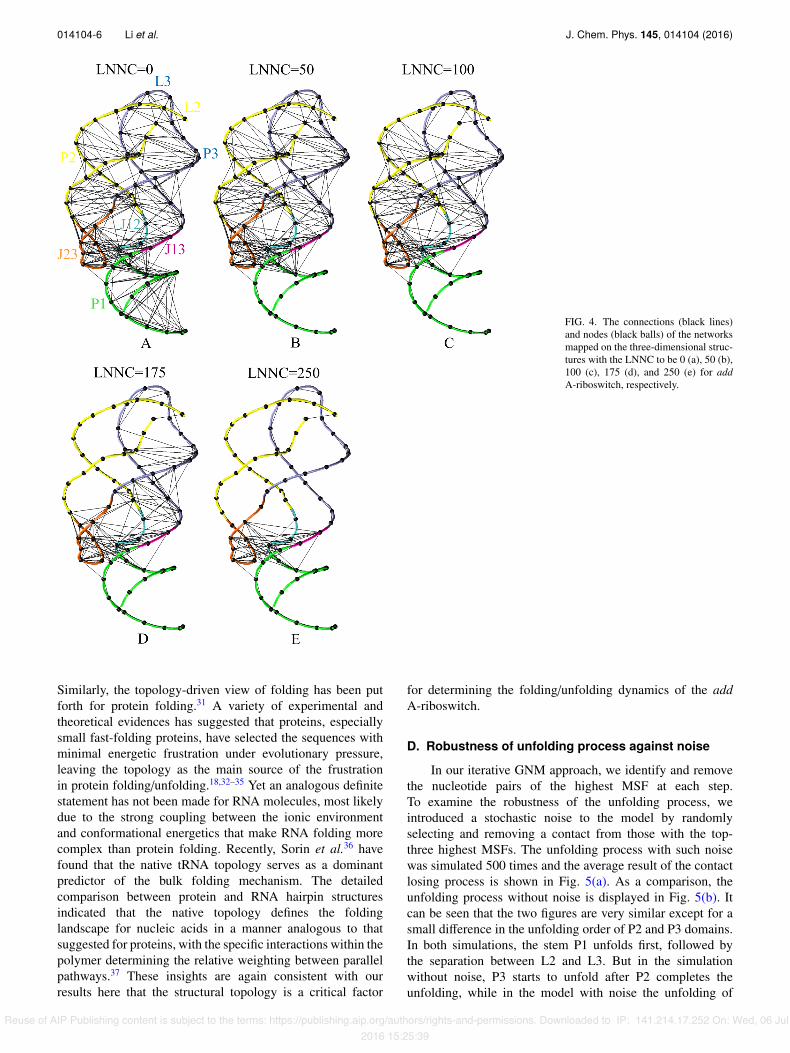
The GNM based calculations showed that the riboswitchmolecule becomes unstable and tends to unfold in the absenceof adenine. To examine the unfolding pathway of the ri-boswitch, we monitored the loss of the native contacts betweennucleotides in the course of unfolding simulation based onthe iterative GNM, which starts from the native riboswitchstructure but removing the adenine ligand. Fig. 3 showsthe snapshots of the native contact maps with each nativecontact marked by a dot in the maps, where Figs. 3(a)-3(e)run from the loss-number-of-native-contact (LNNC) equalto 0, 50, 100, 175, and 250, respectively. In Figure 4, wepresent 3D conformation for each of the snapshots, withthe connections and nodes of the networks mapped on themolecule (see Fig. 4).
The first unfolding event is the loss of the native contactswithin P1 stem, as shown in Figs. 3(b) and 4(b). Similarobservations were also made in the NMR experiment9,30 andMD simulation,12,16 i.e., the main conformational change uponligand dissociation is the unfolding of P1 helix. This resultmeans that P1 is probably easier to unfold compared to theother parts of the riboswitch.
Next, the native tertiary contacts between L2 and L3loops are lost (Figs. 3(c) and 4(c)), indicating that the twoloops separate from each other. Therefore, the removal of the
ligand not only destabilizes P1 directly but also causes a slightcollapse of the kissing loops (L2-L3) as indicated by the MDstudies.9,12 Allner et al.,9 further suggested that along withthe L2-L3 opening process, a key event is that the stackinginteractions between A24 and G72/A73 are lost and A24becomes exposed to the solvent, losing its function as a hookbetween helices P2 and P3. This insight was also observed inour modeling in which the native contacts between junctionJ12 A23/A24 and J13 A73 disappear, causing the structuralinstability and exposure of the hydrophobic core. After thedisruption of L2-L3 interactions, the native contacts within P2stem, as well as several contacts between J12 and J23 are lost(Figs. 3(d) and 4(d)), which is followed by the unfolding ofP3 stem (Figs. 3(e) and 4(e)). Finally, the add A-riboswitchis fully unwound and the hydrophobic binding pocket iscompletely exposed. There are several contacts among thethree junctions (J12, J23, and J13) remaining till the unfoldingevent is almost over. These retained parts which unfold lastmay fold first in a refolding process due to their lowerfree-energy basins, which was also proposed by Allnér et al.9
for add A-riboswitch with all-atom MD simulations. Overall,despite the simplified modeling, the unfolding pathway of theadd A-riboswitch calculated here is in close agreement withthe experimental data obtained by the single-molecule forcespectroscopy.11
It should be mentioned that the detailed interactionsamong nucleotides have been replaced by constant springsin our coarse-grained GNM and the topological structure istherefore the determinant factor for the results. The closeagreement between the modeling results and the experimentaldata implies that the topology plays an important role inthe folding and unfolding process of add A-riboswitch.
FIG. 3. The contact maps of the native conformation (a) and the conformations with the LNNC to be 50 (b), 100 (c), 175 (d), and 250 (e) for add A-riboswitch,respectively. Each native contact is marked by a dot in the maps. The rectangles labelled with letters (a)–(g) represent the contacts between di↵erent secondarystructures that are indicated in the lower right corner.
Reuse of AIP Publishing content is subject to the terms: https://publishing.aip.org/authors/rights-and-permissions. Downloaded to IP: 141.214.17.252 On: Wed, 06 Jul2016 15:25:39
014104-6 Li et al. J. Chem. Phys. 145, 014104 (2016)
FIG. 4. The connections (black lines)and nodes (black balls) of the networksmapped on the three-dimensional struc-tures with the LNNC to be 0 (a), 50 (b),100 (c), 175 (d), and 250 (e) for addA-riboswitch, respectively.
Similarly, the topology-driven view of folding has been putforth for protein folding.31 A variety of experimental andtheoretical evidences has suggested that proteins, especiallysmall fast-folding proteins, have selected the sequences withminimal energetic frustration under evolutionary pressure,leaving the topology as the main source of the frustrationin protein folding/unfolding.18,32–35 Yet an analogous definitestatement has not been made for RNA molecules, most likelydue to the strong coupling between the ionic environmentand conformational energetics that make RNA folding morecomplex than protein folding. Recently, Sorin et al.36 havefound that the native tRNA topology serves as a dominantpredictor of the bulk folding mechanism. The detailedcomparison between protein and RNA hairpin structuresindicated that the native topology defines the foldinglandscape for nucleic acids in a manner analogous to thatsuggested for proteins, with the specific interactions within thepolymer determining the relative weighting between parallelpathways.37 These insights are again consistent with ourresults here that the structural topology is a critical factor
for determining the folding/unfolding dynamics of the addA-riboswitch.
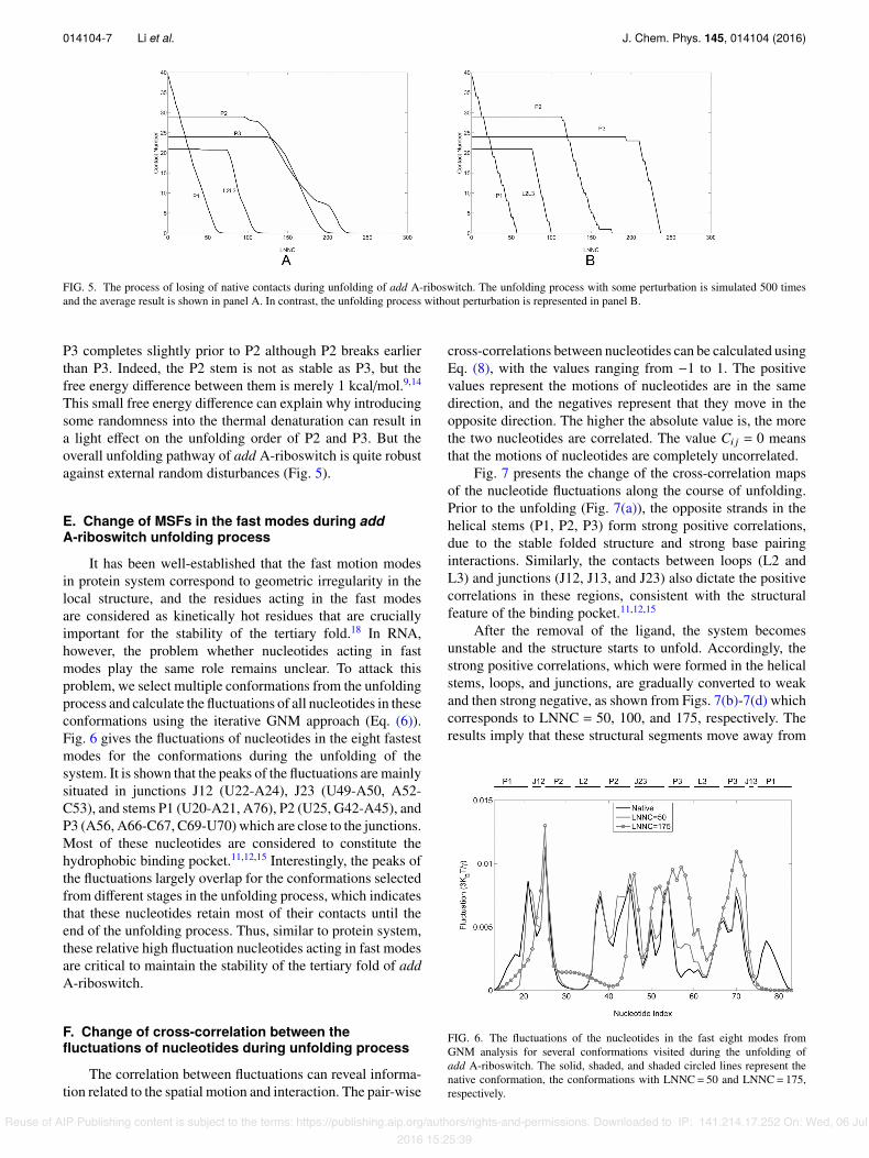
D. Robustness of unfolding process against noise
In our iterative GNM approach, we identify and removethe nucleotide pairs of the highest MSF at each step.To examine the robustness of the unfolding process, weintroduced a stochastic noise to the model by randomlyselecting and removing a contact from those with the top-three highest MSFs. The unfolding process with such noisewas simulated 500 times and the average result of the contactlosing process is shown in Fig. 5(a). As a comparison, theunfolding process without noise is displayed in Fig. 5(b). Itcan be seen that the two figures are very similar except for asmall di↵erence in the unfolding order of P2 and P3 domains.In both simulations, the stem P1 unfolds first, followed bythe separation between L2 and L3. But in the simulationwithout noise, P3 starts to unfold after P2 completes theunfolding, while in the model with noise the unfolding of
Reuse of AIP Publishing content is subject to the terms: https://publishing.aip.org/authors/rights-and-permissions. Downloaded to IP: 141.214.17.252 On: Wed, 06 Jul2016 15:25:39
014104-7 Li et al. J. Chem. Phys. 145, 014104 (2016)
FIG. 5. The process of losing of native contacts during unfolding of add A-riboswitch. The unfolding process with some perturbation is simulated 500 timesand the average result is shown in panel A. In contrast, the unfolding process without perturbation is represented in panel B.
P3 completes slightly prior to P2 although P2 breaks earlierthan P3. Indeed, the P2 stem is not as stable as P3, but thefree energy di↵erence between them is merely 1 kcal/mol.9,14
This small free energy di↵erence can explain why introducingsome randomness into the thermal denaturation can result ina light e↵ect on the unfolding order of P2 and P3. But theoverall unfolding pathway of add A-riboswitch is quite robustagainst external random disturbances (Fig. 5).
E. Change of MSFs in the fast modes during addA-riboswitch unfolding process
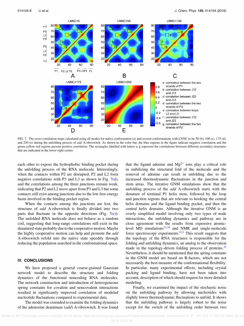
It has been well-established that the fast motion modesin protein system correspond to geometric irregularity in thelocal structure, and the residues acting in the fast modesare considered as kinetically hot residues that are cruciallyimportant for the stability of the tertiary fold.18 In RNA,however, the problem whether nucleotides acting in fastmodes play the same role remains unclear. To attack thisproblem, we select multiple conformations from the unfoldingprocess and calculate the fluctuations of all nucleotides in theseconformations using the iterative GNM approach (Eq. (6)).Fig. 6 gives the fluctuations of nucleotides in the eight fastestmodes for the conformations during the unfolding of thesystem. It is shown that the peaks of the fluctuations are mainlysituated in junctions J12 (U22-A24), J23 (U49-A50, A52-C53), and stems P1 (U20-A21, A76), P2 (U25, G42-A45), andP3 (A56, A66-C67, C69-U70) which are close to the junctions.Most of these nucleotides are considered to constitute thehydrophobic binding pocket.11,12,15 Interestingly, the peaks ofthe fluctuations largely overlap for the conformations selectedfrom di↵erent stages in the unfolding process, which indicatesthat these nucleotides retain most of their contacts until theend of the unfolding process. Thus, similar to protein system,these relative high fluctuation nucleotides acting in fast modesare critical to maintain the stability of the tertiary fold of addA-riboswitch.
F. Change of cross-correlation between thefluctuations of nucleotides during unfolding process
The correlation between fluctuations can reveal informa-tion related to the spatial motion and interaction. The pair-wise
cross-correlations between nucleotides can be calculated usingEq. (8), with the values ranging from �1 to 1. The positivevalues represent the motions of nucleotides are in the samedirection, and the negatives represent that they move in theopposite direction. The higher the absolute value is, the morethe two nucleotides are correlated. The value Ci j = 0 meansthat the motions of nucleotides are completely uncorrelated.
Fig. 7 presents the change of the cross-correlation mapsof the nucleotide fluctuations along the course of unfolding.Prior to the unfolding (Fig. 7(a)), the opposite strands in thehelical stems (P1, P2, P3) form strong positive correlations,due to the stable folded structure and strong base pairinginteractions. Similarly, the contacts between loops (L2 andL3) and junctions (J12, J13, and J23) also dictate the positivecorrelations in these regions, consistent with the structuralfeature of the binding pocket.11,12,15
After the removal of the ligand, the system becomesunstable and the structure starts to unfold. Accordingly, thestrong positive correlations, which were formed in the helicalstems, loops, and junctions, are gradually converted to weakand then strong negative, as shown from Figs. 7(b)-7(d) whichcorresponds to LNNC = 50, 100, and 175, respectively. Theresults imply that these structural segments move away from
FIG. 6. The fluctuations of the nucleotides in the fast eight modes fromGNM analysis for several conformations visited during the unfolding ofadd A-riboswitch. The solid, shaded, and shaded circled lines represent thenative conformation, the conformations with LNNC= 50 and LNNC= 175,respectively.
Reuse of AIP Publishing content is subject to the terms: https://publishing.aip.org/authors/rights-and-permissions. Downloaded to IP: 141.214.17.252 On: Wed, 06 Jul2016 15:25:39
014104-8 Li et al. J. Chem. Phys. 145, 014104 (2016)
FIG. 7. The cross-correlation maps calculated using all modes for native conformation (a) and several conformations with LNNC to be 50 (b), 100 (c), 175 (d),and 250 (e) during the unfolding process of add A-riboswitch. As shown in the color bar, the blue regions in the figure indicate negative correlation and thegreen-yellow-red regions present positive correlation. The rectangles labelled with letters a–g represent the correlations between di↵erent secondary structuresthat are indicated in the lower right corner.
each other to expose the hydrophobic binding pocket duringthe unfolding process of the RNA molecule. Interestingly,when the contacts within P2 are disrupted, P2 and L2 formnegative correlations with P3 and L3 as shown in Fig. 7(d),and the correlations among the three junctions remain weak,indicating that P2 and L2 move apart from P3 and L3 but somecontacts still exist among junctions due to the low free-energybasin involved in the binding pocket region.
When the contacts among the junctions are lost, thestructure of add A-riboswitch is finally divided into twoparts that fluctuate in the opposite directions (Fig. 7(e)).The unfolded RNA molecule does not behave as a randomcoil, suggesting that long-range correlations still exist in thedenatured state probably due to the cooperative motion. Maybethe highly cooperative motion can help and promote the addA-riboswitch refold into the native state speedily throughreducing the population searched in the conformational space.
IV. CONCLUSIONS
We have proposed a general coarse-grained Gaussiannetwork model to describe the structure and foldingdynamics of the functional noncoding RNA molecules.The network construction and introduction of heterogeneousspring constants for covalent and noncovalent interactionsresulted in significantly improved correlation of modelednucleotide fluctuations compared to experimental data.
The model was extended to examine the folding dynamicsof the adenosine deaminase (add) A-riboswitch. It was found
that the ligand adenine and Mg2+ ions play a critical rolein stabilizing the structural fold of the molecule and theremoval of adenine can result in unfolding due to theincreased thermodynamic fluctuations in the junction andstem areas. The iterative GNM simulations show that theunfolding process of the add A-riboswitch starts with thedenature of terminal P1 helix stem, followed by the loopand junction regions that are relevant to hooking the centralhelix domains and the ligand binding pocket, and then thecentral helix domains. Although the iterative GNM is anoverly simplified model involving only two types of nodeinteractions, the unfolding dynamics and pathway are inclose agreement with the results from expensive atomic-level MD simulations12,16 and NMR and single-moleculeforce spectroscopy experiments.9,11 This result suggests thatthe topology of the RNA structures is responsible for thefolding and unfolding dynamics, an analog to the observationmade in the topology-driven folding process of proteins.31
Nevertheless, it should be mentioned that the spring constantsin the GNM model are based on B-factors, which are notnecessarily the best measure of the conformational flexibility.In particular, many experimental e↵ects, including crystalpacking and ligand binding, have not been taken intoaccount, description of which should request for more detailedmodeling.
Finally, we examined the impact of the stochastic noiseon the unfolding pathway by allowing nucleotides withslightly lower thermodynamic fluctuations to unfold. It showsthat the unfolding pathway is largely robust to the noiseexcept for the switch of the unfolding order between two
Reuse of AIP Publishing content is subject to the terms: https://publishing.aip.org/authors/rights-and-permissions. Downloaded to IP: 141.214.17.252 On: Wed, 06 Jul2016 15:25:39
014104-9 Li et al. J. Chem. Phys. 145, 014104 (2016)
central helix domains that involves similar free energy basins.The calculations on the cross-correlation between nucleotidefluctuations further confirmed the unfolding pathway revealedby fluctuation-based simulations. Additionally, the nucleotidesnear the hydrophobic pocket with higher fluctuations in fastmodes remain a high number of contacts throughout theunfolding process and are very important for maintaining thestability of the folded state. Overall, this study demonstratesa new simplified but e�cient Gaussian network modelingapproach to reveal the binding and folding dynamics ofthe adenosine deaminase A-riboswitch which can be readilyextended for other RNA molecules.
ACKNOWLEDGMENTS
This work was supported in part by grants from theChinese Natural Science Foundation (Grant Nos. 11474013,31171267, and 11204267), the Beijing Natural Science Foun-dation (Grant No. 4152011), the China Scholarship Council(Grant No. 201308110231), and the National Institutes ofGeneral Medical Sciences (Grant Nos. GM083107 andGM116960).
1D. L. Lafontaine, Nat. Struct. Mol. Biol. 22, 11 (2015).2A. Serganov and E. Nudler, Cell 152, 17 (2013).3R. R. Breaker, Cold Spring Harbor Perspect. Biol. 4, a003566 (2012).4J. R. Mellin and P. Cossart, Trends Genet. 31, 150 (2015).5F. J. Grundy, S. C. Lehman, and T. M. Henkin, Proc. Natl. Acad. Sci. U. S. A.100, 12057 (2003).
6M. Mandal, M. Lee, J. E. Barrick, Z. Weinberg, G. M. Emilsson, W. L.Ruzzo, and R. R. Breaker, Science 306, 275 (2004).
7W. C. Winkler, A. Nahvi, A. Roth, J. A. Collins, and R. R. Breaker, Nature428, 281 (2004).
8M. Mandal and R. R. Breaker, Nat. Rev. Mol. Cell Biol. 5, 451 (2004).9O. Allnér, L. Nilsson, and A. Villa, RNA 19, 916 (2012).
10V. Kumar, T. Endoh, K. Murakami, and N. Sugimoto, Chem. Commun. 48,9693 (2012).
11K. Neupane, H. Yu, D. A. Foster, F. Wang, and M. T. Woodside, NucleicAcids Res. 39, 7677 (2011).
12M. Sharma, G. Bulusu, and A. Mitra, RNA 15, 1673 (2009).13A. Serganov, Y. R. Yuan, O. Pikovskaya, A. Polonskaia, L. Malinina, A. T.
Phan, C. Hobartner, R. Micura, R. R. Breaker, and D. J. Patel, Chem. Biol.11, 1729 (2004).
14J. C. Lin and D. Thirumalai, J. Am. Chem. Soc. 130, 14080 (2008).15R. Rieder, K. Lang, D. Graber, and R. Micura, ChemBioChem 8, 896
(2007).16U. D. Priyakumar and A. J. MacKerell, J. Mol. Biol. 396, 1422 (2010).17Z. Gong, Y. Zhao, C. Chen, and Y. Xiao, J. Biomol. Struct. Dyn. 29, 403
(2011).18J. G. Su, C. H. Li, R. Hao, W. Z. Chen, and C. X. Wang, Biophys. J. 94, 4586
(2008).19M. Liu, S. Wang, T. Sun, J. Su, Y. Zhang, J. Yue, and Z. Sun, PLoS One 7,
e40441 (2012).20I. Bahar, B. Erman, and T. Haliloglu, Phys. Rev. Lett. 79, 3090 (1997).21L. W. Yang, X. Liu, C. J. Jursa, M. Holliman, A. J. Rader, H. A. Karimi, and
I. Bahar, Bioinformatics 21, 2978 (2005).22W. K. Olson, M. Esguerra, Y. Xin, and X. Lu, Methods 47, 177 (2009).23R. L. Jernigan, M. C. Demirel, and I. Bahar, Int. J. Quantum Chem. 75, 301
(1999).24A. Kloczkowski, J. E. Mark, and B. Erman, Macromolecules 22, 1423
(1989).25L. W. Yang, A. J. Rader, X. Liu, C. J. Jursa, S. C. Chen, H. A. Karimi, and
I. Bahar, Nucleic Acids Res. 34, W24 (2006).26D. Leipply and D. E. Draper, Biochemistry 50, 2790 (2011).27C. D. Stoddard, S. D. Gilbert, and R. T. Batey, RNA 14, 675 (2008).28J. K. Soukup and G. A. Soukup, Curr. Opin. Struct. Biol. 14, 344
(2004).29M. Mandal and R. R. Breaker, Nat. Struct. Mol. Biol. 11, 29 (2004).30O. M. Ottink, S. M. Rampersad, M. Tessari, G. J. Zaman, H. A. Heus, and
S. S. Wijmenga, RNA 13, 2202 (2007).31D. Baker, Nature 405, 39 (2000).32N. Koga and S. Takada, J. Mol. Biol. 313, 171 (2001).33C. Clementi, P. A. Jennings, and J. N. Onuchic, Proc. Natl. Acad. Sci. U. S. A.
97, 5871 (2000).34O. V. Galzitskaya and A. V. Finkelstein, Proc. Natl. Acad. Sci. U. S. A. 96,
11299 (1999).35E. Alm and D. Baker, Proc. Natl. Acad. Sci. U. S. A. 96, 11305 (1999).36E. J. Sorin, B. J. Nakatani, Y. M. Rhee, G. Jayachandran, V. Vishal, and V. S.
Pande, J. Mol. Biol. 337, 789 (2004).37E. J. Sorin, Y. M. Rhee, B. J. Nakatani, and V. S. Pande, Biophys. J. 85, 790
(2003).38See supplementary material at http://dx.doi.org/10.1063/1.4954992 for the
fluctuations of nucleotides of add A-riboswitch with and without Mg2+
ions.
Reuse of AIP Publishing content is subject to the terms: https://publishing.aip.org/authors/rights-and-permissions. Downloaded to IP: 141.214.17.252 On: Wed, 06 Jul2016 15:25:39