applied clinical neuropsychology an...
TRANSCRIPT

Applied Clinical Neuropsychology
An Introduction
Jan Leslie Holtz, PhD

Copyright © 2011 Springer Publishing Company
All rights reserved.
No part of this publication may be reproduced, stored in a retrieval system, or transmitted in any form or by any means, electronic, mechanical, photocopying, recording, or otherwise, without the prior permission of Springer Publishing Company, LLC, or autho-rization through payment of the appropriate fees to the Copyright Clearance Center, Inc., 222 Rosewood Drive, Danvers, MA 01923, 978-750-8400, fax 978-646-8600, [email protected] or on the Web at www.copyright.com.
Springer Publishing Company, LLC11 West 42nd StreetNew York, NY 10036www.springerpub.com
Acquisitions Editor: Nancy HaleCover Design: David LevyComposition: Absolute Service, Inc.
ISBN: 978-0-8261-0474-8E-book ISBN: 978-0-8261-0475-5
10 11 12 13/ 5 4 3 2 1
The author and the publisher of this work have made every effort to use sources believed to be reliable to provide information that is accurate and compatible with the standards generally accepted at the time of publication. Because medical science is continu-ally advancing, our knowledge base continues to expand. Therefore, as new information becomes available, changes in procedures become necessary. We recommend that the reader always consult current research and specific institutional policies before per-forming any clinical procedure. The author and publisher shall not be liable for any special, consequential, or exemplary damages resulting, in whole or in part, from the readers’ use of, or reliance on, the information contained in this book. The publisher has no responsibility for the persistence or accuracy of URLs for external or third-party Internet Web sites referred to in this publication and does not guarantee that any content on such Web sites is, or will remain, accurate or appropriate.
CIP data is available from the Library of Congress.
Special discounts on bulk quantities of our books are available to corporations, professional associations, pharmaceutical com-panies, health care organizations, and other qualifying groups. If you are interested in a custom book, including chapters from more than one of our titles, we can provide that service as well.
For details, please contact:Special Sales Department, Springer Publishing Company, LLC11 West 42nd Street, 15th Floor, New York, NY 10036-8002Phone: 877-687-7476 or 212-431-4370; Fax: 212-941-7842Email: [email protected]
Printed in the United States of America by Bang Printing Company.
Instructors Resources (Learning Objectives, PowerPoints, Image Bank, and Test Bank) for this book are available from [email protected]
vii
Brief Contents
Preface xvii
1. Introduction to Clinical Neuropsychology 1
2. The Nervous System: Structure and Function 31
3. Neuropathology: Neurodegenerative Disorders 61
4. Neuropathology: Acquired Disorders 89
5. Issues in Clinical Neuropsychological Practice 119
6. Interviewing in Clinical Neuropsychology 141
7. Tests of Intellectual Abilities 171
8. Tests of Memory Functioning 193
9. Neuropsychological Test Batteries 217
10. Differential Diagnosis 241
11. Prognosis and Treatment Planning 281
12. Cognitive and Memory Rehabilitation 305
13. Individual Therapy 329
14. Family and Group Therapy 355
15. Psychopharmacology 377
16. Pediatric Neuropsychology 407
17. Geriatric Neuropsychology 439
18. The Future of Clinical Neuropsychology 459
Glossary 475
Index 501

ix
Contents
Brief Contents viiPreface xvii
1. Introduction to Clinical Neuropsychology
Key Terms 1Learning Objectives 1Topical Outline 2Case Vignette 2What Is Clinical Neuropsychology 4Historical Background 4Ancient Hypotheses to Modern Theories of Brain Functioning 5
Neolithic Period or Stone Age 5The Egyptians 6Ancient Greeks 8The Romans 10The Middle Ages (500–1400) 11Renaissance Europe (1400–1600) 1218th Century: Localization Theory 1319th-Century Advances 14
Localization of Brain Functioning Areas: Higher Cortical Areas 15The Development of Clinical Neuropsychology as a Profession 19Clinical Neuropsychology as a Subspecialty of Clinical Psychology 20
Methodology Specific to Clinical Neuropsychology 21
Subjects 21Techniques Dealing With Subject Variables 22
Imaging Techniques in the Study of the Human Brain 22
Electrical Techniques 23Radiological Techniques 24Dynamic Brain Imaging 25Brain Stimulation 26
Summary 27Questions for Further Study 27References 28
2. The Nervous System: Structure and Function
Key Terms 31Learning Objectives 31Topical Outline 32Case Vignette 32Central Nervous System 33Prenatal Development 33
External Factors 33Development of the Central Nervous System 34Prenatal Neuronal Development 36The Neuron 38
Structure of the Neuron 38Glial Cells 47Central Nervous System: Cortical Structures 48The Brain 50
Lobes of the Brain 51Other Cortical Structures 52
Peripheral Nervous System 56The Spinal Cord 57Summary 58Questions for Further Study 58References 59
3. Neuropathology: Neurodegenerative Disorders
Key Terms 61Learning Objectives 61Topical Outline 61Case Vignette 62Degenerative Disorders 64
Cortical Dementias 64Alzheimer’s Type Dementia 65

x Contents
Frontotemporal Dementias 67Dementia With Lewy Bodies 68Subcortical Dementias 68Parkinson’s Disease/Parkinsonism 69Huntington’s Disease 70Progressive Supranuclear Palsy 71
Progressive Disorders of the Central Nervous System 72
Multiple Sclerosis 72Normal Pressure Hydrocephalus 73
Cerebrovascular Disorders 74Cerebrovascular Accident 74Transient Ischemic Attacks 75Vascular Dementia 76Hypertension 76Migraine Headaches 77
Brain Tumors 78Primary Brain Tumors 78
Gliomas 78Central Nervous System Lymphoma 79
Secondary Brain Tumors 80Metastatic Brain Tumors 80
Metabolic and Endocrine Disorders 80Diabetes Mellitus 80Hypothyroidism 81Liver Disease 81Uremia 81
Seizures 81Epilepsy 81
Summary 83Questions for Further Study 84References 84
4. Neuropathology: Acquired Disorders
Key Terms 89Learning Objectives 89Topical Outline 89Case Vignette 90Traumatic Brain Injury/Head Injury 92
Open Head Injury 93Closed Head Injury 94Postconcussion Syndrome 96
Toxic Conditions 97Alcohol-Related Disorders 98Street Drugs (Illegal Substances) 100
Marijuana 100Cocaine 101Narcotics 102
Methamphetamine 103Phencyclidine and Ketamine 104
Social Drugs 105Caffeine 105Nicotine 106
Environmental and Industrial Neurotoxins 108Solvents and Fuels 108Pesticides 108Metals 109Gulf War Syndrome 109
Oxygen Deprivation 110Medical Emergencies 110Hypoxia at High Altitude 110Chronic Oxygen Deprivation 110Carbon Monoxide Poisoning 111
Nutritional Deficiencies 111Malnutrition in Children 111Nutritional Deficiencies in Older Adults 111Nutritional Difficulties and Eating Disorders 112Nutritional Difficulties and Chemical Use 112
Seizures 112Infectious Processes 113
HIV Infection and AIDS 113Herpes Simplex Encephalitis 113Lyme Disease 114Chronic Fatigue Syndrome 114
Summary 114Questions for Further Study 115References 115
5. Issues in Clinical Neuropsychological Practice
Key Terms 119Learning Objectives 119Topical Outline 119Case Vignette 120Ethical Issues in the Practice of Clinical Neuropsychology 121Ethical Principles of Psychology 122Sanctions for Violation of Ethical Principles 127Availability of Resources 128
Private Insurance Programs 128Government Programs 128
Patient Characteristics 131Gender 131Age 131Ethnicity 132Premorbid Intellectual Level 132

Contents xi
Premorbid Personality Factors 132Social Support Network 133
Characteristics of Patient’s Difficulties 133Type and Nature of Problem Presented 133Time Since the Illness or Injury 133
Qualities of Examination Structure and Examination 134
Obtaining the Best Performance From the Patient 134Room Characteristics: Size and Decor 135Accessibility to Those With Disabilities 135Qualities of the Examiner 135
Use of a Psychometrist in Clinical Neuropsychology 135Prescription Privileges for Psychologists 136Summary 137Questions for Further Study 138References 138
6. Interviewing in Clinical Neuropsychology
Key Terms 141Learning Objectives 141Topical Outline 141Case Vignette 142Referral Source 146Presenting Complaint 148Structured Interviews 148Unstructured Interviews 151Diagnostic Interviews 152Collateral Interviews 152Interview Setting 154The Interviewer 155Use of a Translator 156Purpose and Outcome of the Interview 157Termination of the Interview 157Use of Interview Forms 158Ethnic and Cultural Issues in Interviewing 158Summary 169Questions for Further Study 169References 169
7. Tests of Intellectual Abilities
Key Terms 171Learning Objectives 171Topical Outline 171Case Vignette 172
Psychometric Theory 174Test Construction 175Reliability 176Validity 177
Ethics in Testing and Assessment 178History of Intellectual Assessment 179Definition of Intelligence 181Test Administration 183Individual Tests of Intelligence 185
The Binet Scales 185The Wechsler Scales 186
Summary 190Questions for Further Study 190References 190
8. Tests of Memory Functioning
Key Terms 193Learning Objectives 193Topical Outline 193Case Vignette 194Types of Memory and Their Functions 196
Sensory Memory 197Immediate, Short-Term, or Working Memory 197Long-Term Memory 198
Process of Memory Functioning 198Sensory Memory 198Working Memory 198Long-Term Memory 199
Retrieval of Information From Long-Term Memory 200Difficulties With Memory 200
Amnesia 200Tests of Memory Impairment 201Tests of Attention and Concentration 201
Orientation 201Awareness Interview 202Temporal Orientation Test 202Personal Orientation Test 202Finger Localization Test 203Standardized Road Map Test of Direction Sense 203Mental Reorientation Test 203Fargo Map Test 203Attention, Concentration, and Tracking 204Reaction Time 204Vigilance 204Continuous Performance Tests 204Digit Span Test 205
xii Contents
Knox Cube Test–Revised 205Test of Variables of Attention 205Vigil Continuous Performance Test 206Paced Auditory Serial Addition Test 206
Tests of Short-Term or Working Memory and Long-Term Memory 206Individual Tests of Long-Term Memory 207
Verbal Automatisms 207Rey Auditory Verbal Learning Test 207California Verbal Learning Test II 208Rey Complex Figure Test and Recognition Trial (RCFT) (Meyers Version) 208Benton Visual Retention Test, Fifth Edition 209
Memory Assessment Batteries 209Wechsler Memory Scale-IV 209Memory Assessment Scale 210Rivermead Behavioral Memory Test (Second Edition) 211Learning and Memory Battery 211
Summary 212Questions for Further Study 212References 213
9. Neuropsychological Test Batteries
Key Terms 217Learning Objectives 217Topical Outline 217Case Vignette 218Halstead-Reitan Neuropsychological Test Battery 220
Category Test 221Tactile Performance Test 222Speech Sounds Perception Test 222Rhythm Test 222Trail Making Test 223Finger Tapping Test 223Reitan-Indiana Aphasia Screening Test 224Reitan-Kløve Sensory Perceptual Examination 224Reitan-Kløve Lateral Dominance Examination 224Strength of Grip 224Additional Tests 225Halstead-Reitan Neuropsychological Test Battery: Summary Measures 225
Luria’s Neuropsychological Investigation 226
The Luria-Nebraska Neuropsychological Battery 227The Boston Process Approach 228Kaplan Baycrest Neurocognitive Assessment 230Delis-Kaplan Executive Function System 231Neuropsychological Assessment Battery 232Dean-Woodcock Neuropsychological Battery 233Forensic Clinical Neuropsychology 234Summary 235Questions for Further Study 235References 236
10. Differential Diagnosis
Key Terms 241Learning Objectives 241Topical Outline 241Case Vignette 242Difficulties in Differential Diagnosis 244Development of the Diagnostic System 245Axes of the Diagnostic and Statistical Manual of Mental Disorders 247
Axis I 248Disorders Usually First Evident in Infancy, Childhood, or Adolescence 248Delirium, Dementia, and Amnestic and Other Cognitive Disorders 251Mental Disorders Due to Medical Condition 252Substance-Related and Substance-Induced Disorders 253Schizophrenia and Other Psychotic Disorders 255Mood Disorders 257Anxiety Disorders 259Somatoform Disorders 261Factitious Disorders 262Dissociative Disorders 263Sexual and Gender Identity Disorders 264Eating Disorders 266Sleep Disorders 267Impulse Disorders-Control Disorders Not Elsewhere Classified 269Adjustment Disorders 270Other Conditions That May Be a Focus of Clinical Attention 271V Codes 271
Axis II 272Personality Disorders 272Mental Retardation 273

Contents xiii
Axis III 274Axis IV 274Axis V 275
Coding 275Summary 278Questions for Further Study 278References 278
11. Prognosis and Treatment Planning
Key Terms 281Learning Objectives 281Topical Outline 281Case Vignette 282Premorbid Patient Factors 287
Intellectual Abilities 287Personality Factors 289Social Support Network 290Age 290Gender 292
Types of Difficulties Expressed by the Patient 293Nature and Extent of Changes 293Course of Illness or Injury 294
Ways to Enhance Recovery 295Spontaneous Recovery 295Recovery of Old Functional Systems 296Development of New Functional Systems 296Changing the Environment 297
Forensic Issues in Prognosis and Treatment Planning 297
Litigation Issues 298Worker Compensation Issues 298Social Security Disability 299
Summary 300Questions for Further Study 300References 301
12. Cognitive and Memory Rehabilitation
Key Terms 305Learning Objectives 305Topical Outline 305Case Vignette 306History 308Theories for Working With Cognitive Impairment 310
Basic Assumptions 310Models of Cognitive Processing 310Measuring Efficacy and Outcome 311
Cognitive Rehabilitation Techniques 313Attention 313Memory 315
Dysexecutive Syndromes 321Unawareness 324Summary 326Questions for Further Study 327References 327
13. Individual Therapy
Key Terms 329Learning Objectives 329Topical Outline 330Case Vignette 330Individual Therapy 333
Ethics 333Setting 335Theories of Therapy 335
Emotional Difficulties With Central Nervous System Disease or Damage 345
Difficulties 345Acquired Brain Injury 345Stroke and Cardiovascular Difficulties 348Cortical and Subcortical Dementias 350
Summary 351Questions for Further Study 352References 352
14. Family and Group Therapy
Key Terms 355Learning Objectives 355Topical Outline 355Case Vignette 356Family Therapy 359
Family Dynamics 360Ethical Issues 361Stages of Family Therapy 362Theories of Family Therapy 364
Object Relations Family Therapy 364Family Systems Family Therapy 365Structural Family Therapy 365Strategic Family Therapy 366
Role of Family Therapy With an Individual With Central Nervous System Difficulties 366
Psychoeducational Function of Family Therapy 367Psychotherapeutic Function of Family Therapy 367

xiv Contents
Presence or Absence of the Patient in Family Therapy 367
Group Therapy 368Introduction 368History 368Principles of Group Therapy 369Ethical Issues 371Theories of Group Therapy 371
Psychoanalytic Group Therapy 372Psychodrama 372Gestalt Group Therapy 373Behavioral Therapy Groups 373
Summary 374Questions for Further Study 374References 374
15. Psychopharmacology
Key Terms 377Learning Objectives 377Topical Outline 377Case Vignette 378Principles of Psychopharmacology 381
History 381Human Subjects Protection 384
Principles of Drug Action 385Pharmacokinetics 386Pharmacodynamics 387Pharmacotherapeutics 388Drug Interactions 389Adverse Drug Reactions 389
Psychotropic Medications 391Antianxiety Medications 392Antidepressant Medications 394Mood Stabilizers 397Antipsychotic Medications 398Psychostimulant Medications 400
Prescription Privileges 401Summary 403Questions for Further Study 404References 404
16. Pediatric Neuropsychology
Key Terms 407Learning Objectives 407Topical Outline 407Case Vignette 408
Children 412Physical Development 412Cognitive Development 414Moral Development 415Emotional Development 416Social Development 417
Developmental Difficulties 419Ethical Issues With Children 419Acquired Central Nervous System Difficulties in Children 420
Traumatic Brain Injury 420Pediatric Brain Tumors 422Child Abuse 423
Neurodevelopmental Disorders 425Mental Retardation (Intellectual Deficiency) 425Fetal Alcohol Syndrome 426Down Syndrome 427Cultural Familial Intellectual Deficiency 427Learning Disabilities 428Pervasive Developmental Disorders 429Attention Deficit/Hyperactivity Disorder 431
Summary 432Questions for Further Study 432References 432
17. Geriatric Neuropsychology
Key Terms 439Learning Objectives 439Topical Outline 439Case Vignette 440Normal Aging 443
The Brain 443The Immune System 444Physical Appearance and Movement 444The Senses 444Circulatory System 445Sexuality 445Cognitive Abilities 446
Theories of Aging 447Ethical Issues 448Difficulties That Occur With Aging 449
Depression 449Dementia 451Elder Abuse 452Substance Abuse and Dependence and Polypharmacy 452Late-Onset Schizophrenia 453

Contents xv
Summary 454Questions for Further Study 454References 455
18. The Future of Clinical Neuropsychology
Learning Objectives 459Topical Outline 459Case Vignette 459The Future of Education and Training in Clinical Neuropsychology 462Changes in the Health Care System and Access to Health Care 465The Changing Demographics of the Population 467
Changes in Our Knowledge of the Functioning of the Central Nervous System 468Future of Prescription Privileges and the Use of Medications With Central Nervous System Difficulties 469Forensic Issues Reprised 470Multicultural Neuropsychology: Neuropsychology Around the World 471Summary 472Questions for Further Study 472References 472
Glossary 475Index 501

xvii
As with many individuals who write textbooks, the inspiration for this text was need. I developed a course approximately 15 years ago to fill the void in our curriculum for an applied course in clinical neuropsychology. To my surprise and distress there was no text available to fit my needs. The majority of texts were excellent biopsychology or physi-ological psychology texts with little or no applied information. Particularly lacking were the diagnosis, assessment, and rehabilitation of various central nervous system difficulties. Graduate texts were very specific and did not cover all of the areas needed in one course. Hence, this text was developed to meet my needs as an academic and for use with upper-division undergraduate students, beginning graduate students, and as a reference for pro-fessionals. The text contains information from my practice in clinical neuropsychology for illustrative purposes.
The text is designed to fit within the average academic semester. In my course I divide the subject matter into four sections: (1) central nervous system structure and function including diseases and disabilities, (2) test theory and evaluation of assessment tools, (3) various forms of rehabilitation, and (4) issues related specifically to the older adult ( geriatrics) and children (pediatrics).
I would like to thank all of my students, colleagues, and friends who have assisted in the preparation of this text. I would also like to thank my family for their assistance and patience in a very long process.
Preface
Instructors Resources (Learning Objectives, PowerPoints, Image Bank, and Test Bank) for this book are available from [email protected]
1
Key Terms
Learning Objectives
1Introduction to Clinical Neuropsychology
clinical neuropsychologyexperimental neuropsychologytrephinationEdwin Smith Surgical Papyrusbrain–behavior relationshipventricular localization hypothesiscell doctrinebrain hypothesis Hippocratic Oathcontralateral controlholistic medicinemind–body problemcardiac hypothesishumorsdualismmonismlocalization of brain functioning
phrenologyneuroplasticityscientific methodasylummental hygiene movementmoral therapydiagnostic classification systemaphasialateralizationequipotentialityprinciple of mass actionsplit-brain studiescorpus callosumVeterans Administration (VA)posttraumatic stress disorder (PTSD)clinical psychologyscientist–practitioner model
case studydouble dissociation techniquelesion approachelectroencephalography (EEG)evoked potentialX-rayangiographycomputed tomography (CT) scansingle photon emission tomography
(SPECT)positron emission
tomography (PET)magnetic resonance imaging (MRI) intracranial brain stimulationtranscranial magnetic stimulation
After reading this chapter, the student should be able to understand:l Important events and key figures in the evolution of brain science from its origins to
the presentl The various hypotheses and explanations for brain functions and brain-behavior relations
throughout historyl The origins of localization theory and the reasons it continues to be an important area
of studyl Factors that led to the formalization and definition of clinical neuropsychology as a field
of study and an applied sciencel Definitions, methodology, and common imaging techniques used in the study of
brain science

2 Chapter 1. Introduction to Clinical Neuropsychology
arvin Martindale is a 36-year-old Caucasian male who was involved in a moving vehicle accident which was not his fault. He was hit head on by another driver who claimed he did not notice that Marvin was directly in front of him. This of course implies that the other
driver was in the wrong lane. The other driver’s vehicle was traveling at approximately 40 mph and was much larger. Marvin was fortunate in that he was wearing a seat belt which prevented serious injury to his torso. However, he was abruptly propelled forward, slamming his head vio-lently into the steering wheel with his brain ricocheting back and forth several times in his skull.
The police arrived at the scene and had to remove him from his car. The paramedics took Marvin to the hospital. He managed to maintain consciousness during the traumatic event, but the EMTs’ protocol still directed them to put Marvin on a backboard to avoid complications. At the hospital he was given various medical tests including the standard MRI and CT. Marvin spent a week in intensive care. After that period of time he was transferred to a rehabilitation unit at the hospital. While in the rehabilitation unit he had intensive physical, occupational, speech, and recreational therapy in addition to rehabilitative nursing, dietetics, and social work services. He did very well with each of these programs. After 1 month a discharge plan was developed by the staff. Due to his closed head injury, which left him with residual cognitive, memory, and emotional difficulties, it was suggested that he should not be left unsupervised when he first returned home.
When it was time to be released, his wife picked him up from the hospital and took him home. Marvin and his wife had three children, two of whom were 18 months old, the eldest being 3. Marvin worked as a manager in a grocery store prior to the accident. In this position he hired and fired employees, oversaw the work of the employees, and completed accounts payable and accounts receivable. Marvin eventually tried to return to his employment but had trouble concen-trating and his temper was difficult to manage at work. After a few weeks of struggling Marvin applied for disability payments.
A few months after the accident Marvin’s wife stated her husband exhibited a much more prominent temper and was more sexual. Marvin’s wife also stated that his judgment was poor and his child care skills had deteriorated. After several months of these adverse behaviors, she decided to file for a divorce.
Marvin was devastated by the loss of his wife, children, and job and looked to his church for answers. Finally, someone there provided him direction and suggested he return to his physician. The physician quickly recognized that Marvin’s continuing difficulties were likely caused by his accident. The physician then referred Marvin to a clinical neuropsychologist to help determine the nature and extent of his difficulties.
MCase Vignette
Topical Outline
l What Is Clinical Neuropsychology?l Historical Backgroundl Ancient Hypotheses to Modern Theories of Brain Functioningl Localization of Brain Functioning Areas: Higher Cortical Areasl The Development of Clinical Neuropsychology as a Professionl Clinical Neuropsychology as a Subspecialty of Clinical Psychology

Chapter 1. Introduction to Clinical Neuropsychology 3
The clinical neuropsychologist interviewed Marvin and completed a series of neuropsycho-logical tests with him. Interview questions included background information about his school, work, and family life, both before his accident and subsequent to it. The information from before and after the accident helped the clinical neuropsychologist determine which deficits were likely caused by the accident. Examples of topical areas covered on tests include memory abilities, concentration abilities, frustration tolerance, and recognition of other emotions. The results of the tests indicated that Marvin was suffering from attention, concentration, and memory deficits which could be explained because the accident injured the parts of his brain (frontal and left temporal areas) where these abilities are localized. Marvin also had difficulty inhibit-ing aggressive and sexual urges which are difficulties sometimes noted in individuals with brain damage. The clinical neuropsychologist was able to explain these impairments to Marvin in a manner that he understood. The clinical neuropsychologist was also able to help Marvin under-stand the reason that his wife divorced him and questioned his child care abilities with reference to his difficulties. The explanation helped Marvin but did not completely alleviate his sadness over his losses.
In order to assist Marvin, the clinical neurophysiologist formulated a treatment plan to address each of his issues. The plan included individual therapy for emotional difficulties, cognitive rehabilitation for attention, concentration, and memory difficulties, and vocational rehabilitation to help Marvin return to a steady form of employment which he was able to do with his disability. Through the aid of the various individuals involved with his case Marvin was able to obtain maxi-mum recovery from his injuries. Individual therapy helped Marvin to label and express his feelings in a manner appropriate to the situation. A series of cognitive rehabilitation tasks were developed to help Marvin cope with cognitive losses. Memory deficits were addressed by the use of notes and various other memory aids such as simplifying and reducing the information to be remembered and linking the information to existing information to form associations. Vocational rehabilitation services matched Marvin’s current physical and mental functioning with job skill requirements and helped him to obtain employment.
The outcome for Marvin was positive. He learned to understand and cope with his emotions and also compensate for his cognitive and memory losses. It was very important to him to be able to go back to work in an environment that was suitable to his level of functioning. Finally, with the aforementioned skills acquired, he was able to convince his wife that he could manage short unsupervised visits with his children. The skills that he learned allowed him to pay attention to all three children and also be aware regarding appropriate activities for them and protect them from potentially dangerous situations.
Marvin’s case is an example of a scenario frequently faced by a clinical neu-ropsychologist. Moving vehicle accidents are the most common reason for traumatic brain injuries which occur more frequently in males than females.
The treatment plan prepared for Marvin is an example of the type of program a clinical neuropsychologist would develop which includes assessment, diagnosis, and treatment.
Throughout this text, we will look at the aforementioned areas: assessment, diag-nosis, and treatment as we explore the field of clinical neuropsychology. All of the case examples contained in this text are from the author’s neuropsychological practice and are included as illustrative examples of the work of the clinical neuropsychologist. In all of the cases identifying information has been removed to preserve the anonymity of the individuals involved. Additional ethical issues will be discussed as they pertain to each particular case.

4 Chapter 1. Introduction to Clinical Neuropsychology
What Is Clinical Neuropsychology?
Clinical neuropsychology is a specialty area in the field of psychology that focuses on how the brain functions within the normal individual and what happens to an individual with brain illness or brain injury. A clinical neuropsychologist looks at patients like Marvin and asks the question “Why does he behave and think as he does?” The field of clinical neurop-sychology is considered applied because it deals with the assessment, diagnosis, and treat-ment of those individuals with brain illness or injury as opposed to looking at only brain structures and functions. The clinical neuropsychologist will use the knowledge available about the brain to work with people who have brain impairment. In our introductory case study the clinical neuropsychologist administered numerous tests to Marvin to assess the performance of various parts of the brain. In addition, the clinical neuropsychologist interviewed the patient and others who knew the patient to determine a diagnosis and formulate a treatment plan. The clinical neuropsychologist in this case study interviewed Marvin and his ex-spouse for specific information regarding his difficulties.
There are other areas within neuropsychology that also study the brain. The field of experimental neuropsychology focuses on brain–behavior relationships within humans and other animals. Experimental neuropsychology does this by describing structures and functions, as opposed to focusing on assessment, diagnosis, and treatment. However, before we can explore the assessment process and the development of a treatment plan for Mar-vin there is much to learn. This introductory chapter will trace the origins of brain science to the modern era. By looking at the history of brain functioning, we will review the vari-ous ways people throughout history thought the brain functioned and how they treated individuals with behavioral difficulties. We will begin by focusing on less scientifically proven techniques used by very early people such as trephining, and we will end with very advanced medical imaging techniques, such as positron-emission tomography (PET), computed tomography (CT), and functional magnetic resonance imaging (fMRI). This chapter will also cover the development of the field of clinical neuropsychology. The reason a historical perspective is necessary is that many of the early questions regarding the structures and functions of the brain remain to this day.
Historical Background
Clinical neuropsychology is a relatively new field of study with a history dating back to the beginning of the 20th century. The term neuropsychology was first used by Sir William Osler on April 16, 1913, in an address entitled “Specialism in the General Hospital” given at the opening ceremony for the Phipps Psychiatric Clinic at Johns Hopkins Hospital (Osler, 1913). Hans-Leukas Teuber (1916–1977) first used the term in a national meeting during a speech at the American Psychological Association (APA) meeting in 1948 (Teuber, 1955). Donald Hebb (1949) used the term as the subtitle of his 1949 book The Organiza-tion of Behavior: A Neuropsychological Theory. During that time period neuropsychology represented the combined interests of many disciplines including psychologists, neurolo-gists, psychiatrists, speech pathologists, and others interested in the relationship between the brain and behavior. As time passed the term became widely used and appeared in the title of Lashley’s writings, The Neuropsychology of Lashley published in 1960 after his death in 1958 (Beach, 1961). The major use of the term neuropsychology was ultimately related to the study of the relationship between the brain and behavior. Most of the subjects for the early studies were animals.
clinical neuropsychology a division of psychology that specializes in the clinical assessment and treatment of patients with brain injury or neurocognitive deficits
experimental neuropsychology the field of psychology that focuses on brain–behavior relationships usually using animals as subjects

Chapter 1. Introduction to Clinical Neuropsychology 5
Even though the field of clinical neuropsychology is relatively recent, the study of the brain, the core of clinical neuropsychology, goes as far back as the start of civilization. We will begin by tracing the study of the brain by the ancients and work through various his-torical explanations of brain functioning. Throughout history philosophers and scientists have tried to understand the reasons people behaved as they did, specifically after brain illness or injury. As stated previously, the reason for the historical study of the brain is to understand how scholars and researchers at different times in history understood the same difficulties facing the clinical neuropsychologist today. The clinical neuropsychologist does this in the modern era with modern tools. After a thorough discussion of the various conceptualizations of brain functioning, a chronological description of the field of clinical neuropsychology is presented.
Ancient Hypotheses to Modern Theories of Brain Functioning
The early study of the brain is explored through archival data and relics from early people. Ancient civilizations provide us some indications of what they viewed as the role of the brain and how individuals with brain difficulties should be or were treated.
NEOLITHIC PERIOD OR STONE AGE
Trephination was an early procedure that involved boring, cutting, scraping, or chisel-ing a piece of bone from the afflicted individual’s skull (see Figure 1.1). The procedure is believed to have developed as a way to relieve the pressure caused by brain swelling. Trephining is estimated to have first occurred approximately 7,000 years ago during the Neolithic period or Stone Age. It is assumed that some of the subjects who received the procedure exhibited behaviors which were not accepted by their culture (Lisowski, 1967). Examples of the types of behaviors not usually accepted by society at the time could include behaviors that resemble the delusions and hallucinations of schizophre-nia or, possibly, behaviors similar to our case study that were secondary to traumatic
trephination the oldest known surgical technique in which a small piece of bone is removed from the skull leaving a hole in the skull; the procedure has been done for medical and religious reasons
FIGURE 1.1 Adult skull from 3500 BCE showing trephination. Source: Natural History Museum, Lausanne.

6 Chapter 1. Introduction to Clinical Neuropsychology
brain injury (TBI). Many accounts of trephining relate the procedure to the release of evil spirits which were thought to reside within the individual’s head (brain). Early people often attributed behaviors to supernatural causes. The boring of a hole allowed the spirits to escape with the hope of returning the individual to his or her original condition.
Archeologists have been able to recover thousands of trephined skulls from various parts of the world. Bereczki and Marcsik (2005) discuss surgical and symbolic trephi-nations found in ancient populations. Surgical trephination completed for medical purposes involved removal of a bony portion of the cranial vault. Successful trephina-tions showing evidence of healing were found in Bronze Age sediments in present-day Hungary and frequently occurred until modern times. Symbolic trephination involved only the external cortical layer and was regarded as a special pagan custom in the Carpathian Basin. Its use disappeared at the beginning of the 12th century with the spread of Christianity. Verona (2003) systematically studied trephined skulls to see if there was a pattern to the use of trephining. He looked at 750 skulls collected from Peru and concluded that the ancient Peruvians did trephine some children and adult women but focused mainly on adult men. He found no preference for the side of the brain trephined and that most trephining occurred in areas we now know as the frontal and upper parietal regions. He also discussed that most trephinations occurred after the individual had received a skull fracture from events such as blows from a club or a projectile from a slingshot. In these instances the procedure would clearly appear to be for medical reasons, not religious rituals.
Clearly, trephining would appear to be a very crude way to treat the brain because it involves exposure of brain tissue to the elements and to various forms of disease and infection. Some individuals who experienced the trephining procedure and survived probably had residual damage caused by the lack of precision in the procedure which may have affected multiple areas of the brain. However, there are other accounts which state that individuals who had undergone the trephination procedure were able to func-tion “normally” after. In fact, many historical references state that the surgeons who practiced trephination were more skilled than originally thought and were aware of the possibilities of infections. There are also accounts of individuals having had multiple trephinations, as well as accounts of individuals who died from the procedure (O’Connor & Walker, 1967).
THE EGyPTIANS
The next indication of how early people conceptualized the brain came from the Egyp-tians as early as the Third Dynasty (2650–2575 BC). They were thought to have been advanced in many and diverse areas but, surprisingly, were not as advanced in their understanding of the brain. The Egyptians’ lack of brain knowledge is shown through examining early Egyptian burial practices. The process of mummification could take as long as 70 days to complete. The reason for the lengthy process was due to the fact that many internal organs, such as the lungs, liver, stomach, and intestines were kept and pre-served in various types of containers related to religious practices. The important point in the study of brain science, in the process of mummification, was that the brain was discarded even though all the other organs were felt to be important. The heart was never removed when the body was prepared for burial because it was considered the seat of the mind and soul (Leca, 1981).
Even though the Egyptians appeared to discard the brain and not understand its function, a contradictory finding arose with the discovery of the Edwin Smith Surgical

Chapter 1. Introduction to Clinical Neuropsychology 7
Papyrus written approximately in the 17th-century BC (Wilkins, 1964). Imhotep is thought to be the founder of Egyptian medicine and the original author of the papyrus. However, there also may be at least three other authors who wrote and/or edited the document. The Edwin Smith Surgical Papyrus (see Figure 1.2) is one of the first accounts of brain–behavior relationships (Breasted, 1930). A brain–behavior rela-tionship exists when a function of the brain is thought to cause or influence a particu-lar behavior.
The papyrus was purchased by Smith in 1862 and contained two sections. Accord-ing to Finger (2000), an eminent historian of brain science, one section of the papyrus is believed to be authentic and the other possibly not. Smith made an attempt to translate the papyrus but never published it. In 1920, after Smith’s death, his daughter gave the document to the New York Historical Society. The Society then asked James Breasted to translate the document, which he completed in 1930. Included in the document are references to head or brain injuries and their treatment. Although the document is called a surgical papyrus, there were no indications that actual surgery was performed. The docu-ment gave reference to what are currently the meninges (the layers of tissue covering the brain) and the cerebrospinal fluid. The papyrus also discussed early ways to determine which patients could be successfully treated, which patients’ status was questionable, and which patients were too severely impaired for treatment. As stated by Finger (2000), this manner of determining the severity of injuries foreshadows our current system of triage, particularly within the military.
Within the Edwin Smith Surgical Papyrus are accounts of 48 individuals with physi-cal injuries and 27 with trauma to the head. As stated earlier, there were no suggestions of operating procedures being involved. Included, however, were ways to reduce intracranial
FIGURE 1.2 A section of the Edwin Smith Surgical Papyrus show-ing the hieratic script. Light and dark text are the result of the use of two different types of ink. Source: Courtesy of The New York Academy of Medicine.
Edwin Smith Surgical Papyrus early Egyptian manuscript which described the techniques used to treat various forms of difficulties including brain trauma
brain–behavior relationship a relationship that exists between certain functions of the brain and overt behaviors

8 Chapter 1. Introduction to Clinical Neuropsychology
hemorrhaging and the removal of fragments of bone from the ear canal and blood clots from the sinuses. The papyrus also included prescriptions for head wounds, including the mixing of fat from lions, hippopotamuses, crocodiles, snakes, and ibexes. The fats were then applied to the patient’s head as soon as possible to make the body uninhabitable to evil spirits. There are also accounts of other supernatural treatments of patients’ difficul-ties, as we recall the Egyptians still believed that illness and other maladies were caused by various deities.
Another papyrus bought by Smith, named the Eber Papyrus (1555 BC) after Smith sold it to Georg Eber in 1873, contains many early prescriptions. The Eber Papyrus is a massive work, 65 feet long, and contains at least 900 prescriptions for ailments in various parts of the body. According to Finger (2000), some of these prescriptions contain ingre-dients that are used at present, whereas other prescriptions included the use of urine and feces. The use of ingredients similar to those of the present suggests some understanding of the workings of the central nervous system, whereas the use of urine and feces again refers back to the supernatural tradition. The Ebers Papyrus is often thought to contain more magical or superstitious forms of healing than the Edwin Smith Surgical Papyrus (Sarton, 1927).
After conquering Egypt, Alexander the Great founded the city of Alexandria about 334 BC. It was intended to be the link between Greece and the Nile Valley. Although supposedly controlled by the Greeks, the city retained its own government. Alexandria was a city associated with learning and philosophy. There were a number of historically important individuals including Herophilus (335–280 BC) and Erasistratus (304–250 BC) working within the city. These individuals were the first to propose the brain as the center of reason. They provided the first accurate and detailed description of the human brain including the ventricles (Tascioglu & Tascioglu, 2005). During this period arose a climate of scientific inquiry free from the prohibitions of Athens which forbade the use of dissection in the study of anatomy and physiology. Finger (2000) suggests that Herophilus and Erasistratus completed most of their work on cadavers and that they also used condemned criminals for vivisection, hoping that physicians could learn new facts about the human body. Vivisection is the dissection of the body, animal or human, while it is still living.
During the same period there arose a theory of brain functioning which continued into the Middle Ages. The theory stated that the fluid-filled compartments of the brain were responsible for higher mental, as well as spiritual processes. The cavities were thought of as cells, the lateral ventricles forming the first cell, the third ventricle the second cell, while the fourth ventricle comprised the third cell. Within the ventricles were believed to reside animal spirits. This theory became known as the ventricular localization hypothesis. Later the theory was termed the cell doctrine because of the aforementioned divisions of the ventricles into cells (Tascioglu & Tascioglu, 2005). As we now are aware, the ventricles are the sites that produce and transport cerebrospinal fluid and have no role in higher order brain functioning. Cerebrospinal fluid, which cushions the brain within the skull, is made in the choroid plexuses and flows through the ventricles and the subarachnoid space, the space between the layers of the brain.
ANCIENT GREEKS
The classical Greeks, like the Egyptians, were interested in accounts of brain–behavior relationships. Heraclitus (540–480 BC), a philosopher of the 6th-century BC, called the mind an enormous space whose boundaries we could never reach (Kirk, Raven, & Scho-field, 1995). Heraclitus stood primarily for the radical idea that the universe is in constant
ventricular localization hypothesis the hypothesis that mental and spiritual processes reside within the ventricular canals
cell doctrine a term synonymous with the ventricular localization hypothesis, i.e., that the ventricles were the location of higher order mental and spatial processes

Chapter 1. Introduction to Clinical Neuropsychology 9
change and that there is an underlying order or reason to the change. He is considered to be, along with Parmenides, the most significant philosopher of ancient Greece until Socrates and Plato.
Pythagoras (582–507 BC), a mathematician, was the first to suggest that the brain was the organ responsible for human thought. With the assistance of other writers these ideas are described in what is now called the brain hypothesis, the idea that the brain is the source of all behavior (Edelstein, 1967). It is difficult to determine which of the Pythago-reans was responsible for the brain hypothesis because few of the original writings exist. The Pythagoreans, followers of Pythagoras, believed in natural science and philosophy. The Pythagoreans lived together in a communal group and followed a strict ethical code of conduct. They also had a code of silence believing man often spoke to his own detriment. It is often suggested that this is the turning point in time between treatments for ailments being strictly related to religious ideas and the beginning of scientific healing.
Years later Hippocrates (460–379 BC), considered to be the founder of modern medi-cine, further expanded the understanding of the brain. Hippocrates is probably best known for the oath he demanded from physicians working with him, which is now referred to as the Hippocratic Oath. However, history tells us that it may not have been Hippocrates himself but a group of writers who wrote the Hippocratic Collection and who also composed the Hippocratic Oath. It is a sacred oath that at least some physicians at the time swore they would follow. The Hippocratic Oath stated that as physicians they would respect and prac-tice medicine to the best of their abilities and that they would not aid in suicide, perform abortions, or make personal information public (Jones, 2003). The statement has changed somewhat in modern times but most physicians agree to the principles involved.
Hippocrates believed, as a central tenet, that the brain controlled all sensing and movements. Hippocrates was the first to indicate that damage to one side of the brain affected the other side of the body. The modern way of expressing this principle is contralateral control. Many of Hippocrates’ ideas were clearly contradictory with other conceptualizations of his time, which suggested that behavior was controlled by divine causes. Hippocrates and his followers, as described by Finger (2000), believed that a physician should be an astute student of nature and an expert craftsman, rather than a god-inspired priest. By this statement, Hippocrates not only removed himself from the religious description of the brain and heart but also began the use of observation as a tool of science. In terms of treatment of the brain and body, Hippocrates and his followers stressed the benefits of a sound body, a healthy environment, and exercise. Above all, according to Hippocrates, the patient was to be treated as a whole, not an assemblage of parts. Hence, Hippocrates was a physician who practiced holistic medicine, a belief that the body, mind, and soul must be addressed for successful treatment of the patient. Hip-pocrates foreshadowed the changes in treatment of mental patients in the 1700s–1800s in which the main goal was to treat mental illness with a combination of therapy and healthy living habits, such as adequate diet, sleep, and exercise. In addition to the change from supernatural to a more naturalistic approach to dysfunction and treatment came a new way of conceptualizing disorder. Borrowing somewhat from the Pythagoreans came the idea of balance between the humors: blood, yellow bile, phlegm, and black bile. Each of these was associated with a specific element: air with blood, fire with yellow bile, water with phlegm, and earth with black bile. In addition, each substance was associated with a particular organ: blood with the heart, yellow bile with the liver, phlegm with the brain, and black bile with the spleen. In the Hippocratic Collection there are many references to the imbalance of humors as the cause of various ailments. The treatments for difficul-ties caused by imbalances of humors were procedures to restore the balance of the humors such as bloodletting.
brain hypothesis the hypothesis that the brain is the source of human thought and behavior
Hippocratic Oath an agreement that Hippocrates demanded of physicians ensuring that they would do no harm in their quest to appropri-ately treat their patients
contralateral control the premise that one side of the brain controls the motor and sensory functions of the opposite side of the body
holistic medicine a type of medical practice that treats the entire patient; it involves physical, psycho-logical, and spiritual aspects of healing
10 Chapter 1. Introduction to Clinical Neuropsychology
Plato (420–347 BC), a student of Socrates and philosopher of human behavior, thought that the soul was divided into three functions: appetite, reason, and temper, which resided within the brain. Plato chose the brain because the brain was closest to the heavens. Plato also discussed the mind–body question, which has continued to this day to be a major philosophical issue. The mind–body question discusses the essence of the mind. The mind–body question also addresses the connection between what was thought of as immaterial (soul) with something thought to be material (body). Plato took this concept further by describing physical health as the harmony between the mind and body. This is somewhat similar to the Hippocratic physicians’ view of holistic medicine. In addi-tion, historians credit Plato with some of the earliest references to mental health (Finger, 2000). The concept introduced by Plato suggested that a balance between all parts of life would lead to good mental health, a concept with strikingly modern qualities.
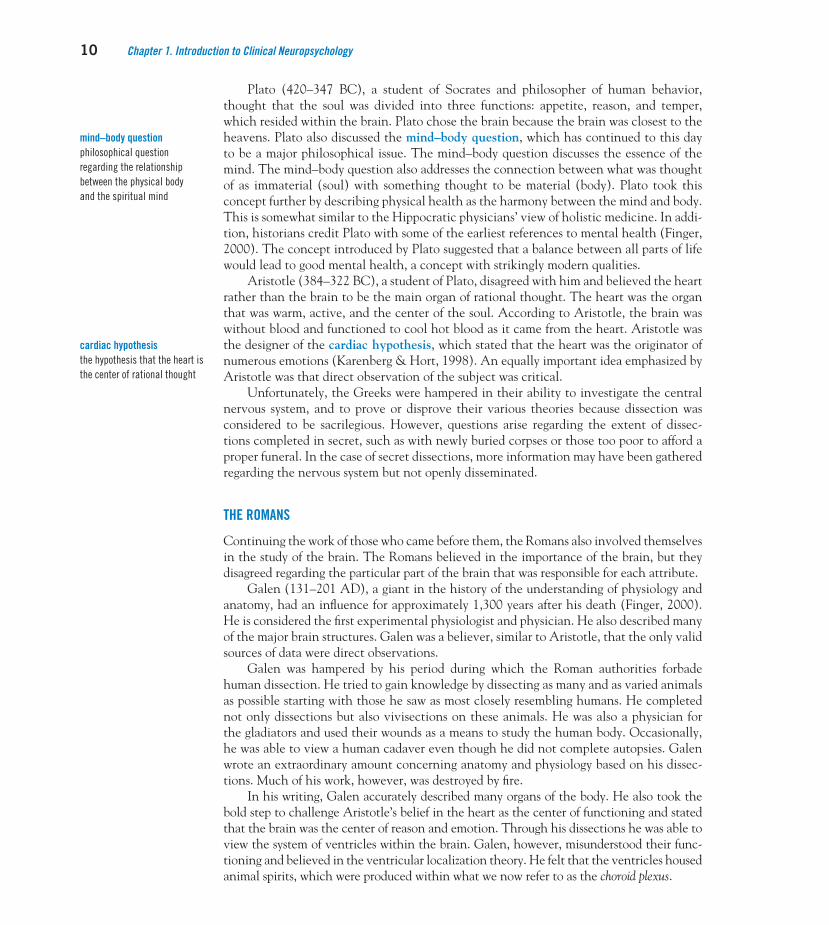
Aristotle (384–322 BC), a student of Plato, disagreed with him and believed the heart rather than the brain to be the main organ of rational thought. The heart was the organ that was warm, active, and the center of the soul. According to Aristotle, the brain was without blood and functioned to cool hot blood as it came from the heart. Aristotle was the designer of the cardiac hypothesis, which stated that the heart was the originator of numerous emotions (Karenberg & Hort, 1998). An equally important idea emphasized by Aristotle was that direct observation of the subject was critical.
Unfortunately, the Greeks were hampered in their ability to investigate the central nervous system, and to prove or disprove their various theories because dissection was considered to be sacrilegious. However, questions arise regarding the extent of dissec-tions completed in secret, such as with newly buried corpses or those too poor to afford a proper funeral. In the case of secret dissections, more information may have been gathered regarding the nervous system but not openly disseminated.
THE ROMANS
Continuing the work of those who came before them, the Romans also involved themselves in the study of the brain. The Romans believed in the importance of the brain, but they disagreed regarding the particular part of the brain that was responsible for each attribute.
Galen (131–201 AD), a giant in the history of the understanding of physiology and anatomy, had an influence for approximately 1,300 years after his death (Finger, 2000). He is considered the first experimental physiologist and physician. He also described many of the major brain structures. Galen was a believer, similar to Aristotle, that the only valid sources of data were direct observations.
Galen was hampered by his period during which the Roman authorities forbade human dissection. He tried to gain knowledge by dissecting as many and as varied animals as possible starting with those he saw as most closely resembling humans. He completed not only dissections but also vivisections on these animals. He was also a physician for the gladiators and used their wounds as a means to study the human body. Occasionally, he was able to view a human cadaver even though he did not complete autopsies. Galen wrote an extraordinary amount concerning anatomy and physiology based on his dissec-tions. Much of his work, however, was destroyed by fire.
In his writing, Galen accurately described many organs of the body. He also took the bold step to challenge Aristotle’s belief in the heart as the center of functioning and stated that the brain was the center of reason and emotion. Through his dissections he was able to view the system of ventricles within the brain. Galen, however, misunderstood their func-tioning and believed in the ventricular localization theory. He felt that the ventricles housed animal spirits, which were produced within what we now refer to as the choroid plexus.
mind–body question philosophical question regarding the relationship between the physical body and the spiritual mind
cardiac hypothesis the hypothesis that the heart is the center of rational thought

Chapter 1. Introduction to Clinical Neuropsychology 11
Galen also believed in the earlier theory that the functions of the body and brain were based on a balance of bodily fluids or humors (blood, yellow bile, phlegm, black bile). Galen’s belief in the four humors (see Figure 1.3) led to many of the treatments suggested for various disorders. During this time period, Galen was very interested in the study of stroke or what was then termed apoplexy. He believed that stroke resulted either from an accumulation of a thick cold humor (such as phlegm or black bile) in the ventricles or from obstructions of the flow of animal spirits.
THE MIDDLE AGES (500–1400)
The early Egyptians, Greeks, and Romans were followed in their study of the brain by many theorists spanning the subsequent centuries, which collectively fall into the histori-cal period in the Western world termed the Middle Ages or Dark Ages.
The time period began with a rudimentary understanding of the brain as the organ of thought and emotion. However, the proposed structures and functions of the brain were inaccurate as many of the earlier writings were unavailable and not based on scien-tific knowledge of the workings of anatomy and physiology. Not being able to study the human body through dissection during this period also led to many of these anatomical misunderstandings. During the Middle Ages, there was a return to superstitious beliefs regarding the causes of many of the difficulties people exhibited. Salient among these were the torturous practices leveled against those believed to be possessed by demons, which we now know may have been afflicted by brain impairments. Examples of symptoms often mistaken for possession by the devil include visual and auditory hallucinations and delusions of g randeur or persecution, commonly noted in schizophrenia.
During the later part of the Middle Ages, the works of Aristotle were rediscovered and translated (between 1200 and 1225), and made available to an expanded audience. His views were accepted as sacred and any questioning was unacceptable. His views were particularly in agreement with the time due to his heavy emphasis on the heart and his nonreliance on any scientific methodology because during this period the church was considered the ultimate authority on all matters.
The initial move away from the ventricular localization theory started in the 13th century. Albertus Magnus (1206–1280) theorized that behavior resulted from a combina-tion of brain structures including the cortex, the midbrain, and the cerebellum. It is very interesting, historically, to note that Magnus was a Dominican monk because at that time clergy were not thought to stray from recognized doctrine.
DryHot
Wet Cold
EARTH
Black Bile(Spleen)Autumn
FIREYellow Bile
(Liver)Summer
AIR
Blood(Heart)Spring
Phlegm(Brain)Winter
WATER
FIGURE 1.3 The theory of four elements expanded to include dual qualities and single associated humors, body organs, and even seasons of the year.
humors the belief that a balance of bodily fluids including blood, mucus, and yellow and black bile were responsible for the functioning of the body and the brain

12 Chapter 1. Introduction to Clinical Neuropsychology
In general, there was a stagnation of new learning during the Middle Ages. Natural philosophers, such as Avicenna, had access to the Greek and Roman books of science. The Arabs and Nestorian Christians venerated, collected, and translated the works of Hippo-crates, Aristotle, and Galen (Finger, 1994). They based their own medical practices on these classics. The works of Middle Eastern scientists and healers only became familiar to Europeans at the end of the Middle Ages. Most of the information became available when Europeans went south to conquer Moorish Spain. The material then spread to France and Italy and began a revival of interest in anatomy, physiology, and medicine.
RENAISSANCE EUROPE (1400–1600)
The Renaissance is generally considered to have begun in Italy in the mid-14th century and ended during the 16th century. The Renaissance marked the end of medieval Europe and allowed intellectual freedom to flourish. This ushered in a period of significant change scientifically, artistically, and socially. Included was a rapid expansion of knowledge of anatomy and physiology supported by reacquisition of earlier texts, which remained active in Arabic thought when Europe was in the Dark Ages. Surprisingly, one of the major factors in the start of the Renaissance was the plague of the 1300s, the Black Death. This pandemic led to a questioning of the existing religious, political, and social structures and subsequently led to freer inquiry and thought. Labor had become scarce, which loosened the ties that had kept workers shackled to their land or their employers. The net result was a society in which the pursuit of knowledge became acceptable. The results were dramatic as evidenced by the rapid expansion of science and the arts.
In the late 15th century, Leonardo da Vinci (1452–1519) conducted several hundred human dissections on cadavers in secret due to religious prohibition against autopsies. He drew detailed diagrams of the human body from these dissections. da Vinci conducted experiments on cattle to determine the true structure of the ventricles. The information da Vinci learned from his cattle experiments demonstrated that the actual function of the ventricles did not correspond to the thinking at the time. However, after completing all of his dissections and work with cerebrospinal fluid, he continued to believe in the ventricular localization hypothesis of brain functioning. It is a striking historical question as to why he held to a theory that he was able to disprove.
In 1543, Andreas Vesalius (1514–1564) published the first accurate book on human anatomy entitled On the Workings of the Human Body. It was one of the most important medical science books ever written (Idowu, Malomo, & Osuagwu, 2006). He completed his work through dissections and careful observations and ultimately proved that Galen’s views on ventricular flow were incorrect. Vesalius began the history of public dissection allowing medical students and doctors to view the procedure in a manner foreshadowing current medical practices. Even though the church retained authority over the soul, he took the risk to expose the rest of the body to scrutiny. Through his work, he found fault with the ventricular localization hypothesis and the movement of animal spirits. He also claimed to find at least 200 errors in the anatomical works of Galen (Finger, 2000). He was able to show that to truly understand the workings of the human body one must study humans, not other animals. Vesalius’ ideas were not well received by the public or the scientific community at that time.
During the 17th century, scientists were looking for a single site for the functioning of the mind. The philosopher Rene Descartes (1596–1630) disagreed with the tripartite soul introduced by Plato. He believed in a complete separation of the mind and body. He felt that the mind was immaterial and without substance, whereas, the body functioned similar to a machine. Descartes also dealt with the mind–body problem or the question

Chapter 1. Introduction to Clinical Neuropsychology 13
of the relationship between the two entities. The complete separation of mind and body is referred to as dualism. Monism states that there are no fundamental differences and a unified set of laws underlie nature. Descartes was a dualist who erroneously speculated that mental processes resided within the pineal gland. His idea was that the pineal gland is the only structure not composed of bilaterally symmetrical halves. Presently, the pineal gland is not fully understood, but most researchers believe that it is involved with sleep regulation and melatonin production. However, it lies near the ventricular system and in Descartes’ thinking, he may have attempted to relate this to the earlier ventricular local-ization hypothesis. He also viewed the cortex as a covering of the pineal body.
Thomas Willis (1621–1675), known for his study of blood circulation and for whom the Circle of Willis is named, also studied brain function. In 1664, he published Cerebri Anatome, a work without equal at the time, which was mainly devoted to the study of the brain (O’Connor, 2003). Willis used clinical evidence from living patients with move-ment disorders and observed degeneration in the various structures at autopsy to back his claims. He also described sensations residing within the corpus striatum (Meyer & Hierons, 1964). He stated that the cerebral gyri controlled memory and will. According to Willis, imagination was also a cerebral function located in the corpus callosum. The corpus striatum was thought to be related to sensation and movement. The cerebellum was thought to control the voluntary and involuntary systems. At that point in history, the pons and medulla were considered to be part of the cerebellum.
Willis was the first person in the post-Renaissance period to divide the brain into functional parts based on comparative anatomy, theory, and clinical practice. Although he did not accurately localize various abilities, his writings became a strong impetus for others to look at the functional working of individual brain areas.
Following the ideas of Willis, Emanuel Swedenborg (1688–1772) concluded that the cerebral cortex was the source of understanding, thinking, judging, and willing. He also went further than Willis and stated that certain functions were represented at different anatomical sites on the cortex. Swedenborg saw the localization of function as the way to understand the difficulties which arose with patients with various types of pathologies. He took his ideas further to include other structures. However, most of his ideas were not accepted or published during his lifetime. One reason he was not accepted was that Swe-denborg began to have visions and eventually felt his calling to be in theology. Leaving science for theology was not accepted by many in the scientific community at that time (Akert & Hammond, 1962).
18TH CENTURy: LOCALIZATION THEORy
Localization of brain functioning is one of the most interesting questions that began to be studied by Swedenborg and others during this period and continues to the modern era. The localization of function refers to the idea that the brain has certain functions which are localized or located within specific areas. Franz Joseph Gall (1758–1828) began to write about this idea in 1810. He stated that certain physiological characteristics of indi-viduals appeared to reflect their intellectual or cognitive capabilities.
Gall correlated 27 faculties of the mind with skull features and located these abili-ties on maps of both hemispheres. He became an early advocate of the idea of cortical localization of function. The theory of phrenology was developed from Gall’s ideas. The theory of phrenology suggested that abilities were so localized that they would appear as protuberances on the skull. A person could exercise a particular ability by rubbing or mas-saging that area and develop more of the particular ability or trait. The ideas Gall proposed appealed to the average person and phrenology began to be practiced in the salons of
dualism the view that within each p erson resides two entities, a mind with mental properties and a body with physical properties
monism the view that there is only one basic and fundamental reality, that all existence is this one reality; hence, the mind and body operate according to the same principles
localization of brain functioning the theory that certain abilities are localized to certain areas of the brain
phrenology inaccurate theory developed by Gall which stated that bumps on the head related to certain abilities residing within the brain; the theory led to belief in reading the bumps and increasing abilities by rubbing the corresponding bumps
14 Chapter 1. Introduction to Clinical Neuropsychology
Europe. The influence of phrenology lasted many years and regrettably had a widespread impact leading the study of the brain in a nonproductive direction.
In addition to the theory of phrenology, which has been considered to be highly inaccurate, Gall was responsible for several significant discoveries in neuroanatomy and neurophysiology. In essence, his work was recognized for some of the earliest views on the idea of localization of functions. Gall, through his dissections, proposed that the cortex and its sulci and gyri were functioning parts of the brain and not just coverings of the pineal body. He also stated that a large pathway, the pyramidal tract, leads from the cortex to the spinal cord, implying that the cortex sends information to the spinal cord to com-mand movement of the muscles. Gall and his colleagues also discovered the role of the corpus callosum in the communication between the hemispheres.
Gall’s student Johann Spurzheim (1776–1832) worked with him studying phrenology for 9 years. The two parted company because Spurzheim felt there were no bad or evil functions as described by Gall (Carlson, 1958). He contended that bad traits were caused by underdevelopment of the specific functions.
Pierre Flourens (1794–1867) disputed Gall stating there was no localization of function within the cortex. Flourens supported his opinion through studying animals usually with very small brains, which if ablated would destroy more than one function. Ablation was a type of surgery in which removing part of the brain led to generalized, not localized, disor-ders of behavior. He proclaimed there was no specific localization of ability, but rather the amount or extent of tissue damage is what mattered. In other words, the greater the mass of impaired tissue, the more dysfunctional the individual will appear. Flourens also stated that the brain operated in an integrated fashion, not with discrete functions. Without knowing it, Flourens was describing the modern term neuroplasticity, which states that various brain areas are able to take over functions for one another when an area is injured or destroyed. The reason that neuroplasticity is possible is that the brain functions as a whole, similar to the way Flourens described. Flourens also believed that the cerebellum was responsible for coordinated movement and that the medulla was required for basic life functioning.
19TH-CENTURy ADVANCES
The 19th century was a time of great advances in many areas of psychology, which would ultimately make an impact on the study of the brain. The scientific method became a reality in psychology with the development of the first laboratories. Wilhelm Wundt (1832–1920) is credited with the first psychology laboratory in Germany in 1879. Soon thereafter, others developed in various parts of Europe and the United States. The scientific method refers to the reliance on the procedures of science as a means of understanding, as opposed to theorizing without any practical data to validate the theory. The advantages of the scien-tific method are the ability to manage or control all parts of the experiment, which leaves nothing to chance. Through the use of the scientific method researchers began to be able to make cause and effect statements for the first time. The researcher could say that A caused B because no other variable could have done so in a controlled situation. The scientific method allowed the researcher to look at the structure and function of the workings of the brains of lower animals. Through the process of scientifically looking at lower animals, the scientist was able to relate findings to humans. However, many scientists felt it was a large leap to go from the functioning of animal brains to the functioning of the human brain.
Along with the scientific method, another movement occurred beginning in France and Great Britain, which fought for better treatment of individuals who were mentally ill (or suffered from brain impairment). Phillipe Pinel (1745–1826), a French physician, was shocked by what he saw as brutality toward the mentally ill. Objectionable practices
scientific method a method of research in which a problem is identified, a hypothesis is formulated, and relevant data are gathered; from these data, cause–effect relationships can be stated
neuroplasticity the brain’s natural ability to form new connections to com-pensate for injury or changes in one’s environment

Chapter 1. Introduction to Clinical Neuropsychology 15
included not only incarcerating patients with prisoners, but also punishment such as chain-ing individuals to walls for behaviors over which they clearly had no control, such as delu-sions and hallucinations. Pinel became head of two asylums or mental hospitals, Bicêtre and Salpêtrierè. Pinel’s ideas for change included the use of kindness and humanity in the treatment of the patients. These principles of treatment led to better lives for the patients.
At the same time as Pinel, William Tuke (1732–1822) began to improve the care of patients in England. Simultaneously, in America, other individuals such as Eli Todd (1769–1833) began to pursue better treatment for the mentally ill. Dorothea Dix (1802–1887) traveled all over the United States campaigning for reform. Clifford Beers (1900–1979) authored a text in 1908 entitled A Mind That Found Itself: An Autobiography (Beers, 1908). This book chronicles Beers’ experience with bipolar disorder and his treatment. The move-ment that these individuals initiated was termed the mental hygiene movement. Along with this movement came the development of moral therapy, which referred to the humane care and treatment of patients.
At the same time as changes were occurring in the treatment of the mentally ill, attempts were begun to formulate diagnostic classification systems. Emil Kraepelin (1856–1926), writing in 1913, was one of the first individuals to describe mental illness and cat-egorized it based on what was termed endogenous (curable) versus exogenous (incurable). The terms have since been defined as biochemical versus stress induced, respectively. His work foreshadowed our current diagnostic classification system. The diagnostic system which is currently in use is the Diagnostic and Statistical Manual of Mental Disorders, Fourth Edition, Text Revision (DSM-IV-TR) published by the American Psychiatric Association; it will be discussed in greater detail in a subsequent chapter (American Psy-chiatric Association, 2000). At this point, suffice it to say that it is a way to classify mental disorders similar to the manner in which physicians classify physical difficulties using the International Classification of Diseases (ICD-10). The advantages of a diagnostic classi-fication system are multifold and include appropriate treatment, research, communication among professionals, and payment for services.
While all of these events were unfolding, Charles Darwin (1809–1882) was concep-tualizing the origin of the species. Darwin’s ideas radically changed the way people under-stood our relationship with other animals. His theory of evolution and the belief that all living things have a common ancestry was an impetus for the study of lower animals with relation to understanding human functioning. Darwin stressed the survival value of outward expression of emotions by animals and humans. He also believed that the human mind contained primitive inclinations that were held in check by higher mental functions. Many of Darwin’s supporters gave credence to the ideas promoted by therapists. Darwin stated that the expression of feelings had survival value, while therapists state that the expression of feelings leads to better mental health and functioning of the individual.
Localization of Brain Functioning Areas: Higher Cortical Areas
Localization of functioning, as stated previously, began to be a topic of interest in the early 1800s. Gall and, subsequently, Swedenborg investigated various types of localization; how-ever, it took a longer time to look into the localization of higher cortical functioning. Much of the impetus for looking at higher cortical functioning came from the aforementioned world events, which included the growing concern for the treatment of individuals with various dif-ficulties. These difficulties had begun to be seen as residing within the cerebral cortex.
Paul Broca (1824–1880) is often given credit for the discovery of localization of language within the left hemisphere. His work will be discussed later in this section.
mental hygiene movement the movement to treat psychiatric patients with kindness and dignity; it instigated the release of mental patients from prison and the building of mental hospitals
moral therapy therapy created for mental patients based on the ideas of the mental hygiene movement; kindness and respect were the main components
diagnostic classification system a system for classifying medical and psychiatric disorders; it lists symptoms of a particular disorder and various other important facts for diagnosis; in psychology, it usually refers to the DSM-IV-TR published by the American Psychiatric Association
asylum an early institution specializing in the care of the mentally ill
16 Chapter 1. Introduction to Clinical Neuropsychology
However, his work clearly was based on those who came before him, Jean-Baptiste Bouil-laud (1796–1881), Simon Alexandre Ernest Aubertin (1825–1893), as well as Marc Dax (1771–1837) and his son Gustave Dax (1815–1874).
Jean-Baptiste Bouillaud was a well-respected French scientist who made all of his assertions based on clinical data and/or autopsies. In 1825, after examining data from a large number of cases, he asserted that the brain had several special organs. One of the special organs was related to speech and difficulties with speech were evident when the specific area was damaged (Stookey, 1963). The difficulties described and the impaired brain areas clearly foreshadow Broca’s discoveries.
Simon Alexandre Ernest Aubertin (1825–1881), Bouillard’s son-in-law, was also a French physician. He also argued from clinical cases that there were specific higher order cognitive functions localized within certain areas.
Marc Dax (1771–1837), was another French neurologist who discovered through clinical practice the link between the damage to the left cerebral hemisphere and the loss of the ability to produce speech. Dax wrote two papers in 1836, one entitled Observations Tending to Prove the Constant Coincidence of Disturbances of Speech With a Lesion of the Left Hemisphere of the Brain and Lesions of the Left Half of the Encephalon Coincident With the Forgetting of Signs of Thinking (Roe & Finger, 1997). He died the following year without publishing his findings.
Gustave Dax (1815–1874), while studying medicine in the 1860s, published his father’s works along with his own findings (Buckingham, 2006). The Dax work was pub-lished 6 weeks before Broca’s paper was published with both stating similar conclusions.
The localization of brain function was further expanded by the work of Paul Broca (1824–1880). Broca examined two clinical cases in detail of individuals who were unable to speak coherently, but were able to understand the spoken word. After their deaths, he examined their brains and found defects or destruction of tissue in exactly the place he had theorized. The area in the posterior, lower region of the left frontal lobe became known as Broca’s area. Broca is also credited with articulating the concept of aphasia, which literally means the inability to use or comprehend language. Broca was one of the first localization-ist psychologists, meaning that he believed that certain abilities were located in specific brain areas. Broca also helped perpetuate the idea that verbal abilities were confined to the left hemisphere. In essence, Broca discovered through clinical cases that language produc-tion was localized to the left hemisphere. This principle became known as lateralization. In contrast, speech is a motor function not specifically localized in a similar manner.
As can be seen, there were many researchers who, through clinical case studies, noticed that verbal abilities resided within the left hemisphere. It remains a historical question who discovered them first and who should receive credit for their discoveries.
Several years after Broca wrote, another researcher, Carl Wernicke (1848–1904), described a second language area of the brain. He was able to discover the new area through the study of dysfunctions in his patients’ abilities. This second area was located in the temporal lobe somewhat to the posterior and inferior to Broca’s area. Damage here led to a particular dysfunction, the inability to make sense with language even though the utterances were grammatically correct whether spoken or written. Wernicke’s discovery led to the belief that the strict localizationists were not correct, that is, expressive language is located in the frontal lobes whereas, receptive language is located in the temporal lobes. Language, in general, does not totally reside in one locale.
Following the discoveries of Broca’s and Wernicke’s areas (see Figure 1.4), another area between the two was discovered. The difficulty was caused by damage to the nerve fibers connecting Wernicke’s and Broca’s areas in the arcuate fasciculus. Damage to this area resulted in what we now term conduction aphasia or the inability in both reception of
aphasia an impairment of the ability to use or comprehend language, usually acquired as a result of a stroke or other brain injury; it may involve difficulties with spoken, written, or gestured language
lateralization the idea that certain abilities reside in one side of the brain or the other; for the majority of individuals, verbal abilities reside in the left hemisphere and spatial abilities reside in the right hemisphere
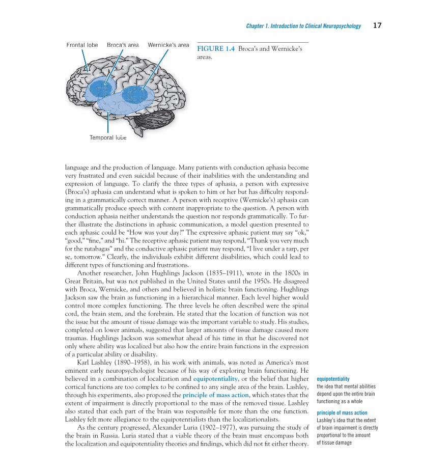
Chapter 1. Introduction to Clinical Neuropsychology 17
language and the production of language. Many patients with conduction aphasia become very frustrated and even suicidal because of their inabilities with the understanding and expression of language. To clarify the three types of aphasia, a person with expressive (Broca’s) aphasia can understand what is spoken to him or her but has difficulty respond-ing in a grammatically correct manner. A person with receptive (Wernicke’s) aphasia can grammatically produce speech with content inappropriate to the question. A person with conduction aphasia neither understands the question nor responds grammatically. To fur-ther illustrate the distinctions in aphasic communication, a model question presented to each aphasic could be “How was your day?” The expressive aphasic patient may say “ok,” “good,” “fine,” and “hi.” The receptive aphasic patient may respond, “Thank you very much for the rutabagas” and the conductive aphasic patient may respond, “I live under a tarp, per se, tomorrow.” Clearly, the individuals exhibit different disabilities, which could lead to different types of functioning and frustrations.
Another researcher, John Hughlings Jackson (1835–1911), wrote in the 1800s in Great Britain, but was not published in the United States until the 1950s. He disagreed with Broca, Wernicke, and others and believed in holistic brain functioning. Hughlings Jackson saw the brain as functioning in a hierarchical manner. Each level higher would control more complex functioning. The three levels he often described were the spinal cord, the brain stem, and the forebrain. He stated that the location of function was not the issue but the amount of tissue damage was the important variable to study. His studies, completed on lower animals, suggested that larger amounts of tissue damage caused more traumas. Hughlings Jackson was somewhat ahead of his time in that he discovered not only where ability was localized but also how the entire brain functions in the expression of a particular ability or disability.
Karl Lashley (1890–1958), in his work with animals, was noted as America’s most eminent early neuropsychologist because of his way of exploring brain functioning. He believed in a combination of localization and equipotentiality, or the belief that higher cortical functions are too complex to be confined to any single area of the brain. Lashley, through his experiments, also proposed the principle of mass action, which states that the extent of impairment is directly proportional to the mass of the removed tissue. Lashley also stated that each part of the brain was responsible for more than the one function. Lashley felt more allegiance to the equipotentialists than the localizationalists.
As the century progressed, Alexander Luria (1902–1977), was pursuing the study of the brain in Russia. Luria stated that a viable theory of the brain must encompass both the localization and equipotentiality theories and findings, which did not fit either theory.
Temporal lobe
Frontal lobe Broca’s area Wernicke’s area
pTemporal lobeporal lobelobel ll b
FFFFFrontal lobbeeFrontal lobbeetal lobbee eaeaeaeaeaareareareaareaarooocaocaa’sa’sscaa s oocoocBrrBrrBrr s areareas as asske’ske ske ske sckckckckniccccninininiWernWernWernWernWernWWWWW FIGURE 1.4 Broca’s and Wernicke’s areas.
equipotentiality the idea that mental abilities depend upon the entire brain functioning as a whole
principle of mass action Lashley’s idea that the extent of brain impairment is directly proportional to the amount of tissue damage

18 Chapter 1. Introduction to Clinical Neuropsychology
Luria described each area of the central nervous system as being involved in one or more brain functions. The first area regulated the arousal level of the brain and proper muscle tone. The second area played a role in the reception and analysis of sensory information from the external and internal environments. The third area was involved with planning, executing, and verifying behavior (Luria, 1966).
At approximately the same time that Luria was studying the functions of various areas of the brain, Roger Sperry (1913–1994) began studying split-brain subjects, those in whom the corpus callosum had been severed. The corpus callosum is the large band of nerves that joins the right and left hemispheres of the brain. Sperry dispelled work stat-ing that the corpus callosum served no function. Sperry studied split-brain subjects and demonstrated important consequences for each hemisphere after separation of the corpus callosum. During independent testing of the hemispheres, the left hemisphere was more verbal, rational, and analytical, while the right was more spatial and emotional. Sperry received a Nobel Prize for his work in 1981.
During the time of Sperry’s initial work, Ward Halstead (1908–1968) developed the first neuropsychology laboratory in 1935 at the University of Chicago. While other researchers were studying and theorizing about brain structure and function, Halstead began the arduous task of assessing brain impairment. He not only attempted to develop tests to measure brain impairment, but also he wanted his tests to be reliable and valid measures of the constructs that he was studying (Halstead, 1947). Halstead worked almost exclusively with neurology patients, those individuals who had diseases or dam-age to their central nervous systems, and developed assessment devices which differenti-ated between patients with brain damage and those without. Halstead developed, in collaboration with his student Ralph Reitan, the Halstead-Reitan Neuropsychological Test Battery. The Halstead-Reitan Neuropsychological Test Battery has been the most popular and most widely used fixed test battery in the United States and abroad for approximately the past 50 years.
At the same time that Halstead and Reitan were working, Luria as stated previously, also developed his theory in Russia. Luria’s works were translated into English and pub-lished in the United States by Anne-Lise Christensen (1927–). Charles Golden (1949–), while at the University of Nebraska, developed the Luria-Nebraska Neuropsychological Battery based on Luria’s original ideas of brain functioning. There is some disagreement within the field regarding whether the Luria-Nebraska Neuropsychological Battery truly reflects Luria’s ideas regarding brain functioning. As will be described in the chapter on test batteries, the Halstead-Reitan Neuropsychological Test Battery and the Luria-Nebraska Neuropsychological Battery have been the most commonly used test batteries. Each battery has its own strengths and weaknesses and each follows a separate way of conceptualizing brain functioning.
The two test batteries were developed in different manners and the directions for administering test items reflects this. The Halstead-Reitan Neuropsychological Test Battery was designed to be administered in a standardized fashion to all subjects and allowed for comparability between subjects, whereas the Luria-Nebraska Neuropsycho-logical Battery was designed to fit the needs of the individual patient and administration of test items could be altered to fit individual needs. There is less comparability of scores across individuals with this approach. Currently, other test batteries are being devel-oped and will be discussed, as well as the issue of the use of a composite of individual tests as opposed to a test battery. After a brief glance at the two major fixed test bat-teries, you might begin to guess which one, if either, was given to Marvin. Considering Marvin was in a moving vehicle accident, which may often lead to legal issues, he was given the Halstead-Reitan Neuropsychological Test Battery. This battery is more useful
split-brain studies studies conducted on humans with severed corpus callosums to determine the extent of com-munication between the two hemispheres
corpus callosum a large mass of myelinated axons connecting the right and left hemispheres; it functions to allow communication between the two hemispheres

Chapter 1. Introduction to Clinical Neuropsychology 19
in forensic situations due to the manner in which it was constructed. He could also have been administered a more flexible test battery, which would allow the choice of tests to be governed by his presenting symptoms.
The Development of Clinical Neuropsychology as a Profession
At the same time the aforementioned tests were being developed, world events were shap-ing the need for clinical neuropsychologists and their expertise and services. It is crucial to the understanding of the work of the clinical neuropsychologist to understand the events which led to the development of the field of clinical psychology. Clinical neuropsychology is considered to be a subspecialty of clinical psychology.
World War I (1914–1918) was the first war where many individuals who would have died in earlier wars were saved by advancements in medical practice. However, these individuals often had residual damage from warfare believed to be related to the noise, shaking of the brain, and various other difficulties which occurred dependent on the job of the soldier. Collectively, these individuals were said to suffer from shell shock, a term given because of the force exerted by the explosions of large munitions.
Economically, the Great Depression (1929–1941) caused a tremendous number of people to become poor, underfed, or homeless. Many of these individuals we could assume suffered from depression, substance abuse, posttraumatic stress disorder (PTSD), or various central nervous system illnesses or injury.
World War II (1939–1945) provided a strong impetus for the services of clinical neuropsychologists. Advances in wartime medicine allowed even more wounded service people to return home than in World War I. These survivors often returned with physi-cal and mental difficulties causing them impairment in their daily lives. At this point in time, the soldier’s psychiatric ailments were often termed battle fatigue. The similarities in the symptoms seen in veterans of the Great Wars and victims of the Great Depression were impetuses for the eventual creation of the term posttraumatic stress disorder.
The creation of the Veterans Administration (VA) in 1930, primarily in response to the needs of World War I veterans, led to increased training for clinical neuropsycholo-gists. The VA was created to service the physical and mental health needs of those who had served in the armed forces. As the study of the brain continued, it became clear that warfare could cause brain damage and that assessment, diagnosis, and rehabilitation ser-vices were needed (van der Kolk, 1997).
The Korean War (1950–1953) furthered the involvement of clinical neuropsychol-ogy within the VA because, again, many more individuals returned home with obvious impairments. The VA system became one of the primary trainers and employers of clinical neuropsychologists.
The Vietnam Conflict (1960–1973) and its political aftermath paved the way for additional services for veterans, but not until 10 years after the soldiers’ reentry. Along with the activism of Vietnam Veterans’ groups for the treatment of emotional issues came the naming of soldiers’ difficulties as PTSD in 1985. Posttraumatic stress disorder means that an individual has experienced a stressor beyond the capacity for most people to endure. The symptoms develop after the stressor and may include flashbacks of trauma, dreams, exaggerated startle response, and hypervigilance. Research on the symptoms of PTSD clearly shows brain impairment is possible particularly in the areas of memory and new learning (van der Kolk, 1997).
After the establishment of PTSD as a diagnostic category, the application of the term to other difficulties including survivors of rape, incest, and natural disasters was begun. All
Veterans Administration (VA) a U.S. government agency created to service the physical and mental health needs of those who had served in the armed forces
posttraumatic stress disorder (PTSD) a mental disorder occurring after a traumatic event outside the range of usual human experience and characterized by symptoms such as reliving the event, reduced involvement with others, and exaggerated startle response; earlier terms for PTSD include shell shock and battle fatigue

20 Chapter 1. Introduction to Clinical Neuropsychology
of these traumatic events also have been shown to have brain sequela. Current examples of individuals who could develop PTSD would include first responders such as police and fire fighters and survivors of hurricanes, tornados, and other types of natural disasters.
These historical examples relate to the development of the field of clinical psychol-ogy and its subspecialty clinical neuropsychology. Clinical neuropsychology began within the general medical service of most facilities. It was greatly influenced by the interac-tions between neurology, neurosurgery, psychiatry, and other medical disciplines. At the present time, clinical neuropsychologists are often assigned their primary responsibilities through the medical service.
Clinical Neuropsychology as a Subspecialty of Clinical Psychology
Ward Halstead began the first clinical neuropsychology laboratory in the 1930s. After the Second World War, the VA was expanded and the need for clinical neuropsychologists was great. However, during this period, the education for clinical neuropsychologists was more experiential than academic. The majority of early individuals who became interested in clinical neuropsychology had their primary training in clinical psychology.
Clinical psychology is the branch of psychology dealing with mental illness. Training for professionals was not standardized until the late 1940s as a consequence of a major meeting entitled the Boulder Conference held at the University of Colorado in Boulder, Colorado. This meeting led to the historic Boulder model (Raimy, 1950). The Boulder model developed the framework for the training of clinical psychologists. The guidelines were termed the scientist–practitioner model. These guidelines stated that to be accred-ited by the APA, a doctoral program in clinical psychology must contain a prescribed number of classes or credits in the scientific bases of behavior and a certain number of classes or credits in the practice of psychology. This model is still upheld in the accredita-tion of PhD programs. APA recognizes clinical neuropsychology as a separate and distinct field of study. Division 40 (Clinical Neuropsychology) of APA was created in 1980. The APA defines a clinical neuropsychologist as follows:
A Clinical Neuropsychologist is a professional psychologist who applies principles of assessment and intervention based upon the scientific study of human behavior as it relates to normal and abnormal functioning of the central nervous system. The Clinical Neuropsychologist is a doctoral-level psychology provider of diagnostic and intervention services who has demonstrated competence in the application of such principles for human welfare following:
A. Successful completion of systematic didactic and experiential training in neuro-psychology and neuroscience at a regionally accredited university;
B. Two or more years of appropriate supervised training applying neuropsychological services in a clinical setting;
C. Licensing and certification to provide psychological services to the public by laws of the state or province in which he or she practices;
D. Review by one’s peers as a test of these competencies. (APA, 1989)
Division 40 of the APA considers the attainment of the American Board of Profes-sional Psychology (ABPP) or the American Board of Clinical Neuropsychology (ABCN) diplomat status in Clinical Neuropsychology as the clearest evidence of competence as a
clinical psychology a branch of psychology devoted to the assessment, diagnosis, and treatment of mental and behavioral disorders
scientist–practitioner model guidelines that state that to be accredited by the APA, a clinical or counseling doctoral program must contain a prescribed number of classes or credits in the scientific bases of behavior and a certain number of classes or credits in the practice of psychology
Chapter 1. Introduction to Clinical Neuropsychology 21
clinical neuropsychologist, assuming that all of the criteria have been met (www.theabcn.org/abpp-diploma/index).
There are three major organizations that exist for the perpetuation of clinical neu-ropsychology. These three organizations are Division 40 (Clinical Neuropsychology), as already described, of the APA, The International Neuropsychological Society (INS) and The National Academy of Neuropsychology (NAN). These organizations function to further the development of the field.
The INS was founded in 1967. It currently has more than 4,500 members through-out the world. It holds two professional meetings per year, one in the United States and the other abroad. The INS is a multidisciplinary organization, which has as its goal the enhancement of communication among the scientific disciplines that contribute to the understanding of the brain–behavior relationship.
The NAN was founded in 1975. Current membership totals more than 3,300 members from 24 countries. The goals of NAN are to advance the scientific study of brain–behavior relationships using neuropsychological techniques, develop standards of practice, develop training guidelines, provide continuing education, and provide an information resource in neuropsychology.
In order to find out more about the actual workings of clinical neuropsychologists, Sweet, Moberg, and Suchy (2000) reviewed a 10-year follow-up survey of clinical neurop-sychologists working in private practice and institutions. The sample included 422 respon-dents, 60.2% ABPP’s and 41.4% general Division 40 members of APA. The PhD was by far the most common degree for both groups with 9 out of 10 holding this type of degree in both groups. The field of study where it was granted was primarily clinical psychology. The majority of the respondents did agree with the statement by the APA and felt that clinical neuropsychology is a subspecialty of clinical psychology. Respondents described the need for broad predoctoral training in clinical psychology, as well as specialty training and an internship in clinical neuropsychology.
Sweet et al. (2000) included journals subscribed to by clinical neuropsychologists as examples of interest areas and expertise. The top six most commonly subscribed to journals in 1999 were Archives of Clinical Neuropsychology, Neuropsychology, The Clinical Neuropsychologist, Journal of the International Neuropsychological Society, Journal of Clinical and Experimental Neuropsychology, and Neuropsychology Review. The number of journals specifically related to clinical neuropsychology gives an indication of the depth and breadth of the field according to the authors.
METHODOLOGy SPECIFIC TO CLINICAL NEUROPSyCHOLOGy
Clinical neuropsychology, being an applied field, has methodological issues similar to other applied fields. An applied field means that knowledge taken from research and clinical practice is used in the treatment of patients. Clinical neuropsychology has also shared methodology with other disciplines.
SubjectsOne of the main questions for the clinical neuropsychologist is the type of subject from which research findings have been taken. As can be seen from the historical background, many studies of the brain have come from individuals who had sustained traumatic inju-ries or suffered from various illnesses. They have been excellent subjects to assist the clini-cal neuropsychologist to understand the effects of damage to particular areas and to assist in developing rehabilitation methods. Investigation of a single individual with central

22 Chapter 1. Introduction to Clinical Neuropsychology
nervous system illness or injury is termed the case study method. The case study method has added a wealth of information to the clinical neuropsychological literature.
It is very challenging to compare the difficulties of one individual to others, even with similar injuries because other extraneous variables may confound the research results. Exam-ples of confounding variables which could cause problems with comparability of subjects include age, gender, socioeconomic status, social support network, access to physical medi-cine, and rehabilitative services. Marvin’s case is a good example. He has a brain injury caused by an automobile accident and many things are known about brain injury from motor vehicle accidents. There are other specifics to Marvin’s case which may make it difficult to develop a treatment plan based only on case study information. Examples of confounding variables in Marvin’s case include being a 36-year-old White male from a lower middle class background, high school educated, lacking support from his ex-wife and having few friends as a social support network. Many researchers try to overcome these difficulties exemplified by Mar-vins’s case by gathering groups of individuals with as similar as possible injuries and matching background factors. By having many individuals with similar difficulties, the clinical neurop-sychologist is able to see the pattern of strengths and weaknesses with various difficulties. This information is very important in developing a treatment plan for rehabilitation.
Techniques Dealing With Subject VariablesThe double dissociation technique was developed to help researchers determine when cog-nitive factors are independent. As described by Shallice (1988), a double dissociation occurs when lesions have opposite or dissimilar effects on two distinct cognitive functions. For example, lesion 1 causes difficulty with cognitive function 1 but not cognitive function 2, while lesion 2 causes a problem with cognitive function 2 but not cognitive function 1. We make an assumption that the functions are independent because the changes in one do not affect the other. A commonly given example to illustrate the double dissociation technique is the study of individuals with Broca’s aphasia and Wernicke’s aphasia. It is clear from the studies of these two groups that the abilities, which reside in these areas, do not cause each other, even though they may affect one another greatly. Hans-Lukas Teuber (1955) used the double dissociation technique to demonstrate lateralization of function. Lesions in the left hemisphere of right-handed people produce deficits in language functions not evident with lesions in the right hemisphere. Lesions in the right hemisphere produce deficits in spatial tests not evident with left-hemisphere lesions.
Another approach often referred to as the lesion approach involves the use of lesions in humans and other animals. Specific lesions may be made in humans and animals who have all of the extraneous variables eradicated by having similar genetic qualities and environments. This is the closest we are able to come to showing the effects on behavior of specific lesions. When the lesion approach is used with humans it is usually in areas where the individual has some form of difficulty for which the lesion should help eradicate the difficulty. Lesion studies in animals are different. Lesions are often used to create deficits in animals and observe functioning afterward. Care and treatment of research animals is always kept foremost in the researcher’s mind when completing lesion studies.
IMAGING TECHNIQUES IN THE STUDy OF THE HUMAN BRAIN
In addition to the choice of subjects for research in clinical neuropsychology, there are also issues related to the use of various techniques for studying the human brain. As time has passed, the clinical neuropsychologist’s role has changed from one in which diagno-sis was made based on results of testing to one in which testing is used to evaluate the strengths and weaknesses of the individual and help develop an effective treatment plan.
case study a detailed analysis of an individual focusing on his or her history and current life situation; it is often a way to study individuals with central nervous system illness or injury
double dissociation technique a research technique in which lesions have opposite or dis-similar effects on two distinct cognitive functions; it was developed to help determine when cognitive functions are independent
lesion approach the lesioning of animals and sometimes humans to study the effects of the lesion on various brain functions

Chapter 1. Introduction to Clinical Neuropsychology 23
The advent of advanced medical imaging techniques has allowed the diagnosis to be made much quicker and then corroborated by neuropsychological testing. However, there are times and circumstances where medical techniques are not able to view difficulties or the deficits are so small and the determination of diagnosis is greatly enhanced by performance on neuropsychological tests.
Electrical TechniquesElectroencephalography (EEG) is the oldest technique which records brain waves and their pattern as a person completes different tasks (see Figure 1.5). The use of the EEG began in 1928 when Hans Berger (1873–1941) used it to measure brain waves. Berger’s intention was to find evidence for telepathy, the unscientific manner of communica-tion between individuals. He did not find what he desired but discovered that the brain waves of a sleeping brain were very different from an awake brain. Researchers have since used this technology to record and measure the activity of nerve cells in the brains of individuals who are normal and those suffering from various difficulties and diseases. The EEG measures the firing of nerve cells, which tend to fire in a synchronous pattern leading to alpha, beta, theta, and delta waveforms.
The EEG was the first technique to record the electrical activity of the brain. How-ever, it is limited in that it does not reveal the contents of thought but reveals only that the person is thinking. Other techniques building on EEG technology have produced more sophisticated brain wave evaluations. One manner in which to expand on the EEG is to employ a computer to map the activity of the brain’s electrical activity as it is occurring in the individual’s head. The techniques have been termed brain electrical activity mapping (BEAM). It produces a color-coded map of the brain as various areas exhibit activation.
A further advance using EEG technology is to record changes in the EEG signal in response to a sensory stimulus. This procedure has been termed an evoked potential (EP) or an event-related potential (ERP). The difficulty with this procedure is that multiple
FIGURE 1.5 Characteristic EEG recordings. Brain wave patterns correspond to differ-ent states of consciousness in humans. Source: Adapted from Kolb, B., & Whishaw, I. Q. (2009). Fundamentals of Human Neuropsychology, 6th ed. New York: Worth Publishers.
(A) AWAKE OR EXCITED—beta rhythm
(B) RELAXED, WITH EYES CLOSED—alpha waves
(C) DROWSY—slowed frequency, increased-amplitude waves
(D) ASLEEP—slower, higher-amplitude delta waves
(E) DEEP SLEEP—even slower and higher-amplitude waves
(F) COMA—further slowing
Time (sec)Time (sec)1 2 3 4 5 6 71 2 3 4 5 6 7
electroencephalography (EEG) a record of brain wave patterns as an individual completes a task
evoked potential the ability to record changes in EEG activity in response to sensory stimuli; the response is termed an evoked potential or event-related potential

24 Chapter 1. Introduction to Clinical Neuropsychology
recordings need to be made and an average computed because of the multitude of signals which are produced by the brain. The EEG is particularly useful in the recording of seizure activity, which registers as a very nonsynchronous pattern. The EEG is also used to detect brain wave patterns in individuals with sleep disorders due to the fact that different sleep states are associated with particular forms of electrical activity. It is the main diagnostic tool used in sleep laboratories. In addition, the EEG is effective in diagnosis of individuals with TBI in which the seizure threshold may have changed.
Radiological TechniquesThe first radiological recordings of brain structure and function came from X-rays. Early X-ray pictures of the brain were very crude and did not capture the essence of the brain difficulties only external boundaries. The X-rays were absorbed by different structures to different degrees; hence, the images of bone were clearer than soft tissue, which was clearer than the blood vessels and ventricles. A second difficulty was that it produced only a two-dimensional image of the three-dimensioned brain. X-rays also introduced radiation into the brain. At the time they were introduced, the safety range for radiation had not been determined. However, now it is thought that the radiation the brain is exposed to using the X-ray is minimal.
A method of enhancing the X-ray is termed pneumoencephalography. In this pro-cedure a small amount of cerebrospinal fluid is removed and replaced by air. The X-rays are taken as the air moves up the spinal cord. Because of the presence of air the ventricles stand out clearly. This procedure is not used very often as it is outdated and painful to the patient. A procedure similar to pneumoencephalography is angiography with the excep-tion that a substance that absorbs X-rays is injected into the bloodstream. This procedure reveals the conditions of the blood vessels. It can be a dangerous and painful procedure and has been replaced by newer technologies.
The CT scan was developed in the early to mid-1980s and is based on the X-ray principle (see Figure 1.6). An X-ray beam is passed through the same area from many
X-ray an imaging method in which radiation effects the density of different parts of the brain to various degrees, which then appear on the X-ray film
angiography the use of radiopaque substances to allow visualiza-tion of blood vessels; it is considered a form of X-ray
computed tomography (CT) scan a computer-assisted imaging process based on multiple X-ray images of the brain; it provides a three-dimensional perspective of the brain with clear differen-tiation of brain structures
(A) CT scan (C) Reconstruction, lateral view(B) Horizontal section
Lesion
Posterior
Anterior
Lesion
Lesion
Plane of sectionin parts A and B
FIGURE 1.6 Computed tomography. (A) Horizontal CT scan of a subject with Broca’s aphasia. The region at the left is the location of the lesion. (B) A schematic representation of (A) with the area of the lesion shown in blue. (C) A reconstruction of the brain, showing a lateral view of the left hemisphere with the lesion shown in black. Source: Adapted from Kolb, B., & Whishaw, I. Q. (2009). Fundamentals of Human Neuropsychology, 6th ed. New York: Worth Publishers.

Chapter 1. Introduction to Clinical Neuropsychology 25
different angles thus creating many different images which may be combined using com-puter technology. The computer is able to produce a three-dimensional view of the three-dimensional brain, which had been lacking with previous technologies. Cerebrospinal fluid, brain tissue, blood, and hard tissue all appear as different shades on a CT scan and allow a much better view of diseased or dysfunctional areas. However, the CT scan does introduce radiation into the individual’s system. A more advanced form of CT scan is termed an enhanced CT and involves injecting a contrast agent to provide better visu-alization of brain structures. The difficulties include that this procedure is more invasive and patients have been known to react poorly to the contrast agent.
Single photon emission tomography (SPECT) is similar to CT but is much simpler and inexpensive. Radiolabeled probes are injected into the patient. Tissues will absorb the probes as they circulate in the bloodstream. As the camera rotates around the patient, it picks up photon emissions and relays this information to a computer which converts the information into a film representation. The radiolabeled probes do not have to be synthe-sized but are commercially available. The pictures using this technique are less clear and it takes longer to get them than with PET. It is important to understand that SPECT does not provide views of brain structures but rather shows metabolic activity of various parts of the brain.
Dynamic Brain ImagingPositron emission tomography (PET) was the first technique to follow the CT scan using a different form of technology (see Figure 1.7). It allows the researcher to determine the amount of a particular substance being used by a specific brain section. The substance is usually radioactive labeled glucose or oxygen, which is metabolized by the brain and the radioactivity is later recorded by a special detector. The PET scan measures metabolic activity of different brain regions with the idea that the more active regions will use more glucose. Positron emission tomography is the only procedure through which research-ers can examine the cerebral glucose use and oxygen metabolism three dimensionally. Positron emission tomography has proven to be effective in the diagnosis of head trauma, brain tumors, and stroke through suppression of metabolic activity even when the brain structures appear normal using MRI or CT.
Magnetic resonance imaging (MRI) does not use radiation and for that reason has become favored over the CT scan. The MRI machine broadcasts a radio frequency (RF) pulse that specifically affects hydrogen atoms. This pulse is directed toward the area of the
FIGURE 1.7 PET scanner and resultant images.
positron emission tomography (PET) a technique used to visualize brain activity based on cerebral blood flow; it tracks the metab-olism of glucose, oxygen, and/or neurotransmitters
magnetic resonance imaging (MRI) a brain imaging technique requiring the use of magnetic fields; gives a clearer image than CT scan and is less dangerous because it does not use radiation
single photon emission tomography (SPECT) a technique that measures the emission of single photons of a given energy from radioactive probes; these emissions are used to construct images of the probes located within the body, thereby detailing the flow of blood in a given area

26 Chapter 1. Introduction to Clinical Neuropsychology
body being examined. Some of the RF pulse’s energy is captured by protons and alters their physical characteristics. The magnets of the machine alternate between being on and off and, during the period that the magnets are turned off, the energy absorbed by the protons is slowly released. This release of energy is observed by coils in the machine that send the signals to a computer for processing into a two-dimensional or three-dimensional picture (see Figure 1.8). The images garnered from the MRI are also much clearer than from CT scan. MRIs allow for a three-dimensional view of the brain. Individuals with loose metal imbedded in their bodies cannot have MRIs because the magnetic fields would move the body metal (e.g., heart pacemakers, etc.) However, metal, attached to live tissue, such as in the case of dental fillings, is allowable.
While MRI gives excellent pictures of the brain, it is often of interest to observe how the brain metabolizes or uses certain nutrients such as glucose or how oxygen is distributed. This is very important because the brain functions using these substances. It also is neces-sary to determine deficits when there is an obstruction of these nutrients. To accomplish this, a substance is injected into the circulation with a tracer, which can then be recorded via computer. A tracer is an inert substance used as a transport.
Neuronal activity changes are measured by fMRI, which accompanies changes in cere-bral blood flow and blood oxygenation. Based on these, the researcher is able to infer the activity levels of various brain regions. The fMRI is an adaptation of an MRI scanner in such a manner as to allow detection of increased or decreased blood flow in particular areas of the brain. In comparison to SPECT, PET, and CT, fMRI does not involve radiation exposure.
Brain StimulationIntracranial brain stimulation is the process through which actual brain tissue is stimulated. Early studies indicated that movements may be elicited by stimulating the motor cortex, sen-sations by stimulating the sensory cortex, and that disruption of cognitive activities such as speech may occur through stimulating the speech centers of the brain. Electrical brain simula-
FIGURE 1.8 Magnetic resonance imaging. The subject is placed in a long metal cylin-der that has two sets of magnetic coils arranged at right angles and a coil that surrounds the head. This coil perturbs the static magnetic fields to produce an image of a horizontal section through the head (right). Source: Courtesy of Intermountain Medical Imaging, Boise, Idaho.
intracranial brain stimulation stimulation of brain tissue used in treatment of various central nervous system diseases, such as Parkinson’s disease

Chapter 1. Introduction to Clinical Neuropsychology 27
tion is used less as a diagnostic tool and more as an adjunct to various forms of treatment. Electrical brain stimulation has been very helpful in the treatment of Parkinson’s disorder. The difficulty with this procedure is that it is invasive needing the skull to be opened to insert the electrode. This procedure is therefore used rarely and when other options may have failed.
Transcranial magnetic stimulation is a procedure in which the brain is able to be stimulated through the skull. The original use for this procedure was by neurosurgeons to stimulate brain tissues and monitor the condition during brain surgery. It has currently been used in a manner similar to intracranial brain stimulation as a treatment for various disorders. The advantage to this technique is that it is noninvasive.
As can be seen, there are various techniques that can be used to investigate the struc-ture and functioning of the brain. In Marvin’s case, both CT and an MRI were used. The CT scan gave an overall view of the structures of his brain to determine as best and as clearly as possible the structures which were affected by the impact of the motor vehicle accident. The MRI with the use of contrast dye was able to further evaluate the use of oxygen and glucose by his brain and to determine if there were any impediments to its flow to the brain. The use of multiple techniques is often employed to correctly ascertain the difficulties the individual is experiencing.
transcranial magnetic stimulation procedure in which the brain is stimulated through the skull
Summary
The text and the first chapter began with a case study of an individual who sustained a closed head injury from a motor vehicle accident. Unfortunately, this is a fairly common difficulty with which the clinical neuropsychologist works. The case study also highlights many of the roles and functions which the clinical neuropsychologist performs and will be explored in the remainder of the text.
The chapter includes a brief history of the understanding of brain functioning through-out time. The time periods are divided not only by the historical events but also by the types of technology which occurred in various locales during various periods. The study of brain functioning is important because many of the historical issues remain as current concerns. However, in juxtaposition, many of the early themes of structure and function of the brain have been disproved.
The chapter also includes an introduction to the field of applied clinical neuropsychology and its place within the broader umbrella of clinical psychology. Techniques particular to research in clinical neuropsychology are discussed in this chapter. Also included are general research techniques, which are shared with experimental neuropsychology and imaging techniques shared with the multiple disciplines in the area of the neurosciences.
Questions for Further Study
1. Which questions are left unanswered regarding the functions of the brain? Explain the current theories regarding their functions.
2. Explain the changes that may have occurred in the role of the clinical neuropsycholo-gist with the advent of advanced imaging techniques.
3. As best as you are able to speculate, what will be the strengths and weaknesses of the individual in the case study in 5 years and in 10 years? What types of assistance will he need, if any?

28 Chapter 1. Introduction to Clinical Neuropsychology
Akert, K., & Hammond, M. P. (1962). Emmanual Swedenborg (1688–1772) and his contributions to neurology. Medical History, 6, 255–266.
American Board of Clinical Neuropsychology. (2010). Retrieved July 11, 2010, from http://www .theabcn.org/abpp_diploma/index.html
American Psychiatric Association. (2000). Diagnostic and statistical manual of mental disorders (4th ed., text rev.). Washington, DC: Author.
American Psychological Association. (1989). Definition of a clinical neuropsychologist. The Clinical Neuropsychologist, 3(1), 22.
Beach, F. (1961). Karl Spencer Lashley: June 7, 1890–August 7, 1958. Biographical Memoirs of the National Academy of Science, 36, 162–204.
Beers, C. W. (1908). A mind that found itself: An autobiography. New York: Longmans, Green, and Co.Bereczki, Z., & Marcsik, A. (2005). Trephined skulls from ancient populations in Hungary. ACTA
Medica Lituanica, 12(1), 65–69.Breasted, J. H. (1930). The Edwin Smith Surgical Papyrus. Chicago: The University of Chicago Press.Buckingham, H. W. (2006). The Marc Dax (1770/1877)/Paul Broca (1824–1880) controversy over
priority in science: Left hemisphere specificity for seat of articulate language and for lesions that cause aphemia. Clinical Linguistics and Phonetics, 20(7–8), 613–619.
Carlson, E. T. (1958). The influence of phrenology on early American psychiatric thoughts. American Journal of Psychiatry, 115, 535–538.
Edelstein, L. (1967). Ancient medicine: Selected papers of Ludwig Edelstein. Baltimore: Johns Hopkins University Press.
Executive Committee of APA. (1989). Definition of a clinical neuropsychologist: The following statement was adopted by the executive committee of division 40 at the APA meeting on August 12, 1988. The Clinical Neuropsychologist, 3(1), 22.
Finger, S. (1994). Origins of neuroscience: A history of explorations into brain function. New York: Oxford University Press.
Finger, S. (2000). Minds behind the brain: A history of the pioneers and their discoveries. New York: Oxford University Press.
Halstead, W. C. (1947). Brain and intelligence: A quantitative study of the frontal lobes. Chicago: University of Chicago Press.
Hebb, D. O. (1949). The organization of behavior: A neuropsychological theory. New York: John Wiley & Sons, Inc.
Idowu, O. E., Malomo, A. O., & Osuagwu, F. C. (2006). Lecciones de historia anatomia humana desde el origen hasta el renacimiento [Lessons from history: Human anatomy, from the origins to the Renaissance]. International Journal of Morphology, 24(1), 99–104.
Jones, D. A. J. (2003). The Hippocratic oath: Its contents and the limits to its adaptation. Catholic Medical Quarterly, 54(3).
Karenberg, A., & Hort, I. (1998). Medieval descriptions and doctrines of stroke: Preliminary analysis of select sources. Part II: Between Galenism and Aristotelism—Islamic theories of apolexy (800–1200). Journal of the History of the Neurosciences, 7(3), 174–185.
Kirk, G. S., Raven, J. E., & Schofield, M. (1995). The presocratic philosophers: A critical history with a selection of texts (2nd ed.). Cambridge, UK: Cambridge University Press.
Leca, A. P. (1981). The Egyptian way of death: Mummies and the cult of the immortal. Garden City, NY: Doubleday.
Lisowski, F. P. (1967). Prehistoric and early historic trepanation. In D. Brothwell & A. T. Sandison (Eds.), Diseases in antiquity (pp. 651–672). Springfield, IL: Charles C. Thomas.
Luria, A. R. (1966). Higher cortical functions in man. New York: Basic Books.Meyer, A., & Hierons, R. (1964). A note on Thomas Willis’ views on the corpus striatum and the
internal capsule. Journal of the Neurological Sciences, 1, 547–554.O’Connor, J. (2003). Thomas Willis and the background to Cerebri Anatome. Royal Society of
Medicine, 96(3), 139–143.
References

Chapter 1. Introduction to Clinical Neuropsychology 29
O’Connor, D. C., & Walker, A. E. (Ed.). (1967). A history of neurological surgery (pp. 1–22). New York: Hafner.
Osler, W. (1913). Specialism in the general hospital. John Hopkins Alumni Magazine.Raimy, V. C. (Ed.). (1950). Training in clinical psychology. Englewood Cliffs, NJ: Prentice Hall.Roe, D., & Finger, S. (1997). Gustave Dax and his fight for recognition: An overlooked chapter in
the early history of cerebral dominance. Journal of the History of the Neurosciences, 5, 228–240.Sarton, G. (1927). Introduction to the history of science. Baltimore: Carnegie Institution of Washington.Shallice, T. (1988). From neuropsychology to mental structure. New York: Cambridge University
Press.Stookey, B. (1963). Jean-Baptiste Bouillaud and Ernest Aubertin: Early studies in cerebral localization
and the speech center. Journal of the American Medical Association, 184, 1024–1029.Sweet, J., Moberg, P., & Suchy, Y. (2000). Ten-year follow-up survey of clinical neuropsychologists:
Part II. Private practice and economics. The Clinical Neuropsychologist, 14(4), 479–495.Tascioglu, A. O., & Tascioglu, A. B. (2005). Ventricular anatomy: Illustrations and concepts from
antiquity to renaissance. Neuroanatomy, 4, 57–63.Teuber, H. L. (1955). Physiological psychology. Annual Review of Psychology, 6, 267–296.van der Kolk, B. A. (1997). The psychobiology of posttraumatic stress disorder. Journal of Clinical
Psychiatry, 58(9), 16–24.Verona, J. W. (2003). Trepanation in prehistoric South America: Geographic and temporal trends
over 2,000 years. In R. Arnott, S. Finger, & C. U. M. Smith (Eds.), Trepanation: History, discovery, theory (pp. 223–236). Lisse, The Netherlands: Swets & Zeitlinger.
Wilkins, R. H. (1964). Edwin Smith surgical papyrus. Journal of Neurosurgery, 5, 240–244.