applications of magnetic resonance imaging (mri) and computed tomography ct) lecture 1 f33ab5
TRANSCRIPT

Applications of Magnetic Resonance Imaging (MRI)
and Computed Tomography CT)
Lecture 1
F33AB5


What are CT and MRI?
• CT uses X-rays to produce tomographs (images of slices)
• MRI uses magnetic fields to probe the intrinsic magnetisation of hydrogen nuclei
http://www.mri-ny.com/scannersound.html
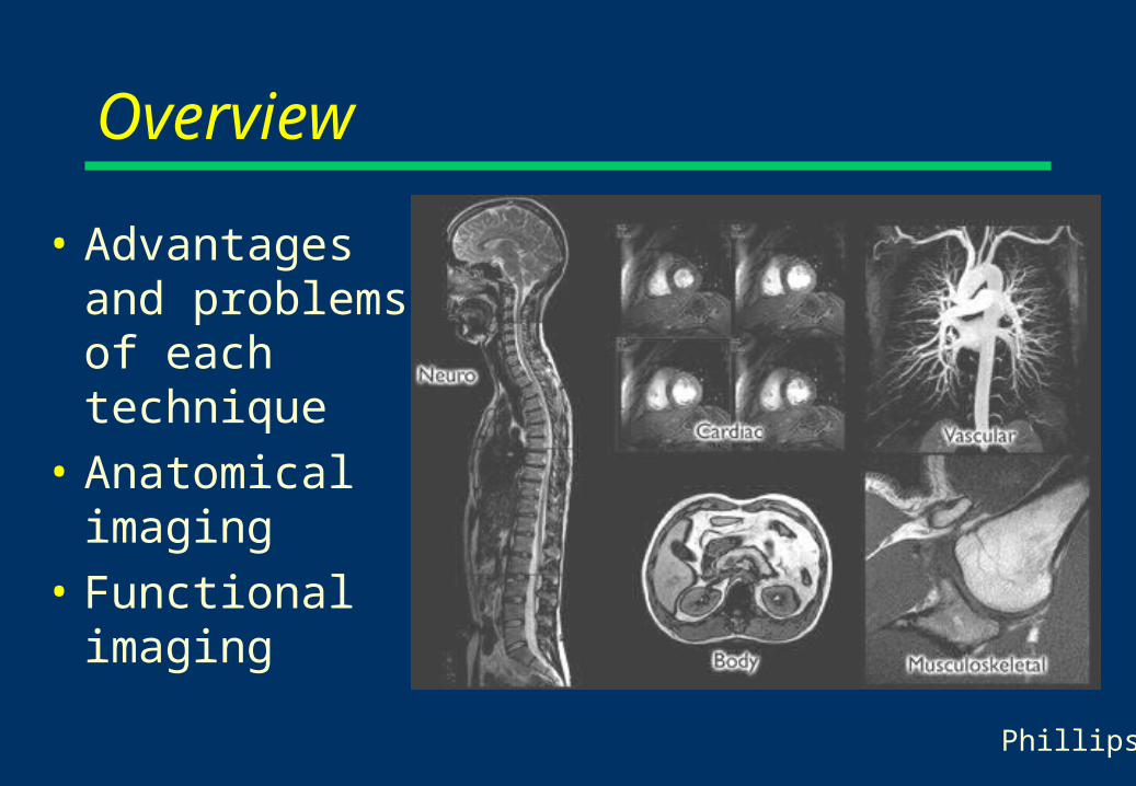
Overview
• Advantages and problems of each technique
• Anatomical imaging
• Functional imaging
Phillips

Problems of CT
• Dose (fluroscopy/dynamic mode not possible)
• (Speed- improving)
• (3D- now available using helical scanning)
• Artefacts behind bone
Advantages of CT
• (Limited) soft tissue contrast
• Spatial measurements exact (if set up correctly)

Problems of MRI
• Not for people who are claustrophobic• Not for people with metal in their bodies• Susceptibility differences (eg between air and
tissue) cause distortions in most sequences, compromising surgical planning
• Can be slow (not EPI), can have motion artefacts
• Can be expensive (£750k)
Advantages of MRI
• Excellent (and controllable) soft tissue contrast
• Much functional information
• Steerable imaging planes
• Safe
• Hugely versatile
Anatomical- CT
• Intracranial bleeds
• Radiotherapy planning– low geometric distortion– CT contrast relates to radiation attenuation
• Stereotactic surgery– low geometric distortion
• Angiography

Anatomical- CT• Intracranial bleeds• Radiotherapy planning
– low geometric distortion– CT contrast relates to
radiation attenuation
• Stereotactic surgery– low geometric distortion
• Angiography
Chronic subdural haematoma
http://www.radiology.co.uk/xrayfile/xray/tutors/cttrauma/tutor.htm

Anatomical- CT
• Intracranial bleeds
• Radiotherapy planning– low geometric distortion– CT contrast relates to radiation attenuation
• Stereotactic surgery– low geometric distortion
• Angiography
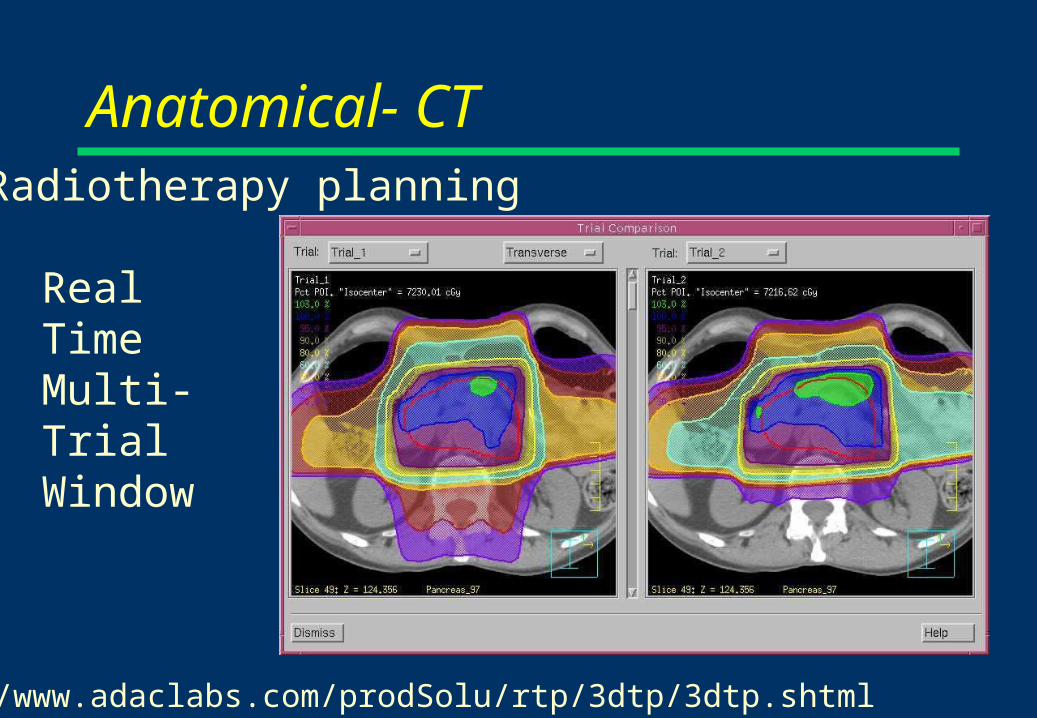
Real Time Multi-Trial Window
http://www.adaclabs.com/prodSolu/rtp/3dtp/3dtp.shtml
Anatomical- CTRadiotherapy planning

Anatomical- CT
Dose distribution along path shown as histogram colored according to the volumes of interest.
http://www.uke.uni-hamburg.de/institute/imdm/idv/publikationen/car1993/
Radiotherapy planning

Anatomical- CT
• Intracranial bleeds
• Radiotherapy planning– low geometric distortion– CT contrast relates to radiation attenuation
• Stereotactic surgery– low geometric distortion
• Angiography

Anatomical- CTMRI CT
CT generally has better geometric accuracyPatient a metal sterotactic frame, ( 'spots' around the head in the images). Streaking artifacts on the CT scans, because of beam-hardening effects.
Brain with a deep central tumour
Dr Paul Morgan, from Academic Radiology

Anatomical- CT
• Intracranial bleeds
• Radiotherapy planning– low geometric distortion– CT contrast relates to radiation attenuation
• Stereotactic surgery– low geometric distortion
• Angiography

Anatomical- CT
Left carotid artery showing aneurysm
Angiography

Anatomical- CT
Ascending aortic aneurysm
Angiography

Anatomical MRI• Head (grey/white matter contrast)
– Tumours – Multiple sclerosis– Myelination in childhood
• Orthopaedic (no bone artefacts)– Spine (sagittal views)
• Great vessels (no contrast agent)
• Bone and soft tissue tumours and disease
• Fluroscopy and Microscopy

Fetal Brain
Placenta
Fetal Lung Fetal Liver
Anatomical MRIFetal imaging-brain

L R
Liver
Spleen
Kidneys
Meal in fundus
Meal in antrumSpinal cord
Anatomical MRI

Anatomical MRI• Head (grey/white matter contrast)
– Tumours – Multiple sclerosis– Myelination in childhood
• Orthopaedic (no bone artefacts)– Spine (sagittal views)
• Great vessels (no contrast agent)• Bone and soft tissue tumours and disease• Fluroscopy and Microscopy
MRI gives flexible contrast
Anatomical MRI• Head (grey/white matter contrast)
– Tumours – Multiple sclerosis– Myelination in childhood
• Orthopaedic (no bone artefacts)– Spine (sagittal views)
• Great vessels (no contrast agent)
• Bone and soft tissue tumours and disease
• Fluroscopy and Microscopy

Anatomical MRI
Orthopaedic MRI (sports injury)

Anatomical MRI• Head (grey/white matter contrast)
– Tumours – Multiple sclerosis– Myelination in childhood
• Orthopaedic (no bone artefacts)– Spine (sagittal views)
• Great vessels (no contrast agent)
• Bone and soft tissue tumours and disease
• Fluroscopy and Microscopy

MR Functional imaging Angiography
Pulmonary arteries
http://www.cardiac-mri.com

Anatomical MRI• Head (grey/white matter contrast)
– Tumours – Multiple sclerosis– Myelination in childhood
• Orthopaedic (no bone artefacts)– Spine (sagittal views)
• Great vessels (no contrast agent)
• Bone and soft tissue tumours and disease
• Fluroscopy and Microscopy

Anatomical MRI• Head (grey/white matter contrast)
– Tumours – Multiple sclerosis– Myelination in childhood
• Orthopaedic (no bone artefacts)– Spine (sagittal views)
• Great vessels (no contrast agent)
• Bone and soft tissue tumours and disease
• Fluroscopy and Microscopy

Functional MRI
Cardiac MRI
End diastole
http://www.cardiac-mri.com

MR Functional imaging Fluroscopy

MRI microscopy
Pharmaceutical Pharmaceutical Dosage FormDosage Form
Castor Bean Seedling
Aplysia Neuron
MaterialsMaterials Plants (Plants (in vivoin vivo)) Excised samples Excised samples ((in vitroin vitro))
Professor Bowtell
Anatomical MRI and CT• Abdominal cancer
– rectal– prostate– cervical, uterine– bladder– breast
• Brain cancer (meninges)• Congential heart disease• Dementia

CT Functional Imaging
• CT is not a very functional modality
• However with contrast agents it can measure– perfusion– angiography– renography
• But- this all requires dynamic repeated scanning… dose is a problem
MRI is a Functional Imaging Technique• Perfusion• Tracers
– Blood brain barrier permeability– Lung function– Molecular imaging?
• Physical properties of tissues– microstructure from relaxation times– microstructure from diffusion– elastic properties
• fMRI- brain activation

MRI is a Functional Imaging Technique• Perfusion• Tracers
– Blood brain barrier permeability– Lung function– Molecular imaging?
• Physical properties of tissues– microstructure from relaxation times – microstructure from diffusion– elastic properties
• fMRI- brain activation

Perfusion rateml/100g/min
>1000500-1000300-500<100
MR Functional imaging- Perfusion

MR Functional imaging Blood brain barrier permeability

MRI is a Functional Imaging Technique• Perfusion• Tracers
– Blood brain barrier permeability– Lung function– Molecular imaging?
• Physical properties of tissues– microstructure from relaxation times– microstructure from diffusion– elastic properties
• fMRI- brain activation

Lung ventilation using hyperpolarized helium
MR Functional imaging Tracers
Dr Owers-Bradley

MRI is a Functional Imaging Technique• Perfusion• Tracers
– Blood brain barrier permeability– Lung function– Molecular imaging?
• Physical properties of tissues– microstructure from relaxation times microstructure from
diffusion– elastic properties
• fMRI- brain activation

MRI is a Functional Imaging Technique• Perfusion• Tracers
– Blood brain barrier permeability– Lung function– Molecular imaging?
• Physical properties of tissues– microstructure from relaxation times– microstructure from diffusion– elastic properties
• fMRI- brain activation
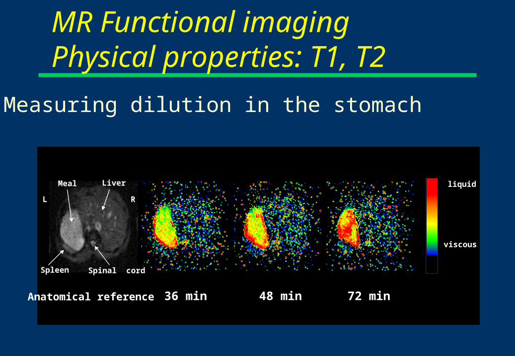
liquid
viscous
36 min 48 min 72 min
Spleen Spinal cord
L R
LiverMeal
Anatomical reference
MR Functional imaging Physical properties: T1, T2
Measuring dilution in the stomach

MRI is a Functional Imaging Technique• Perfusion• Tracers
– Blood brain barrier permeability– Lung function– Molecular imaging?
• Physical properties of tissues– microstructure from relaxation times– microstructure from diffusion– elastic properties
• fMRI- brain activation

MR Functional imaging Diffusion• Staging stroke
• White matter tracts (diffusion anisotropy)
lesion

MRI is a Functional Imaging Technique• Perfusion• Tracers
– Blood brain barrier permeability– Lung function– Molecular imaging?
• Physical properties of tissues– microstructure from relaxation times – microstructure from diffusion– elastic properties
• fMRI- brain activation

MRI is a Functional Imaging Technique• Perfusion• Tracers
– Blood brain barrier permeability– Lung function– Molecular imaging?
• Physical properties of tissues– microstructure from relaxation times – microstructure from diffusion– elastic properties
• fMRI- brain activation

Unit 5
Unit 1
Both digits
Unit 7
Unit 8
Both units
1
2
3 4
FA I
SA I
5
6
8
7
MR Functional imaging fMRI
Which part of your brain senses touch? Dr Francis
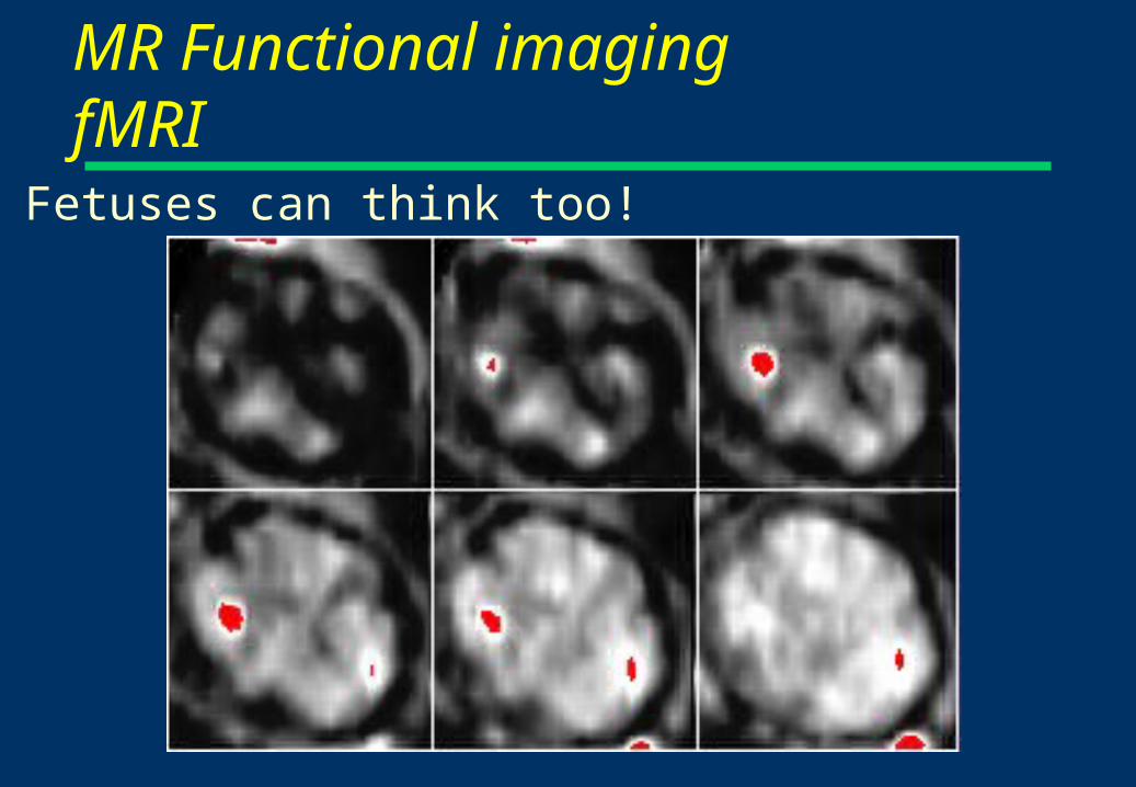
Fetuses can think too!
MR Functional imaging fMRI


An MRI study day