application of direct-sequencing peptide proteomics to … · application of direct-sequencing...
TRANSCRIPT

Application of direct-sequencing peptide proteomics to the characterization of antagonistic (endogenous and exogenous) proteins in
cereal grains
Adam Koziol
Thesis submitted to the Faculty of Graduate and Postdoctoral Studies
in partial fulfillment of the requirements for a doctoral degree in Biochemistry
Department of Biochemistry, Microbiology and Immunology
Faculty of Medicine
University of Ottawa
© Adam Koziol, Ottawa, Canada, 2013

In a Wheat Field, Evening Shadows James Edward Hervey MacDonald
Canada, 1929

ii
Abstract
The cereal seed plays a crucial role in society – both in the “food as medicine” paradigm,
but also in food security. It is the starch and proteins present in the seed that lend it
importance in these dissimilar anthropomorphic activities. This thesis investigation first
characterized the post-translational processing of the potential diabetogen, wheat
globulin-3. Globulin-3-like peptides were observed primarily in the embryo. These
peptides varied significantly in their molecular masses and isoelectric points, as
determined by two dimensional electrophoresis and immunoblotting. Five major
polypeptide spots were sequenced by mass spectrometry, allowing for the development of
a model of the post-translational events contributing to the globulin-3 processing profile.
Three separate investigations of starch granules from different cereal species were
performed. In the first series of experiments, pathogen-susceptible maize kernels were
injected with either conidia of the fungal pathogen Fusarium graminearum or sterile
water controls. Proteins in the desiccated fungal remnants on the surface of the kernels as
well as in the endosperm and embryo tissues of the control and infected kernels were
isolated and these proteomes were sequenced using tandem mass spectrometry.
Approximately 250 maize proteins were identified. These proteins were classified into
functional categories. There was an increased representation of defense proteins in the
both the embryo and endosperm tissues of infected maize samples. The proteome of the
fungal remnants was composed of 18 proteins. Several of these proteins were categorized
as being involved in the metabolism of plant-sourced molecules, or in stress response.
The second series of experiments detail the investigation of commercially prepared rice
and maize starches using tandem mass spectrometry. The majority of identified proteins,
iii
in both rice and maize samples, were involved in either carbohydrate metabolism or
storage. Markers for seed maturity and for starch mobilization were also documented.
Finally, the third series of experiments investigated the non-host proteomes present in
commercially-prepared starches. Non-host proteins from a variety of species, including
Homarus americanus were found in the starch samples. This documentation of H.
americanus proteins in these starch samples may have food safety implications with
regards to shellfish allergies.

iv
Acknowledgements
I would like to thank my family for always being there for me. I could not have done this
without them.
I am grateful to my supervisor, Illimar Altosaar. He gave me this opportunity, and
supported me every step of the way. A supervisor alone does not make a lab, and the
companionship and camaraderie of the various Altosaar lab members over the years has
been very much appreciated.
The assistance both in terms of materials and resources I received from the employees of
Agriculture and Agri-Food Canada was invaluable.
My TAC members have changed as my doctoral training project has developed over time.
I am thankful for the assistance and guidance from every TAC member, and other faculty
members over the course of my studies.
Finally, I cherish the friendships, both new and existing, that have made this experience
fantastic at times, and just bearable at others, but have always made my life better.

v
Table of Contents
Abstract ............................................................................................................................... ii
Acknowledgements ............................................................................................................ iv
Table of Contents ................................................................................................................ v
List of Figures .................................................................................................................... xi
List of Tables ..................................................................................................................... xii
Chapter 1 ............................................................................................................................. 1
Introduction ......................................................................................................................... 1
1.1 Preamble – Research outline ..................................................................................... 2
1.2 The Seed .................................................................................................................... 2
1.2.1 Evolution ............................................................................................................ 2
1.2.2 Physiology of fruit and seed structures .............................................................. 3
1.3 Storage proteins ......................................................................................................... 5
1.3.1 Globulins ............................................................................................................ 6
1.3.2 Globulins and human health ............................................................................... 9
1.3.3 Globulin summary ............................................................................................ 12
1.4 Starch granules ........................................................................................................ 12
1.4.1 Starch granule surface ...................................................................................... 15
1.4.2 Starch granule summary ................................................................................... 18
1.5 Plant/pathogen interactions ..................................................................................... 18
Summary of plant/pathogen interactions ................................................................... 22
1.6 Mass spectrometry ................................................................................................... 22
1.6 Research Hypotheses and Objectives ...................................................................... 30
Chapter 2 ........................................................................................................................... 34
Seed storage proteins of the globulin family are cleaved post-translationally in wheat embryos ............................................................................................................................. 34
2.1 Abstract ................................................................................................................... 35
2.2 Findings ................................................................................................................... 36
2.3 Results ..................................................................................................................... 38
2.3.1 Glo-3 antigenically-related proteins co-isolate with wheat globulins .............. 38
2.3.2 The Glo-3-related proteins are primarily located in the embryo ...................... 38
vi
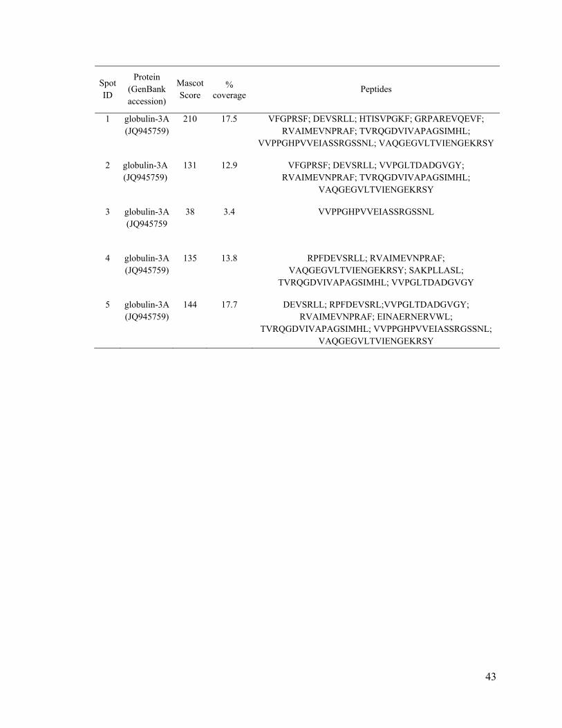
2.3.3 Identification of selected Glo-3-related polypeptides ...................................... 41
2.3.4 Characterization of selected Glo-3-related polypeptides ................................. 42
2.4 Discussion ............................................................................................................... 46
2.4.1 Type 1 diabetes ................................................................................................. 46
2.4.2 Characterization of Glo-3 ................................................................................. 47
2.4.3 Post-translational processing of Glo-3 ............................................................. 48
2.5 Conclusion ............................................................................................................... 52
2.6 Methods ................................................................................................................... 53
2.6.1 Wheat seed protein extraction and sample preparation .................................... 53
2.6.2 Sample preparation for 1D separation .............................................................. 54
2.6.3 Sample preparation for 2D separation .............................................................. 54
2.6.4 1D SDS-PAGE protein fractionation ............................................................... 54
2.6.5 Immunoblot analysis ........................................................................................ 55
2.6.6 Two-dimensional gel electrophoresis (2DE) .................................................... 56
2.6.7 Liquid chromatography tandem mass spectrometry (LC-MS/MS) .................. 57
2.6.8 N-terminal sequencing ..................................................................................... 58
Chapter 3 ........................................................................................................................... 59
Taking stock of the protein remnants on the battlefield between host and pathogen: Maize CL30-Fusarium interactome ............................................................................................. 59
3.1 Abstract ................................................................................................................... 60
3.2 Introduction ............................................................................................................. 61
3.3 Results ..................................................................................................................... 63
3.3 Discussion ............................................................................................................... 65
3.3.1 Percent representation ...................................................................................... 68
3.3.2 Proteinase inhibitors ......................................................................................... 68
3.3.3 Chitinases ......................................................................................................... 69
3.3.4 Xylanase inhibitors ........................................................................................... 70
3.3.5 Ribosome inactivating proteins ........................................................................ 71
3.3.6 Peroxidases ....................................................................................................... 72
3.3.7 Fusarium on the surface ................................................................................... 73
3.3.8 Fusarium metabolism proteins ......................................................................... 74
vii
3.3.9 Fusarium stress response .................................................................................. 75
3.4 Conclusions ............................................................................................................. 76
3.5 Methods ................................................................................................................... 77
Chapter 4 ........................................................................................................................... 80
The starch granule associated proteomes of commercially purified starch reference materials from rice and maize ........................................................................................... 80
4.1 Abstract ................................................................................................................... 81
4.2 Introduction ............................................................................................................. 82
4.3 Experimental procedures ......................................................................................... 85
4.3.1 Sampling ........................................................................................................... 85
4.3.2 Starch granule preparation ................................................................................ 85
4.3.3 Peptide preparation ........................................................................................... 85
4.3.4 Chromatography and mass spectrometry ......................................................... 86
4.3.5 Protein identification ........................................................................................ 87
4.4 Results ..................................................................................................................... 88
4.4.1 Starch granule associated proteins in rice ........................................................ 88
4.4.2 Starch granule associated proteins in maize ..................................................... 92
4.5 Discussion ............................................................................................................... 97
4.5.1 Starch granule associated proteins ................................................................. 100
4.5.2 Starch biosynthetic enzymes .......................................................................... 101
4.5.3 Orthophosphate dikinase ................................................................................ 102
4.5.4 Starch mobilization ......................................................................................... 102
4.5.5 14-3-3 proteins ............................................................................................... 103
4.5.6 Storage proteins .............................................................................................. 105
4.6 Conclusions ........................................................................................................... 106
Chapter 5 ......................................................................................................................... 108
Commercially produced rice and maize starches contain non-host proteins, as shown by mass spectrometry ........................................................................................................... 108
5.1 Abstract ................................................................................................................. 109
5.2 Introduction ........................................................................................................... 110
5.3 Methods ................................................................................................................. 112
viii
5.3.1 Protein extraction ........................................................................................... 112
5.3.2 Protein separation ........................................................................................... 113
5.4 Results and Discussion .......................................................................................... 114
5.5 Conclusion ............................................................................................................. 118
Chapter 6 ......................................................................................................................... 120
General Discussion of Major Findings ............................................................................ 120
6.1 General Discussion of Chapter 2 ........................................................................... 121
6.1.1 Glo-3 expression pattern and food safety ....................................................... 121
6.1.2 Post-translational endoproteolytic cleavage of Glo-3 .................................... 123
6.2 General Discussion of Chapter 3 ........................................................................... 124
6.2.1 Proteins in desiccated fungal remnants .......................................................... 124
6.2.2 Maize proteome response to fungal infection ................................................ 125
6.3 General Discussion of Chapter 4 ........................................................................... 126
6.4 General Discussion of Chapter 5 ........................................................................... 127
6.5 Translational medicine .......................................................................................... 128
References ....................................................................................................................... 130
Appendix A: Supplemental Tables and Figures .............................................................. 156
Appendix B: Curriculum Vitae ....................................................................................... 159
Appendix C: Reuse permissions ...................................................................................... 161

ix
List of Abbreviations
1D: one-dimensional 2D: two-dimensional 3D: three-dimensional ABA: abscisic acid ADP: adenine-diphosphate glucose AGPase: ADP-glucose pyrophosphorylase Beg: barley embryo globulin BCA: bicinchoninic acid BLAST: basic local alignment search tool Bt1: brittle 1 CBB: Coomassie Brilliant Blue CHAPS: 3-([3-cholamidopropyl]dimethylamino)-1-propanesulfonate CID: collision-induced dissociation DAP: days after pollination DON: deoxynivalenol DTT: dithiothreitol GBSS: granule bound starch synthase Glo-3: globulin-3 ESI: electrospray ionization FDR: false discovery rate G1P: glucose-1-phosphate G6P: glucose-6-phosphate HR: hypersensitive response HRP: horseradish peroxidase IEF: isoelectric focusing IgE: immunoglobulin E IgG: immunoglobulin G IPG: immobilized pH gradient KEGG: Kyoto Encyclopedia of Genes and Genomes LC: liquid chromatography LDS: linoleate diol synthase LEA: late embryogenesis protein Ma: million years ago MS: mass spectrometry MS/MS: tandem mass spectrometry NCBI: National Center for Biotechnology Information NCBInr: National Center for Biotechnology Information non-redundant OYE: old yellow enzyme OISB: Ottawa Institute of Systems Biology PGM: phosphoglucomutase PI: proteinase inhibitor pI: isoelectric point PPDK: orthophosphate dikinase PR: pathogenesis-related

x
PSV: protein storage vacuole Q: mass-resolving quadrupole q: radio frequency quadrupole Reg: rice embryo globulin RIP: ribosome inactivating protein ROS: reactive oxygen species S: Svedberg unit SBE: starch branching enzyme SDBE: starch debranching enzyme SDH: sorbitol dehydrogenase SGAP: starch granule associated protein SGS: starch granule surface SOD: superoxide dismutase SS: starch synthase SuSy: sucrose synthase T1D: type 1 diabetes TBP: tributylphosphine TCA: trichloroacetic acid TMV: Tobacco mosaic virus TOF: time of flight UDP: uracil diphosphate UDPG: uracil diphosphate glucose UGPase: UTP-glucose-1-phosphate uridylyltransferase USD: United States dollars UTMB: University of Texas Medical Branch WDEIA: wheat-dependent exercise-induced anaphylaxis

xi
List of Figures
Figure 1.1: Ovule morphology. ........................................................................................... 4
Figure 1.2: Three-dimensional structure of jack bean 7S globulin trimers. ........................ 7
Figure 1.3: Immunolocalization of globulin-3 in developing wheat embryos. ................. 10
Figure 1.4: Scanning electron micrographs of wheat starch granules. .............................. 14
Figure 1.5: Organization of amylopectin and starch granules. .......................................... 16
Figure 1.6: Channels in starch granules may contain proteins. ......................................... 17
Figure 1.7: Damaged starch granules of spring wheat infected with Fusarium. ............... 23
Figure 1.8: Functional distribution of proteins identified in plastids. ............................... 24
Figure 1.9: Schematic of a triple quadrupole mass spectrometer. ..................................... 27
Figure 1.10: Types of peptide fragment ions observed in an MS/MS spectrum. .............. 29
Figure 2.1: SDS-PAGE and immunoblot analysis of AC Barrie salt-soluble proteins. .... 39
Figure 2.2: 2D gels and immunoblot analysis of AC Barrie salt-soluble proteins. ........... 40
Figure 2.3: Model of Glo-3 endoproteolytic processing. .................................................. 45
Figure 3.1: Percent representation of maize functional protein categories. ...................... 64
Figure 4.1: Functional classification of identified proteins. .............................................. 90
Figure 4.2: Workflow and validation of proteome characterization. .............................. 104
Figure S1: Silver stained SDS-PAGE of protein extracts. ............................................. 157
xii
List of Tables
Table 1.1: Families of PR proteins. ................................................................................... 20
Table 2.1: MS/MS sequencing results of selected gel spots of wheat 7S globulins ......... 42
Table 3.1: Defense proteins in maize samples. ................................................................. 66
Table 3.2: Proteome of desiccated fungi on the surface of infected maize kernels. ......... 67
Table 4.1: Proteins involved in carbohydrate metabolism from Oryza sativa starch. ...... 91
Table 4.2: Proteins involved in cellular processes from Oryza sativa starch. ................... 93
Table 4.3: Seed storage proteins from Oryza sativa starch. .............................................. 94
Table 4.4: Proteins involved in carbohydrate metabolism from Zea mays starch. ........... 96
Table 4.5: Proteins involved in cellular processes from Zea mays starch. ........................ 98
Table 4.6: Seed storage proteins from Zea mays starch. ................................................... 99
Table 5.1: Non-host protein contaminants associated to rice starch granule. ................. 115
Table 5.2: Non-host proteins identified in maize starch samples. ................................... 117
Table S.1: Proteins isolated from commercially purified wheat starch. ......................... 158
1
Chapter 1 Introduction

2
1.1 Preamble – Research outline
Written in manuscript format, the experiments detailed in this thesis use mass
spectrometry techniques (MS) to investigate two separate tissues within the cereal seed:
the protein matrix and starch granules. To provide a biological overview for these
experiments, the introduction is divided into four distinct sections. The first section
provides a summary of seed evolution and physiology, the second section details the
components of the protein matrix, with an emphasis on the wheat storage globulins.
Starch granule development, morphology, and previous proteomic studies are presented
in the third section, while the fourth section provides insight into the mass spectrometry
techniques used in the experiments performed for this thesis.
1.2 The Seed 1.2.1 Evolution
Elksinia polymorpha, the first known plant to bear seeds, evolved in the Late Devonian
(Famennian), 365 million years ago (Ma) (Gerrienne et al., 2004). It has been
hypothesized, however, that seeds could have evolved as early as between 385 and 365
Ma in vascular plants that belonged to the paraphyletic group termed Pteridospermae, or
seed ferns. The seed ferns evolved from progymnosperms, which employed pteridophytic
reproduction (spores), but also featured certain vegetative features common to seed ferns
(Linkies et al., 2010). The transition from spores to seeds as a reproduction mechanism
required the evolution of three individual traits: heterospory, integuments, and pollen-
receiving structures (Taylor and Taylor, 2009). Heterospory is the specialization of spores

3
into haploid male-like microspores and female-like megaspores, and integuments are
protective layers around the nucellus (Esau, 1977).
1.2.2 Physiology of fruit and seed structures
The common features of all seeds are that, independent of plant lineage, seeds are
composed of an embryo, a protective seed coat, and a nutrient source (Linkies et al.,
2010). In the case of gymnosperms, seeds are not covered by an ovary, and are attached
to the cone scales (< Greek gymnospermos “naked seed”). In contrast, angiosperm seeds
are enclosed by the ovary (< Greek angeion “vessel” and sperma “seed”) (Figure 1.1).
The angiosperm cereal Triticum aestivum (wheat) is one of the most economically
important crop plants, as approximately 60% of the proteins and calories consumed
globally by humans are derived from wheat. As the United States Department of
Agriculture estimates that more than 356 million metric tons of wheat are consumed
annually, wheat is a good model seed system to study. The majority of calories, for both
the embryo, and for human consumption, from harvested wheat are derived from the
endosperm, which accounts for approximately 83% of total seed weight (Pomeranz et al.,
1970). Endosperm tissue arises from double fertilization events that are unique to
angiosperms. During the fertilization of the egg cell, a fertilization of a second cell of the
megagametophyte by two different sperm cells occurs, leading to the formation of the
triploid, nutritive, and supportive tissue that supplies energy to the embryo post-wintering
(Floyd and Friedman, 2000). Through an initial cell enlargement and endopolyploidy
phase, followed by a cellularization phase, the endosperm tissue is able to grow at very

4
Figure 1.1: Ovule morphology.a, Angiosperm ovule. b, Gymnosperm ovule. Legend: i, integument (covering); ii, inner integument; m, micropyle (opening); oi, outer integument; s, stalk. Reproduced from Frohlich and Chase, 2007 with permission Macmillan Publishers Ltd: Nature.

5
rapid rates (Olsen, 2004). The endosperm cells are composed of 50-90% compact, high
density starch granules, and seed storage proteins.
1.3 Storage proteins
Seed storage proteins serve as sources of carbon, nitrogen, and sulphur source for
germinating embryos (Shewry and Tatham, 1990). Additionally, the nutritional value of
the seeds for human consumption is dependent on the amino acid balance of the storage
proteins. For example, storage proteins high in the amino acids lysine, tryptophan and
methionine, are considered to have a very good amino acid balance (Borlaug, 1983). Seed
proteins are sequestered in an apoptotic, anhydrous protein matrix or in protein bodies
called protein storage vesicles (Hara-Nishimura et al., 1998). Intended to be digested and
subsequently taken up by the sprouting grain, they are now anthropomorphically
classified based on their solubility in certain solvents. Albumins are soluble in water and
dilute buffers, globulins are soluble in salt solutions, prolamins are soluble in 70%
alcohol, and glutelins are soluble in dilute acids or bases (Osborne, 1908). Certain storage
proteins are soluble in more than one solvent or have sequences and/or structures closely
related to proteins of another solubility class. For example, the α-globulins of wheat,
maize and rice are 18-25 kDa proteins that are soluble in saline solutions, but share
significant sequence identity with a high molecular weight glutenin from wheat (Gu et al.,
2004; Shorrosh et al., 1992; Woo et al., 2001). Additionally, rice glutelins are soluble in
NaOH, but have related 3D structures to the 11S globulins of oat (Robert et al., 1985;
Robert et al., 1985).
6
Depending on the plant species, the relative proportions of the seed storage proteins can
be significantly different. In legumes such as chick pea, the majority of the storage
proteins are globulins, with distributions of total protein as follows: albumin (8-12%),
globulin (53-60%), prolamin (3-6%) and glutelin (19-24%) (Dhawan et al., 1991). This is
very different than the distribution of storage proteins in wheat, where albumins and
globulins represent 15% of total endosperm proteins, and prolamins (53%), and glutelins
(32%) are the most predominant proteins (Shewry and Halford, 2002).
1.3.1 Globulins
The globulin family of seed storage proteins is of particular interest, as dietary exposure
to certain members of this protein family have been implicated in the development of type
1 diabetes in certain mammals (MacFarlane et al., 2003). There are two categories of
globulins based on their sedimentation coefficients: 7-9S and 11-13S (Danielsson, 1949).
Orthologs to the 7S and 11S globulin protein families, vicilins and legumins, respectively,
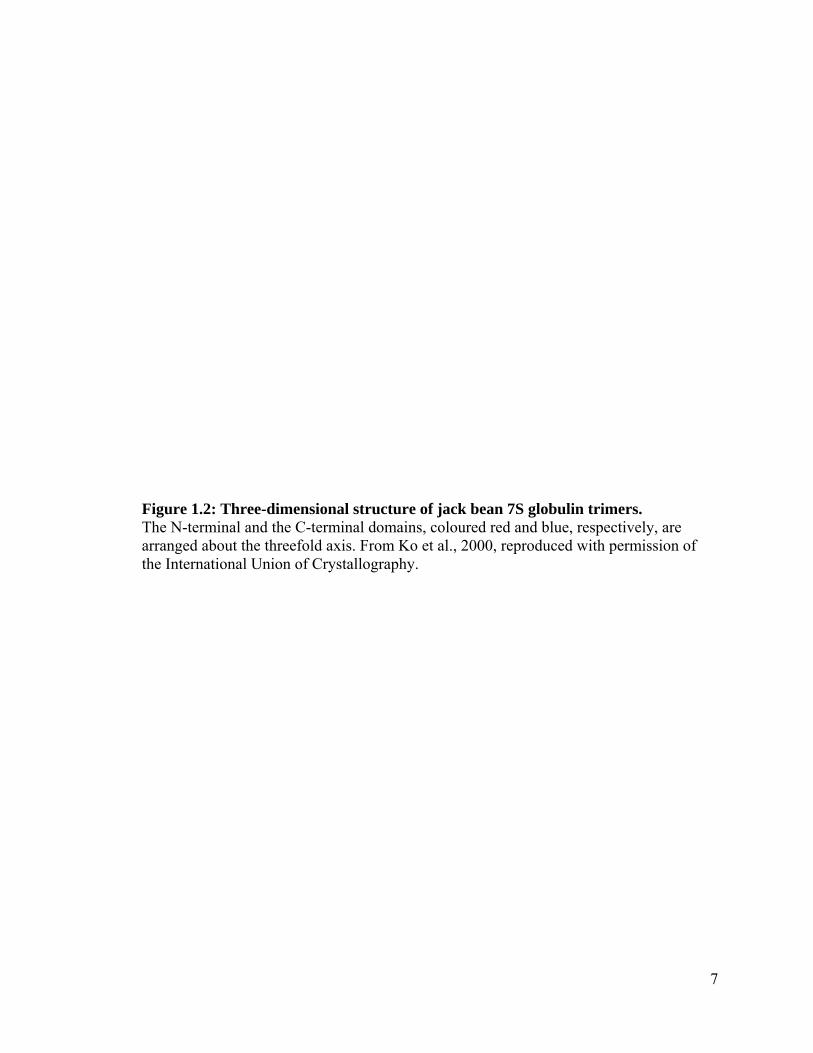
were first identified in the Leguminoseae (Derbyshire et al., 1976). Globulins are
encoded by multigene families, and have characteristic β-barrel cupin domains (Dunwell,
1998) (Figure 1.2). The 7-9S globulins are arranged as homo- or hetero-trimers
(Lawrence et al., 1994), while the 11-13S globulins are assembled into hexameric
configurations (Adachi et al., 2001). Both the globulins and the legumins undergo
significant post-translational modifications, including glycosylation and endoproteolytic
cleavage prior to their storage in seed protein storage vesicles in the embryo (Hara-
Nishimura et al., 1998).
7
Figure 1.2: Three-dimensional structure of jack bean 7S globulin trimers. The N-terminal and the C-terminal domains, coloured red and blue, respectively, are arranged about the threefold axis. From Ko et al., 2000, reproduced with permission of the International Union of Crystallography.

8
In cereal crops, there have been several studies characterizing the 7S globulins in barley
and maize, (Heck et al., 1993; Kriz, 1989), and more recently in Brachypodium
distachyon (Larre et al., 2010). However, these proteins are still poorly characterized in
wheat and oats. The lack of information on oat globulins is unusual, as oats are the first
foodstuff to be allowed a medical health claim by the U.S. Food and Drug Administration
stating that consumption of soluble fiber from whole oats reduces the risk of heart
disease. The expression of 7S globulins is primarily in the embryo and aleurone layer
(Sun et al., 1996), with little to no evidence of globulin expression in the endosperm (Loit
et al., 2009). Expression of globulins is transcriptionally regulated by the phytohormone
abscisic acid (ABA) (Rivin and Grudt, 1991). As globulins are expressed primarily in the
embryo and contain increased levels of nitrogen-rich amino acids arginine, lysine,
aspartic acid and glutamic acid (Zhu et al., 2006), it is hypothesized that the globulins
have evolved to be used as an easily exploitable source of amino groups and energy for
germinating embryos (Shewry and Tatham, 1990).
There are two members of the 7S globulin protein family present in multiple cereal
species: globulin 1 (Glbl) and globulin 2 (Glb2) in maize, barley embryo globulins (Begl
and Beg2) in barley and rice embryo globulins (Regl and Reg2) in rice (Sun et al., 1996;
Yupsanis et al., 1990). Previous studies on Glb1, Beg1 and Reg1 have shown that these
proteins are all encoded by single copy genes (Belanger and Kriz, 1989; Heck et al.,
1993; Sun et al., 1996). However, there is evidence to suggest that Beg1 may belong to a
small multigene family (Zhang et al., 2004). The expression patterns of the members of
the protein family can be significantly different, as the rice embryo globulins as well as

9
Begl transcripts are highly expressed during early germination, whereas Beg2 transcript
levels decrease during the germination process (Zhang et al., 2004).
In wheat, there are three members of the globulin protein family: globulin-1, -2 and -3, as
well as triticin (Gomez et al., 1988; Singh et al., 1991). Globulin-3A is a potential food
allergen (Larre et al., 2011), and has been associated with an increased risk for the
development of type 1 diabetes (MacFarlane et al., 2003) and celiac disease (Taplin et al.,
2011) in susceptible individuals. The globulin-3 gene family in wheat cultivar Glenlea
contains three individual globulin-3 genes: Glo-3A, Glo-3B, and Glo-3C (Loit et al.,
2009). These genes share a moderate to high degree of nucleotide sequence identity (73-
93%) and are expressed exclusively in the embryo and aleurone layer (Figure 1.3).
Previous investigations into the post-translational endoproteolytic cleavage of the
globulin-3 family have shown that these proteins can be processed at internal cleavage
sites to yield polypeptides with a range of molecular weights and pI values (Dupont et al.,
2011; Singh et al., 2001). To gain a more precise understanding of the processing events
that orchestrate our dietary proteomes, a targeted study of these processed proteins is
required to understand their role in the development of human diseases.
1.3.2 Globulins and human health
Wheat seed storage proteins, including globulin-3, and the γ-gliadins have been
associated with the development of wheat allergies, wheat sensitivity and celiac disease
(Tatham and Shewry, 2008). Certain occupations have higher incidences of wheat
allergies; baker's asthma and rhinitis can affect as many as 30% of professional bakers

10
Figure 1.3: Immunolocalization of globulin-3 in developing wheat embryos. Panel A: wheat aleurone (al) cells were positively stained while endosperm (es) cells were unstained when sections were stained with Glo-3A antibodies. Panel (C): wheat embryonic (em) tissue stained with Glo-3A antibodies. Green staining in seed coat (sc) shows unspecific staining. Green (examples indicated with arrowheads) indicates positive staining. Reproduced with permission from Loit et al., 2009.


11
(Bittner et al., 2008). Though these allergies were recognized by the Romans, it was not
until the 1970s that wheat albumins were found to be reactive with immunoglobulin E
(IgE) antibodies from individuals suffering from baker’s asthma (Baldo and Wrigley,
1978). The allergies in these individuals are responses to the inhalation of wheat flour and
dust particles.
Type 1 diabetes (T1D) is an autoimmune disease characterized by the destruction of the
insulin producing β-cells in the pancreatic islets of Langerhans and the loss of glucose
homeostasis (Jahromi and Eisenbarth, 2007). It has been hypothesized that a
dysfunctional gut barrier may be involved in disease onset in certain individuals
(Maurano et al., 2005). While the incidence of T1D in developed countries has risen by
3% annually since 1960 (Onkamo et al., 1999), there is still no definitive cause of this
disease. Identified risk genes impart genetic susceptibility to T1D, but there is under 50%
concordance in monozygotic twins (Todd, 2010). Therefore, the development of T1D
likely relies on environmental triggers, such as diet. Alternatively, the hygiene hypothesis
states that the reduced exposure to infectious agents during childhood is responsible for
the increased prevalence of T1D (Yazdanbakhsh et al., 2002). In current societies, with
heightened standards for sanitation, healthcare and diet, the natural development of the
immune system can be repressed by the lack of exposure to T1D protective factors, such
as symbiotic prokaryotes and certain parasites (Todd, 1991).
Celiac disease, also known as gluten enteropathy, or sprue, is a hereditary immune system
disorder (Fasano, 2001). The intestinal epithelium and the tight intercellular junctions in
healthy individuals are intact and do not permit dietary antigens to pass the gut barrier
(Heyman et al., 2012). However, the tight junction system becomes damaged in subjects
12
with celiac disease (Shan et al., 2002). In these individuals, the immune systems can be
stimulated by dietary antigens, such as gliadins that pass through the tight junctions (Tjon
et al., 2010). An increased expression of the tight junction modulating protein zonulin
may have a role in the degradation of the tight junctions (Lammers et al., 2008). Celiac
disease is one of the most common genetic diseases, with worldwide prevalence of
approximately 0.3-1% (Catassi et al., 1994).
1.3.3 Globulin summary
As the mechanisms of the development of diseases such as type 1 diabetes and celiac
disease are still poorly understood, the implication of dietary antagonists is an important
avenue to explore. Further research is necessary to discover how globulin-3 is involved
with disease development, and whether the post-translation processing of globulin-3 is a
factor in the development of these diseases.
1.4 Starch granules
Photosynthesis fixes carbon dioxide using the reductant generated through the light
reactions of photosynthesis. The reductant can be subsequently oxidized to support the
energy needs of plant growth and development. Energy generation and consumption
needs vary depending on light levels and the developmental stage of plants. Therefore the
capacity to store energy as inert starch granules in the form of complex polysaccharides
composed of monomeric glucose molecules linked by glycosidic bonds is a necessity.
13
Starch typically accumulates in photosynthetically active chloroplasts or in amyloplasts in
non-photosynthetic tissues, which specialize in starch storage (Tetlow, 2011).
Starch formation in wheat amyloplasts is associated with stromules (stroma-filled tubules)
during the development of the endosperm in cereals (Natesan et al., 2005). It was
determined that A-type granules (>15 µM) are initiated in undifferentiated plastids in the
coenocytic endosperm (Bechtel and Wilson, 2003). Following cellularization of the
endosperm, B-type granules (5-15 µM) form in stromules protruding from plastids
containing A-type granules in the subaleurone layer. C-type granules (< 5 µM) are
initiated in branched stromules that form cytosolic networks (Bechtel and Wilson, 2003)
(Figure 1.4). Similar development of starch granules and stromules was observed in cells
of the central endosperm (Bechtel and Wilson, 2003). Starch granules are composed of
98-99% amylose and amylopectin molecules (Buléon et al., 1998).These polysaccharide
chains are composed of glucose monomers linked by -1,4 or -1,6 glycosidic bonds
(Ball and Morell, 2003). Amylose contains 99% -1,4 bonds and 1% -1,6 linkages and
therefore is essentially a long, linear molecule compared to amylopectin, which contains
95% -1,4 linkages and 5% -1,6 linkages (Tester et al., 2004). The crystalline structure
of starch granules is attributed to the dense packing of the double helices of amylopectin
(Perez and Bertoft, 2010) (Figure 1.5). The ratio of amylose to amylopectin can range
significantly between starch granules: from under 1% amylose in waxy starches to greater
than 70% amylose in high amylose starches (Buléon et al., 1998). The composition of
most starch granules ranges from 20-35% amylose (Tester et al., 2004). The size, shape,
and composition of starch granules can vary between species. Wheat starch granules are
composed of 36% amylopectin (Morrison et al., 1994), and have a bimodal size

14
Figure 1.4: Scanning electron micrographs of wheat starch granules. Panels A and B are mature wheat endosperm tissue (Large A-type granules (>15 m), medium B-type granules (5–15 m) and small C-type granules (<5 m) are present in panels A and B. B is a magnified image of the boxed portion in A. C shows starch granules isolated from mature endosperm. D shows a fractured starch granule, revealing the layers of starch. Reproduced from Rahman et al., 2000 with permission from Elsevier.


15
distribution: type A granules, range from 15 to 35 µm in diameter with a lenticular shape
and the smaller type B granules range from 2 to 10 µm with a spherical beach ball shape
(Tester et al., 2004). In contrast, rice starch granules have an unimodal size distribution,
are approximately 3 to 8 µm in size (Cledat et al., 2004) and are composed of 47-51%
amylopectin (Qi et al., 2003). During development, polyhedral compound granules are
generated through the interactions of the growing granules (Jane et al., 1994). The shape,
size and composition of starch granules all have impacts on the bioutilization, the end use
properties as well as on the nutritional benefits of starch for both embryo and predators.
The nature of the starch granule surface, and particularly the presence of surface lipids
and proteins, has many significant effects on the properties of the starch.
1.4.1 Starch granule surface
The starch granule surface is characterized by the presence of certain features, including
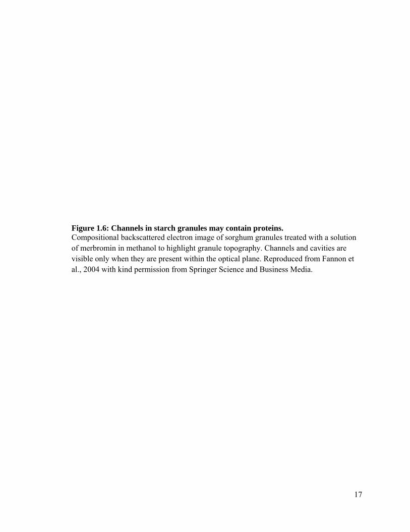
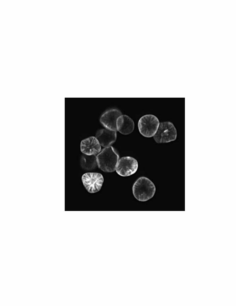
pores, protrusions, and channels (Fannon et al., 2003; Fannon et al., 2004) (Figure 1.6).
The occurrence, size and number of these topological features is dependent on the
species, tissue, and starch granule type (Huber and BeMiller, 2000). In addition to these
topological features, proteins have been observed on the starch granule surface using
atomic force microscopy (Baker et al., 2001) and other techniques, including mass
spectrometry (Wall et al., 2010) and immunofluorescence (Lauriere et al., 1986). These
starch granule associated proteins (SGAPs) include a number of proteins of varying
molecular weight, including several isoforms of the starch granule bound starch synthases
(GBSS), starch synthases (SS) and starch branching enzymes (SBE)

16
Figure 1.5: Organization of amylopectin and starch granules. (A) Schematic view of a starch granule with its succession of amorphous and crystalline growth rings. (B) Relation between a section of a crystalline growth ring of the granule and the molecular organization of amylopectin. (C) The succession of ten alternating lamellae in context to the primary structure of a portion of an amylopectin molecule. Each line represents an α-1,4 linked glucan chain. The chains are hooked together by α-1,6 branches. The dotted line delimits the sections appearing in the crystalline and amorphous lamellae. (D) Relation of a part of the primary structure depicted in (C) to the secondary structure of a single cluster displaying the double helical structures. Reproduced from Perez et al., 2010 with permission from John Wiley and Sons.

17
Figure 1.6: Channels in starch granules may contain proteins. Compositional backscattered electron image of sorghum granules treated with a solution of merbromin in methanol to highlight granule topography. Channels and cavities are visible only when they are present within the optical plane. Reproduced from Fannon et al., 2004 with kind permission from Springer Science and Business Media.

18
(Rahman et al., 2007) as well as several other proteins, many of which are still poorly
characterized. For instance, an abundant and poorly characterized unnamed wheat SGAP
with a molecular weight of approximately 30 kDa is present both on the surface and
within the granule (Baldwin, 2001). The function of the protein is unknown, but it has
been hypothesized that it may be metabolized by the germinating embryo. The well
documented SGAP puroindoline is the major determinant of wheat grain softness, an
important property in determining the end-use quality of wheat (Morris, 2002).
Puroindolines have also been shown to have antimicrobial properties and have likely
evolved as a defence mechanism (Capparelli et al., 2005; Krishnamurthy et al., 2001).
1.4.2 Starch granule summary
Starch granules can therefore be regarded as unique intracellular compartments of
endosperm cells that consist of the metabolic and regulatory proteins for starch synthesis
and degradation. The proteins localized to this compartment either contain a specific
binding domain for starch or directly interact with a protein or group of proteins bound to
granules. Since these proteins and the starch granules store large amounts of energy in
their chemical bonds, there are important safeguards, such as the innate immune system,
in place to protect them from predation.
1.5 Plant/pathogen interactions
To protect their energy reserves (both starch granules and storage proteins), and,
similarly, the embryo from predators and pathogens, plants have evolved layers of

19
resistance mechanisms. These mechanisms include physical barriers, such as the cuticle,
and chemical barriers, including antimicrobial molecules, such as phytoanticipins
(Osbourn, 1996). In addition to these “first-line” defenses, plants have inducible
resistance mechanisms that are activated upon the sensing of pathogen infection (Durrant
and Dong, 2004). These inducible mechanisms include the initiation of the hypersensitive
response (HR), the cross-linking of cell walls, and the accumulation of reactive oxygen
species (ROS), secondary metabolites, and pathogenesis-related (PR) proteins (Sels et al.,
2008). The PR proteins are proteins that are either undetectable or present at low
concentrations in healthy tissues, but are induced following pathogen infection (van Loon
et al., 2006).
The PR proteins were originally described in Nicotiana tabacum (tobacco) as proteins
upregulated in the hypersensitive response to Tobacco mosaic virus (TMV) (van Loon
and van Strien, 1999). There are now 17 recognized families of PR proteins (Table 1.1).
The PR families are numbered based on the order of their discovery (van Loon et al.,
2006). While not all PR families have been demonstrated to have direct roles in defense,
many of the families have well documented antimicrobial activities. PR families such as
PR-3, -4, -8, and -11 (chitinases), PR-5 (thaumatin), PR-12 (defensin), PR-13 (thionin),
and PR-14 (lipid-transfer protein) catalyze the degradation of specific microbial
macromolecules. Chitinases catalyze the cleavage of the β-1,4-glycosidic bonds of the
fungal cell wall structural molecules chitin and chitosan (Jitonnom et al., 2011).
Collectively, PR-5, -12, -13, -14 catalyze the degradation of microbial cell walls, though
these families have different target molecules (Dubreil et al., 1998; Lay and Anderson,
2005).

20
Table 1.1: Families of PR proteins.
Adapted from van Loon et al., 2006.
Family Properties
PR-1 Unknown
PR-2 β-1,3-glucanase
PR-3 Chitinase type I, II, IV, V, VI, VII
PR-4 Chitinase type I, II
PR-5 Thaumatin-like
PR-6 Proteinase-inhibitor
PR-7 Endoproteinase
PR-8 Chitinase type III
PR-9 Peroxidase
PR-10 Ribonuclease-like
PR-11 Chitinase, type 1
PR-12 Defensin
PR-13 Thionin
PR-14 Lipid-transfer protein
PR-15 Oxalate oxidase
PR-16 Oxalate-oxidase-like
PR-17 Unknown
21
The functions of the PR proteins are not limited solely to defense. The PR-2 proteins have
antimicrobial function through their ability to catalyze the cleavage of 1,3-β-D-glucosidic
linkages of the glucans that are prevalent in fungal cell walls (Leubner-Metzger and
Meins, 1999). However, a diverse assortment of roles for this family in uninfected plants
have been established, including, but not limited to embryogenesis (Helleboid et al.,
1998), mobilization of energy reserves (Fincher and Stone, 1993), fruit ripening (Hinton
and Pressey, 1980), and cold tolerance (Hincha et al., 1997). Other PR families also have
diverse roles unrelated to plant defense, therefore careful study is required to document
all the functions of the PR families.
Successful fungal pathogen infection is characterized by the secretion of fungal enzymes
that catalyze the degradation of the plant’s physical barriers, such as the cuticle and cell
walls (Phalip et al., 2005). Enzymes secreted by Fusarium graminearum, the fungal
pathogen responsible for causing Gibberella ear rot in maize, include lipases and
cutinases that degrade the cuticle (Feng et al., 2005; Jenczmionka and Schafer, 2005;
Kang and Buchenauer, 2000) and pectinases that modify plant cell walls (Kikot et al.,
2009), allowing easier access of other fungal enzymes, such as cellulases and xylanases,
to plant cell components (Reignault et al., 2008). These fungal cell wall-degrading
enzymes facilitate the colonization of wheat spikes prior to fungal grain infection
(Wanjiru et al., 2002).
Following the penetration of a cereal plant seed’s physical barriers, the pathogen will
target the energy-rich endosperm tissue (Kikot et al., 2009). Fungal enzymes such as
proteinases and amylases catalyze the degradation of the seed’s storage proteins and
starch granules, respectively (Schwarz et al., 2001). For example, following infection of
22
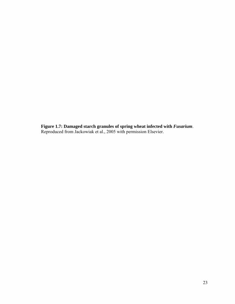
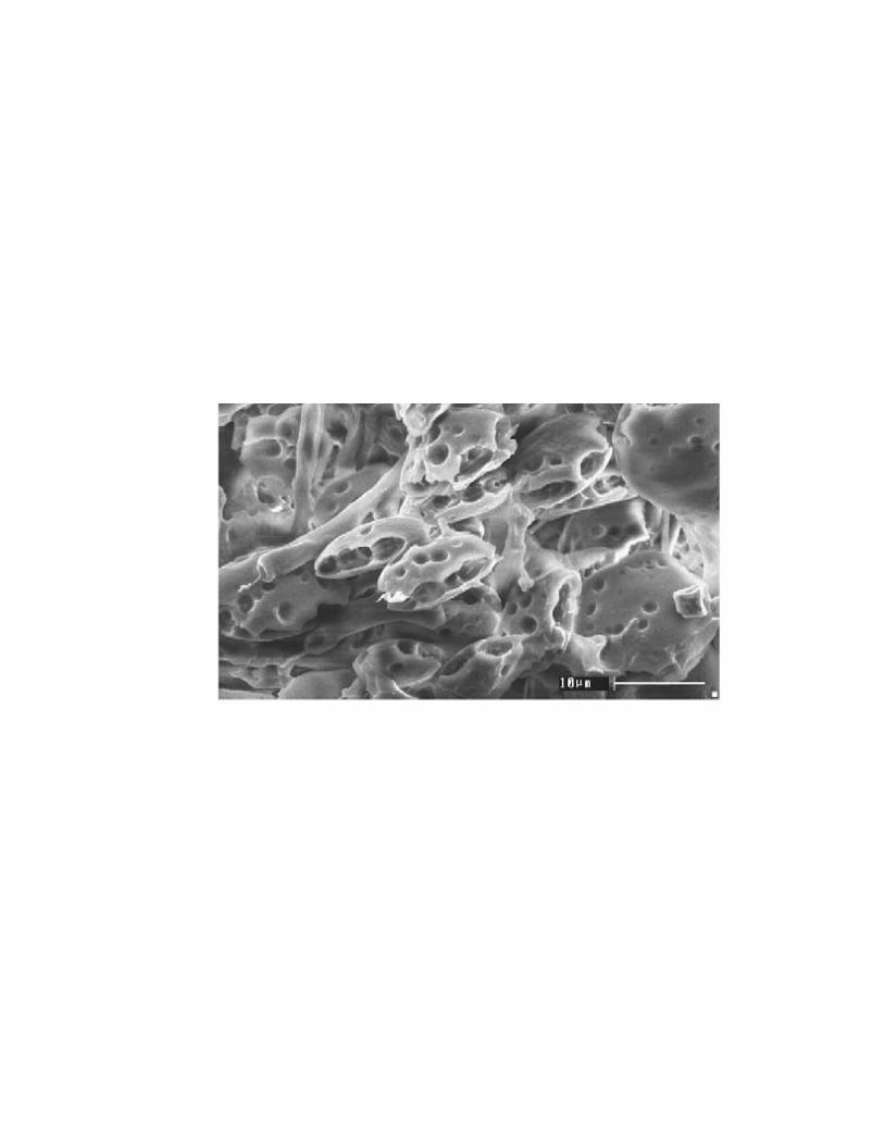
endosperm tissue with F. graminearum, starch granules were digested to a great degree
and the intracellular protein matrix was fully degraded (Jackowiak et al., 2005) (Figure
1.7).
Summary of plant/pathogen interactions
Plants and pathogens are continuously evolving new mechanisms for the defense or
infection of plant tissues, respectively. This ongoing evolutionary arms race between
pathogen and plant can have very large economic impacts. Over $1 billion USD of crops
are lost to F. graminearum infection every year (Goswami and Kistler, 2004), indicating
the study of these plant/pathogen interactions using state of the art tools, such as mass
spectrometry, can have both economic impacts and benefits for human health by
increasing food security.
1.6 Mass spectrometry
The proteomes of wheat endosperm (Vensel et al., 2005), wheat and potato amyloplasts
(Andon et al., 2002; Stensballe et al., 2008) and maize starch-granules (Grimaud et al.,
2008) have been catalogued using high-throughput mass spectrometry techniques. The
proteins identified in these studies were classified based upon their biochemical roles in
the cell.
In wheat amyloplasts, many of the identified proteins have roles in metabolism, as well as
protein destination and storage (Balmer et al., 2006) (Figure 1.8). It is interesting to note
23
Figure 1.7: Damaged starch granules of spring wheat infected with Fusarium. Reproduced from Jackowiak et al., 2005 with permission Elsevier.

24
Figure 1.8: Functional distribution of proteins identified in amyloplasts from wheat endosperm. Reproduced from Balmer et al., 2006 with permission from Oxford University Press.


25
that over one third of the proteins identified in the study were unknown or hypothetical
proteins. This underscores the necessity of gene discovery and functional analysis studies
for large-scale proteomic analysis. The starch granule associated proteome of water
washed soft (pathogen resistant) and hard wheat (pathogen susceptible) cultivars have
previously been investigated (Wall et al., 2010). In addition to the many unknown and
hypothetical proteins, there were multiple proteins with antimicrobial activities identified.
As these samples were collected from field grown plants that were deemed fit for harvest,
a natural progression of this work is to study grains intentionally infected with pathogens
and investigating the subsequent plant/pathogen interactions at the proteomic level.
Furthermore, the proteomes of commercially-available starch has been studied to test the
suitability of the starch for patients with celiac disease (Kasarda et al., 2008). The authors
discovered many proteins that had not previously been believed to be associated with the
starch granule surface, showing that careful study is required when documenting proteins
in foods destined for human consumption.
The term mass spectrometry refers to a broad range of analytical techniques used for the
analysis of molecules based on their mass to charge ratio. As mass spectrometry covers
such a broad range of techniques, only the techniques used in the experiments in this
thesis will be covered. These techniques include the electrospray ionization (ESI) delivery
system, the time of flight (TOF) mass analyzer and mass selection and detection
components, and the QStar QqTOF triple quadrupole tandem mass spectrometer. For this
latter instrument, Q refers to a mass-resolving quadrupole, and lower-case q refers to a
radio frequency-only quadrupole or hexapole collision cell (Chernushevich et al., 2001).
This instrument is a tandem mass spectrometer, as there are two mass selection
26
quadrupoles (Q1 and Q3) separated by ion fragmentation in a collision chamber (q2).
When performing peptide sequencing experiments, mixtures of peptides are first
separated by liquid chromatography and ionized using electrospray ionization. This
technique involves three separate stages: droplet formation, droplet shrinkage and
gaseous ion formation (Smith et al., 1990). To promote droplet shrinkage, peptides are
suspended in volatile solvents such as acetonitrile. During droplet formation, liquid is
extruded from a capillary tip in a "Taylor cone" shape due to electrostatic forces and is
subsequently aerosolized (Figure 1.9). The droplet size of this aerosolized liquid is
controlled by the inclusion of certain compounds, such as acetic or formic acid, that
increase the conductivity of the peptide mixture as well as the applied potential, the flow
rate of the solvent, and the diameter of the capillary. As the droplets shrink, gaseous ions
pass into the mass spectrometer (Cech and Enke, 2001).
A characteristic of ESI is that the gaseous ions can each have multiple charged residues.
Multiple charges reduce the mass to charge ratio of the protein, allowing for the analysis
of high molecular weight proteins that are normally outside the usable mass range of the
mass spectrometer (Dongre et al., 1996). For mass analysis, the QStar hybrid QqTOF
MS/MS consists of three quadrupoles linked to a TOF mass analyzer (Figure 1.9). The
first quadrupole (Q1) is responsible for initial ion selection, allowing for the selection of a
specific mass range. The second quadrupole (q2) is a non-mass filtering radio frequency
quadrupole that serves as a collision cell where ions selected in Q1 can be broken down
into smaller ions through collision-induced dissociation (CID) with an inert gas such as
argon, nitrogen or helium. The third quadrupole (Q3) allows for further ion selection
before the selected ions are delivered to the TOF mass analyzer (Aebersold and Mann,

27
Figure 1.9: Schematic of a triple quadrupole mass spectrometer. Legend: Q1, quadrupole 1, q, quadrupole 2, Q3 quadrupole 3, CID, collision-induced dissociation. From broadinstitute.org/files/shared/proteomics/tq.jpg. Last viewed September 29, 2012.


28
2003). As ions enter the TOF chamber, the ions are accelerated by an electric field of
known strength. Due to this acceleration, all ions with identical charge will have the same
kinetic energy. As lighter particles are able to accelerate to higher speeds than heavy
particles, an ion’s velocity is dependent on the mass-to-charge ratio. As the time an ion
takes to reach an ion detector at a known distance can be measured, this time and the
experimental parameters are used to calculate the mass-to-charge ratio of the ion (Takats
et al., 2004).
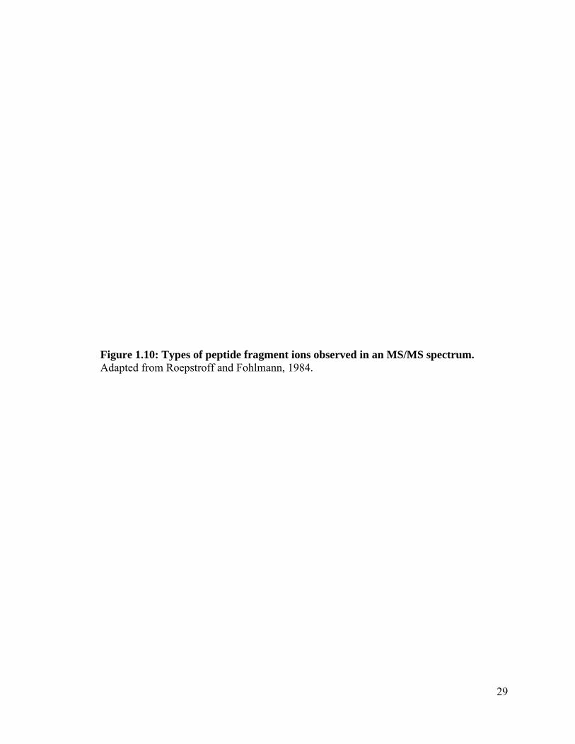
When analyzing peptide samples using a tandem mass spectrometer, the fragmentation of
positively charged ions occurs in a predictable pattern due to the tendency for the amide
bond of peptides to be cleaved during ion fragmentation (Kelleher et al., 1999). The
fragment containing the N-terminus is referred to as a b-ion, while the fragment
containing the C-terminus is referred to as a y-ion (Roepstorff and Fohlman, 1984)
(Figure 1.10). The differences in mass between the fragmented ions compared to the mass
of the ion prior to fragmentation allows for calculation of the amino acid sequence of the
peptide. Based on this deduced amino acid sequence, it is possible to identify the protein
from which this ion was generated. The MS/MS peptide spectra generated from liquid
chromatography (LC) MS/MS analyses are analyzed using search engines such as Mascot
(Perkins et al., 1999). The search algorithms used by Mascot compare experimentally
obtained fragmentation spectra to theoretical fragmentation spectra calculated from a
database of protein sequences, such as the National Center for Biotechnology Information
(NCBI) non-redundant protein database. The scoring system used by Mascot is
probability-based. The total score is the probability that the observed match is a random
event, with the scores reported as -10*LOG10(P), where P is the absolute probability. To
29
Figure 1.10: Types of peptide fragment ions observed in an MS/MS spectrum. Adapted from Roepstroff and Fohlmann, 1984.


30
determine whether the calculated protein score is significant, Mascot computes an
expectation value for every hit. The expectation value is defined as the number of
matches with equal or better scores that are expected to occur by chance alone. This
threshold is commonly set at p<0.05, therefore any expectation values below 0.05 are
considered significant. Another technique to ensure the significance of the Mascot
matches are decoy database searches. These searches use the same protein databases as
the standard query, but the sequences have been randomized or reversed. This allows for
the calculation of the false discovery rates (FDR) in an experiment (Elias et al., 2005).
The FDR is calculated as follows: FDR = FP / (FP + TP), where FP is the number of
matches in the decoy database and TP is the number of matches in the target database.
1.6 Research Hypotheses and Objectives
Rationale 1
Previously, globulin-3A in wheat cultivar AC Barrie was shown to have implications in
the development of type 1 diabetes in genetically susceptible individuals. As AC Barrie
and Glenlea are closely related hexaploid cultivars, it is feasible that the globulin-3 family
in AC Barrie is similar to the previously documented globulin-3 proteins in Glenlea.
Additionally, the post-translational endoproteolytic processing of globulins have been
shown to be similar to the post-translational processing observed in barley.

31
Hypothesis 1
Globulin-3 in wheat undergoes post-translational processing, including glycosylation and
endoproteolytic processing, similar to 7S proteins in other species such as soy and barley.
Objectives
1. Determine the expression pattern of globulin-3 in wheat embryo and
endosperm tissues.
2. Sequence the proteolytically-cleaved proteins separated on two-
dimensional SDS-PAGE gels.
3. Create a model for proteolytic cleavage of the globulin-3 preproprotein.
Rationale 2
Use of a pathogen-susceptible maize genotype and the mechanical kernel injection
technique both promote a fuller expression of the fungal pathogen proteome. Under these
optimal pathogen growth conditions, the fungus will take up nutrients from the plant and
will express enzymes to metabolize the nutrients. Additionally, the inducible plant
defence proteins as well as fungal proteins will be present and interacting within both the
endosperm and embryo of the host tissues.
Hypothesis 2
Proteins sequenced from the desiccated fungal remnants on the surface of maize kernels
can provide insight into the biochemical processes occurring in the fungus prior to post-

32
infection desiccation. Host defense-related proteins will be present in both the endosperm
and embryo tissues, allowing for a better understanding of plant/pathogen interactions.
Objectives
1. Isolate and purify proteins from desiccated fungal remnants on the surface
of infected maize kernels as well as proteins from the endosperm and
embryo tissues of infected and control kernels.
2. Sequence the proteomes of these tissues.
3. Classify proteins based on their biochemical roles.
Rationale 3
The certificates of analysis for commercially available starch samples give basic
measurements of the non-starch components of the samples, such as pH and percentages
of moisture, protein, and lipid, but not the full proteome of the starch samples. There is
1% protein in these starches that is undocumented, thus requiring deeper inspection using
mass spectrometry techniques.
Hypothesis 3
The protein component of commercially purified starch samples could affect the outcome
of other experiments in which the starches will serve as defined baseline substrates. The

33
investigation of these trace proteomes will allow for the determination of protein-sourced
contributions to the macroscopic characteristics of these starches.
Objectives
1. Purify the starch granule-associated proteins in commercially-purified
starch.
2. Sequence these proteins using tandem mass spectrometry.
3. Classify the proteins based on biochemical characteristics.
Rationale 4
The water used to process the starch during commercial purification can contaminate
starch samples with non-host organisms, and can affect the end quality of the starch. For
example, proteins from microbial pathogens have been documented on the surface of
starch granules in lab-scale purified wheat starch.
Hypothesis 4
The source of any non-host proteins present in the commercial starch preparations is the
water used during the extraction of the starch granules.
Objectives
1. Isolate and sequence the proteins present in commercially prepared rice
and maize starches.
2. Document any non-host protein present in the starch samples.

34
Chapter 2
Seed storage proteins of the globulin family are cleaved post-translationally in wheat embryos
Koziol AG, Loit E, McNulty M, MacFarlane AJ, Scott FW, Altosaar, I
Published in: BMC Research Notes 2012, 5:385 (28 July 2012)
Contribution of Authors:
AGK performed bioinformatic analyses, analyzed data, assembled all figures and tables,
drafted and edited the manuscript. EL helped draft the first manuscript, aided in editing,
and helped with experiments. MM performed the electrophoresis experiments,
contributed to the initial writing and subsequent editing. AJM, FWS provided the
globulin-3 antibodies and edited the manuscript. FWS participated in the study design. IA
designed the study, provided all reagents and funding, and edited the manuscript.

35
2.1 Abstract
Background: The 7S globulins are plant seed storage proteins that have been associated
with the development of a number of human diseases, including peanut allergy. Immune
reactivity to the wheat seed storage protein globulin-3 (Glo-3) has been associated with
the development of the autoimmune disease type 1 diabetes in diabetes-prone rats and
mice, as well as in a subset of human patients.
Findings: The present study characterized native wheat Glo-3 in salt-soluble wheat seed
protein extracts. Glo-3-like peptides were observed primarily in the wheat embryo. Glo-3-
like proteins varied significantly in their molecular masses and isoelectric points, as
determined by two dimensional electrophoresis and immunoblotting with anti-Glo-3
antibodies. Five major polypeptide spots were identified by mass spectrometry and N-
terminal sequencing as belonging to the Glo-3 family.
Conclusions: These results in combination with our previous findings have allowed for
the development of a hypothetical model of the post-translational events contributing to
the wheat 7S globulin profile in mature wheat kernels.

36
2.2 Findings
The 7S globulins, orthologs of the vicilins of the Leguminoseae, are salt-soluble storage
proteins that accumulate during seed development (Debiton et al., 2011; Jerkovic et al.,
2010). Vicilins were first described by Osborn and Campbell in 1898 as a class of seed
storage proteins in Vicia faba (horse bean) (Osborne and Campbell, 1898). Both the
vicilins and the legumins, distinguishable by their sedimentation coefficients of 7-9S and
11-13S, respectively (Danielsson, 1949), contain characteristic β-barrel cupin domains
(Dunwell, 1998). The 7S globulins are translated as preproproteins that, following the co-
translational cleavage of the signal peptide, assemble into homo- or heterotrimers
(Lawrence et al., 1994) within the lumen of the endoplasmic reticulum (Chrispeels,
1991). Prior to accumulation in seed protein storage vesicles, the trimers undergo post-
translational processing, which includes glycosylation and partial endoproteolytic
cleavage (Herman and Larkins, 1999, Heck et al., 1993).
Exposure to a number of wheat seed proteins can induce a number of immune-mediated
diseases including gluten sensitive enteropathy (celiac disease) (Di Sabatino and Corazza,
2009), Baker’s asthma and wheat-dependent exercise-induced anaphylaxis (WDEIA) in
predisposed individuals (Tatham and Shewry, 2008). The Triticum aestivum (wheat)
storage protein WP5212, later named globulin-3 (Glo-3), has been demonstrated to be a
potential food allergen (Larre et al., 2011), identified as the first candidate wheat protein
associated with the development of type 1 diabetes (T1D) (MacFarlane et al., 2003), and
now celiac disease (Taplin et al., 2011). We recently identified the genomic origins of
three Glo-3 genes, Glo-3A, B and C in the wheat cultivar Glenlea (Loit et al., 2009).

37
Immunofluorescence studies have localized the Glo-3 gene products to the developing
wheat seed embryo and aleurone layer (Loit et al., 2009).
Few studies have sought to characterize wheat 7S globulins because they were thought to
be minor storage proteins with little contribution to the bread-making properties of wheat
flour (Fabijanski et al., 1985; Robert et al., 1985). However, 7S proteins, based on their
sedimentation coefficient, have been characterized in barley and maize, and more
recently, two Glo-3-like sequences have been identified in the model cereal
Brachypodium distachyon (Larre et al., 2010). In addition to cultivar Glenlea, Glo-3
proteins have been observed in cultivars Butte 86 and Recital (Dupont et al., 2011;
Tasleem-Tahir et al., 2011), indicating that Glo-3 is well-conserved in wheat, thus
deserving more attention.
Due to its documented role in the development of T1D, we initiated the present study to
characterize the Glo-3-related proteins and peptides in wheat cultivar AC Barrie, the
original source of WP5212 (MacFarlane et al., 2003). We hypothesized that Glo-3
undergoes post-translational processing, including glycosylation and endoproteolytic
processing, similar to 7S proteins in other species. Therefore, we sought to characterize
the expression and the distribution of Glo-3 antigenically related proteins by Mr and pI in
the embryo and endosperm of AC Barrie, and to link observed protein fragments with
their corresponding endoproteolytic cleavage events.

38
2.3 Results 2.3.1 Glo-3 antigenically-related proteins co-isolate with wheat globulins
To characterize the Glo-3 antigenically-related proteins in whole AC Barrie seeds,
globulins were extracted, following the classical method (Khavkin et al., 1978; Robert et
al., 1985). The globulin-enriched fraction was separated by 1D SDS-PAGE and
immunoblots were probed with polyclonal rabbit antibodies specific for Glo-3A (Figure
2.1) (Loit et al., 2009). The four most intense protein bands, as resolved by SDS-PAGE,
had relative mobilities of 33-36, 47-53 and 64-65 and 66-68 (doublet) kDa. The Glo-3
antigenically-related proteins had comparable Mr to these intense bands (33-37, 47-53,
64-68 kDa). Pre-immune serum and secondary/tertiary antibody controls were negative
for immunoreactivity with the Glo-3-related proteins (Figure 2.1).
2.3.2 The Glo-3-related proteins are primarily located in the embryo
Protein expression levels of 7S globulins have been shown to be highest in the embryo
and aleurone layers, while almost absent in the endosperm (Burgess and Shewry, 1986;
Sun et al., 1996; Thijssen et al., 1996). To study the expression of Glo-3 proteins, AC
Barrie endosperm and embryo salt-soluble protein fractions were compared by two-
dimensional (2D) electrophoresis according to pI and Mr, followed by immunoblotting
using anti Glo-3 antibodies (Figure 2.2). The embryo protein fraction was noticeably
more complex than the endosperm fraction, with 287 spots detected by GE Healthcare
ImageQuant TL Colony Version 7.0 in the CBB R-250-stained 2D polyacrylamide gel of
the embryo protein fraction compared to the 122 spots detected in the 2D gel of the

39
Figure 2.1: SDS-PAGE and immunoblot analysis of AC Barrie salt-soluble proteins. The salt-soluble fraction from AC Barrie seeds was separated under reducing conditions by SDS-PAGE (12% polyacrylamide) and stained with CBB R-250. Standard lane (M) is Precision Plus Protein (Bio-Rad). Proteins were immunoblotted with polyclonal anti-Glo-3 antiserum at a 1:10,000 dilution, with pre-immune serum (1:10,000), or with secondary and tertiary antibodies alone.


40
Figure 2.2: 2D gels and immunoblot analysis of AC Barrie salt-soluble proteins. Salt-soluble globulins were extracted from AC Barrie wheat seed endosperm (panels A, B) and embryo-enriched (panels C, D, E) fractions and separated by 2-DE. Proteins were stained with CBB R-250 (panels A, C) or transferred to nitrocellulose and probed with polyclonal rabbit anti-Glo-3 antiserum (panels B, D, E). Marker lanes (M) are Pre-stained Benchmark (Invitrogen). Molecular masses shown on immunoblots are approximations. Spots chosen for mass spectrometry are labelled 1-5 and marked with arrows (panel D), and represent a sampling of the major observed molecular masses (~30 kDa and ~50 kDa) with isoelectric points in the acidic (pH 3), neutral (pH 6-7) and basic (pH 9-10) regions. Circled spots are non-specific spots common to blots probed with pre-immune serum and anti-Glo-3 antibodies.


41
endosperm protein fraction (Figure 2.2, panels A, C). Analysis of the immunoblots
revealed 91 spots corresponding to antigenically-related Glo-3-related proteins in the
embryo protein fraction, and 46 spots in the endosperm protein fraction (Figure 2.2,
panels B, D). On the basis of the increased anti-Glo-3 immunoreactivity with the salt-
soluble embryo protein fraction, further studies focused on the AC Barrie embryo. One
immunoreactive spot, with Mr 57 kDa and pI 5.8, was common between blots probed with
anti-Glo-3 specific antibodies (circled in Figure 2.2, panels B and D) and with pre-
immune serum (data not shown). This spot was considered non-specific for Glo-3
immunoreactivity.
Of the 91 anti-Glo-3 immunoreactive spots in the salt-soluble embryo protein fraction, 59
spots corresponded to the four dominant bands identified in the 1D immunoblot (Mr 33-
37, 47-53, 64-65, and 66-68 kDa) (Figure 2.1 and Figure 2.2, panel D). Twelve spots in
the Mr 33-37 kDa range and 23 spots in the Mr 47-53 kDa range were observed with 20
spots having pI values between 7.5 and 9.5. Twenty-four spots were in the Mr range of
the 64-65 and 66-68 kDa doublet, with 21 spots having pI values between 7.5 and 9.5. As
71 of the 91 spots had pI values between 7 and 10, the salt-soluble embryo protein faction
was resolved on a 2D gel with a pH range of 7-10 (Figure 2.2, panel E). There were 103
Glo-3 immunoreactive spots identified within this narrower pH range, with 70 of the 103
spots with Mr of 33-37, 47-53, 64-65, or 66-68 kDa.
2.3.3 Identification of selected Glo-3-related polypeptides
To confirm that the antigenic epitopes detected by the anti-Glo-3 antibodies were specific
to Glo-3, five anti-Glo-3-immunoreactive spots from the salt-soluble embryo protein

42
fraction were excised from the 2D gel, and analyzed by mass spectrometry (LC- MS/MS)
(Figure 2.2, panel D; numbers indicate location of spots). The spots excised were chosen
as they had a wide range of Mr and pI values, and they fell outside the predicted Mr and pI
values of proglobulin-3 (GenBank Accession JQ945759) (Mr 66.6 kDa and pI 8.5, as
calculated using the Expasy Compute pI/Mw tool). Mass spectrometry results are
summarized in Table 1. All five spots were identified as Glo-3A by interrogating the non-
redundant NCBI database.
2.3.4 Characterization of selected Glo-3-related polypeptides
To study the post-translational processing of Glo-3, three spots were analyzed with N-
terminal sequencing. One sequence (Spot 1) may be N-terminally blocked because no
information could be obtained, despite protein visualization after amido black staining.
The N-terminal sequence of Spot 3 was determined to be SRDTFNLL, which matched
the Glo-3 (GenBank Accession JQ945759) sequence starting at amino acid residue 337.
Spot 4 was difficult to visualize following protein transfer and staining. As determined by
N-terminal sequencing, the first two residues could not be resolved (X) and the last
residue was reported as either arginine (R) or glutamic acid (E). Using arginine as the last
residue, the resulting sequence XXHGDSRR matched the findings of Singh et al., and the
Glo-3B sequence (GenBank accession FJ439136) starting at residue 117.
The post-translational endoproteolytic cleavage events of preproglobulin-3 that would be
required to yield polypeptides with Mr and pI corresponding to the sequenced spots are
Table 2.1: MS/MS sequencing results of selected gel spots of wheat 7S globulins
43
Spot ID
Protein (GenBank accession)
Mascot Score
% coverage
Peptides
1 globulin-3A(JQ945759)
210 17.5 VFGPRSF; DEVSRLL; HTISVPGKF; GRPAREVQEVF; RVAIMEVNPRAF; TVRQGDVIVAPAGSIMHL;
VVPPGHPVVEIASSRGSSNL; VAQGEGVLTVIENGEKRSY
2 globulin-3A (JQ945759)
131 12.9 VFGPRSF; DEVSRLL; VVPGLTDADGVGY; RVAIMEVNPRAF; TVRQGDVIVAPAGSIMHL;
VAQGEGVLTVIENGEKRSY
3 globulin-3A(JQ945759
38 3.4 VVPPGHPVVEIASSRGSSNL
4 globulin-3A(JQ945759)
135 13.8 RPFDEVSRLL; RVAIMEVNPRAF; VAQGEGVLTVIENGEKRSY; SAKPLLASL;
TVRQGDVIVAPAGSIMHL; VVPGLTDADGVGY
5 globulin-3A(JQ945759)
144 17.7 DEVSRLL; RPFDEVSRL;VVPGLTDADGVGY; RVAIMEVNPRAF; EINAERNERVWL;
TVRQGDVIVAPAGSIMHL; VVPPGHPVVEIASSRGSSNL; VAQGEGVLTVIENGEKRSY
44
summarized in Figure 2.3. In addition to the Mr and pI of the spots in Figure 2.2, panel D,
the location of the MS sequenced peptides within globulin-3 (black bars), N-terminal
sequence data (purple bars - when available), as well as the location of the epitopes used
to generate the polyclonal anti-Glo-3 antibodies (red bars) were considered. The size and
location of the signal sequence was predicted by TargetP 1.1 (Emanuelsson et al., 2000;
Nielsen et al., 1997), (http://www.cbs.dtu.dk/services/TargetP/). Proteins from previous
studies (Dupont et al., 2011; Singh et al., 2001) were included to demonstrate that the
methods used for the determination of the processing events in the current study are
applicable to previously published findings and that our observed processing corresponds
to the processing inferred in the literature. The processing of Spot 3 (Figure 2.3) is
supported by the N-terminal sequence that matched cleavage site 3 (SRDTFNLL), as well
as the MS sequenced peptide in the C-terminal vicilin domain segment, and an observed
Mr 28-30 kDa and pI 6.2-7.0. Spot 3 corresponds to the cleavage of proglobulin-3A at
cleavage site 3, with predicted Mr 28.6 kDa and pI 5.76. The processing of Spot 4 is
supported by N-terminal sequence data that match the previously documented cleavage
site 2 (XXHGDSRR) (Singh et al., 2001). Spot 4 corresponds to cleavage of proglobulin-
3A at cleavage sites 2 and 4, with predicted Mr 35.1 kDa and pI 9.14. Spot 2 lacked N-
terminal sequence data, but as with Spot 4, contained MS sequenced peptides present in
the N-terminal vicilin domain segment and an observed Mr 31-35 kDa. Cleavage at both
sites 1 and 3, or 2 and 4 yield products with Mr of approximately 35 kDa. The epitopes
used for the generation of the antiglobulin-3 antibody are present in the middle and C-
terminal segments. Neither of those epitope sites is present in products created from
cleavage sites 1 and 3, so therefore Spot 2 corresponds to cleavage at sites 2

45
Figure 2.3: Model of Glo-3 endoproteolytic processing. The observed Mr, pI, MS/MS sequence data, and N-terminal sequence data from this study and previous studies (Dupont et al., 2011) were reconciled with theoretical peptides created by the endoproteolytic cleavage of preproglobulin-3 (GenBank Accession JQ954759). A linear representation of preproglobulin-3 is shown with approximate locations of potential cleavage sites, labelled 1-4. The protein domains are represented as follows: signal sequence (SS) (white); N-terminal sequence (grey); vicilin domain (divided into three segments by cleavage sites – N-terminal segment (green), middle segment (blue), C-terminal segment (orange). Red boxes correspond to the location of the linear epitopes used when creating the anti-Glo-3 polyclonal antibodies. Black boxes correspond to the location of MS/MS sequenced peptides. Purple boxes correspond to sequence obtained from N-terminal sequencing. The observed and expected molecular masses (kDa) and isoelectric points (pI) of the resulting polypeptides following endoproteolytic cleavage is indicated on the right. Additionally, the expected processing of proteins inferred by previous studies (Dupont et al., 2011) and (Singh et al., 2001) has been demonstrated.
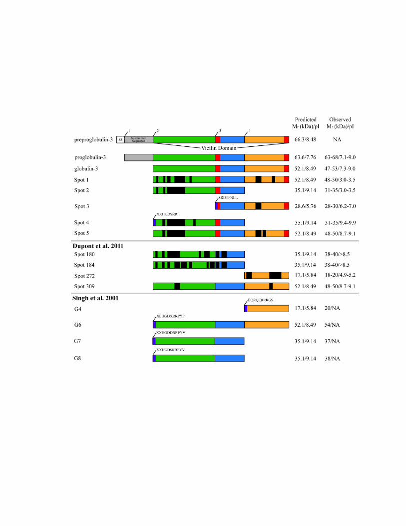

46
and 4. Spots 1 and 5 lack N-terminal sequence data, but had MS sequenced peptides that
localized to the N-terminal and C-terminal segments of the vicilin domain, and exhibited
relative mobilities of 48-50 kDa (Figure 2.3). The pI of Spot 1 was 3.0-3.5, while the pI
of Spot 5 was 8.7-9.1 Both Spot 1 and Spot 5 correspond to the theoretical cleavage of
preproglobulin-3 at cleavage site 2 with predicted Mr 52.1 kDa and pI 8.49.
2.4 Discussion 2.4.1 Type 1 diabetes
Inflammation associated with the T-cell-mediated autoimmune disease T1D results in the
loss of the insulin-producing β cells in the pancreatic islets of Langerhans (Eizirik et al.,
2009). While the incidence of T1D has steadily increased in developed countries over the
past 60 years, a definitive cause of T1D has yet to be elucidated (Todd, 2010). While
many risk genes for T1D have been identified, it has been proposed that most T1D-
related genes are not highly penetrant and that T1D is actually a complex disease
requiring both genetic susceptibility and the exposure to environmental risk factors
(MacFarlane et al., 2009). Serum IgG antibodies from human patients with T1D, but not
from matched control groups, are able to bind Glo-3 in vitro, indicating that Glo-3 may be
a candidate risk factor for the development of T1D in certain individuals (MacFarlane et
al., 2003). We therefore chose to characterize the Glo-3-related proteins in the wheat
cultivar AC Barrie with respect to protein maturation in developed seeds.
47
2.4.2 Characterization of Glo-3
When the amino acid sequences of preproglobulin-3A from the wheat cultivars AC Barrie
and Glenlea are compared, there are five amino acid substitutions out of 588 residues
(Loit et al., 2009). However, the theoretical isoelectric points of these Glo-3 proteins are
8.48, and 7.78 in AC Barrie and Glenlea, respectively. The difference in pI of 0.70 is due
to the substitution of two arginine residues (R43Q and R102H) in the AC Barrie Glo-3.
The AC Barrie salt-soluble Glo-3-related proteins demonstrate a range of isoelectric
points concentrated in the basic range (pH 7-9) when separated by 2DE (Figure 2.2, panel
E). As there are three Glo-3 genes in the wheat cultivar Glenlea (Loit et al., 2009), it is
feasible that AC Barrie would also have three Glo-3 genes, as both AC Barrie and
Glenlea are closely related hexaploid wheat cultivars (McCallum and DePauw, 2008).
The charge trains of gel spots with similar Mr and a range of pI values at the major size
groups (Figure 2.2) are likely due to post-translational processing and modifications, as
previously discussed (Robert et al., 1985), as well as amino acid substitutions between the
multiple immunologically related Glo-3 proteins in AC Barrie, or from artifacts generated
during the execution of the extraction and examination protocols (Deng et al., 2012) .
When the wheat cultivar AC Barrie salt-soluble globulin fraction was resolved by 1D
SDS-PAGE under reducing conditions, bands of three major size ranges of approximately
64-70 kDa, 47-53 kDa and 33-37 kDa were visible (Figure 2.1); referred to as 65 kDa, 50
kDa and 35 kDa, respectively. These three major wheat globulin groups are characteristic
of the 7S globulins described in other studies (Burgess and Shewry, 1986; Robert et al.,
1985). Intriguingly, while Spot 3 had an observed Mr of 28-30 kDa (Figure 2.2D), no
distinct band is visible in this region in Figure 2.1, though a faint band is visible just

48
below the 35 kDa band. The 28-30 kDa band is likely less intense than the major bands,
as Spot 3 is the only significant immunoreactive spot at 28-30 kDa, while the doublet at
65 kDa comprises several spots and is still markedly less intense than the 50 and 35 kDa
bands, which both include several intense spots.
The 7S globulins are enriched in the embryo and aleurone layer of wheat and other
cereals (Heck et al., 1993; Khavkin et al., 1978; Kriz, 1999; Yupsanis et al., 1990).
Consistent with our previous findings (Loit et al., 2009), immunoblots probed with anti-
Glo-3 polyclonal serum revealed that 7S globulins are expressed at low levels in the salt-
soluble fraction of wheat endosperm (Figure 2.1). In the embryo-enriched fraction, Glo-3
related proteins are restricted to each of the major size groups, suggesting that these Glo-
3-related proteins have similar post-translational processing patterns as the previously
characterized 7S globulins.
2.4.3 Post-translational processing of Glo-3
The 7S storage proteins in other plants undergo a series of post-translational
modifications which include limited endoproteolytic events (Gatehouse et al., 1983;
Sharma et al., 2010; Spencer et al., 1983). Glycosylation is frequently observed in 7S
globulins as N-linked complex glycans (Sturm et al., 1987), but is not required for proper
folding or export to the protein storage vacuole (PSV) (Chrispeels et al., 1982). A
previous study has shown that wheat 7S globulins bind the lectin concanavalin A,
although the exact nature and extent of this binding is unknown (Robert et al., 1985).
Additionally, other cleavage events may occur at the N-terminus of Glo-3 as observed for
49
maize Glb1 (Schwartz, 1979). These post-translational modifications, among others,
likely contribute to the heterogeneity of the observed isoelectric points as well as the wide
range of observed molecular masses among the Glo-3-related proteins.
The MS/MS sequencing of the numbered spots in Figure 2.2, panel D, all returned Glo-
3A sequences when queried against the non-redundant NCBI protein database, indicating
that, similar to orthologous 7S storage proteins in other species (Heck et al., 1993), the
peptide spots in Figure 2.2, panel D were derived from a common precursor protein. The
Glo-3-related proteins found at the acidic end of the pH spectrum, including the proteins
in spots 1 and 2, exhibited a pI of approximately 3.0 (Figure 2.2, panel D). The low pI of
these proteins could be due to protein precipitation during the rehydration of the IPG
strips (Sanchez et al., 1997), as similar spots are visible at pH 7.0 in the immunoblot of
the 2D gel of pH 7-10 (Figure 2.2, panel E).
Considering features such as the location of the MS/MS sequenced peptides within Glo-3,
N-terminal sequence data (when available), as well as the Mr and pI of the spots, we were
able to deduce the likely cleavage pattern of the preproglobulin-3 precursor protein
(Figure 2.3) that resulted in the globulin-3 polypeptide spectrum recognized by anti-Glo-3
antibodies (Figure 2.2, panel D). It must be noted that the antibodies used to probe the
globulin fraction were raised against two linear epitopes from the vicilin domain of
WP5212: SRDTFNLLEQRPKIAN and RGDEAVEAFLRMATA (MacFarlane et al.,
2003; Scott, 2007). These epitopes are located in the middle (blue) and C-terminal
(orange) vicilin domain segments (Figure 2.3). Therefore, N-terminal polypeptides
created by the endoproteolytic cleavage at site 3 may not be recognized by the anti-Glo-3
antibodies used in this study.

50
The model of endoproteolytic cleavage presented in Figure 2.3 suggests that the Glo-3-
related proteins of varying molecular masses and isoelectric points originate from 7S
globulin precursors possessing a signal peptide, an N-terminal segment and a vicilin
domain (Figure 2.3). These precursors belong to a multigene family whose members
differ slightly in sequence, with varying length N-terminal segments (Lawrence et al.,
1994). We propose that the Glo-3 protein, prior to co-translational removal of the signal
sequence, is a ~66 kDa monomer (Figure 2.3). After signal sequence removal (cleavage
site 1), the protein becomes a ~64 kDa holoprotein. Three internal cleavage sites have
been identified that, when processed, can yield polypeptides with a range of Mr and pI
values (Figure 2.3). Spot 3 (Figures 2, panel D, Figure 2.3) was observed at Mr 28-30 kDa
and pI 6.2-7.0, and Spot 4, which has a similar Mr (31-35 kDa), a more basic pI (9.4-9.9),
and a different N-terminus than Spot 3. The N-terminal sequence determined for Spot 3
SRDTFNLL represents a novel cleavage site for globulin-3. The location of this cleavage
site is consistent with processing by vacuolar processing enzymes (Ariizumi et al., 2011),
as an aspartic acid residue precedes the N-terminal sequence of Spot 3. These findings
suggest that these ~30 kDa globulin-3-related polypeptides could arise from different
processing events of the same precursor, as was observed for barley Beg1 (Heck et al.,
1993; Yupsanis et al., 1990). Curiously, there were no proteins identified in the
immunoblotting experiments that corresponded to the product of processing at cleavage
site 4, as was documented in other studies that sequenced globulin-3 (Figure 2.3 and
(Dupont et al., 2011; Singh et al., 2001; Tasleem-Tahir et al., 2011)). Potentially, as the
antiglobulin-3 antibodies are polyclonal and were prepared by the co-immunization of
rabbits with a mixture of two separate peptides (Sancho et al., 2008), it is possible that the

51
antibodies recognize only one of the two peptides. However, further study on the binding
of these antibodies to processed globulin-3 proteins is still required in order to achieve a
more definitive understanding.
When compared to the Glo-3A, Glo-3B, and Glo-3C sequences previously published by
our group (Loit et al., 2009), each of the spots contained at least one peptide in Table 1
that mapped solely to the Glo-3A coding sequence (GenBank Accession JQ945759 - data
not shown). As the Glo-3A protein coding sequence is 99% identical between wheat
cultivars Glenlea (GenBank accession ACJ65514) and AC Barrie (JQ945759), the Glo-
3B and Glo-3C proteins may share a high percentage identity between these two cultivars.
Therefore, we attributed all spots to Glo-3A. However, on close inspection of the Glo-3B
(FJ439136) genomic sequence, there are several insertion/deletion events (data not
shown) that would yield a protein of similar molecular weight as Glo-3A (66.3 kDa).
Further sequencing of Glo-3 cDNA clones from both AC Barrie and Glenlea cultivars is
necessary to determine the transcribed sequence of Glo-3.
The endoproteolytic processing events outlined here are likely part of a series of post-
translational events that lead to the maturation of the Glo-3-like proteins, also similar to
those observed for barley Beg1 (Heck et al., 1993). The identification of wheat Glo-3
holoproteins (~65-70 kDa) by immunoblot analysis suggests that the endoproteolytic
modifications described are partial and may not be prerequisites for proper folding,
transport, targeting, and storage of the Glo-3 proteins. Alternatively, as the wheat genome
has not been sequenced, the spots with Mr 65-70 kDa may correspond to proteins in the
globulin-3 family that encode proteins recognizable by the antiglobulin-3 antibodies, but
are processed in a different manner than the proteins observed in this study. Further

52
sequencing and study are required in order to fully catalogue the globulin-3 family. The
presence of polypeptides of ~50 kDa and ~30-38 kDa (Singh et al., 2001 and this study)
with the same N-termini reinforce the idea that not every hypothesized set of
endoproteolytic processing events occurs, and some could occur with varying degrees of
processing. Such maturation of vicilins has not only medical implications, but also
implications for the quality of foods rich in vicilins following processing and production
(e.g. wheat, soybean (Kaviani et al., 2011; Sancho et al., 2008)).
2.5 Conclusion
With a greater understanding of the endoproteolytic processing events that lead to the
maturation of the Glo-3 family of proteins observed in the salt-soluble embryo protein
fraction, we can now refine the research of Glo-3 to certain domains present only in the
polypeptides that are associated with T1D or celiac disease following endoproteolytic
processing. In addition, the specific breeding or genetic modification of wheat could be
performed to minimize any potential disease- or food quality-related protein or peptide
content compared to that of existing wheat cultivars, as is underway for the deletion of the
conglycinin α' subunit from the soybean proteome (Rayhan et al., 2011; Stanojevic et al.,
2011).

53
2.6 Methods 2.6.1 Wheat seed protein extraction and sample preparation
For each extraction, 4 g of whole Triticum aestivum AC Barrie seeds or 50-100 embryos
or endosperm, which were dissected as in (Walker-Simmons, 1987) were employed.
Seeds were initially ground in a domestic coffee grinder. Finer powder was obtained by
hand milling using a mortar and pestle under liquid nitrogen. Wheat powder was mixed
with 10 volumes of fresh ice-cold acid denaturing solution (10% (w/v) trichloroacetic
acid (TCA) and 0.05% (w/v) dithiothreitol (DTT) in acetone). Samples were stirred at
4oC for 1 h and left at -20oC overnight. The resulting suspension was centrifuged for 30
min at 35,000 x g at 4oC (Beckman Avanti J-25, rotor JA 25.50). The supernatant was
decanted and the pellet resuspended in ice-cold acetone containing 0.05% (w/v) DTT.
The mixture was extracted by incubation for 1 h at -20oC. The suspension was
centrifuged for 20 min at 35,000 x g at 4oC, the supernatant decanted and the pellet dried
on ice. Dried powder was extracted with 15 ml of 1 M NaCl solution (1.0 M NaCl; 0.05
M Tris; pH 8.0). The mixture was stirred for 1 h at room temperature and centrifuged
(27,000 x g for 30 min at 22oC). Supernatants were collected and the extraction with 1 M
NaCl solution was repeated. The supernatants were pooled and dialysed against 5 changes
of ddH2O for 24 h at 4oC. Precipitates were collected by centrifugation at 35,000 x g at
4oC for 45 min. Pellets were drained and extracted for either one-dimensional (1D) or
two-dimensional protein separation.

54
2.6.2 Sample preparation for 1D separation
Pellets were resuspended in 1 M NaCl solution. Samples were centrifuged at 20,000 x g
for 1 h at 22oC and quantified by the bicinchoninic acid (BCA) assay (Smith et al., 1985).
Supernatants were aliquoted and stored at -80oC until use.
2.6.3 Sample preparation for 2D separation
Proteins from pellets were extracted with 1.5 - 3 ml rehydration buffer (8M/2M deionized
urea/thiourea, 2% 3-([3-cholamidopropyl]dimethylamino)-1-propanesulfonate (CHAPS),
50 mM DTT, 0.0005% bromophenol blue) containing pH 3-10 ampholytes (Bio-Rad).
Following sonication, samples were centrifuged at 200,000 x g for 1 h at 22oC in the
Beckman TL-100 ultracentrifuge. Supernatants were collected, quantified by Bradford
method (Bradford, 1976), aliquoted and stored at -80oC until use. Throughout the 2D
separation process, care was taken not to heat the urea/thiourea-containing solutions
above 30oC to avoid carbamylation of amino groups which can lead to artifactual spot
heterogeneity (Shaw and Riederer, 2003).
2.6.4 1D SDS-PAGE protein fractionation
Protein extracts were fractionated under SDS-PAGE reducing conditions. Protein samples
were combined in a 1:1 (v/v) ratio with 2X sample buffer (4% SDS, 20% glycerol, 0.12
M Tris (pH 6.8), 10% (v/v) -mercaptoethanol, 0.01% bromophenol blue). Samples were
boiled for 5-10 min and centrifuged at 20,000 x g for 10 min. All samples were loaded on

55
discontinuous (5% stacking, 10%-12% resolving) 1.5 mm SDS polyacrylamide gels
(Laemmli, 1970). The SDS running buffer, adjusted to pH 8.3 with NaOH, consisted of
25 mM Tris base, 0.19 M glycine and 0.1% SDS. Electrophoresis was performed in the
Mini-PROTEAN 3 System (Bio-Rad) at 100-150 V (400 mA) until dye front reached the
bottom of the gel. Gels were either stained with Coomassie Brilliant Blue (CBB) R-250
or were used for immunoblot analysis.
2.6.5 Immunoblot analysis
Proteins from SDS-PAGE were transferred under semi-dry conditions by means of the
Trans-Blot SD Semi-Dry Transfer Cell (Bio-Rad). Gels were rinsed 5-10 min in semi-dry
transfer buffer (24 mM Tris, 192 mM glycine and 15% methanol) and transferred to
nitrocellulose membranes (Bio-Rad) also soaked in transfer buffer. Electroblotting
proceeded for 1-1.5 h at 11 V (400 mA). Once complete, the transfer was verified by
staining membranes with Ponceau S (0.2% (w/v) in 1% glacial acetic acid) followed by
several washes of ddH2O. Prior to immunoblotting, membranes were destained with
TBST buffer (10 mM Tris-HCl (pH 7.3), 0.1 M NaCl and 0.5% Tween-20) and were
incubated 30 min with shaking, in 5% skim milk powder in TBST buffer at room
temperature. Membranes were incubated with polyclonal rabbit anti-Glo-3 antibody
(MacFarlane et al., 2003) (diluted 1:10,000 in 5% skim milk powder in TBST and 0.05%
NaN3) and left overnight at 4oC with gentle rocking. Next day, membranes were washed 4
x 5 min in TBST, and incubated 1 h in biotin-SP-conjugated AffiniPure goat anti-rabbit
IgG (H+L) secondary antibody (Jackson ImmunoResearch Laboratories, Inc., West
Grove, PA) with a dilution of 1:100,000. This incubation was followed by another set of

56
washes in TBST and a final incubation in affinity purified goat anti-biotin-horseradish
peroxidase (HRP)-conjugated tertiary antibody (Cell Signaling Technology, Inc. Danvers,
MA) for 1 h. Following 4 x 5 min washes, membranes were treated with ECL™ Western
Blotting Detection Reagents (Amersham Biosciences, Piscataway, NJ) for 1 min, exposed
to Kodak BioMax Light Film (Fisher Scientific, Nepean, ON). Film was developed by the
Kodak X-OMAT 2000A Processor.
2.6.6 Two-dimensional gel electrophoresis (2DE)
Proteomic analysis of the AC Barrie salt-soluble proteins was conducted by 2DE. The
proteins were separated in the first dimension by isoelectric focusing (IEF) according to
their isoelectric points (pI). Extracts were applied to linear immobilized pH gradient
(IPG) 7 cm strips with pH ranges 3-10 and 7-10 (Bio-Rad, Mississauga, ON). A protein
load of 100-150 μg was applied to strips for staining and immunoblot applications; 250
μg for mass spectrometry and N-terminal sequencing applications. Strips were covered in
mineral oil and rehydrated overnight in a reswelling tray in 125-150 μl volumes of sample
diluted in rehydration buffer. For strips in the pH ranges 3-10, DTT was used as a
reducing agent; tributylphosphine (TBP) was used for strips in the pH range of 7-10.
Rehydrated strips were focused in the PROTEAN IEF Cell (Bio-Rad) according to the
following program: step1, 250 V (30 min), linear ramp; step2, 4000 V (2 h), linear ramp;
step3, 4000 V (10,000 Vh), rapid ramp. Immediately following isoelectric focusing, the
strips were either stored at -80oC for future use or equilibrated prior to running the second
dimension. Strips were thawed, if necessary, and reduced for 15 min in 2 ml of
equilibration buffer (6 M urea, 2% SDS, 20% glycerol, 0.375 M Tris-HCl, pH 8.8
57
containing 2% (wt/vol) DTT or 5 mM TBP). A second step, one of alkylation, was
performed for 15 min in the same equilibration buffer with the exception that 2.5% (w/v)
iodoacetamide was substituted for reducing agents DTT or TBP. Overlay agarose (Bio-
Rad) was used to seal the equilibrated strips at the top of 1 mm vertical resolving gels
(10% or 12% polyacrylamide). Second dimension SDS-PAGE was performed using the
Bio-Rad MiniProtean 3 at 100 V (400 mA); runs were terminated when the bromophenol
dye front had reached the end of the gel. Protein spots were counted using ImageQuant
TL Colony Version 7.0 (GE Healthcare).
2.6.7 Liquid chromatography tandem mass spectrometry (LC-MS/MS)
Gels were carefully manipulated under a laminar flow hood at all times to reduce keratin
contamination. Following separation in the second dimension, proteins were detected
with Bio-Safe Coomassie (Bio-Rad) according to manufacturer’s instructions and
destained with multiple changes of ddH2O. Selected spots were excised from the gel by
means of a clean scalpel blade and subjected to mass spectrometric analysis (Ottawa
Institute of Systems Biology, OISB). Proteins were digested in-gel with chymotrypsin as
previously described (Wilm et al., 1996). Trypsin digests were avoided because of the
high frequency of arginine and lysine residues in the reported wheat globulin sequences
(Kriz, 1999; Loit et al., 2009). Peptides were separated by liquid chromatography on an
Agilent 1100 Series HPLC System (Agilent Technologies, Palo Alto, CA) and applied by
electrospray to a QSTAR Pulsar quadrupole-TOF mass spectrometer (ABI/MDS Sciex,
Concord, ON) as described in (Vasilescu et al., 2005). Resulting peptide masses were
used to interrogate the non-repetitive NCBI protein database (06/10/2011; 1,4,324,397

58
sequences; 4,906,523,086 residues) using Mascot software (version 2.3) (Matrixscience
Ltd.) as in our previous studies (Koziol et al., 2012; Wall et al., 2010). Fixed
modifications were set for carbamidomethyl (C) and variable modifications for oxidation
(M). One missed cleavage was allowed. Peptide and MS/MS mass tolerances permitted
were ±100 ppm and 0.2 Da, respectively.
2.6.8 N-terminal sequencing
Gels used for N-terminal sequencing were treated for wet transfer to PVDF membranes.
Briefly, the gels were soaked in CAPS (3-(cyclohexylamino)-1-propanesulfonic acid)
electroblotting buffer (10 mM CAPS/NaOH pH 11 and 10% methanol) for 5 min.
Sequiblot PVDF (Bio-Rad) membranes were wet in 100% methanol, soaked in CAPS
electroblotting buffer along with Whatman 3MM paper sheets and transblot sponges. The
blotting sandwich was assembled and run in the Mini Trans-Blot Cell (Bio-Rad) at 50 V
(170 mA) for 45 min. After disassembly, the membranes were rinsed thoroughly with
ddH2O, saturated in 100% methanol and stained for a minimum of 1 min in amido black
stain (0.1% amido black, 1% acetic acid and 40% methanol). Membranes were rinsed in
multiple changes of ddH2O and air dried before excision of spots. Spots of interest were
subjected to Edman degradation using the 494 cLC PROCISE Sequencing System
(Applied Biosystems; Foster City, CA) for high sensitivity (femtomole quantities) N-
terminal protein sequencing (University of Texas Medical Branch, UTMB, Biomolecular
Resource Facility Core).

59
Chapter 3
Taking stock of the protein remnants on the battlefield between host and pathogen: Maize CL30-Fusarium interactome
Koziol AG, Reid L, Altosaar I
Submitted to: Journal of Proteomics
Contribution of authors:
AGK contributed to the study design, performed all experiments, analyzed data,
contributed all figures, and wrote and edited the manuscript. LR contributed to study
design, provided field samples, and edited the manuscript. IA contributed to the study
design, data analysis and editing.

60
3.1 Abstract
The economic losses associated with gibberella ear rot make necessary a better
understanding of both the pathogen, Fusarium graminearum, and its host, Zea mays. The
F. graminearum susceptible inbred maize line CL30 was exposed to water controls or
conidial suspensions of F. graminearum through kernel inoculations, and the host plants
were allowed to grow to maturity. Proteins were extracted separately from the maize
embryo and endosperm tissues, as well as from the fungal remnants on the surface of the
maize kernels. The proteins were sequenced using direct-sequencing LC-MS/MS. We
identified 250 maize proteins with high confidence. These proteins were classified into
several functional categories, including: late embryogenesis abundant, seed storage,
metabolism, and defense. There was a significantly increased representation of defense
proteins in both the embryo and endosperm tissues of infected maize samples. The
proteome of the fungal remnants on the surface of the maize kernels was composed of 18
proteins that were present after desiccation. Several of these proteins were categorized as
being involved in the metabolism of plant-sourced molecules, or in stress response. Taken
together, these data should help dissect the interactive system between host and pathogen
during the important time period of grain maturation and harvest.

61
3.2 Introduction
Gibberella ear rot and stalk rot in maize, as well as Fusarium head blight in wheat, barley,
and oats are caused through the infection of these crops by the fungal pathogen Fusarium
graminearum (Holbert et al., 1923; Miller et al., 1985; Reid et al., 1992; Takeda and
Heta, 1989; Trail, 2009). Not only do these diseases reduce grain yields (Kazan et al.,
2012), but F. graminearum also synthesizes mycotoxins, such as deoxynivalenol (DON),
fusarin C, autofusarin, and zearalenone (Logrieco et al., 2002; Taylor et al., 2008). One of
the best studied mycotoxins, DON, is a virulence factor that aids in fungal colonization,
and also poses serious human health risks (Harris et al., 1999). For instance, DON inhibits
eukaryotic protein biosynthesis, and upon ingestion, causes nausea, vomiting, and
convulsions (Goswami and Kistler, 2004; Vesonder et al., 1981), with long-term exposure
to DON being linked to immune system suppression and certain neurological disorders
(Bennett and Klich, 2003; Pestka and Bondy, 1994). Consequently, the concentration of
DON in food products is tightly regulated, with limits of 1 μg DON per gram of food for
human consumption (Trail, 2009).
The sequence of the four chromosomes of the F. graminearum strain PH-1 (teleomorph
Gibberella zeae) genome was published in 2007 (Cuomo et al., 2007). The genome is
36.45 Mb and codes for 13 321 predicted proteins
(www.broadinstitute.org/annotation/genome/fusarium_group/GenomeStats.html; last
viewed 7/09/2012). Compared to other sequenced fungal genomes, F. graminearum has
an overrepresentation of certain protein categories, including hydrolytic enzymes (Cuomo
et al., 2007), which may have implications in pathogenesis. The infection process is
initiated for gibberella ear rot when ascospores or conidia are introduced to exposed silks
62
or punctured kernels (Koehler, 1942; Miller et al., 2007; Sutton, 1982). Following
germination of the spores, the fungus can then colonize the host plant (Mesterhazy et al.,
2012). However, in response to fungal infection, plants have several inducible defense
strategies, including the strengthening of cell walls and the production of antimicrobial
proteins (Campo et al., 2004; Reid et al., 1992; Reid et al., 1994).
We have previously examined the starch granule-associated proteomes of wheat (Wall et
al., 2010), and of commercially prepared rice and maize starches (Koziol et al., 2012;
Koziol et al., 2012). In these experiments, it was presumed that the grains used in the
preparation of starches were healthy and free of obvious fungal contamination. The use of
CL30, a susceptible host genotype of maize, and the mechanical kernel injection
technique promotes a fuller expression of the foreign proteome. While there have been
proteomic sequencing studies of F. graminearum within the host following infection, to
our knowledge, the proteome of the fungal remnants on the surface of maize seeds at
maturity has not previously been determined. We hypothesized that the proteins
sequenced within these fungal remnants could provide insight into the biochemical
processes occurring in the fungus prior to desiccation, and that even though CL30 is
susceptible to infection by F. graminearum, we would be able to identify host defense-
related proteins present in both the endosperm and embryo tissues. To that end, we
initiated a gel-free protein sequence project using LC-MS/MS to investigate the interplay
of the proteomes of host and pathogen.

63
3.3 Results
Kernels of the inbred maize line, CL30, were injected at 10-15 days post-silk emergence
with either water controls or conidial suspensions of F. graminearum and allowed to
develop to maturity. Following harvest, kernels from both treatments were collected, and
the proteomes of discrete tissues from the kernels, as well as the remnants of F.
graminearum on the surface of the maize seeds were sequenced using LC-MS/MS.
Sequences were analyzed and classified by biochemical function.
Cumulatively, 250 plant-sourced proteins were identified in the water control or
Fusarium treated embryo and endosperm samples. These proteins were classified based
upon their molecular function, with the classes as follows: maturation; storage; stress
tolerance; metabolism; nucleic acid binding; protein folding, synthesis and transport; oil
body-associated; miscellaneous; unknown; and defense. This classification is shown in
Figure 3.1. When comparing the percent representation between the control and infected
samples, the largest differences in percent representation include nucleic acid binding
(11.8% in control and 5.4% in the infected) and oil body associated (5.9% in the control
and 2.7% in the infected) in the embryo samples. The storage (4% in the control and 7.5%
in the infected), stress tolerance (16% in the control and 7.5% in the infected) and
metabolism (10.7% in the control and 18.9% in the infected) proteins in the endosperm
also showed differences in percent representation. Additionally, the defense proteins had
different representations in both the embryo and endosperm samples.
64
Figure 3.1: Percent representation of maize functional protein categories. Proteins were isolated from both the Fusarium infected and control embryo and endosperm tissues and sequenced by LC-MS/MS. Functional categories are based on KEGG classifications (Kanehisa and Goto, 2000).
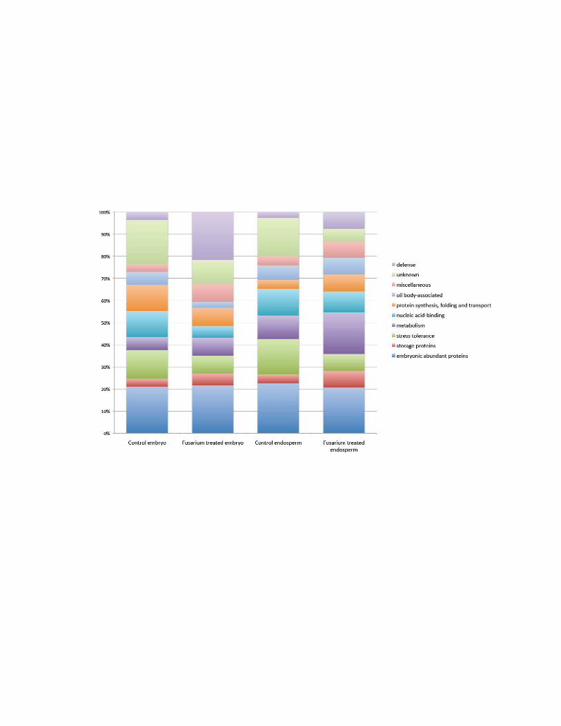
65
That both the embryo and endosperm samples had different representation of defense
proteins was of particular interest. The percentage representation of defense proteins in
the control embryo, infected embryo, control endosperm, and infected endosperm
samples was 3.5%, 21.6%, 2.7%, and 7.5%, respectively. The defense proteins identified
in each sample are shown in Table 3.1, which includes GenBank accession number,
protein name, species, Mascot score, percent coverage, and the number of peptide hits
(and unique hits). The percent coverage was calculated by adding together the total
number of amino acids residues present in all the peptide hits attributed to a particular
protein, and dividing this sum by the total length of the protein in GenBank. There were
five classes of defense proteins identified: chitinases; ribosome inhibiting proteins;
proteinase inhibitors; proteases; and peroxidases.
We discovered 18 proteins that were not degraded upon the desiccation of the fungal
remnants on the surface of the maize kernels (Table 3.2). Of the 18 proteins identified, 10
were attributed to F. graminearum, and four proteins were attributed to F. oxysporum. A
putative linoleate diol synthase from F. graminearum had the highest Mascot score (349).
Other enzymes involved in the processing of raw nutrients include glycoside hydrolase
and xylanase. Additionally, we identified ubiquitous proteins, proteins involved with
stress tolerance, metabolism, as well as proteins with unknown functions.
3.3 Discussion
The economic losses from all F. graminearum diseases are in excess of $1 billion
annually through the reduction in grain yield and certain traits such as grain quality

66
Table 3.1: Defense proteins in maize samples.
Sample Accession Protein Mascot Score
% Coverage
Peptides, Uniques
H2O treated maize embryo
NP_001147325 subtilisin-chymotrypsin inhibitor CI-1B 139 38.6 2,2
ACJ62806 ribosome-inactivating protein 106 12.3 3,3
AAT39992 chitinase, class GH19 101 7.2 2,1
Fusarium treated maize embryo
ACG36543 peroxidase 12 209 13.6 4,4
NP_001140805 uncharacterized protein LOC100272880 - pepsin retropepsin-like 202 16.1 4,4
AAT39991 chitinase, class GH19 159 14.2 4,3
NP_001105541 chitinase chem5 precursor - contains GH18 hevamine XIP I class III domain 150 8.9 2,2
NP_001142312
uncharacterized protein LOC100274481 - contains GH18 hevamine XIP I class III domain
147 9.9 3,3
NP_001232812 trypsin/factor XIIA inhibitor precursor 99 47.8 3,3
NP_001146946 cysteine proteinase inhibitor B 79 14.0 2,2
CAN72184 hypothetical protein VITISV_042598 - retropepsin like 78 3.1 4,3
H2O treated maize endosperm
CAA49723 protein b-32 – ribosome inhibiting protein 237 21.5 5,2
NP_001149472 endochitinase A precursor, class GH19 120 11.8 2,2
Fusarium treated maize endosperm
ACJ62815 ribosome-inactivating protein 206 20.6 4,2
ABA34115 maize protease inhibitor 111 38.4 2,2
NP_001142312
uncharacterized protein LOC100274481 precursor - putative xylanase inhibitor XIP-I, or class III plant chitinase
92 7.1 2,2
NP_001142211 uncharacterized protein – contains ricin-type β-trefoil domains 77 5.8 2,2

67
Table 3.2: Proteome of desiccated fungi on the surface of infected maize kernels.
Accession Protein Species Mascot Score
% Coverage
Peptides, Uniques
XP_382844 hypothetical protein FG02668.1 – putative linoleate diol synthase
Gibberella zeae PH-1
349 10.1 10,8
XP_381869 hypothetical protein FG01693.1 Gibberella zeae PH-1
220 5.4 5,5
XP_389762 hypothetical protein FG09586.1 - putative phosphatidylinositol/phosphatidylglycerol transfer protein
Gibberella zeae PH-1
194 26.7 5,5
AAR02398 glycoside hydrolase 7 Gibberella zeae 183 7.8 4,3
XP_388897 SODC_NEUCR superoxide dismutase Gibberella zeae PH-1
171 15.8 2,1
EGU80493 hypothetical protein FOXB_08953 – putative Cu-Zn superoxide dismutase
Fusarium oxysporum Fo5176
137 16.9 2,1
XP_381699 hypothetical protein FG01523.1 – putative aldo-keto reductase
Gibberella zeae PH-1
132 17.9 4,4
XP_381422 hypothetical protein FG01246.1 – putative old yellow enzyme
Gibberella zeae PH-1
131 2.6 3,3
ABU48779 translation elongation factor 1-alpha Wickerhamomyces rabaulensis
126 15.9 2,2
XP_001538961 ubiquitin/79aa fusion protein Ajellomyces capsulatus NAm1
121 37.0 3,1
XP_391289 hypothetical protein FG11113.1 – putative bicupin, oxalate decarboxylase
Gibberella zeae PH-1
121 9.6 3,3
CAD92004 calmodulin Fusarium oxysporum
117 32.2 3,2
EGU72061 hypothetical protein FOXB_17429 Fusarium oxysporum Fo5176
110 36.4 2,1
XP_003044867 predicted protein - ribosomal protein S11 Nectria haematococca mpVI 77-13-4
103 22.7 3,3
AAV98254 putative xylanase 2 Gibberella zeae 99 8.2 2,2
XP_390596 hypothetical protein FG10420.1 Gibberella zeae PH-1
95 21.4 3,3
EGU83490 hypothetical protein FOXB_05994 - contains stress-induced bacterial acidophilic repeat motif
Fusarium oxysporum Fo5176
86 33.8 2,1
XP_003007386 60S ribosomal protein L26-1 Verticillium albo-atrum VaMs.102
82 32.3 3,2
68
(Goswami and Kistler, 2004). Previous studies have investigated the proteomic profiles of
both pathogen and host at various stages of infection (Geddes et al., 2008; Mohammadi et
al., 2011; Shin et al., 2011; Zhou et al., 2006), but we were interested in the proteomes of
both the field desiccated fungal remnants on the surface of the seed at harvest and in the
tissues of the mature seeds themselves.
3.3.1 Percent representation
The sessile nature of plants forces their pathogen defense systems to be particularly
robust (The Arabidopsis Genome Initiative, 2000). It has previously been shown that
certain defense proteins are upregulated following pathogen infection (Walter et al.,
2010). Our data confirms these findings, as there was an increase in the number of
sequenced proteins associated with defense in both the infected embryo and infected
endosperm tissues compared to the control samples (Figure 1). The number of identified
defense proteins increased 617% in the infected embryo, and 278% in the infected
endosperm. The lower percentage increase of defense proteins in the endosperm is due to
the apoptotic (and therefore less responsive) nature of the endosperm (Young and Gallie,
2000).
3.3.2 Proteinase inhibitors
Proteinase inhibitors (PIs) are abundantly expressed in storage tissues; comprising up to
10% total protein content (Habib and Fazili, 2007). PIs act by forming complexes with
proteolytic enzymes (Sels et al., 2008). These complexes reduce the activity of the

69
proteolytic enzymes, and therefore are important in regulating a number of important
biological functions influenced by proteolytic enzymes, including signal transmission,
seed dormancy, and apoptosis (Haq et al., 2004). In addition to regulating host
biochemical processes, PIs have a proposed role in plant defense. Upon infection, fungi
secrete proteases that degrade cell walls to facilitate colonization, as well as digest plant
proteins down to the amino acids required for fungal metabolism (Dunaevskii et al.,
2005). In response to fungal infection, plant PIs are upregulated, and bind and reduce
fungal protease activity (Ryan, 1990). We identified four PIs (Table 3.2). Both the
infected embryo and endosperm tissues had more represented PIs compared to the water
injected control samples. One PI identified in Fusarium-treated embryo was a
trypsin/factor XIIA inhibitor (GenBank accession NP_001232812). This PI, in addition to
inhibiting proteases, also demonstrates α-amylase inhibitor activity. These dual function
inhibitors are particularly effective in defending against fungal pathogens, as the
degradation of both proteins and starch granules is inhibited – severely limiting the
sources of energy to the growing fungal hyphae.
3.3.3 Chitinases
Chitinases hydrolytically cleave the β-1,4-glycosidic bonds in both chitin and chitosan,
which are important structural components of fungal cell walls (Jitonnom et al., 2011).
The majority of identified plant chitinases are endochitinases that hydrolyze bonds within
the chitin, chitosan, or other molecules, as opposed to exochitinases, which catalyze the
cleavage of bonds at the non-reducing end of chitin polymers (Huang et al., 2012).
Chitinases play an important role in plant defense against fungal pathogens. Following

70
infection, chitinases are upregulated (van Loon et al., 2006), and hydrolyze exposed chitin
polymers in fungal hyphal tips, inhibiting fungal growth (Grover, 2012). Plant chitinases
are grouped into two separate families, based on features of their catalytic domains:
GH18, which includes chitinase Classes III and V, and GH19, which includes chitinase
Classes I, II, and IV (Santos et al., 2008). The GH18 and GH19 families likely do not
share a common ancestral protein, as these families have significantly different amino
acid sequences and three-dimensional structures (Ubhayasekera, 2011). Class IV
chitinases (GenBank accessions AAT39991, AAT39992, NP_001149472) are
distinguished by the presence of an N-terminal carbohydrate-binding domain
(Ubhayasekera et al., 2009). Class IV chitinases were found in both infected and control
samples.
3.3.4 Xylanase inhibitors
Xylan is composed of linked β-1,4-linked D-xylose groups that can be substituted by
different residues, including L-arabinose (Juge, 2006). Xylans and their substituted
derivative molecules are the most abundant hemicellulose molecule in cereals,
representing up to 25% of the dry weight of maize (Pordesimo et al., 2005). Secreted
fungal endoxylanases catalyze the degradation of xylans into poly- and oligosaccharides
by hydrolyzing the β-1,4-linkages between xylopyranoside residues in xylans (Prade,
1996). There are two classes of fungal xylanase: GH10 and GH11 that differ in size, 3D
structure and catalytic properties (Biely et al., 1997). We identified a putative xylanase in
our sequencing of the Fusarium remnants on the seed surface (Table 3.2). This 40 kDa
GH10 xylanase is characterized by broad substrate specificity and a resistance to certain
71
xylanase inhibitor proteins (XIPs) (Goesaert et al., 2004).The two XIP-like proteins we
identified (Table 3.2 - GenBank accessions NP_0011005541 and NP_001142312) have
been shown to able to inhibit both GH10 and GH11 xylanase classes. Class GH10 is
inhibited through XIPs binding to the xylanase substrate-binding pocket, while class
GH11 is inhibited through XIPs binding to the active site (Payan et al., 2004). In addition
to sharing identity with XIPs, these XIP-like proteins we identified have sequence
identity to class III chitinases, though these proteins have modified active sites that lack
the ability to catalyze the degradation of chitin (Payan et al., 2003). The discovery of
XIPs in only the Fusarium treated samples agrees with previous studies that demonstrated
that XIPs in maize were upregulated following F. graminearum infection (Mohammadi et
al., 2011).
3.3.5 Ribosome inactivating proteins
Cereal ribosome inactivating proteins (RIPs) are rRNA N-glycosidases that inactivate
ribosomes by catalyzing the irreversible depurination of a single adenine residue in the
conserved elongation factor-binding α-sarcin loop of 28S rRNA molecules (Barbieri et
al., 1993). Accordingly, protein synthesis is arrested in these depurinated ribosomes.
There are three types of RIPs. The maize RIP1 protein (b-32) is a type 3 RIP, as it is
expressed as inactive precursors that, following proteolytic removal of N-terminal,
internal, and C-terminal domains, yield two chains, which interact non-covalently to form
an active form that is 10 000 times more active than the precursor (Walsh et al., 1991).
Unlike many of the other pathogen resistance gene products, b-32 is not upregulated in
response to pathogen infection; it is expressed in the endosperm concurrently with the
72
deposition of storage proteins (Balconi et al., 2010). This is reflected in Table 3.1, as the
RIP proteins are found in both the control and Fusarium infected samples.
3.3.6 Peroxidases
Oxidative burst is the rapid and transient production of reactive oxygen species (ROS) in
response to pathogen infection (Angel Torres, 2010). There are three major proposed
functions of the ROS during oxidative burst: 1) generating a toxic environment that
inhibits pathogen growth, 2) reinforcing cell walls to prevent pathogen spreading, and 3)
activating signaling cascades to induce pathogen response genes (Passardi et al., 2005).
There are several pathways that work synergistically to produce ROS during oxidative
burst. From these intertwined pathways, we identified a single peroxidase in our survey.
This class III peroxidase (GenBank accession ACG36543) was present in the Fusarium
treated embryo sample. Class III peroxidases are thermostable glycoproteins with broad
substrate specificity that are localized to vacuoles and cell walls (Almagro et al., 2009).
The importance of class III peroxidases in response to pathogen attack was demonstrated
in Arabidopsis, where transgenic lines with reduced transcript levels of class III
peroxidases had a reduced oxidative burst and had an increased susceptibility to pathogen
infections (Bindschedler et al., 2006). Our discovery of ACG36543 in only the Fusarium
treated embryo reinforces previous findings that this particular peroxidase is upregulated
in response to pathogen infection (Mohammadi et al., 2011) and may be sufficient to
convey protection due to its multifunctional properties (Passardi et al., 2005).
73
Fungal pathogens can mitigate the effects of the ROS expressed during an infected host’s
oxidative burst through the action of several different proteins. These protein families
include peroxidases, catalases and superoxide dismutases (Mayer et al., 2001). We
identified two copper-zinc (Cu-Zn) superoxide dismutases (SOD) amongst the Fusarium
remnants (Table 3.2). There are four classes of SOD, which can be differentiated by their
metal co-factors: 1) Cu-Zn, 2) Mn, 3) Fe, and 4) Ni (Mayer et al., 2001). All classes of
SOD catalyze the dismutation of •O2 and •OH to O2 and H2O2 (Hwang et al., 2002).
Previously, F. graminearum Cu-Zn SOD proteins have been identified in infected
Triticum aestivum (Paper et al., 2007; Zhou et al., 2006), indicating that this protein is
highly expressed by F. graminearum during infection.
3.3.7 Fusarium on the surface
The proteins identified in the fungal remnants on the surface of the maize kernels could
be divided into four main categories depending on their biological functions: 1) Proteins
responsible for the metabolism of plant molecules (linoleate diol synthase, glycoside
hydrolase), 2) ROS resistance (superoxide dismutase), 3) constitutive expressed proteins
(elongation factors, ribosomal proteins, calmodulin, and others), and 4) hypothetical
proteins with no assigned function. As we identified individual 18 proteins in our
desiccated Fusarium samples, these proteins may still be present as they are either very
highly expressed or resistant to degradation.
74
3.3.8 Fusarium metabolism proteins
Up to 70% of the total oil content from maize seeds is linoleic acid (Jellum, 1970). This
unsaturated n-6 fatty acid can be metabolized by Fusarium by the putative linoleate diol
synthase (LDS) (Table 3.2) in the following reaction: linoleic acid is dioxygenated to
(8R)-hydroperoxylinoleate and subsequently isomerized to (7S,8S)-dihydroxylinoleate
(Hornsten et al., 1999). Proteins that share sequence identity with LDS have been found
in several filamentous fungi (Soanes et al., 2008) and have been hypothesized to be
involved with the mobilization of storage lipids (Huber et al., 2002). Gene deletion
studies in the rice-blast disease causing Magnaporthe oryzae demonstrated that LDS is
not required for successful infection or sporulation, possibly due to the presence of
redundant linoleic acid metabolizing pathways (Jerneren et al., 2010).
The degradation of different glycosides is performed by the family of glycoside
hydrolases that catalyze the hydrolysis of glycosidic linkages, freeing sugar moieties. The
glycoside cellulose is a chain of β-(1→4) linked D-glucose units. There are two classes of
fungal glycoside hydrolases that degrade cellulose: 1) endoglucanases that catalyze the
cleavage of cellulose polymers at random locations, and 2) cellobiohydrolases that
hydrolyze the non-reducing ends of cellulose chains (Munoz et al., 2001). The glycoside
hydrolase 7 we identified is a cellobiohydrolase. As the most predominant glycoside in
maize stalks (approximately 50% of dry weight (Appenzeller et al., 2004)) is cellulose,
glycoside hydrolases are necessary for Fusarium to degrade plant cell walls to facilitate
colonization and nutrient uptake (Yoshizawa et al., 2002).

75
3.3.9 Fusarium stress response
Old Yellow Enzyme (OYE) is a flavin-dependent NADPH-oxidoreductase that was first
described in 1932 by Warburg & Christian in 1932 from brewers’ bottom yeast. Proteins
with sequence similarity to OYE have been identified in a broad phylogenetic range of
organisms, including bacteria, fungi, plants, and mammals (Williams and Bruce, 2002).
While the physiological role of OYE is, as of yet, unknown, a number of hypothesized
functions has been proposed, including oxidative stress response (Lee et al., 1999). OYE
is upregulated in the presence of hydrogen peroxide (Fitzpatrick et al., 2003) and can be
oxidized by many different compounds, including ketones and aldehydes (Trotter et al.,
2006). These data together with our discovery of OYE in the fungal remnants indicates
that it may play a protective role during plant colonization.
Plant late embryogenesis proteins (LEA) are a diverse group of hydrophilic proteins
associated with abiotic stress tolerance - most notably desiccation and cold stress
(Tolleter et al., 2010). There are three main groups of LEA proteins in plants that are
distinguishable by the presence of sequence motifs (Tunnacliffe and Wise, 2007). The
majority of proteins within each group have only minimal secondary structure when in
solution, though they have increased folding when in a dry state (Tolleter et al., 2007). It
has been shown that LEA proteins accumulate during the final stages of embryo
maturation (Galau et al., 1986). This accumulation of LEA proteins in mature seeds is
reflected in this study, because in each of the maize samples investigated the percent
representation was greater than 20% (Figure 1). While the function of LEA proteins in
stress tolerance is unknown, it has been hypothesized that they have several different
functions, including stabilizing membranes (Steponkus et al., 1998) or desiccation-

76
sensitive proteins (Goyal et al., 2005), preventing cellular water loss (Garay-Arroyo et al.,
2000), or by binding ions or ROS (Tunnacliffe and Wise, 2007).
In addition to plants, LEA proteins have been discovered in prokaryotes, protists,
invertebrates, and certain invertebrates (Tolleter et al., 2007). All fungal LEA-like
proteins are group 3 LEAs, which are characterized by the presence of multiple repeats of
an 11 amino acid residue motif (Tunnacliffe and Wise, 2007). We identified a single
LEA-like protein in the fungal remnants (Table 3.2). This protein also shares 96%
sequence identity with a conidiation-specific protein. Conidiation-specific proteins are
expressed during conidia (spore) formation, which is triggered by low nutrient or water
levels (White and Yanofsky, 1993). It is therefore likely that this LEA-like protein was
expressed in the Fusarium examined during seed maturation and desiccation in order to
form the spores necessary for the biological dispersal of the parental Fusarium genomic
sequence. As other fungal species propagate in a similar manner, the proteins on the
surface of the kernels sourced to fungal species other than F. graminearum (Table 3.2)
may be present due to the damage from the inoculation of the maize kernels. This damage
potentially allowed for the limited colonization by opportunistic fungi that would
otherwise not have been able to colonize maize.
3.4 Conclusions
The approximately 250 maize proteins identified were classified into functional
categories, including: late embryogenesis abundant, seed storage, metabolism, and
defense, with an increased representation of defense proteins in both the embryo and
77
endosperm tissues of infected samples. Most of the proteome of the fungal remnants was
categorized as being involved in the metabolism of plant-sourced molecules, or in stress
response. Taken together, these data should help dissect the interactive system between
host and pathogen during the important time period of grain maturation and harvest.
Understanding the interplay of pathogen and host is important in developing pathogen
resistant breeding lines. Greater knowledge of host defense systems will allow for the
fine-tuning of the polygenic disease resistance present in maize. Finally, the proteins
present in the fungal remnants may be targets for fungicides or for rapid biological
screening of grain shipments for presence of pathogen infection.
3.5 Methods
F. graminearum susceptible Zea mays inbred line CL30 was planted on the Central
Experimental Farm in Ottawa, ON, Canada for the 2010 growing season. Conidial
suspensions of F. graminearum strain DAOM 180378, obtained from the Canadian
Collection of Fungal Cultures, Ottawa, ON were prepared to 5 × 10⁵ conidia/mL as
described in (Reid et al., 1996). Maize kernels were inoculated with either the conidial
suspensions or sterile water controls using a four-pin automatic kernel inoculator as per
Reid, et al., (, 1997). This inoculation was timed to the blister to early milk stages of
kernel development; approximately 10-15 days post-silk emergence. Three ears of both
the fungal and water treated maize were harvested at maturity.
Kernels were harvested and suspended in protein extraction buffer (10mM Tris-HCl pH
7.8, 0.5 mM EDTA, 10 mM KCl, 1mM PMSF). Kernels were agitated for 30 seconds,

78
allowed to settle. The supernatant was decanted into a fresh tube, and sonicated on ice at
20 amplitude microns 20 times in three second bursts. Cellular debris was pelleted by
centrifugation at 14 000 g for one min at 4 C, and supernatant transferred to a fresh tube
and stored at -80 C. The kernels were surface sterilized in a 10% solution of sodium
hypochlorite for 5 min, washed 5 times in ddH2O and dried in a laminar flowhood.
Kernels were incubated overnight in 0.3% (w/v) sodium metabisulphite and 85% (v/v)
lactic acid at 37 C. Embryo and endosperm tissues were manually dissected from the
pericarp using a razor blade. Isolated tissues were ground in a mortar and pestle and
suspended in protein extraction buffer. The solution was filtered through a 63 μ mesh and
centrifuged at 14 000 g for 10 min at 4 C. The pellet was washed two times with protein
extraction buffer, two times with ice cold 95% ethanol, and two times with ice cold
acetone. The pellets were allowed to dry in a laminar flowhood. Protein extraction buffer
was added to the pellets and allowed to incubate for 30 min at room temperature. The
solution was centrifuged at 14 000 g for ten min, and the supernatant was collected.
Along with the protein samples previously stored at -80 C, the samples were
supplemented with four volumes of ice cold acetone and stored at -20 C overnight to
precipitate proteins. The samples were centrifuged at 14 000 g for twenty min and washed
twice with ice cold acetone and dried in a laminar flowhood.
Pellets were resuspended in 100 μL ddH2O. Protein concentration was determined using
the Bradford assay (Bradford, 1976). Two micrograms of each protein sample was
subjected to mass spectrometric analysis at the Ottawa Institute of Systems Biology,
OISB. Protein samples were digested with trypsin, and digested peptides were separated
by liquid chromatography on an Agilent 1100 Series HPLC System (Agilent
79
Technologies, Palo Alto, CA). Separated peptides were ionized by electrospray to a
QSTAR Pulsar quadrupole-TOF mass spectrometer (ABI/MDS Sciex, Concord, ON) as
described in (Vasilescu et al., 2005). Mascot software (version 2.3) (Matrixscience Ltd.)
was used to interrogate the non-repetitive NCBI protein database (12/20/2011; 16 392
747 sequences; 5 641 810 382 residues) with the taxonomy filter set to either ‘other
fungi’ (3 787 334 sequences) or ‘other green plants’ (3 614 282 sequences).
Carbamidomethyl (C) and oxidation (M) were set to fixed variable modifications,
respectively. One missed cleavage was permitted. Tolerances were set for both peptide
(±100 ppm) and MS/MS mass tolerances (0.2 Da). Decoy searches were performed, and
false positive rates were approximately 1%.
Molecular function was assigned to proteins using the Kyoto Encyclopedia of Genes and
Genomes (KEGG) at http://www.genome.jp/kegg/kegg2.html (last viewed 7/09/2012)
(Kanehisa and Goto, 2000; Kanehisa et al., 2006; Kanehisa et al., 2010). Peptides
corresponding to unknown or hypothetical proteins in the NCBI database were classified
based on sequence similarity by BLAST searches (Altschul et al., 1990).

80
Chapter 4 The starch granule associated proteomes of commercially purified starch reference materials from rice and maize
Koziol AG, Marquez BK, Huebsch MP, Smith JC, Altosaar, I
Published in: Journal of Proteomics 75: 993-1003 (2012)
Contribution of authors:
AGK contributed to the study design, performed experiments except LC-MS/MS,
analyzed data, contributed all figures, wrote the manuscript, and participated in editing
the manuscript. BKM aided in experiments, analyzing data, and editing. MPH performed
LC-MS/MS. JS participated in analyzing data and editing. IA participated in study design,
data analysis, and editing.

81
4.1 Abstract
Commercially available reference materials are integral components of many
experimental protocols, as it is critical to compare one’s results to those derived from
well-characterized standards. Most reference materials are well defined, with all their
components being catalogued. However, certain reference materials, such as
commercially prepared starch samples, can have undefined components, potentially
limiting their usefulness as standards. The proteome of commercially prepared starch has
not been documented, and to that end, we initiated a mass spectrometry-based survey of
the proteins associated with starch granules in commercially prepared rice and maize
starch samples. We performed direct trypsin treatments of starch samples and sequenced
both the water-soluble peptides liberated into the aqueous supernatant and the peptides
released from the starch granule surface by isopropanol solvent washing. We discovered
that the majority of proteins, in both rice and maize samples, were involved in either
carbohydrate metabolism or storage. We also documented proteins that are markers for
seed maturity and for starch mobilization.

82
4.2 Introduction
Starch is a versatile and renewable commodity that is exploited as food and biofuel, as
well as in industry during the production of chemicals, paper, binders, and adhesives
(Roper, 2002). Worldwide, almost 300 billion tons of starch is manufactured per year,
with 90% originating from cereal crops (Halford et al., 2011; Tetlow, 2011). These crops,
which include rice and maize, produce seeds that contain up to 70% of their dry weight as
starch (Dale and Housley, 1986). Having such concentrated reserves of starch so readily
available, it is therefore not surprising that as an extractable component, rice starch was
used as far back as ancient Egypt for the manufacture of papyrus (Bostock and Riley,
1855). Cereal starch is synthesized in amyloplasts, and stored in the endosperm as semi-
crystalline granules consisting of amylose and amylopectin (Zeeman et al., 2010). Both
these polymers contain -(14) glycosidic linkages, however, amylopectin which makes
up 75% of the mass of most starch granules, contains approximately 5-6% of randomly
distributed -(16) glycosidic linkages (Grimaud et al., 2008). These linkages introduce
branch points in the amylopectin chains that allow for the clustering of chains into tightly
packed arrays, and, consequently, the semi-crystalline nature of starch (Perez and Bertoft,
2010).
The origins of starch synthesis can be traced to the common ancestor of the
Archaeplastida (Patron and Keeling, 2005). Following an endosymbiotic event between a
heterotrophic eukaryotic host and a cyanobacterium, which gave rise to the
Archaeplastida, the genomic reorganization in the new, photosynthetic eukaryotic
organism merged polyglucan synthesis pathways of bacterial and eukaryotic origins
(Deschamps et al., 2008). Cytosolic starch synthesis pathways evolved from these

83
reorganization events, and it was with the evolution of the light-harvesting complexes,
which are responsible for light capture and photoprotection (Koziol et al., 2007) that
starch synthesis was relocated to plastids (Tetlow, 2011). It has been proposed that this
relocation was prompted by the requirements for ATP, generated via starch degradation,
to overcome protoporphyrin IX-induced oxidative stress (Deschamps et al., 2008).
In addition to starch, the endosperm of cereal seeds contains proteins, lipids,
carbohydrates, and other organic and inorganic compounds (Lopes and Larkins, 1993).
Most of the proteins found in seeds are storage proteins, which act as sinks of amino acids
for germinating embryos (Shewry and Halford, 2002). However, structural and metabolic
proteins, and starch granule associated proteins (SGAPs) are present as well (Lowy et al.,
1981). Many of these proteins are enzymes involved in starch biosynthesis, and include
adenosine 5 diphosphate-glucose pyrophosphorylase, granule-bound starch synthase
(GBSS), soluble starch synthase, starch branching enzyme, starch debranching enzyme,
and plastidial starch phosphorylase (Jeon et al., 2010). Other proteins are involved in
defense, transport, signaling, protein folding, and stress response (Balmer et al., 2006;
Wall et al., 2010).
Despite SGAP being a term in the biochemical lexicon since 1986, SGAPs have only
been investigated on a case-by-case approach (Baldwin, 2001; Greenwell and Schofield,
1986). Regardless of this lack of scientific precision, commercial starch samples have
nevertheless still been frequently used as reference materials in experiments. The rice and
maize starch samples we examined here have been used in a broad range of experiments
that, for example, have studied starch granule structure using light and electron
microscopy (Baldwin et al., 1994), the enzymatic digestibility of starch granules by

84
amylases and glucosidases (Erban et al., 2009), the production of ceramic materials with
differing levels of porosity through the use of starch additives (Diaz and Hampshire,
2004), the pasting properties of floridean starch in algae (Yu et al., 2002), and the use of
sonication treatments during starch separation (Zhang et al., 2005), among others.
The certificates of analysis for these commercially available starch samples give basic
measurements of the non-starch components of the samples, such as pH and percentages
of moisture, protein, and lipid, but in this current age of systems biology, it is important to
be able to catalogue all the detectable components of a sample. It has previously been
stated that the presence of proteins and lipids within purified starch samples have an
effect on the rate of granule hydrolysis by amylases and glucoamylases (Williamson et
al., 1992). Therefore, it is possible that the protein component of commercially purified
starch samples could affect the outcome of other experiments in which the starches will
serve as defined baseline substrates. It is for this reason that we initiated a tandem mass-
spectrometry-based survey of commercially available starch reference materials to
catalogue their trace proteomes. We have previously investigated the SGAPs in wheat
(Wall et al., 2010; Wall et al., 2010) using strict washing protocols at a laboratory scale.
Here, we have investigated the proteomes of rice and maize starch granules following
commercial starch purification.

85
4.3 Experimental procedures 4.3.1 Sampling
Rice and maize starch samples were purchased from Sigma-Aldrich (St. Louis, MO). In
order to confirm random sampling, 1 kg rice starch (Cat. No. S7260, Lot No. 107K0052)
and 2 kg maize starch (Cat. No. S4126, Lot No. 015K0144) reagent bottles were gently
agitated, and four separate 50 mg aliquots from each bottle were removed, with gentle
agitation between each sampling step.
4.3.2 Starch granule preparation
To disperse the granules and allow for granule swelling, 350 μL of ddH2O was added to
200 mg of starch. The samples were incubated for 30 min at room temperature with
gentle agitation. To prepare samples for trypsin treatment, ammonium bicarbonate was
added to the samples to a final concentration of 100 mM. Trypsin (T6567, Sigma-
Aldrich) was suspended in 1 mM HCl to a concentration of 1 μg/μL according to the
manufacturer’s directions. Starch samples were incubated with 5 μg of trypsin overnight
at 37 C with gentle mixing.
4.3.3 Peptide preparation
The peptides associated with the starch granule surface were collected according to our
modified protocol (Wall et al., 2010). Briefly, the trypsin-treated granules were
centrifuged at 18,000 x g for 1 min. This aqueous supernatant was transferred to a fresh

86
tube. The pellet, which consisted of trypsin-treated starch granules and peptides, was
washed five times with a ten-fold excess of ddH2O to remove any residual water-soluble
proteins. Following water washing, proteins remaining on the starch granule surface were
extracted by adding 350 μL 50% (v/v) isopropanol, 50 mM NaCl and gently mixing for
45 min at room temperature. The samples were centrifuged at 18,000 x g for 1 min, and
the supernatant was collected. The peptides from both this isopropanol fraction and the
reserved aqueous supernatant were dried in a Speed Vac (Speed Vac Concentrator model
number SVC 100H; Savant Instruments, Inc. Hicksville, NY). Peptide pellets were
resuspended in 40 μL of ddH2O, purified using ZipTips with C18 resin (Millipore,
Bedford, MA) to remove salt and residual starch, and dried again in a Speed Vac.
Peptides were resuspended in 40 μL of 0.1% formic acid.
4.3.4 Chromatography and mass spectrometry
To determine the number and nature of proteins in the samples, peptides were loaded into
7 cm by 200 μm inner diameter trap columns, fritted, and packed in-house with 5 cm of
5 μm Magic C18AQ reversed-phase packing material (Michrom Bioresources, Auburn,
CA) using custom pressure vessels constructed in-house and washed with 100 uL of 0.1%
formic acid. The column was then connected to a 75 um inner diameter Picofrit tip (New
Objective, Woburn, MA) also packed with 5 cm of Magic C18AQ reversed phase
material. Peptides were eluted at a flow rate of approximately 250 nL/min using an HP
1090 HPLC (Agilent Technologies, Santa Clara, CA) and the following gradient of
acetonitrile containing 0.1% formic acid over 90 min: 0 min 2%, 3 min 5%, 8 min 10%,

87
70 min 35%, 75 min 80%, 83.5 min 100% (wash step), 84 min 2%, 90 min 2%
(re-equilibration steps).
Eluted peptides were ionized by nanoelectrospray ionization, and analyzed using a
QSTAR XL QqTOF mass spectrometer (AB Sciex, Foster City, CA) operating in
information-dependent acquisition mode. Mass analysis included a 1 s survey scan
followed by two 2 s and two 3 s tandem mass spectrometric scans on the most intense
peaks in the spectrum. Masses could be sequenced twice before being added to an
exclusion list for 90 s.
4.3.5 Protein identification
Tandem mass spectrometric data were interpreted using Mascot software (version 2.3)
(Matrixscience Ltd., Boston, MA) using a custom database consisting of all proteins from
the 20100704 comprehensive National Center for Biotechnology Information non-
redundant (NCBInr) protein database (11 365 658 sequences; 3 876 535 693 residues).
Mass tolerances were set to ±50 ppm and ±0.2 Da for the peptide and fragment ion
spectra, respectively; one missed cleavage was considered. Oxidation of methionine
residues was selected as a variable modification; no fixed modifications were selected
because the tryptic shaving did not involve an alkylation step. An automated decoy search
was performed, and false positive rates were approximately 1%. Each match from the
database was verified manually; data were deemed acceptable if at least three successive
y- or b-ions were present (or y++ or b++ ions if the charge state of the peptide was > 2+).
Only proteins with two or more peptide-based identifications with Mascot scores ≥ 55
88
were included to minimize the chance of matches occurring due to random events.
Peptides corresponding to unannotated sequences in the database were classified based
upon sequence homology by BLAST searches (Altschul et al., 1990). Molecular function
of the proteins matched from the database was inferred using the Kyoto Encyclopedia of
Genes and Genomes (KEGG) at http://www.genome.jp/kegg/kegg2.html (Kanehisa and
Goto, 2000; Kanehisa et al., 2006; Kanehisa et al., 2010). For proteins that corresponded
to multiple pathways, the most suitable pathway was chosen based upon the context of
the protein being found on the starch granule surface.
4.4 Results
Commercial rice and maize starch samples were treated with trypsin. Peptides generated
from the proteolytic digestion of proteins associated with the starch granule surface (SGS)
were isolated with aqueous or isopropanol/salt solvents. The peptides were sequenced by
tandem mass spectrometry, and MS/MS data were interpreted using Mascot software. The
molecular functions of proteins identified by Mascot were determined using KEGG.
4.4.1 Starch granule associated proteins in rice
Using Mascot, peptides from 107 proteins were identified in the rice starch sample. We
determined that 90 of these proteins were plant-sourced with 60 proteins identified in the
aqueous fraction and 30 identified in the isopropanol fraction. The remaining 17 non-
plant proteins were from a phylogenetically diverse range of prokaryotes and eukaryotes
and may be present in the starch samples as remnants from pathogen attacks (Wall et al.,
89
2010) or contaminants from the commercial purification of the starch granules. The
percentage representation of the molecular functions of the proteins determined using
KEGG is shown in Figure 4.1A and are as follows: seed storage proteins (41%),
carbohydrate metabolism (34%), cellular proteins (8%), defence proteins (10%), and
other metabolism proteins (7%). The carbohydrate metabolism-related proteins can be
subdivided into starch and sucrose metabolism (81%), glycolysis/gluconeogenesis (11%),
pyruvate metabolism (7%), fructose and mannose metabolism (4%), and glyoxylate and
dicarboxylate metabolism (4%).
We identified 21 proteins classified as carbohydrate metabolism proteins in the rice starch
samples. Of these 21 proteins, 17 were found in the aqueous fraction and four were found
in the isopropanol fraction. These proteins are presented in Table 4.1, which includes
GenBank accession number, protein name, Mascot score, number of peptide hits (and
unique peptide hits), percent protein coverage of the peptide hits, and the carbohydrate
metabolism subcategories. Percent protein coverage of the peptide hits was calculated by
dividing the number of amino acid residues spanned by the assigned peptides by the total
number of amino acid residues of the full-length protein. Proteins generated from
peptides in the isopropanol fraction are presented in bold font. Within carbohydrate
metabolism subcategories, we found 17 proteins related to starch and sucrose metabolism,
including sucrose synthase, granule-bound starch synthase, branching enzyme, ADP-
glucose pyrophosphorylase, pullulanase, α-glucosidase, and α-1,4-glucan phosphorylase.
Other carbohydrate metabolism-related proteins identified included sorbitol
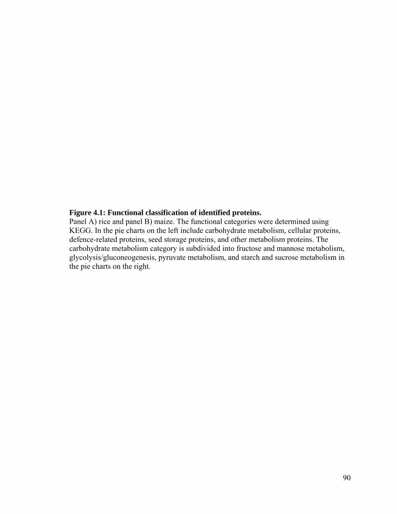
90
Figure 4.1: Functional classification of identified proteins. Panel A) rice and panel B) maize. The functional categories were determined using KEGG. In the pie charts on the left include carbohydrate metabolism, cellular proteins, defence-related proteins, seed storage proteins, and other metabolism proteins. The carbohydrate metabolism category is subdivided into fructose and mannose metabolism, glycolysis/gluconeogenesis, pyruvate metabolism, and starch and sucrose metabolism in the pie charts on the right.

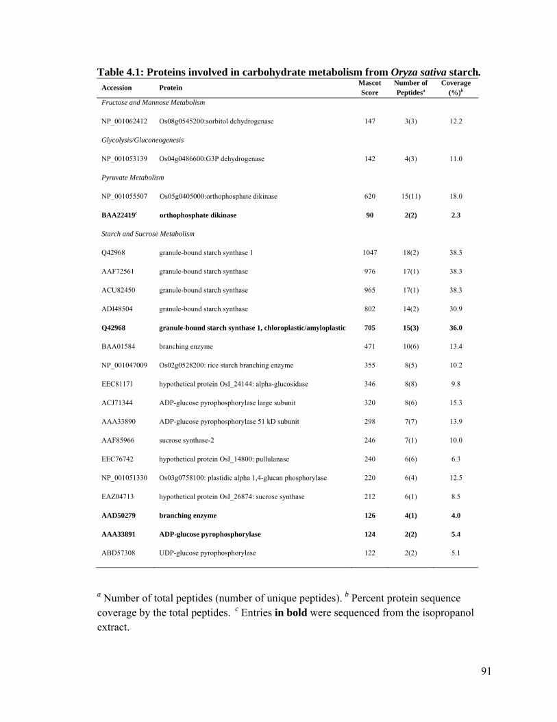
91
Table 4.1: Proteins involved in carbohydrate metabolism from Oryza sativa starch. Accession Protein
Mascot Score
Number of Peptidesa
Coverage (%)b
Fructose and Mannose Metabolism
NP_001062412 Os08g0545200:sorbitol dehydrogenase 147 3(3) 12.2
Glycolysis/Gluconeogenesis
NP_001053139 Os04g0486600:G3P dehydrogenase 142 4(3) 11.0
Pyruvate Metabolism
NP_001055507 Os05g0405000:orthophosphate dikinase 620 15(11) 18.0
BAA22419c orthophosphate dikinase 90 2(2) 2.3
Starch and Sucrose Metabolism
Q42968 granule-bound starch synthase 1 1047 18(2) 38.3
AAF72561 granule-bound starch synthase 976 17(1) 38.3
ACU82450 granule-bound starch synthase 965 17(1) 38.3
ADI48504 granule-bound starch synthase 802 14(2) 30.9
Q42968 granule-bound starch synthase 1, chloroplastic/amyloplastic 705 15(3) 36.0
BAA01584 branching enzyme 471 10(6) 13.4
NP_001047009 Os02g0528200: rice starch branching enzyme 355 8(5) 10.2
EEC81171 hypothetical protein OsI_24144: alpha-glucosidase 346 8(8) 9.8
ACJ71344 ADP-glucose pyrophosphorylase large subunit 320 8(6) 15.3
AAA33890 ADP-glucose pyrophosphorylase 51 kD subunit 298 7(7) 13.9
AAF85966 sucrose synthase-2 246 7(1) 10.0
EEC76742 hypothetical protein OsI_14800: pullulanase 240 6(6) 6.3
NP_001051330 Os03g0758100: plastidic alpha 1,4-glucan phosphorylase 220 6(4) 12.5
EAZ04713 hypothetical protein OsI_26874: sucrose synthase 212 6(1) 8.5
AAD50279 branching enzyme 126 4(1) 4.0
AAA33891 ADP-glucose pyrophosphorylase 124 2(2) 5.4
ABD57308 UDP-glucose pyrophosphorylase 122 2(2) 5.1
a Number of total peptides (number of unique peptides). b Percent protein sequence coverage by the total peptides. c Entries in bold were sequenced from the isopropanol extract.
92
dehydrogenase, glyceraldehyde phosphate, fructose-bisphosphate aldolase, and
orthophosphate dikinase.
There were five proteins identified in rice starch with a broad range of functions. These
proteins were termed cellular proteins, of which, three were in the aqueous fraction and
two were in the isopropanol fraction (Table 4.2). There were four proteins classified as
folding, sorting and degradation proteins, including the endosperm lumenal binding
protein, which was found in both aqueous and isopropanol fractions, heat shock protein
70, and protein disulfide-isomerase. There was one protein (elongation factor-1 alpha)
associated with translation.
The most abundant class of proteins in rice starch was the seed storage proteins. There
were 25 seed storage proteins identified in the rice starch samples (Table 4.3). Of these,
there were 21 glutelin, one globulin, and three prolamin proteins. There were 14 seed
storage proteins found in the aqueous fraction and 11 seed storage proteins in the
isopropanol fraction.
4.4.2 Starch granule associated proteins in maize
In the maize starch samples, peptides from 96 proteins were identified in the aqueous
fraction and 41 proteins were identified in the isopropanol fraction, yielding 137 proteins
in total. Of these 137 proteins, 109 were identified as plant-sourced, with the remaining

93
Table 4.2: Proteins involved in cellular processes from Oryza sativa starch. Accession Protein
Mascot Score
Number of Peptidesa
Coverage (%)b Molecular Functions
AAB63469 endosperm lumenal binding protein 707 12(7) 21.3 Folding, sorting and degradation
NP_001066486 Os12g0244100: heat shock 70 protein 178 4(2) 7.2 Folding, sorting and degradation
AAX85991 protein disulfide isomerase 142 4(4) 8.2 Folding, sorting and degradation
CAA34756c elongation factor-1 alpha 104 2(1) 5.0 Translation
AAA62325 endosperm lumenal binding protein 97 2(2) 3.8 Folding, sorting and degradation
a Number of total peptides (number of unique peptides). b Percent protein sequence coverage by the total peptides. c Entries in bold were sequenced from the isopropanol extract.

94
Table 4.3: Seed storage proteins from Oryza sativa starch.
Accession Protein Mascot Score
Number of Peptidesa
Coverage (%)b
EEC73106 hypothetical protein Osl_07091:glutelin 689 15(14) 42.3
NP_001046512 Os02g0268100:glutelin 665 12(7) 31.8
EEC72839c hypothetical protein OsI_06572:glutelin 625 13(1) 35.0
CAA33838 unnamed protein product:glutelin 567 13(1) 33.5
EAY75915 hypothetical protein Osl_03835:glutelin 565 12(4) 32.9
EEC72839 hypothetical protein OsI_06572:glutelin 556 12(7) 28.1
NP_001046512 Os02g0268100:glutelin 554 11(7) 24.6
EEC73106 hypothetical protein OsI_07091:glutelin 519 13(12) 38.7
BAA00462 pre-proglutelin 479 11(0) 26.7
EEC75527 hypothetical protein OsI_12139:glutelin 470 10(1) 19.0
EAY85201 hypothetical protein OsI_06564:glutelin 469 9(8) 19.0
EEC75527 hypothetical protein OsI_12139:glutelin 468 12(9) 24.2
1311273A hypothetical protein OsI_0656:glutelin 463 11(0) 26.7
CAA38211 glutelin 457 10(1) 25.4
BAA00462 prepro-glutelin 413 12(0) 33.1
1311273A glutelin 411 12(1) 33.1
AAO22140 glutelin precursor 382 7(5) 15.8
EAY85201 hypothetical protein OsI_06564:glutelin 339 9(2) 20.2
EAY75915 hypothetical protein OsI_03835:glutelin 337 10(3) 28.6
AAO22140 glutelin precursor 292 6(4) 15.4
CAA29507 glutelin 306 7(1) 39.4
BAA11129 prolamin 291 4(4) 49.0
BAA11129 prolamin 278 5(5) 53.0
CAA52764 11S globulin 138 3(1) 6.3
AAV43828 putative prolamin 7 123 2(2) 18.9
a Number of total peptides (number of unique peptides). b Percent protein sequence coverage by the total peptides. c Entries in bold were sequenced from the isopropanol extract.

95
28 proteins being attributed to a phylogenetically diverse range of organisms. The
molecular functions of the plant-sourced proteins were determined using KEGG, and the
percentage representation of the major classifications is shown in Figure 4.1B. The major
classifications include: carbohydrate metabolism-related proteins (50%), seed storage
proteins (16%), cellular proteins (12%), defence-related proteins (12%), and other
metabolism-related proteins (10%). Due to the high percentage representation of
carbohydrate metabolism-related proteins, this category was further divided into: starch
and sucrose metabolism (47%), glycolysis/gluconeogenesis (34%), pyruvate metabolism
(11%), and fructose and mannose metabolism (8%).
In contrast to the 34% in the rice starch samples, we identified 50% of the proteins in the
maize starch samples as being involved in carbohydrate metabolism. Maize starch
contained 18 proteins associated with starch and sucrose metabolism, including sucrose
synthase, granule-bound starch synthase, branching enzyme, ADP-glucose
pyrophosphorylase, UTP-glucose-1-phosphate uridylyltransferase, and glucose-1-
phosphate adenylyltransferase large subunit 1 (Table 4.4). While the maize starch
samples had only one protein, with three isoforms, involved in fructose and mannose
metabolism (sorbitol dehydrogenase), there were 13 proteins corresponding to
glycolysis/gluconeogenesis, including fructose-bisphosphate aldolase,
phosphoglucomutase, triosephosphate isomerase, glyceraldehyde-3-phosphate
dehydrogenase, 3-phosphoglycerate kinase, and brittle 1. Additionally, there were four
proteins found in the maize starch samples involved with pyruvate metabolism. These
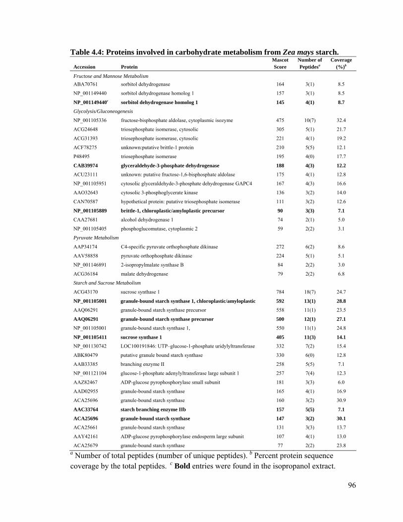
96
Table 4.4: Proteins involved in carbohydrate metabolism from Zea mays starch.
Accession Protein Mascot Score
Number of Peptidesa
Coverage (%)b
Fructose and Mannose Metabolism
ABA70761 sorbitol dehydrogenase 164 3(1) 8.5
NP_001149440 sorbitol dehydrogenase homolog 1 157 3(1) 8.5
NP_001149440c sorbitol dehydrogenase homolog 1 145 4(1) 8.7
Glycolysis/Gluconeogenesis
NP_001105336 fructose-bisphosphate aldolase, cytoplasmic isozyme 475 10(7) 32.4
ACG24648 triosephosphate isomerase, cytosolic 305 5(1) 21.7
ACG31393 triosephosphate isomerase, cytosolic 221 4(1) 19.2
ACF78275 unknown:putative brittle-1 protein 210 5(5) 12.1
P48495 triosephosphate isomerase 195 4(0) 17.7
CAB39974 glyceraldehyde-3-phosphate dehydrogenase 188 4(3) 12.2
ACU23111 unknown: putative fructose-1,6-bisphosphate aldolase 175 4(1) 12.8
NP_001105951 cytosolic glyceraldehyde-3-phosphate dehydrogenase GAPC4 167 4(3) 16.6
AAO32643 cytosolic 3-phosphoglycerate kinase 136 3(2) 14.0
CAN70587 hypothetical protein: putative triosephosphate isomerase 111 3(2) 12.6
NP_001105889 brittle-1, chloroplastic/amyloplastic precursor 90 3(3) 7.1
CAA27681 alcohol dehydrogenase 1 74 2(1) 5.0
NP_001105405 phosphoglucomutase, cytoplasmic 2 59 2(2) 3.1
Pyruvate Metabolism
AAP34174 C4-specific pyruvate orthophosphate dikinase 272 6(2) 8.6
AAV58858 pyruvate orthophosphate dikinase 224 5(1) 5.1
NP_001146891 2-isopropylmalate synthase B 84 2(2) 3.0
ACG36184 malate dehydrogenase 79 2(2) 6.8
Starch and Sucrose Metabolism
ACG43170 sucrose synthase 1 784 18(7) 24.7
NP_001105001 granule-bound starch synthase 1, chloroplastic/amyloplastic 592 13(1) 28.8
AAQ06291 granule-bound starch synthase precursor 558 11(1) 23.5
AAQ06291 granule-bound starch synthase precursor 500 12(1) 27.1
NP_001105001 granule-bound starch synthase 1, 550 11(1) 24.8
NP_001105411 sucrose synthase 1 405 11(3) 14.1
NP_001130742 LOC100191846: UTP–glucose-1-phosphate uridylyltransferase 332 7(2) 15.4
ABK80479 putative granule bound starch synthase 330 6(0) 12.8
AAB33385 branching enzyme II 258 5(5) 7.1
NP_001121104 glucose-1-phosphate adenylyltransferase large subunit 1 257 7(4) 12.3
AAZ82467 ADP-glucose pyrophosphorylase small subunit 181 3(3) 6.0
AAD02955 granule-bound starch synthase 165 4(1) 16.9
ACA25696 granule-bound starch synthase 160 3(2) 30.9
AAC33764 starch branching enzyme IIb 157 5(5) 7.1
ACA25696 granule-bound starch synthase 147 3(2) 30.1
ACA25661 granule-bound starch synthase 131 3(3) 13.7
AAY42161 ADP-glucose pyrophosphorylase endosperm large subunit 107 4(1) 13.0
ACA25679 granule-bound starch synthase 77 2(2) 23.8 a Number of total peptides (number of unique peptides). b Percent protein sequence coverage by the total peptides. c Bold entries were found in the isopropanol extract.

97
included pyruvate orthophosphate dikinase, 2-isopropylmalate synthase B, and malate
dehydrogenase.
We found nine cellular proteins in the maize starch sample (Table 4.5). There were three
proteins identified that were involved with folding, sorting and degradation. These
proteins included heat shock protein 70, protein disulfide-isomerase, and thioredoxin H-
type. We found one elongation factor, elongation factor 2-gamma 3, which was classified
as a translation-related protein. Additionally, we identified histone H4 and two proteins
with fasciclin domains associated with cell adhesion. The highest scoring cellular protein
in maize starch was the 14-3-3 protein associated with the cell cycle.
There were 12 seed storage proteins identified in the maize starch samples (Table 4.6). Of
these, there were four zein, three legumin, two globulin, one vicilin, and two glycinin G1
proteins. Of these 12 proteins, six were found in the aqueous fraction and six were found
in the isopropanol fraction.
4.5 Discussion
We have previously examined the starch granule proteome in hard and soft wheats
following very strict washing procedures (Wall et al., 2010a; Wall et al., 2010b). Here,
we have examined two different species, rice, with starch granules composed of 23.6%
amylose (Tan, 2003) and maize with starch granules composed of 23.1% amylose
(Uthumporn et al., 2010), whose granules have been processed in large-scale, commercial
environments. The protocols for commercial starch processing are optimized for high-
throughput processing at low cost, rather than the absolute removal of all non-starch
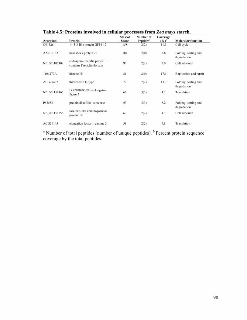
98
Table 4.5: Proteins involved in cellular processes from Zea mays starch.
a Number of total peptides (number of unique peptides). b Percent protein sequence coverage by the total peptides.
Accession Protein Mascot Score
Number of Peptidesa
Coverage (%)b Molecular function
Q01526 14-3-3-like protein GF14-12 158 2(2) 11.1 Cell cycle
AAC36132 heat shock protein 70 104 2(0) 3.8 Folding, sorting and degradation
NP_001105408 endosperm specific protein 1 – contains Fasciclin domain
97 2(2) 7.0 Cell adhesion
1101277A histone H4 81 2(0) 17.6 Replication and repair
ACG29437 thioredoxin H-type 77 2(2) 13.9 Folding, sorting and degradation
NP_001151465 LOC100285098 – elongation factor 2
66 3(3) 4.2 Translation
P52588 protein disulfide-isomerase 63 3(3) 8.2 Folding, sorting and degradation
NP_001151356 fasciclin-like arabinogalactan protein 10
62 2(2) 4.7 Cell adhesion
ACG36145 elongation factor 1-gamma 3 58 2(2) 4.8 Translation

99
Table 4.6: Seed storage proteins from Zea mays starch.
a Number of total peptides (number of unique peptides). b Percent protein sequence coverage by the total peptides. c Entries in bold were sequenced from the isopropanol extract.
Accession Protein Mascot Score
Number of Peptidesa
Coverage (%)b
AAL16994 legumin 1 213 4(1) 10.6
1802402A globulin 2 209 4(4) 13.8
CAA41809 vicilin-like embryo storage protein 200 4(2) 9.1
P04776 glycinin G1 190 4(4) 9.1
NP_001104865 legumin 1 180 4(1) 10.6
ABV71978c z1D alpha zein protein 179 3(1) 21.0
ABV71937 z1A alpha zein protein 155 3(1) 23.8
AAL16994 legumin 1 128 3(3) 52.6
ABV71937 z1A alpha zein protein 110 2(2) 10.9
1107201E zein M36M361 108 2(2) 6.4
AAC31468 globulin 1 80 2(2) 7.4
P04776 glycinin G1 64 2(2) 3.8
100
fractions (Tester et al., 2007). We were therefore interested in determining whether direct
trypsinolysis could be used to access and identify the proteins that remain associated with
the starch granules following commercial-scale processing (workflow shown in Figure
4.2A). In order to identify hydrophobic peptides on the starch granule surface, we used an
isopropanol extraction protocol adapted from Morris et al. (1994). While this protocol
was optimized for the isolation of the wheat SGAP puroindoline, an amphipathic protein,
we were able to identify certain peptides that remained associated to the SGS following
commercial starch processing and subsequent in-lab washing. While it is possible that
these associations are artifactual in nature, it is feasible that they are physiologically
important and are therefore worthy of further study.
4.5.1 Starch granule associated proteins
Thoroughly water washing starch granules from cereal seeds in a laboratory setting can
reduce the total protein content to 0.25% (Skerritt et al., 1990), whereas commercial
starches have protein contents of approximately 0.4% (Appelqvist and Debet, 1997). The
endosperm-sourced proteins of cereal grains can be divided into three distinct categories:
1) seed storage proteins, 2) cellular or amyloplastic proteins, or 3) starch granule
associated proteins (SGAPs). The SGAPs are defined as proteins that are located at the
surface and/or interior of starch granules (Baldwin, 2001). The most abundant SGAP
family, granule-bound starch synthase (GBSS), comprises up to 85% of the total internal
SGAPs (Echt and Schwartz, 1981), and is involved in starch synthesis through the
addition of uridine diphosphate glucose or adenine diphosphate glucose to growing
glucan polymers (Baba et al., 1987; Denyer et al., 1996; Hanashiro et al., 2008). Due to

101
the abundance of GBSS, it is appropriate that we found five different GBSS proteins in
rice starch and an additional ten GBSS proteins in maize starch.
4.5.2 Starch biosynthetic enzymes
The enzymes involved in starch metabolism are well documented. For a comprehensive
review, see Tetlow, 2011. Both rice and maize starch samples had a high percentage
representation (34% and 50%, respectively) of proteins involved with carbohydrate
metabolism (Figures 4.1A and 4.1B). This is consistent with previous studies that
examined amyloplast and starch granule-associated proteomes in Triticum aestivum
(Andon et al., 2002) and Solanum tuberosum (Denyer et al., 1996). Many of the proteins
in the starch biosynthetic pathway (Keeling and Myers, 2010) were identified in our study
(Tables 4.1 and 4.4). These proteins have been assembled in Figure 4.2B to demonstrate
how much of the biosynthetic machinery of proteinaceous origin is still present in
commercially processed starches. In the endosperm of monocots, other than GBSS,
soluble starch synthase, and the starch branching and de-branching enzymes, and a
plastidial form of ADP-glucose pyrophosphorylase (AGPase), starch biosynthetic
enzymes are located in the cytoplasm (Tetlow, 2011). It is likely that during the
dessication of the endosperm, these cytoplasmic enzymes became associated with the
remnants of the amyloplast membrane or to the hydrophobic surface of the starch
granules (Wall et al., 2010a; Wall et al., 2010b).

102
4.5.3 Orthophosphate dikinase
It has been proposed that the composition of the storage protein fractions and the starch-
protein balance is controlled, in part, by orthophosphate dikinase (PPDK) (Mechin et al.,
2007). Given that starch turnover and carbon allocation are integral in coordinating
metabolism with growth and that starch content is negatively correlated with biomass
(Sulpice et al., 2009), PPDK may also be involved in starch-biomass balance. The
classical role of PPDK in both C3 and C4 plants is in catalyzing the reversible reaction of
pyruvate, ATP and phosphate to phosphoenolpyruvate, AMP and diphosphate (Chastain
and Chollet, 2003). In maize, the expression of PPDK is upregulated at 21 days after
pollination (DAP) (Mechin et al., 2007), while the expression of PPDK in rice is highest
from 5 – 15 DAP, after which, PPDK is rapidly degraded or inactivated through
phosphorylation. This pool of inactivated PPDK is present in mature seeds, and may play
a role in developmental processes during seed germination (Chastain et al., 2006). Future
analysis of the starch granule-associated phosphoproteome will allow for the
determination of phosphorylation levels of PPDK in these starch samples.
4.5.4 Starch mobilization
To utilize their stored carbon reserves, plants must be able to degrade their starch
granules to oligosaccharides and monosaccharides. However, of the 59 proteins identified
in our survey as being involved with carbohydrate metabolism, only two were involved in
the mobilization of starch (Table 4.1). Those enzymes, plastidic -1,4-glucan
-phosphorylase and -glucosidase were both found in the rice starch samples.

103
Glucosidase is involved in the hydrolysis of α-1,4 glucosidic linkages (Smith et al., 2005),
while plastidic -1,4-glucan phosphorylase acts by converting the terminal glycosyl units
of the non-reducing ends of linear oligosaccharide glucan chains to glucose 1-phosphate
(Duwenig et al., 1997). While the well-studied -amylase is involved in the direct
degradation of the polysaccharides that make up starch granules, -glucosidase is
generally believed to preferentially hydrolyze oligosaccharides (Hanes, 1932). There is,
however, evidence that -glucosidase can act synergistically with -amylase to enhance
granule degradation in barley (Sun and Henson, 1990). While -glucosidase expression is
high during seed development in barley, the expression decreases to basal levels seeds
upon maturity (Fincher, 1989). Our ability to detect these starch mobilizing enzymes in
only the rice samples may indicate that some of the rice kernels were harvested prior to
maturity.
4.5.5 14-3-3 proteins
The 14-3-3 protein family consists of 28–33-kDa acidic polypeptides that are
ubiquitously expressed in eukaryotes (Morrison, 2009). The 14-3-3 proteins form homo-
or hetero-dimers that are able to bind a broad range of ligands due to the specific
phospho-serine/phospho-threonine binding activity of these 14-3-3 dimers (Fu et al.,
2000). However, not all 14-3-3 binding is phosphorylation dependent (Aitken, 2006). The
ligands with which the dimers of 14-3-3 have been demonstrated to interact include
transcription factors, cytoskeletal proteins, signaling molecules, and biosynthetic
enzymes, among others (Morrison, 2009). While it has been hypothesized that the
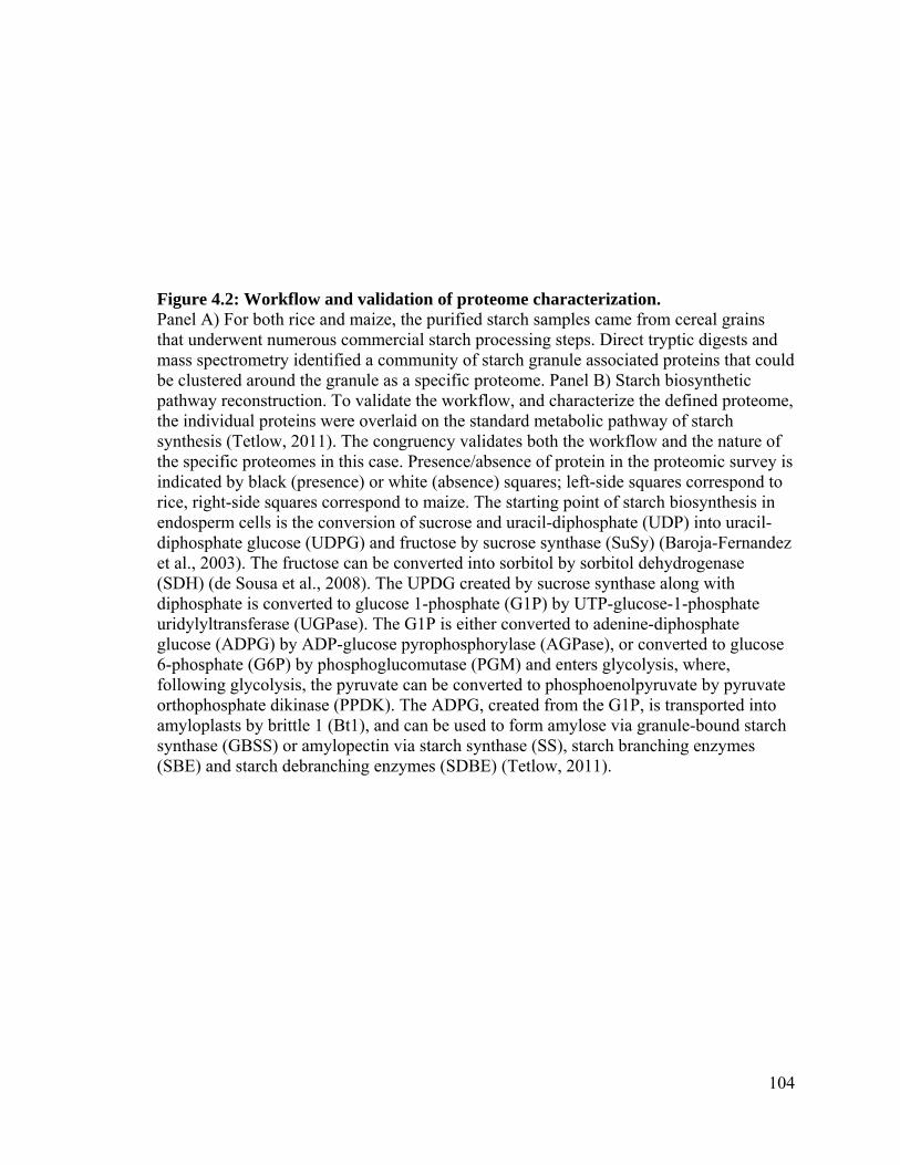
104
Figure 4.2: Workflow and validation of proteome characterization. Panel A) For both rice and maize, the purified starch samples came from cereal grains that underwent numerous commercial starch processing steps. Direct tryptic digests and mass spectrometry identified a community of starch granule associated proteins that could be clustered around the granule as a specific proteome. Panel B) Starch biosynthetic pathway reconstruction. To validate the workflow, and characterize the defined proteome, the individual proteins were overlaid on the standard metabolic pathway of starch synthesis (Tetlow, 2011). The congruency validates both the workflow and the nature of the specific proteomes in this case. Presence/absence of protein in the proteomic survey is indicated by black (presence) or white (absence) squares; left-side squares correspond to rice, right-side squares correspond to maize. The starting point of starch biosynthesis in endosperm cells is the conversion of sucrose and uracil-diphosphate (UDP) into uracil-diphosphate glucose (UDPG) and fructose by sucrose synthase (SuSy) (Baroja-Fernandez et al., 2003). The fructose can be converted into sorbitol by sorbitol dehydrogenase (SDH) (de Sousa et al., 2008). The UPDG created by sucrose synthase along with diphosphate is converted to glucose 1-phosphate (G1P) by UTP-glucose-1-phosphate uridylyltransferase (UGPase). The G1P is either converted to adenine-diphosphate glucose (ADPG) by ADP-glucose pyrophosphorylase (AGPase), or converted to glucose 6-phosphate (G6P) by phosphoglucomutase (PGM) and enters glycolysis, where, following glycolysis, the pyruvate can be converted to phosphoenolpyruvate by pyruvate orthophosphate dikinase (PPDK). The ADPG, created from the G1P, is transported into amyloplasts by brittle 1 (Bt1), and can be used to form amylose via granule-bound starch synthase (GBSS) or amylopectin via starch synthase (SS), starch branching enzymes (SBE) and starch debranching enzymes (SDBE) (Tetlow, 2011).


105
assembly of the enzyme complexes responsible for starch biosynthesis is mediated by the
phosphorylation-dependent binding of 14-3-3 (Tetlow, 2011), there is no record of 14-3-3
being found in mature seeds. Previously, 14-3-3 isoforms have been found in maize
starch granules isolated from immature pollen (Datta et al., 2002), as well as in
Arabidopsis (Sehnke et al., 2001) and barley (Alexander and Morris, 2006). Isoforms of
GBSS, soluble starch synthase, and starch branching enzyme, as well as the starch-
degrading enzyme -amylase can be bound by barley grain 14-3-3 proteins (Alexander
and Morris, 2006). The particular isoform of 14-3-3 identified in this study is GF14-12
(Table 4.5), which was first cloned in 1994 and has been shown to form DNA/protein
complexes (de Vetten and Ferl, 1994). This discovery of a 14-3-3 protein in mature seeds
supports the role of 14-3-3 proteins in the assembly of starch biosynthetic complexes.
4.5.6 Storage proteins
The protein content of cereal endosperm can be divided into four categories based on
their solubility in solvents. These Osborne fractions include albumins, soluble in water;
globulins, soluble in dilute salt solutions; prolamins, soluble in alcohol/water solutions;
and glutelins, soluble in weak acids/bases (Osborne, 1908). Rice endosperm is 7-8%
protein (Puchongkavarin et al., 2005), which can be divided into 3.8–8.8% albumin, 9.6–
10.8% globulin, 2.6–3.3% prolamin and 66–78% glutelin (Cagampan et al., 1966). In
maize, the prolamins, represented by the zein family, are more prevalent, while the
glutelins are less predominant, depending on the variety of maize used in the production
of the maize starch samples. The percentage representation of maize endosperm proteins
is usually 4-8% albumin, 3-4% globulin, 47-55% prolamin, and 38-45% glutelin (Alais

106
and Linden, 1991). The protein bodies in rice endosperm are tightly associated with the
starch granule surface (Tanaka et al., 1980), and are difficult to remove during starch
granule purification, even with extended soaking in solutions of dilute base (Wang and
Wang, 2001). The distribution of the seed storage proteins in the rice starch sample
favored glutelin, with 84% of the seed storage proteins being identified as glutelins (Table
4.3). Moreover, we found 21 different glutelins associated with the commercial rice
starch granule preparations. Given that the current genome sequence database (GenBank)
of the rice cultivar Nipponbare contains 22 members of the rice glutelin gene family, it is
remarkable that such a high proportion of these are clustered close to the rice granule
surface and had not been removed by commercial starch processing and purification. On
the other hand, the maize starch samples had an intriguingly high representation of
globulins (25%) compared to the prolamins (zeins) (33%) (Table 4.6). This divergence
from the expected ratios may be an artefact of the commercial starch purification
conditions employed. Future experiments may allow for the determination of the
sequence of storage protein isoforms with strong starch-adhering properties. These
isoforms could potentially be used to breed varieties of cereals with “customizable” starch
adherence that impacts bread-making characteristics or other industrial attributes.
4.6 Conclusions
We were able to look at molecular snapshots of the endosperm proteins that remained
associated to starch granules following the commercial processing of starch from two
different species, both with 23% amylose content. We were, consequently, able to glean
insight into the physico-chemical properties of the protein remnants that make up the
107
intra-granular packing or protein matrix of the mature seeds of two commercially
important plants: rice and maize. These proteinaceous remnants could affect the yield of
pure starch granules during wet milling and subsequent purification in addition to
affecting the experiments in which commercially prepared starches are used as reference
materials. Furthermore, the differences inferred between the intracellular architecture and
molecular dynamics of rice (C3 plant) and maize (C4 plant) should be instructive to those
engineering the maize C4 pathway into rice (Hibberd et al., 2008). Finally, we also found
markers of seed maturity in the rice starch samples. These markers indicate that both
immature and mature seeds were used in the production of the rice starch samples.

108
Chapter 5 Commercially produced rice and maize starches contain non-host proteins, as shown by mass spectrometry
Koziol AG, Marquez BK, Huebsch MP, Smith JC, Altosaar, I
Published in: Journal of Cereal Science, DOI: 10.1094/CCHEM-04-12-0043-N
Contribution of authors:
AGK contributed to the study design, performed experiments except LC-MS/MS,
analyzed data, wrote the manuscript, and participated in editing the manuscript. BKM
aided in experiments, analyzing data, writing the manuscript, and editing. MPH
performed LC-MS/MS. JS participated in analyzing data and editing. IA participated in
study design, data analysis, and editing.

109
5.1 Abstract
To evaluate the presence of contaminating, non-host proteins in commercially prepared
rice and maize starch samples, we initiated a direct sequencing mass spectrometric
proteomics survey. We discovered non-host proteins from a variety of species, including
Phytophthora cinnamomi, Homarus americanus, and Ovis aries. Our documentation of
H. americanus proteins in these starch samples may have food safety implications with
regards to shellfish allergies. We hypothesize that these proteins were introduced to the
starch samples via process wash water used in the milling and deproteination steps in the
commercial preparation of the starches analyzed. The introduction of non-host proteins
during commercial processing of starch samples that are used routinely in analytical
studies indicates that these studies are using impure materials. Therefore, further study
and documentation of the starch samples is required to ensure that all components of the
samples are properly catalogued.
110
5.2 Introduction
In plants, glucose molecules are linked for many reasons, including reducing the osmotic
potential of free sugars, sequestering and storing glucose units for diurnal/season
metabolic needs, and providing dense granules for gravitropism of statoliths (Ball and
Morell, 2003; Caspar and Pickard, 1989; Tetlow, 2011). The linked chains of glucose
occur as two distinct polymers: amylose, which is primarily α(14) linked glucosyl
units, with occasional α(16) branches, and amylopectin, while still predominantly
α(14) linked glucosyl units, contains a much higher proportion of α(16) branches
compared to amylose (Grimaud et al., 2008). Together, amylose and amylopectin are
assembled into starch granules (SGs). The SGs in storage organs, such as the endosperm
of grains or the storage parenchyma cells of tubers, act as reservoirs for the carbohydrates
synthesized during photosynthesis, and have internal semi-crystalline growth rings that
are the result of periodic differences in the rate of glucose polymerization during starch
synthesis (Perez and Bertoft, 2010).
While the biochemical dissection of SGs in planta has progressed at a fine molecular
scale (Graf and Smith, 2011; Yandeau-Nelson et al., 2011), the concomitant analytics of
the molecular toolkit for furthering starch-based biofuels or foods has lagged (Shi and
Gao, 2011; Shu et al., 2006). Starch extraction on an industrial scale, usually by wet
milling, involves several steps, including the removal of the pericarp, germ, fibres, and
protein matrix, as well as the purification and drying of the starch granules (Sun, 2005).
Generally, wet milling includes a steeping step to allow for easy separation of the germ
and to loosen the protein matrix within the endosperm. The steeping process involves
fermentation by lactic acid bacteria and yeast (Cabrales et al., 2006; Singh et al., 1999).

111
The seeds are ground gently and the germ is separated from the endosperm based on
differences in density in hydrocyclones. The endosperm slurry is ground again, and
passed through a series of fine screens to remove fibres and the protein matrix. Starch is
purified from any remaining protein based on density in a series of cyclones. The isolated
starch granules are then dried prior to being packaged as dry powder and transported
(BeMiller and Whistler, 2009).
Commercially extracted starch is ubiquitously present in both industrial and culinary
applications. The industrial applications include papermaking, adhesives, bioplastics,
biofuel (Kaur et al., 2012; Pitak and Rakshit, 2011; Tasic and Veljkovic, 2011), while
modified starches and starch sugars are important food additives (BeMiller, 2011). We
have previously documented the remnants of pathogenic microbial proteins on the surface
of starch granules in lab-scale purified wheat starch; however, we did not observe
proteins from organisms from soil- or water-dwelling organisms (Wall et al., 2010). In
our recent study to characterize the proteomes of commercially prepared reference
starches from leading biochemical reagent suppliers, we were able to document many
host-sourced proteins that remain in starch samples following industrial processing
(Koziol et al., 2012). In addition to these host proteins, we discovered proteins from a
broad taxonomic range of organisms. It has previously been stated that the purity of the
water used to process the starch during commercial purification can contaminate starch
samples, and affects the end quality of the starch (Sun, 2005). These findings serve as a
caveat for all aspects of biomass research as well as food science and food processing
where such biochemical reagents have been assumed to be pure starch. We therefore
112
hypothesized that the non-host proteins present in the commercial starch preparations
were sourced from the water used during the extraction of the starch granules.
5.3 Methods
Methods were as published by Koziol et al (2012). Briefly, rice (cat. no.S7260-1KG, lot
no. 107K0052) and maize (cat. no.S4126-2KG, lot no. 015K0144) starches were obtained
from Sigma-Aldrich (St. Louis, MO). Prior to sampling, reagent bottles were gently
agitated, and four separate 50 mg aliquots from each bottle were removed, with gentle
shaking between each sampling step.
5.3.1 Protein extraction
The sampled starches were suspended in 350 μL of ddH2O and incubated for 30 min with
gentle agitation. Ammonium bicarbonate was added to the samples to a final
concentration of 100 mM. To proteolytically digest the proteins present in the samples, 5
μg of 1 μg/μL trypsin (T6567, Sigma-Aldrich) in 1 mM HCl was added. Samples were
incubated overnight at 37ºC with gentle agitation. To collect the peptides in the
supernatants, samples were centrifuged (Heraeus Sepatech Biofuge A 1302; 18 000 x g, 1
min) and the supernatants were transferred into fresh tubes. Peptides in the supernatants
were vacuum-dried (Speed Vac Concentrator SVC 100H; Savant Instruments, Inc.
Hicksville, NY) for 3 hours. Starch pellets were washed five times in distilled water, were
suspended in protein extraction buffer (50% isopropanol (v/v), 50 mM NaCl), and were
gently mixed for 45 min. Samples were centrifuged (2 500 x g, 5 min). Supernatants were
transferred to new tubes. Peptides in the supernatants were vacuum-dried as described

113
above. Starch pellets were discarded. To purify and concentrate the protein fractions,
vacuum-dried peptides were resuspended in 40 μL ddH2O. Peptide suspensions were
purified using ZipTips (Millipore, Bedford MA, cat. no. ZTC18M096). The
manufacturer’s protocol was repeated eight times per sample. Resulting peptide solutions
were vacuum-dried as described above.
5.3.2 Protein separation
Peptide separation was accomplished using a 7 cm by 200 µm inner diameter trap column
coupled to a 5 cm by 75 µm Picofrit nanoelectrospray tip (New Objective, Woburn, MA)
packed with 5 μm Magic C18AQ resin (Michrom Bioresources, Auburn, CA). Using a tee
union prior to the columns, the flow from an HP 1090 HPLC (Agilent Technologies,
Santa Clara, CA) was split to pass the following gradient of acetonitrile containing 0.1%
formic acid at a flow rate of approximately 250 nL/min: 0 min 2%, 3 min 5%, 8 min 10%,
70 min 35%, 75 min 80%, 83.5 min 100% (wash step), 84 min 2%, 90 min 2% (re-
equilibration steps). Eluted peptides were analyzed using a QSTAR XL QqTOF mass
spectrometer (AB Sciex, Foster City, CA) operating in information-dependent acquisition
mode. Mass analysis included a 1 s survey scan followed by two 2 s and two 3 s tandem
mass spectrometric scans on the most intense peaks in the spectrum for each cycle.
Masses could be sequenced twice before being added to an exclusion list for 90 s.
Tandem mass spectrometric data were interpreted using Mascot software (version 2.3)
(Matrixscience Ltd., Boston, MA) using a custom database containing all proteins from
the 20100704 comprehensive National Center for Biotechnology Information non-
redundant (NCBInr) protein database (11,365,658 sequences; 3,876,535,693 residues).
One missed cleavage was allowed, and mass tolerances were set to ±50 ppm for peptides

114
and ±0.2 Da for fragment ion spectra. Oxidation of methionine residues was selected as a
variable modification, while no fixed modifications were selected. An automated decoy
search was performed, and the rate of false positives was approximately 1%. Each match
from the database was verified manually and spectra were only used if at least three
successive y- or b-ions were present. Only proteins with two or more peptide-based
identifications with Mascot scores ≥ 55 (p < 0.05) were included to decrease the chance
of matches occurring due to random events.
5.4 Results and Discussion
To better understand the source and nature of the proteins present in commercial starch
samples, our approach was applied to both commercially prepared rice and maize
starches. These commercial starches have been recently characterized at the level of the
host plant proteome (Koziol et al., 2012), but proteins of non-plant origin had not been
investigated. Surprisingly, three non-host proteins were identified in the rice samples, and
three different non-host proteins were identified in the maize samples (Tables 5.1 and
5.2). Mascot and manual NCBI BLAST searches revealed that these non-host proteins
were of microbial and animal origin. The prevalence of the non-host proteins in the
samples was approximately 5% calculated by occurrence in Mascot hit tables (data not
shown) (Koziol et al 2012). Of the non-host proteins identified, only one (glucose
regulated protein/BiP - heat shock protein 70-like) contains a domain that binds
carbohydrates (Di Luccio et al., 2007). It is therefore presumed that the other proteins
bound non-specifically to the starch granules (Nelson Jr. and Lebrun, 1956).
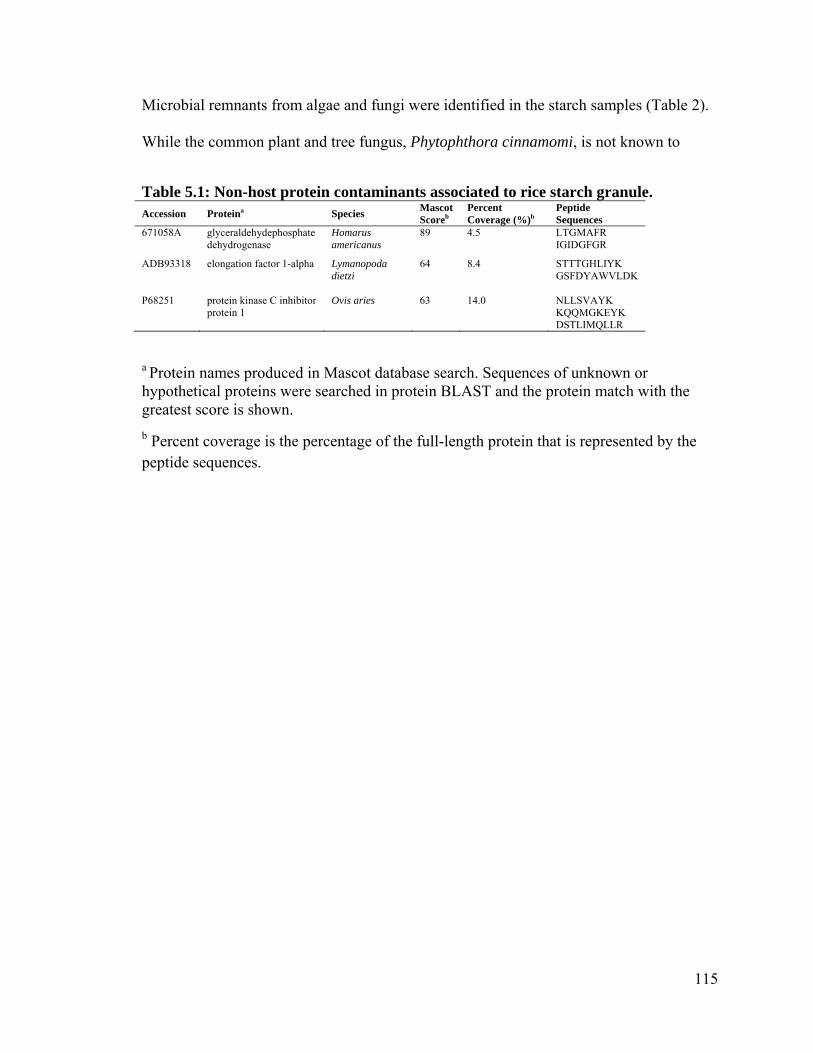
115
Microbial remnants from algae and fungi were identified in the starch samples (Table 2).
While the common plant and tree fungus, Phytophthora cinnamomi, is not known to
Table 5.1: Non-host protein contaminants associated to rice starch granule. Accession Proteina Species
Mascot Scoreb
Percent Coverage (%)b
Peptide Sequences
671058A glyceraldehydephosphate dehydrogenase
Homarus americanus
89 4.5 LTGMAFR IGIDGFGR
ADB93318 elongation factor 1-alpha Lymanopoda dietzi
64 8.4 STTTGHLIYK GSFDYAWVLDK
P68251 protein kinase C inhibitor protein 1
Ovis aries 63 14.0 NLLSVAYK KQQMGKEYK DSTLIMQLLR
a Protein names produced in Mascot database search. Sequences of unknown or hypothetical proteins were searched in protein BLAST and the protein match with the greatest score is shown.
b Percent coverage is the percentage of the full-length protein that is represented by the peptide sequences.

116
target maize, it causes root rot, stem cankers and crown rot in ornamental and forest
species such as cranberry, pear and pine (dos Santos et al., 2011; Hardham, 2005). Being
a water mould, it is common for Phytophthora species to be spread through cereal crops
during irrigation (Hong and Moorman, 2005), allowing for infection of many plants in the
same field. Alternatively, P. cinnamomi may have been introduced to the starch samples
during starch processing. Common steps in wet-milling of cereal grains such as sodium
dioxide steeping and lactic acid fermentation may also provide optimal growing
conditions for thiophilic and acidophilic bacteria that could potentially contaminate starch
samples (Sun, 2005). Such contaminants in wheat starch preparations are known to
include microbial enzymes (Wall et al., 2010), so these current results underscore the
need to be cautious when using such starch preparations in analytical studies, especially
in biomass or biomedical studies, where starch-protein interactions are being queried.
Non-microbial contaminants were also found in the commercial starch samples of rice
and maize. These proteins are listed among the other contaminants in Tables 1 and 2.
Using Mascot and BLAST searches of the non-redundant NCBI database, the
contaminating proteins were found to originate from a variety of species, including
Homarus americanus, Lymanopoda dietzi, and Ovis aries. The proteins found from these
species are conserved across the animal kingdom; elongation factor 1-alpha,
glyceraldehyde phosphate dehydrogenase, protein kinase C inhibitor, and skeletal actin.
The purity of commercial starch is highly dependent on the quality of water used in the
processing streams removing the bran and storage components of the starting flour
feedstock materials. The unexpected presence of proteins from non-host organisms in the
starch samples suggests that cross-contamination between water sources occurred in the

117
Table 5.2: Non-host proteins identified in maize starch samples.
a Protein names produced in Mascot database search. Sequences of unknown or hypothetical proteins were searched in protein BLAST and the protein match with the greatest score is shown.
b Percent coverage is the percentage of the full-length protein that is represented by the peptide sequences.
Accession Proteina Species Mascot Score
Percent Coverage (%)b
Peptide Sequences
CAA53369 glucose regulated protein /BiP - heat shock protein 70-like
Phytophthora cinnamomi 153 5.3 LSQEEIDR VEIIANDQGNR IINEPTAAAIAYGIDK
ACI23566 skeletal muscle actin 3 Homarus americanus 75 9.3
AVFPSIVGR AGFAGDDAPR EEYDESGPGIVHRKCF
BAE48223 heat shock protein 70 Chlorella pyrenoidosa 55 4.2 YKAEDETHRAR IINEPTAAAIAYGLDK
118
starch mills. As Chlorella pyrenoidosa is a fresh water alga (Koziol et al., 2007), it was
likely introduced to the starch samples by the water used in the starch processing.
The ease of starch contamination during commercial processing is disconcerting,
especially since starch is a consumable product. It is therefore possible that a subset of the
contaminants could induce allergic reactions in susceptible individuals. It is the protein
component of foodstuffs that is commonly responsible for the development of food
allergies (Ramesh, 2008). Recently, the prevalence of shellfish allergies in American
children was reported as 17.2% (Gupta et al., 2011). Allergies to shellfish are caused by
the protein tropomyosin (Ramesh, 2008). While we did not observe tropomyosin in the
starch samples, we documented proteins attributed to Homarus americanus (American
lobster) in both the rice and maize starch samples. With this discovery of H. americanus
proteins in commercial starch samples, a risk of triggering shellfish allergies has been
identified. It may, therefore, be prudent to screen industrially-extracted starches more
rigorously for the presence of any potential allergens.
5.5 Conclusion
We have analyzed commercially prepared starches of two agriculturally important crop
species: rice and maize by sampling the starch granule associated proteins via water
washing and isopropanol stripping followed by direct sequencing tandem mass
spectrometry. Unexpectedly, we identified non-host proteins, which included microbial
remnants and non-microbial contaminants on the surface of starch granules of both
species. Six non-host proteins were identified in the starch samples. More importantly,

119
the results demonstrated that this methodology can be used to ensure that potential
allergenic proteins are not present in starch samples to be consumed by animals.

120
Chapter 6 General Discussion of Major Findings

121
6.1 General Discussion of Chapter 2
The work in this thesis combined data from 2D-electrophoresis, immunoblotting , N-
terminal sequencing and MS/MS sequencing of peptides resulting from the
endoproteolytic cleavage of salt-soluble globulin-3 in wheat flour preparation enriched in
embryo tissue. The results show that the globulin-3 family is subjected to post-
translational processing and accumulates in the form of a set of cleavage products.
Previous work that described this protein family was performed on the hexaploid wheat
cultivar Glenlea (Loit et al., 2009), while the work performed here was on cultivar AC
Barrie, the cultivar that was used in the experiments that discovered the putative link
between dietary intake of Glo-3A and the development of type 1 diabetes (MacFarlane et
al., 2003). Immunoblot analysis of proteins from both embryo and endosperm tissues
revealed that the Glo-3 protein family is expressed predominantly in the embryo with
very low expression levels in the endosperm (Figure 2.2). Sequencing protein spots
extracted from 2D PAGE gels of salt-soluble embryo proteins using mass spectrometry
allowed for the development of a model describing the post-translational partial
endoproteolytic cleavage of the preproglobulin-3 family (Figure 2.3).
6.1.1 Glo-3 expression pattern and food safety
Our findings that demonstrated that the expression of Glo-3 was predominantly in the
embryo confirms immunolocalization data of Glo-3 in Glenlea (Loit et al., 2009) and
therefore demonstrates that Glo-3 shares similar expression patterns to 7S globulins in
rice, maize and other cereals (Burgess and Shewry, 1986; Sun et al., 1996; Thijssen et al.,

122
1996). Wheat gluten, first isolated and named by Bartolomeo Beccari in Bologna in 1745
(Beccari, 1745), is today still a generic term that describes a mixture of gliadins and
glutelins; seed storage proteins expressed in the endosperm, but not the embryo (van
Herpen et al., 2008). However, Glo-3 can be present at very low levels in wheat gluten
(MacFarlane et al., 2003); as commercial gluten is often made from whole wheat, which
consists of the endosperm and embryo, as well as the aleurone layer. To study the
potential diabetogenic effects of Glo-3, an investigation is required that compares wheat
gluten that is prepared exclusively from endosperm and therefore does not contain Glo-3
to gluten that is prepared from whole wheat. Stripping or milling off the outer layers of
wheat seeds has been demonstrated to reduce the sensitivity of predisposed individuals to
wheat flour (Handoyo et al., 2008), as these polished seeds contained lower levels of
allergenic proteins. Alternatively, wheat could be genetically transformed to express
lower levels of the globulin-3 protein family. A Glo-3 knockdown/knockout line of wheat
would allow for increased precision in studying the diabetogenic effects of Glo-3. Using
gluten prepared solely from endosperm compared to the entire wheat seed will not only
exclude Glo-3, but also other embryo/aleurone layer specific proteins, making it more
difficult to definitively prove a role for Glo-3 in the development in T1D. Knockout lines
should be identical to the parental lines, with the exception of Glo-3 expression. Maize
lines that are Glb1 and Glb2 null have been shown to be able to germinate normally
(Kriz, 1989), indicating that knocking out the globulin family is not a lethal mutation.
123
6.1.2 Post-translational endoproteolytic cleavage of Glo-3
The endoproteolytic processing of some wheat flour protein families has been
investigated using N-terminal sequencing (Singh et al., 2001) and mass spectrometry
(Dupont et al., 2011), but a concentrated study of the processing of Glo-3 specifically has
not been reported before. This present thesis used multiple characteristics of protein spots
to determine the location of processing events that occurred. The pI and Mr of the spots,
the location of the sequenced peptides within the protein sequence, and the N-terminal
sequence data (when available) were all taken into account when assembling the
processing model (Figure 2.3). As was demonstrated in Figures 2.2 and 2.3, and in
agreement with the literature, the endoproteolytic processing of the Glo-3 protein is
partial. It is still unknown how the proteins are prevented from not being completely
processed at every protease recognition site; though it has been determined that cleavage
is not required for proper folding, transport and storage of these proteins (Heck et al.,
1993).
With a better understanding of the processing of the Glo-3 protein family, it is now
possible to study the diabetogenic effects of the individual polypeptides generated from
the cleavage of proglobulin. The genetic engineering of wheat is now common practice,
although the 200 odd traits modified to date are mostly agronomic. Wheat can therefore
also be bred or engineered to lack certain protease recognition sites or domains within
Glo-3. The wheat lacking either certain domains or cleavage sites can then be used in
animal feeding trials in an attempt to locate a specific region within the protein that
displays increased diabetogenic activity. Given that this type of engineering already

124
promises to benefit celiac patients (Gil-Humanes et al., 2010), the same effort should now
be expended for a much more expensive disease, T1D.
6.2 General Discussion of Chapter 3
This work has increased the understanding of the protein expression by pathogen and host
in water-treated or Fusarium graminearum-infected maize kernels. Previous studies have
investigated host-F. graminearum interactions in actively growing kernels hours or days
after infection (Mohammadi et al., 2011; Zhou et al., 2006), but I was interested in the
proteins present in the host and pathogen proteomes following the desiccation of mature
kernels under field conditions. Our group has previously documented fungal proteins
present in healthy, mature wheat seeds (Wall et al., 2010), I therefore decided to extend
these investigations by sequencing the proteome of healthy, mature maize seeds, as well
as maize seeds with a heavy pathogen load to determine the number and nature of
pathogen and host proteins present under these two conditions. In healthy plants, we
found no evidence of fungal proteins within the seeds. However, we determined that
fungal proteins are present in both the desiccated fungal remnants on the surface of
infected seeds as well as within the seeds’ tissues.
6.2.1 Proteins in desiccated fungal remnants
It was intriguing that we only discovered 18 proteins in the fungal remnants despite the
fact that silver-stained PAGE gels showed a complex banding pattern (Figure S1), and
that there were many fungal proteins sequenced in the endosperm and embryo tissues, it

125
is possible that increased sampling will reveal an increased number of proteins in the
fungal remnants. Alternatively, as the seed contains chaperone proteins that aid in
maintaining protein folding and integrity, these chaperone(s) may have allowed for the
preservation of the fungal proteins within the seed, while the proteins in the fungal
remnants were degraded.
6.2.2 Maize proteome response to fungal infection
This study used a gel-free direct peptide sequencing protocol to investigate the host and
pathogen proteomes present in mature kernels. Previous studies have used 2D
electrophoresis to separate protein spots, with individual spots being excised from the gel
and sequenced with mass spectrometry. These protocols are time- and labour-consuming,
though they allow for quantitation of protein levels and the sequencing of a larger portion
of the proteome when compared to the gel-free methods used in this study. Nevertheless,
the gel-free methods were used here as they are rapid to perform and provide sufficient
proteomic sequence data for analysis.
The study of infected and control maize endosperm and embryo tissues independently has
not previously been performed. The separation of the tissues allowed for the investigation
at a scale smaller than the whole seed. As expected, the endosperm tissue had a small
increase in the number of defense-related proteins present in the infected samples
compared to the controls, while the embryo had a much larger increase in the number of
proteins when the two conditions were compared (Figure 3.1). It would be interesting to
compare rates of appearance of host defense proteins in the two respective tissues, diploid

126
embryo and triploid endosperm now that we know more about how apoptosis is governed
in developing cereal seeds (Chaban et al., 2011; Wang et al., 2012).
6.3 General Discussion of Chapter 4
A previous study investigated the proteomes of wheat starch purified by several different
commercial companies to investigate the suitability of these starches for celiac patients
(Kasarda et al., 2008). In that study, non-seed storage proteins such as α-amylase
inhibitors as well as enolases were identified. We were therefore interested in determining
whether commercially prepared rice and maize starches also contained these non-seed
storage proteins. Commercial preparation of a bulk commodity such as starch implies that
removal of protein is not absolutely complete. This work demonstrated that the
contaminating 1% protein content of commercially prepared rice and maize starches
includes several biochemical classes of proteins, including many proteins involved with
starch metabolism (Figure 4.1A,B).
Additionally, in an unpublished analysis of commercially purified wheat starch with
results shown in the Supplemental Materials of this thesis, I discovered that Glo-3
proteins were present in these starch samples (Table S1).The wheat starch was prepared
and analyzed using the same protocols as the rice and maize starches in Chapter 3 of this
thesis. Although these particular commercially-prepared starches are not intended for
human consumption, the presence of Glo-3 could invalidate certain studies if these
starches are considered “pure starch controls” in animal feeding trials to study type 1
diabetes or celiac disease. Furthermore, starches that are intended for animal feed, such as

127
Peking duck force-feedings, are known to be watched by federal surveillance authorities
for presence of Fusarium toxins, both small molecules like vomitoxin as well as proteins
(Danicke et al., 2004).
6.4 General Discussion of Chapter 5
Using mass spectrometry, I show that there are non-host proteins present in
commercially-prepared rice and maize starch samples. When starches from healthy or
infected wheat or maize seeds are prepared in the lab I discovered that there are certain
non-host proteins present in the wheat starch (Wall et al., 2010), but not in maize starch
(This thesis, chapter 3). This is potentially due to the manner in which the starches are
prepared. The wheat was stored in paper bags after it had been hulled, a procedure that
removes the protective palea and lemma from the seed, allowing for organisms to
colonize the seed surface. During the starch extraction, wheat seeds were sterilized in
hypochlorite and rinsed with distilled water prior to grinding and starch extraction (Wall
et al., 2010). While adequate to remove microorganisms and large debris, it is unlikely
that this protocol would strip away all foreign proteins present on the surface of the seeds.
Conversely, the protocol for extracting maize starch in the laboratory involves a long
steeping step in lactic acid, followed by the careful dissection of the endosperm from the
pericarp and embryo tissues. The removal of the pericarp, and therefore any non-host
surface proteins attached to this tissue, should reduce the number of non-host proteins in
the starch samples. When comparing the commercially prepared starches to these control
starches prepared in the laboratory, the presence of proteins from Homarus americanus,
Chlorella pyrenoidosa, and Ovis aries can therefore be attributed to the commercial

128
starch preparation protocols. While these proteins are present in low concentrations in the
samples, their presence, like Glo-3 in wheat starch, must be taken into account when
interpreting studies using these starches as controls. Low levels of wheat proteins have
been detected in the urine of schizophrenia patients so it is important to develop more
such refined techniques to monitor the presence, biotransformation and migration of food-
derived peptides (Samaroo et al., 2010).
6.5 Translational medicine
With the current high-throughput technologies available today, the phrase “you are what
you eat” is more relevant than ever. Cereal proteins and their degradation products in the
diet are being shown to be involved with an ever increasing number of biochemical
pathways. For instance, studies on celiac disease are able to show, with great precision,
that in predisposed individuals, the partial proteolytic degradation of gliadins by
gastrointestinal enzymes can produce the so-called toxic peptides and the
immunodominant peptides that damage epithelial cells, or activate mucosal T-cell-
mediated adaptive immunity, respectively (Lindfors et al., 2012). Additionally,
“nutropioids”, the food-derived opioid oligopeptides, have been studied for many years
e.g. (Samaroo et al., 2010; Zioudrou et al., 1979). These oligopeptides with µ-opioid
activity are recognized by the µ-opioid receptors (MOR) that are expressed exclusively in
the brain and the small intestine, where they are involved with controlling food intake
levels (Pfluger et al., 2012) and gut motility and bowel movements (Moughan et al.,
2007), respectively. Recently, satiety effects of protein rich diets have been associated
with the antagonism of MORs (Duraffourd et al., 2012).

129
Given that dietary cereal proteins can have effects that range from the development of
diseases and allergies to cravings and satiety, the use of mass spectrometry to study these
proteins is imperative. In this thesis, I used this tool to gain an understanding of the post-
translational endoproteolytic cleavage of Glo-3 and the proteomes of cereals grown under
normal or pathogen-infected conditions. The data generated in this thesis not only
allowed me to test my hypotheses, but will ultimately allow for a greater understanding of
the proteins that are ingested on a daily basis worldwide.
130
References
Adachi, M., Takenaka, Y., Gidamis, A., Mikami, B., and Utsumi, S. (2001). Crystal structure of soybean proglycinin alaB1b homotrimer. J. Mol. Biol. 305, 291-305.
Aebersold, R., and Mann, M. (2003). Mass spectrometry-based proteomics. Nature 422, 198-207.
Aitken, A. (2006). 14-3-3 proteins: A historic overview. Semin. Cancer Biol. 16, 162-172.
Alais, C., and Linden, G. (1991). Food Biochemistry (Nancy, France: Ellis Horwood).
Alexander, R.D., and Morris, P.C. (2006). A proteomic analysis of 14-3-3 binding proteins from developing barley grains. Proteomics 6, 1886-1896.
Almagro, L., Ros, L.V.G., Belchi-Navarro, S., Bru, R., Barcelo, A.R., and Pedreno, M.A. (2009). Class III peroxidases in plant defence reactions. J. Exp. Bot. 60, 377-390.
Altschul, S.F., Gish, W., Miller, W., Myers, E.W., and Lipman, D.J. (1990). Basic local alignment search tool. J. Mol. Biol. 215, 403-410.
Andon, N.L., Hollingworth, S., Koller, A., Greenland, A.J., Yates, J.R., and Haynes, P.A. (2002). Proteomic characterization of wheat amyloplasts using identification of proteins by tandem mass spectrometry. Proteomics 2, 1156-1168.
Angel Torres, M. (2010). ROS in biotic interactions. Physiol. Plantarum 138, 414-429.
Appelqvist, I.A.M., and Debet, M.R.M. (1997). Starch-biopolymer interactions - A review. Food Rev. Int. 13, 163-224.
Appenzeller, L., Doblin, M., Barreiro, R., Wang, H., Niu, X., Kollipara, K., Carrigan, L., Tomes, D., Chapman, M., and Dhugga, K. (2004). Cellulose synthesis in maize: isolation and expression analysis of the cellulose synthase (CesA) gene family. Cellulose 11, 287-299.
Ariizumi, T., Higuchi, K., Arakaki, S., Sano, T., Asamizu, E., and Ezura, H. (2011). Genetic suppression analysis in novel vacuolar processing enzymes reveals their roles in controlling sugar accumulation in tomato fruits. J. Exp. Bot. 62, 2773-2786.
Baba, T., Yoshii, M., and Kainuma, K. (1987). Acceptor molecule of antigranulocytes-bound starch synthase from sweet-potato roots. Starch-Starke 39, 52-56.
Baker, A.A., Miles, M.J., and Helbert, W. (2001). Internal structure of the starch granule revealed by AFM. Carbohydr. Res. 330, 249-256.

131
Balconi, C., Lanzanova, C., and Motto, M. (2010). Ribosome-Inactivating Proteins in Cereals. In Toxic Plant Proteins, Lord, J. M., and Hartley, M. R. eds., (Berlin: Springer) pp. 149-166.
Baldo, B.A., and Wrigley, C.W. (1978). IgE antibodies to wheat-flour components - studies with sera from subjects with baker's asthma or celiac condition. Clin. Allergy 8, 109-124.
Baldwin, P.M. (2001). Starch granule-associated proteins and polypeptides: A review. Starch-Starke 53, 475-503.
Baldwin, P.M., Adler, J., Davies, M.C., and Melia, C.D. (1994). Holes in starch granules - confocal, SEM and light-microscopy studies of starch granule structure. Starch-Starke 46, 341-346.
Ball, S.G., and Morell, M.K. (2003). From bacterial glycogen to starch: understanding the biogenesis of the plant starch granule. Annu. Rev. Plant. Biol. 54, 207-233.
Balmer, Y., Vensel, W.H., DuPont, F.M., Buchanan, B.B., and Hurkman, W.J. (2006). Proteome of amyloplasts isolated from developing wheat endosperm presents evidence of broad metabolic capability. J. Exp. Bot. 57, 1591-1602.
Barbieri, L., Battelli, M., and Stirpe, F. (1993). Ribosome-inactivating proteins from plants. Biochim. Biophys. Acta 1154, 237-282.
Baroja-Fernandez, E., Munoz, F.J., Saikusa, T., Rodriguez-Lopez, M., Akazawa, T., and Pozueta-Romero, J. (2003). Sucrose synthase catalyzes the de novo production of ADP-glucose linked to starch biosynthesis in heterotrophic tissues of plants. Plant and Cell Physiology 44, 500-509.
Beccari, J.B. (1745). De frumento. De Bononiensi Scientiarum et Artium Instituto Atque Academia Commentarii 2, 122-127.
Bechtel, D.B., and Wilson, J.D. (2003). Amyloplast formation and starch granule development in hard red winter wheat. Cereal Chem. 80, 175-183.
Belanger, F.C., and Kriz, A.L. (1989). Molecular characterization of the major maize embryo globulin encoded by the glb1 gene. Plant Physiol. 91, 636-643.
BeMiller, J.N., and Whistler, R.L. (2009). Starch: Chemistry and Technology (Burlington, MA: Academic Press).
BeMiller, J.N. (2011). Pasting, paste, and gel properties of starch-hydrocolloid combinations. Carbohydr. Polym. 86, 386-423.
Bennett, J., and Klich, M. (2003). Mycotoxins. Clin. Microbiol. Rev. 16, 497-516.
132
Biely, P., Vrsanska, M., Tenkanen, M., and Kluepfel, D. (1997). Endo-beta-1,4-xylanase families: differences in catalytic properties. J. Biotechnol. 57, 151-166.
Bindschedler, L.V., Dewdney, J., Blee, K.A., Stone, J.M., Asai, T., Plotnikov, J., Denoux, C., Hayes, T., Gerrish, C., Davies, D.R., Ausubel, F.M., and Bolwell, G.P. (2006). Peroxidase-dependent apoplastic oxidative burst in Arabidopsis required for pathogen resistance. Plant Journal 47, 851-863.
Bittner, C., Grassau, B., Frenzel, K., and Baur, X. (2008). Identification of wheat gliadins as an allergen family related to baker's asthma. J. Allergy Clin. Immunol. 121, 744-749.
Borlaug, N. (1983). Contributions of conventional plant-breeding to food-production. Science 219, 689-693.
Bostock, J., and Riley, H.T. (1855). Pliny the Elder, the Natural History (London: Taylor and Francis).
Bradford, M.M. (1976). Rapid and sensitive method for quantitation of microgram quantities of protein utilizing principle of protein-dye binding. Anal. Biochem. 72, 248-254.
Buléon, A., Colonna, P., Planchot, V., and Ball, S. (1998). Starch granules: structure and biosynthesis. Int. J. Biol. Macromol. 23, 85-112.
Burgess, S.R., and Shewry, P.R. (1986). Identification of homologous globulins from embryos of wheat, barley, rye and oats. J. Exp. Bot. 37, 1863-1871.
Cabrales, L., Niu, Y., Buriak, P., and Eckhoff, S. (2006). Effect of laboratory batch steeping pH on starch yield and pasting properties of selected corn hybrids. Cereal Chem. 83, 22-24.
Cagampan, G.B., Cruz, L.J., Espiritu, S.G., Santiago, R.G., and Juliano, B.O. (1966). Studies on extraction and composition of rice proteins. Cereal Chem. 43, 145-155.
Campo, S., Carrascal, M., Coca, M., Abian, J., and San Segundo, B. (2004). The defense response of germinating maize embryos against fungal infection: a proteomics approach. Proteomics 4, 383-396.
Capparelli, R., Amoroso, M.G., Palumbo, D., Iannaccone, M., Faleri, C., and Cresti, M. (2005). Two plant puroindolines colocalize in wheat seed and in vitro synergistically fight against pathogens. Plant Mol. Biol. 58, 857-867.
Caspar, T., and Pickard, B. (1989). Gravitropism in a starchless mutant of Arabidopsis - implications for the starch-statolith theory of gravity sensing. Planta 177, 185-197.

133
Catassi, C., Ratsch, I.M., Fabiani, E., Rossini, M., Bordicchia, F., Candela, F., Coppa, G.V., and Giorgi, P.L. (1994). Celiac-disease in the year 2000 - exploring the iceberg. Lancet 343, 200-203.
Cech, N.B., and Enke, C.G. (2001). Practical implications of some recent studies in electrospray ionization fundamentals. Mass Spectrom. Rev. 20, 362-387.
Chaban, I.A., Lazareva, E.M., Kononenko, N.V., and Polyakov, V.Y. (2011). Antipodal complex development in the embryo sac of wheat. Russian Journal of Developmental Biology 42, 79-91.
Chastain, C.J., and Chollet, R. (2003). Regulation of pyruvate, orthophosphate dikinase by ADP-/Pi-dependent reversible phosphorylation in C-3 and C-4 plants. Plant Physiology and Biochemistry 41, 523-532.
Chastain, C.J., Heck, J.W., Colquhoun, T.A., Voge, D.G., and Gu, X.Y. (2006). Posttranslational regulation of pyruvate, orthophosphate dikinase in developing rice (Oryza sativa) seeds. Planta 224, 924-934.
Chernushevich, I.V., Loboda, A.V., and Thomson, B.A. (2001). An introduction to quadrupole-time-of-flight mass spectrometry. Journal of Mass Spectrometry 36, 849-865.
Chrispeels, M.J. (1991). Sorting of proteins in the secretory system. Annu. Rev. Plant Physiol. Plant Mol. Biol. 42, 21-53.
Chrispeels, M.J., Higgins, T.J.V., and Spencer, D. (1982). Assembly of storage protein oligomers in the endoplasmic reticulum and processing of the polypeptides in the protein bodies of developing pea cotyledons. J. Cell Biol. 93, 306-313.
Cledat, D., Battu, S., Mokrini, R., and Cardot, P.J. (2004). Rice starch granule characterization by flow cytometry scattering techniques hyphenated with sedimentation field-flow fractionation. J. Chromatogr. A 1049, 131-138.
Cuomo, C.A., Gueldener, U., Xu, J., Trail, F., Turgeon, B.G., Di Pietro, A., Walton, J.D., Ma, L., Baker, S.E., Rep, M., et al. (2007). The Fusarium graminearum genome reveals a link between localized polymorphism and pathogen specialization. Science 317, 1400-1402.
Dale, E.M., and Housley, T.L. (1986). Sucrose synthase activity in developing wheat endosperms differing in maximum weight. Plant Physiol. 82, 7-10.
Danicke, S., Matthaus, K., Valenta, H., and Halle, I. (2004). Effects of Fusarium-toxin contaminated wheat grains and non-starch-polysaccharide (NSP) hydrolyzing enzyme preparation on Pekin duck performance. Archiv Fur Geflugelkunde 68, 199-205.

134
Danielsson, C.E. (1949). Seed globulins of the gramineae and leguminosae. Biochem. J. 44, 387-400.
Datta, R., Chamusco, K.C., and Chourey, P.S. (2002). Starch biosynthesis during pollen maturation is associated with altered patterns of gene expression in maize. Plant Physiol. 130, pp. 1645-1656.
de Sousa, S.M., Paniago, M.D., Arruda, P., and Yunes, J.A. (2008). Sugar levels modulate sorbitol dehydrogenase expression in maize. Plant Mol. Biol. 68, 203-213.
de Vetten, N.C., and Ferl, R.J. (1994). Two genes encoding GF14 (14-3-3) proteins in Zea mays. Structure, expression, and potential regulation by the G-box binding complex. Plant Physiol. 106, 1593-1604.
Debiton, C., Merlino, M., Chambon, C., Bancel, E., Decourteix, M., Planchot, V., and Branlard, G. (2011). Analyses of albumins, globulins and amphiphilic proteins by proteomic approach give new insights on waxy wheat starch metabolism. J. Cereal Sci. 53, 160-169.
Deng, X., Hahne, T., Schroeder, S., Redweik, S., Nebija, D., Schmidt, H., Janssen, O., Lachmann, B., and Waetzig, H. (2012). The challenge to quantify proteins with charge trains due to isoforms or conformers. Electrophoresis 33, 263-269.
Denyer, K., Clarke, B., Hylton, C., Tatge, H., and Smith, A.M. (1996). The elongation of amylose and amylopectin chains in isolated starch granules. Plant Journal 10, 1135-1143.
Derbyshire, E., Wright, D., and Boulter, D. (1976). Legumin and vicilin, storage proteins of legume seeds. Phytochemistry 15, 3-24.
Deschamps, P., Colleoni, C., Nakamura, Y., Suzuki, E., Putaux, J., Buleon, A., Haebel, S., Ritte, G., Steup, M., Falcon, L.I., et al. (2008). Metabolic symbiosis and the birth of the plant kingdom. Mol. Biol. Evol. 25, 536-548.
Deschamps, P., Haferkamp, I., d'Hulst, C., Neuhaus, H.E., and Ball, S.G. (2008). The relocation of starch metabolism to chloroplasts: when, why and how. Trends Plant Sci. 13, 574-582.
Dhawan, K., Malhotra, S., Dahiya, B., and Singh, D. (1991). Seed protein-fractions and amino-acid-composition in gram (Cicer-arietinum). Plant Foods for Human Nutrition 41, 225-232.
Di Luccio, E., Petschacher, B., Voegtli, J., Chou, H., Stahlberg, H., Nidetzky, B., and Wilson, D.K. (2007). Structural and kinetic studies of induced fit in xylulose kinase from Escherichia coli. J. Mol. Biol. 365, 783-798.
Di Sabatino, A., and Corazza, G.R. (2009). Coeliac disease. Lancet 373, 1480-1493.

135
Diaz, A., and Hampshire, S. (2004). Characterisation of porous silicon nitride materials produced with starch. Journal of the European Ceramic Society 24, 413-419.
Dongre, A.R., Jones, J.L., Somogyi, A., and Wysocki, V.H. (1996). Influence of peptide composition, gas-phase basicity, and chemical modification on fragmentation efficiency: Evidence for the mobile proton model. J. Am. Chem. Soc. 118, 8365-8374.
dos Santos, A.F., Tessmann, D.J., Alves, T.C.A., Vida, J.B., and Harakava, R. (2011). Root and crown rot of Brazilian pine (Araucaria angustifolia) caused by Phytophthora cinnamomi. J. Phytopathol. 159, 194-196.
Dubreil, L., Gaborit, T., Bouchet, B., Gallant, D.J., Broekaert, W.F., Quillien, L., and Marion, D. (1998). Spatial and temporal distribution of the major isoforms of puroindolines (puroindoline-a and puroindoline-b) and non specific lipid transfer protein (ns-LTPle(1)) of Triticum aestivum seeds. Relationships with their in vitro antifungal properties. Plant Science 138, 121-135.
Dunaevskii, Y., Tsybina, T., Belyakova, G., Domash, V., Sharpio, T., Zabreiko, S., and Belozerskii, M. (2005). Proteinase inhibitors as antistress proteins in higher plants. Appl. Biochem. Microbiol. 41, 344-348.
Dunwell, J.M. (1998). Cupins: a new superfamily of functionally diverse proteins that include germins and plant storage proteins. Biotechnology & Genetic Engineering Reviews 15, 1-32.
Dupont, F.M., Vensel, W.H., Tanaka, C.K., Hurkman, W.J., and Altenbach, S.B. (2011). Deciphering the complexities of the wheat flour proteome using quantitative two-dimensional electrophoresis, three proteases and tandem mass spectrometry. Proteome Sci. 9, 10.
Duraffourd, C., De Vadder, F., Goncalves, D., Delaere, F., Penhoat, A., Brusset, B., Rajas, F., Chassard, D., Duchampt, A., Stefanutti, A., Gautier-Stein, A., and Mithieux, G. (2012). Mu-opioid receptors and dietary protein stimulate a gut-brain neural circuitry limiting food intake. Cell 150, 377-388.
Durrant, W.E., and Dong, X. (2004). Systemic acquired resistance. Annu. Rev. Phytopathol. 42, 185-209.
Duwenig, E., Steup, M., Willmitzer, L., and Kossmann, J. (1997). Antisense inhibition of cytosolic phosphorylase in potato plants (Solanum tuberosum L.) affects tuber sprouting and flower formation with only little impact on carbohydrate metabolism. Plant Journal 12, 323-333.
Echt, C.S., and Schwartz, D. (1981). Evidence for the inclusion of controlling elements within the structural gene at the waxy locus in maize. Genetics 99, 275-284.

136
Eizirik, D.L., Colli, M.L., and Ortis, F. (2009). The role of inflammation in insulitis and beta-cell loss in type 1 diabetes. Nature Reviews Endocrinology 5, 219-226.
Elias, J., Haas, W., Faherty, B., and Gygi, S. (2005). Comparative evaluation of mass spectrometry platforms used in large-scale proteomics investigations. Nature Methods 2, 667-675.
Emanuelsson, O., Nielsen, H., Brunak, S., and von Heijne, G. (2000). Predicting subcellular localization of proteins based on their N-terminal amino acid sequence. J. Mol. Biol. 300, 1005-1016.
Erban, T., Erbanova, M., Nesvorna, M., and Hubert, J. (2009). The importance of starch and sucrose digestion in nutritive biology of synanthropic acaridid mites: alpha-amylases and alpha-glucosidases are suitable targets for inhibitor-based strategies of mite control. Arch. Insect Biochem. Physiol. 71, 139-158.
Esau, K. (1977). Anatomy of seed plants (London: John Wiley & Sons).
Fabijanski, S., Altosaar, I., Lauriere, M., Pernollet, J.C., and Mosse, J. (1985). Antigenic homologies between oat and wheat globulins. FEBS Lett. 182, 465-469.
Fannon, J.E., Gray, J.A., Gunawan, N., Huber, K.C., and BeMiller, J.N. (2004). Heterogeneity of starch granules and the effect of granule channelization on starch modification. Cellulose 11, 247-254.
Fannon, J.E., Gray, J.A., Gunawan, N., Huber, K.C., and BeMiller, J.N. (2003). The channels of starch granules. Food Science and Biotechnology 12, 700-704.
Fasano, A. (2001). Celiac disease: The past, the present, the future. Pediatrics 107, 768-770.
Feng, J., Liu, G.S., Selvaraj, G., Hughes, G.R., and Wei, Y.D. (2005). A secreted lipase encoded by LIP1 is necessary for efficient use of saturated triglyceride lipids in Fusarium graminearum. Microbiology-SGM 151, 3911-3921.
Fincher, G.B. (1989). Molecular and cellular biology associated with endosperm mobilization in germinating cereal-grains. Annu. Rev. Plant Physiol. Plant Mol. Biol. 40, 305-346.
Fincher, G.B., and Stone, B.A. (1993). Physiology and biochemistry of germination in barley. In Barley: chemistry and technology, MacGregor, A. W., and Bhatty, R. S. eds., (St. Paul, MN: AACC, American Association of Cereal Chemists) pp. 247-295.
Fitzpatrick, T., Amrhein, N., and Macheroux, P. (2003). Characterization of YqjM, an old yellow enzyme homolog fromBacillus subtilisinvolved in the oxidative stress response. J. Biol. Chem. 278, 19891-19897.
137
Floyd, S., and Friedman, W. (2000). Evolution of endosperm developmental patterns among basal flowering plants. Int. J. Plant Sci. 161, S57-S81.
Frohlich, M.W., and Chase, M.W. (2007). After a dozen years of progress the origin of angiosperms is still a great mystery. Nature 450, 1184-1189.
Fu, H.A., Subramanian, R.R., and Masters, S.C. (2000). 14-3-3 proteins: Structure, function, and regulation. Annu. Rev. Pharmacol. Toxicol. 40, 617-647.
Galau, G., Hughes, D., and Dure, L. (1986). Abscisic-acid induction of cloned cotton late embryogenesis-abundant (LEA) messenger-RNAs. Plant Mol. Biol. 7, 155-170.
Garay-Arroyo, A., Colmenero-Flores, J., Garciarrubio, A., and Covarrubias, A. (2000). Highly hydrophilic proteins in prokaryotes and eukaryotes are common during conditions of water deficit. J. Biol. Chem. 275, 5668-5674.
Gatehouse, J.A., Lycett, G.W., Delauney, A.J., Croy, R.R.D., and Boulter, D. (1983). Sequence specificity of the post-translational proteolytic cleavage of vicilin, a seed storage protein of pea (Pisum sativum L). Biochem. J. 212, 427-432.
Geddes, J., Eudes, F., Laroche, A., and Selinger, L.B. (2008). Differential expression of proteins in response to the interaction between the pathogen Fusarium graminearum and its host, Hordeum vulgare. Proteomics 8, 545-554.
Gerrienne, P., Meyer-Berthaud, B., Fairon-Demaret, M., Streel, M., and Steemans, P. (2004). Runcaria, a middle Devonian seed plant precursor. Science 306, 856-858.
Gil-Humanes, J., Piston, F., Tollefsen, S., Sollid, L.M., and Barro, F. (2010). Effective shutdown in the expression of celiac disease-related wheat gliadin T-cell epitopes by RNA interference. Proc. Natl. Acad. Sci. U. S. A. 107, 17023-17028.
Goesaert, H., Elliott, G., Kroon, P., Gebruers, K., Courtin, C., Robben, J., Delcour, J., and Juge, N. (2004). Occurrence of proteinaceous endoxylanase inhibitors in cereals. Biochimica Et Biophysica Acta - Proteins and Proteomics 1696, 193-202.
Gomez, L., Sanchezmonge, R., and Salcedo, G. (1988). A family of endosperm globulins encoded by genes located in group-1 chromosomes of wheat and related species. Molecular & General Genetics 214, 541-546.
Goswami, R., and Kistler, H. (2004). Heading for disaster: Fusarium graminearum on cereal crops. Molecular Plant Pathology 5, 515-525.
Goyal, K., Pinelli, C., Maslen, S., Rastogi, R., Stephens, E., and Tunnacliffe, A. (2005). Dehydration-regulated processing of late embryogenesis abundant protein in a desiccation-tolerant nematode. FEBS Lett. 579, 4093-4098.

138
Graf, A., and Smith, A.M. (2011). Starch and the clock: the dark side of plant productivity. Trends Plant Sci. 16, 169-175.
Greenwell, P., and Schofield, J.D. (1986). A starch granule protein associated with endosperm softness in wheat. Cereal Chem. 63, 379-380.
Grimaud, F., Rogniaux, H., James, M.G., Myers, A.M., and Planchot, V. (2008). Proteome and phosphoproteome analysis of starch granule-associated proteins from normal maize and mutants affected in starch biosynthesis. J. Exp. Bot. 59, 3395-3406.
Grover, A. (2012). Plant chitinases: genetic diversity and physiological roles. Crit. Rev. Plant Sci. 31, 57-73.
Gu, Y., Coleman-Derr, D., Kong, X., and Anderson, O. (2004). Rapid genome evolution revealed by comparative sequence analysis of orthologous regions from four triticeae genomes. Plant Physiol. 135, 459-470.
Gupta, R.S., Springston, E.E., Warrier, M.R., Smith, B., Kumar, R., Pongracic, J., and Holl, J.L. (2011). The prevalence, severity, and distribution of childhood food allergy in the United States. Pediatrics 128, E9-E17.
Habib, H., and Fazili, K.M. (2007). Plant protease inhibitors: a defense strategy in plants. Biotech. Mol. Biol. Rev. 2, 68-85.
Halford, N.G., Curtis, T.Y., Muttucumaru, N., Postles, J., and Mottram, D.S. (2011). Sugars in crop plants. Ann. Appl. Biol. 158, 1-25.
Hanashiro, I., Itoh, K., Kuratomi, Y., Yamazaki, M., Igarashi, T., Matsugasako, J., and Takeda, Y. (2008). Granule-bound starch synthase I is responsible for biosynthesis of extra-long unit chains of amylopectin in rice. Plant and Cell Physiology 49, 925-933.
Handoyo, T., Akagawa, M., Morita, N., Maeda, T., and Mitsunaga, T. (2008). Hypoallergenic characteristics of wheat flour produced by stepwise polishing. Int. J. Food Prop. 11, 243-252.
Hanes, C.S. (1932). Studies on plant amylases. I. The effect of starch concentration upon the velocity of hydrolysis by the amylase of germinated barley. Biochem. J. 26, 1406-1421.
Haq, S., Atif, S., and Khan, R. (2004). Protein proteinase inhibitor genes in combat against insects, pests, and pathogens: natural and engineered phytoprotection. Arch. Biochem. Biophys. 431, 145-159.
Hara-Nishimura, I., Shimada, T., Hatano, K., Takeuchi, Y., and Nishimura, M. (1998). Transport of storage proteins to protein storage vacuoles is mediated by large precursor-accumulating vesicles. Plant Cell 10, 825-836.
139
Hardham, A. (2005). Phytophthora cinnamomi. Molecular Plant Pathology 6, 589-604.
Harris, L., Desjardins, A., Plattner, R., Nicholson, P., Butler, G., Young, J., Weston, G., Proctor, R., and Hohn, T. (1999). Possible role of trichothecene mycotoxins in virulence of Fusarium graminearum on maize. Plant Dis. 83, 954-960.
Heck, G.R., Chamberlain, A.K., and Ho, T.H.D. (1993). Barley embryo globulin-1 gene, beg1- characterization of cDNA, chromosome mapping and regulation of expression. Molecular & General Genetics 239, 209-218.
Helleboid, S., Bauw, G., Belingheri, L., Vasseur, J., and Hilbert, J.L. (1998). Extracellular beta-1,3-glucanases are induced during early somatic embryogenesis in Cichorium. Planta 205, 56-63.
Herman, E., and Larkins, B. (1999). Protein storage bodies and vacuoles. Plant Cell 11, 601-613.
Heyman, M., Abed, J., Lebreton, C., and Cerf-Bensussan, N. (2012). Intestinal permeability in coeliac disease: insight into mechanisms and relevance to pathogenesis. Gut 61, 1355-1364.
Hibberd, J.M., Sheehy, J.E., and Langdale, J.A. (2008). Using C-4 photosynthesis to increase the yield of rice - rationale and feasibility. Curr. Opin. Plant Biol. 11, 228-231.
Hincha, D.K., Meins, F., and Schmitt, J.M. (1997). Beta-1,3-glucanase is cryoprotective in vitro and is accumulated in leaves during cold acclimation. Plant Physiol. 114, 1077-1083.
Hinton, D.M., and Pressey, R. (1980). Glucanases in fruits and vegetables. J. Am. Soc. Hort. Sci. 105, 499-502.
Holbert, J., Burlison, W., Biggar, H., Koehler, B., Dungan, G., and Jenkins, M. (1923). Early vigor of maize plants and yield of grain as influenced by the corn root, stalk, and ear rot diseases. Journal of Agricultural Research 23, 0583-0630.
Hong, C., and Moorman, G. (2005). Plant pathogens in irrigation water: challenges and opportunities. Crit. Rev. Plant Sci. 24, 189-208.
Hornsten, L., Su, C., Osbourn, A., Garosi, P., Hellman, U., Wernstedt, C., and Oliw, E. (1999). Cloning of linoleate diol synthase reveals homology with prostaglandin H synthases. J. Biol. Chem. 274, 28219-28224.
Huang, Q., Xie, X., Liang, G., Gong, F., Wang, Y., Wei, X., Wang, Q., Ji, Z., and Chen, Q. (2012). The GH18 family of chitinases: their domain architectures, functions and evolutions. Glycobiology 22, 23-34.
140
Huber, K.C., and BeMiller, J.N. (2000). Channels of maize and sorghum starch granules. Carbohydr. Polym. 41, 269-276.
Huber, S., Lottspeich, F., and Kamper, J. (2002). A gene that encodes a product with similarity to dioxygenases is highly expressed in teliospores of Ustilago maydis. Molecular Genetics and Genomics 267, 757-771.
Hwang, C., Rhie, G., Oh, J., Huh, W., Yim, H., and Kang, S. (2002). Copper- and zinc-containing superoxide dismutase (Cu/ZnSOD) is required for the protection of Candida albicans against oxidative stresses and the expression of its full virulence. Microbiology 148, 3705-3713.
Jackowiak, H., Packa, D., Wiwart, M., and Perkowski, J. (2005). Scanning electron microscopy of Fusarium damaged kernels of spring wheat. Int. J. Food Microbiol. 98, 113-123.
Jahromi, M.M., and Eisenbarth, G.S. (2007). Cellular and molecular pathogenesis of type 1A diabetes. Cellular and Molecular Life Sciences 64, 865-872.
Jane, J.L., Kasemsuwan, T., Leas, S., Zobel, H., and Robyt, J.F. (1994). Anthology of starch granule morphology by scanning electron-microscopy. Starch-Starke 46, 121-129.
Jellum, M. (1970). Plant introductions of maize as a source of oil with unusual fatty acid composition. J. Agric. Food Chem. 18, 365-370.
Jenczmionka, N.J., and Schafer, W. (2005). The Gpmk1 MAP kinase of Fusarium graminearum regulates the induction of specific secreted enzymes. Curr. Genet. 47, 29-36.
Jeon, J., Ryoo, N., Hahn, T., Walia, H., and Nakamura, Y. (2010). Starch biosynthesis in cereal endosperm. Plant Physiology and Biochemistry 48, 383-392.
Jerkovic, A., Kriegel, A.M., Bradner, J.R., Atwell, B.J., Roberts, T.H., and Willows, R.D. (2010). Strategic distribution of protective proteins within bran layers of wheat protects the nutrient-rich endosperm. Plant Physiol. 152, 1459-1470.
Jerneren, F., Sesma, A., Francheschetti, M., Hamberg, M., and Oliw, E.H. (2010). Gene deletion of 7,8-linoleate diol synthase of the rice blast fungus. Studies on pathogenicity, stereochemistry, and oxygenation mechanisms. J. Biol. Chem. 285, 5308-5316.
Jitonnom, J., Lee, V.S., Nimmanpipug, P., Rowlands, H.A., and Mulholland, A.J. (2011). Quantum mechanics/molecular mechanics modeling of substrate-assisted catalysis in family 18 chitinases: conformational changes and the role of Asp142 in catalysis in ChiB. Biochemistry 50, 4697-4711.

141
Juge, N. (2006). Plant protein inhibitors of cell wall degrading enzymes. Trends Plant Sci. 11, 359-367.
Kanehisa, M., and Goto, S. (2000). KEGG: Kyoto encyclopedia of genes and genomes. Nucleic Acids Res. 28, 27-30.
Kanehisa, M., Goto, S., Furumichi, M., Tanabe, M., and Hirakawa, M. (2010). KEGG for representation and analysis of molecular networks involving diseases and drugs. Nucleic Acids Res. 38, D355-D360.
Kanehisa, M., Goto, S., Hattori, M., Aoki-Kinoshita, K.F., Itoh, M., Kawashima, S., Katayama, T., Araki, M., and Hirakawa, M. (2006). From genomics to chemical genomics: new developments in KEGG. Nucleic Acids Res. 34, D354-D357.
Kang, Z., and Buchenauer, H. (2000). Ultrastructural and immunocytochemical investigation of pathogen development and host responses in resistant and susceptible wheat spikes infected by Fusarium culmorum. Physiol. Mol. Plant Pathol. 57, 255-268.
Kasarda, D.D., Dupont, F.M., Vensel, W.H., Altenbach, S.B., Lopez, R., Tanaka, C.K., and Hurkman, W.J. (2008). Surface-associated proteins of wheat starch granules: suitability of wheat starch for celiac patients. J. Agric. Food Chem. 56, 10292-10302.
Kaur, B., Ariffin, F., Bhat, R., and Karim, A.A. (2012). Progress in starch modification in the last decade. Food Hydrocoll. 26, 398-404.
Kaviani, B., Pourkhalili, S.T., Sajedi, R.H., and Mosadegh, B. (2011). Salt treatment can change composition of glycinin and beta-conglycinin proteins in soybean seed. Plant Omics 4, 228-235.
Kazan, K., Gardiner, D.M., and Manners, J.M. (2012). On the trail of a cereal killer: recent advances in Fusarium graminearum pathogenomics and host resistance. Molecular Plant Pathology 13, 399-413.
Keeling, P.L., and Myers, A.M. (2010). Biochemistry and genetics of starch synthesis. Annual Review of Food Science and Technology 1, 271-303.
Kelleher, N.L., Lin, H.Y., Valaskovic, G.A., Aaserud, D.J., Fridriksson, E.K., and McLafferty, F.W. (1999). Top down versus bottom up protein characterization by tandem high-resolution mass spectrometry. J. Am. Chem. Soc. 121, 806-812.
Khavkin, E.E., Misharin, S.I., Markov, Y.Y., and Peshkova, A.A. (1978). Identification of embryonal antigens of maize: globulins as primary reserve proteins of embryo. Planta 143, 11-20.

142
Kikot, G., Hours, R., and Alconada, T. (2009). Contribution of cell wall degrading enzymes to pathogenesis of Fusarium graminearum: a review. J. Basic Microbiol. 49, 231-241.
Ko, T.P., Day, J., and McPherson, A. (2000). The refined structure of canavalin from jack bean in two crystal forms at 2.1 and 2.0 Ǻngstrom resolution. Acta Crystallographica Section D-Biological Crystallography 56, 411-420.
Koehler, B. (1942). Natural mode of entrance of fungi into corn ears and some symptoms that indicate infection. Journal of Agricultural Research 64, 0421-0442.
Koziol, A.G., Borza, T., Ishida, K., Keeling, P., Lee, R.W., and Durnford, D.G. (2007). Tracing the evolution of the light-harvesting antennae in chlorophyll a/b- containing organisms. Plant Physiol. 143, 1802-1816.
Koziol, A.G., Marquez, B.K., Huebsch, M.P., Smith, J.C., and Altosaar, I. (2012). Commercially produced rice and maize starches contain non-host proteins, as shown by mass spectrometry. Cereal Chemistry 89, 262-267.
Koziol, A.G., Marquez, B.K., Huebsch, M.P., Smith, J.C., and Altosaar, I. (2012). The starch granule associated proteomes of commercially purified starch reference materials from rice and maize. Journal of Proteomics 75, 993-1003.
Krishnamurthy, K., Balconi, C., Sherwood, J.E., and Giroux, M.J. (2001). Wheat puroindolines enhance fungal disease resistance in transgenic rice. Mol. Plant Microbe Interact. 14, 1255-1260.
Kriz, A.L. (1999). 7S globulins of cereal. In Seed Proteins, Shewry, P. R., and Casey, R. eds., (Dordrecht: Kluwer Academic Publishers) pp. 477-498.
Kriz, A.L. (1989). Characterization of embryo globulins encoded by the maize glb genes. Biochem. Genet. 27, 239-251.
Laemmli, U.K. (1970). Cleavage of structural proteins during assembly of head of bacteriophage-T4. Nature 227, 680-685.
Lammers, K.M., Lu, R., Brownley, J., Lu, B., Gerard, C., Thomas, K., Rallabhandi, P., Shea-Donohue, T., Tamiz, A., Alkan, S., et al. (2008). Gliadin induces an increase in intestinal permeability and zonulin release by binding to the chemokine receptor CXCR3. Gastroenterology 135, 194-204.
Larre, C., Lupi, R., Gombaud, G., Brossard, C., Branlard, G., Moneret-Vautrin, D.A., Rogniaux, H., and Denery-Papini, S. (2011). Assessment of allergenicity of diploid and hexaploid wheat genotypes: Identification of allergens in the albumin/globulin fraction. Journal of Proteomics 74, 1279-1289.

143
Larre, C., Penninck, S., Bouchet, B., Lollier, V., Tranquet, O., Denery-Papini, S., Guillon, F., and Rogniaux, H. (2010). Brachypodium distachyongrain: identification and subcellular localization of storage proteins. J. Exp. Bot. 61, 1771-1783.
Lauriere, C., Lauriere, M., and Daussant, J. (1986). Immunohistochemical localization of beta-amylase in resting barley-seeds. Physiol. Plantarum 67, 383-388.
Lawrence, M.C., Izard, T., Beuchat, M., Blagrove, R.J., and Colman, P.M. (1994). Structure of phaseolin at 2·2 Å resolution: implications for a common vicilin/legumin structure and the genetic-engineering of seed storage proteins. J. Mol. Biol. 238, 748-776.
Lay, F.T., and Anderson, M.A. (2005). Defensins - components of the innate immune system in plants. Curr. Protein Peptide Sci. 6, 85-101.
Lee, J., Godon, C., Lagniel, G., Spector, D., Garin, J., Labarre, J., and Toledano, M. (1999). Yap1 and Skn7 control two specialized oxidative stress response regulons in yeast. J. Biol. Chem. 274, 16040-16046.
Leubner-Metzger, G., and Meins, F. (1999). Functions and regulation of plant β-1,3-glucanases (PR-2). In Pathogenesis-related proteins in plants, Datta, S. K., and Muthukrishnan, S. eds., (Boca Raton, Florida: CRC Press LLC) pp. 49-76.
Lindfors, K., Rauhavirta, T., Stenman, S., Maki, M., and Kaukinen, K. (2012). In vitro models for gluten toxicity: relevance for celiac disease pathogenesis and development of novel treatment options. Exp. Biol. Med. 237,
Linkies, A., Graeber, K., Knight, C., and Leubner-Metzger, G. (2010). The evolution of seeds. New Phytol. 186, 817-831.
Logrieco, A., Mule, G., Moretti, A., and Bottalico, A. (2002). Toxigenic Fusarium species and mycotoxins associated with maize ear rot in Europe. Eur. J. Plant Pathol. 108, 597-609.
Loit, E., Melnyk, C.W., MacFarlane, A.J., Scott, F.W., and Altosaar, I. (2009). Identification of three wheat globulin genes by screening a Triticum aestivum BAC genomic library with cDNA from a diabetes-associated globulin. BMC Plant Biology 9, 93.
Lopes, M.A., and Larkins, B.A. (1993). Endosperm origin, development, and function. Plant Cell 5, 1383-1399.
Lowy, G.D.A., Sargeant, J.G., and Schofield, J.D. (1981). Wheat-starch granule protein - the isolation and characterization of a salt-extractable protein from starch granules. J. Sci. Food Agric. 32, 371-377.
144
MacFarlane, A.J., Burghardt, K.M., Kelly, J., Simell, T., Simell, O., Altosaar, I., and Scott, F.W. (2003). A type 1 diabetes-related protein from wheat (Triticum aestivum) - cDNA clone of a wheat storage globulin, Glb1, linked to islet damage. J. Biol. Chem. 278, 54-63.
MacFarlane, A.J., Strom, A., and Scott, F.W. (2009). Epigenetics: deciphering how environmental factors may modify autoimmune type 1 diabetes. Mammalian Genome 20, 624-632.
Maurano, F., Mazzarella, G., Luongo, D., Stefanile, R., D'Arienzo, R., Rossi, M., Auricchio, S., and Troncone, R. (2005). Small intestinal enteropathy in non-obese diabetic mice fed a diet containing wheat. Diabetologia 48, 931-937.
Mayer, A., Staples, R., and Gil-ad, N. (2001). Mechanisms of survival of necrotrophic fungal plant pathogens in hosts expressing the hypersensitive response. Phytochemistry 58, 33-41.
McCallum, B.D., and DePauw, R.M. (2008). A review of wheat cultivars grown in the Canadian prairies. Can. J. Plant Sci. 88, 649-677.
Mechin, V., Thevenot, C., Le Guilloux, M., Prioul, J.L., and Damerval, C. (2007). Developmental analysis of maize endosperm proteome suggests a pivotal role for pyruvate orthophosphate dikinase. Plant Physiol. 143, 1203-1219.
Mesterhazy, A., Lemmens, M., and Reid, L.M. (2012). Breeding for resistance to ear rots caused byFusariumspp. in maize - a review. Plant Breeding 131, 1-19.
Miller, J., Young, J., and Sampson, D. (1985). Deoxynivalenol and Fusarium head blight resistance in spring cereals. Phytopathologische Zeitschrift-Journal of Phytopathology 113, 359-367.
Miller, S.S., Reid, L.M., and Harris, L.J. (2007). Colonization of maize silks by Fusarium graminearum, the causative organism of gibberella ear rot. Canadian Journal of Botany 85, 369-376.
Mohammadi, M., Anoop, V., Gleddie, S., and Harris, L.J. (2011). Proteomic profiling of two maize inbreds during early gibberella ear rot infection. Proteomics 11, 3675-3684.
Morris, C.F. (2002). Puroindolines: the molecular genetic basis of wheat grain hardness. Plant Mol. Biol. 48, 633-647.
Morris, C.F., Greenblatt, G.A., Bettge, A.D., and Malkawi, H.I. (1994). Isolation and characterization of multiple forms of friabilin. J. Cereal Sci. 20, 167-174.
Morrison, D.K. (2009). The 14-3-3 proteins: integrators of diverse signaling cues that impact cell fate and cancer development. Trends Cell Biol. 19, 16-23.
145
Morrison, W.R., Tester, R.F., and Gidley, M.J. (1994). Properties of damaged starch granules .2. Crystallinity, molecular order and gelatinization of ball-milled starches. J. Cereal Sci. 19, 209-217.
Moughan, P.J., Fuller, M.F., Han, K., Kies, A.K., and Miner-Williams, W. (2007). Food-derived bioactive peptides influence gut function. Int. J. Sport Nutr. Exerc. Metab. 17, S5-S22.
Munoz, I., Ubhayasekera, W., Henriksson, H., Szabo, I., Pettersson, G., Johansson, G., Mowbray, S., and Stahlberg, J. (2001). Family 7 cellobiohydrolases from Phanerochaete chrysosporium: crystal structure of the catalytic module of Cel7D (CBH58) at 1.32 Ǻngstrom resolution and homology models of the isozymes. J. Mol. Biol. 314, 1097-1111.
Natesan, S.K.A., Sullivan, J.A., and Gray, J.C. (2005). Stromules: a characteristic cell-specific feature of plastid morphology. J. Exp. Bot. 56, 787-797.
Nelson Jr., R.A., and Lebrun, J. (1956). The requirement for antibody and complement for in vitro phagocytosis of starch granules. J. Hyg. 54, 8-19.
Nielsen, H., Engelbrecht, J., Brunak, S., and vonHeijne, G. (1997). Identification of prokaryotic and eukaryotic signal peptides and prediction of their cleavage sites. Protein Eng. 10, 1-6.
Olsen, O. (2004). Nuclear endosperm development in cereals and Arabidopsis thaliana. Plant Cell 16, S214-S227.
Onkamo, P., Vaananen, S., Karvonen, M., and Tuomilehto, J. (1999). Worldwide increase in incidence of Type I diabetes - the analysis of the data on published incidence trends. Diabetologia 42, 1395-1403.
Osborne, T. (1908). Our present knowledge of plant proteins. Science 28, 417-427.
Osborne, T.B., and Campbell, G.F. (1898). Proteids of the pea. Journal of the American Chemical Society 20, 348-362.
Osbourn, A.E. (1996). Preformed antimicrobial compounds and plant defense against fungal attack. Plant Cell 8, 1821-1831.
Paper, J.M., Scott-Craig, J.S., Adhikari, N.D., Cuomo, C.A., and Walton, J.D. (2007). Comparative proteomics of extracellular proteins in vitro and in planta from the pathogenic fungus Fusarium graminearum. Proteomics 7, 3171-3183.
Passardi, F., Cosio, C., Penel, C., and Dunand, C. (2005). Peroxidases have more functions than a Swiss army knife. Plant Cell Rep. 24, 255-265.

146
Patron, N.J., and Keeling, P.J. (2005). Common evolutionary origin of starch biosynthetic enzymes in green and red algae. J. Phycol. 41, 1131-1141.
Payan, F., Flatman, R., Porciero, S., Williamson, G., Juge, N., and Roussel, A. (2003). Structural analysis of xylanase inhibitor protein I (XIP-I), a proteinaceous xylanase inhibitor from wheat (Triticum aestivum, var. Soisson). Biochem. J. 372, 399-405.
Payan, F., Leone, P., Porciero, S., Furniss, C., Tahir, T., Williamson, G., Durand, A., Manzanares, P., Gilbert, H., Juge, N., and Roussel, A. (2004). The dual nature of the wheat xylanase protein inhibitor XIP-I - Structural basis for the inhibition of family 10 and family 11 xylanases. J. Biol. Chem. 279, 36029-36037.
Perez, S., and Bertoft, E. (2010). The molecular structures of starch components and their contribution to the architecture of starch granules: A comprehensive review. Starch-Starke 62, 389-420.
Perkins, D.N., Pappin, D.J.C., Creasy, D.M., and Cottrell, J.S. (1999). Probability-based protein identification by searching sequence databases using mass spectrometry data. Electrophoresis 20, 3551-3567.
Pestka, J.J., and Bondy, G.S. (1994). Immunotoxic effects of mycotoxins. In Mycotoxins in Grain: Compounds other than Aflatoxin, Miller, J. D., and Trenholm, H. L. eds., (St. Paul, MN: The American Phytopathological Society) pp. 339-358.
Pfluger, P.T., Schriever, S.C., and Tschoep, M.H. (2012). Nutropioids, hedonism in the gut? Cell Metabolism 16, 137-139.
Phalip, V., Delalande, F., Carapito, C., Goubet, F., Hatsch, D., Leize-Wagner, E., Dupree, P., Van Dorsselaer, A., and Jeltsch, J.M. (2005). Diversity of the exoproteome of Fusarium graminearumgrown on plant cell wall. Curr. Genet. 48, 366-379.
Pitak, N., and Rakshit, S.K. (2011). Physical and antimicrobial properties of banana flour/chitosan biodegradable and self sealing films used for preserving fresh-cut vegetables. LWT-Food Science and Technology 44, 2310-2315.
Pomeranz, Y., Carvajal, M., Hoseney, R., and Ward, A. (1970). Wheat germ in breadmaking 1. Composition of germ lipids and germ protein fractions. Cereal Chem. 47, 373-380.
Pordesimo, L., Hames, B., Sokhansanj, S., and Edens, W. (2005). Variation in corn stover composition and energy content with crop maturity. Biomass & Bioenergy 28, 366-374.
Prade, R. (1996). Xylanases: from biology to biotechnology. Biotechnology and Genetic Engineering Reviews 13, 101-131.

147
Puchongkavarin, H., Varavinit, S., and Bergthaller, W. (2005). Comparative study of pilot scale rice starch production by an alkaline and an enzymatic process. Starch-Starke 57, 134-144.
Qi, X., Tester, R.F., Snape, C.E., and Ansell, R. (2003). Molecular basis of the gelatinisation and swelling characteristics of waxy rice starches grown in the same location during the same season. J. Cereal Sci. 37, 363-376.
Rahman, S., Bird, A., Regina, A., Li, Z., Ral, J.P., McMaugh, S., Topping, D., and Morell, M. (2007). Resistant starch in cereals: Exploiting genetic engineering and genetic variation. J. Cereal Sci. 46, 251-260.
Rahman, S., Li, Z., Batey, I., Cochrane, M.P., Appels, R., and Morell, M. (2000). Genetic alteration of starch functionality in wheat. J. Cereal Sci. 31, 91-110.
Ramesh, S. (2008). Food allergy overview in children. Clin. Rev. Allergy Immunol. 34, 217-230.
Rayhan, M.U., Van, K., Kim, D.H., Il Kim, S., Kim, M.Y., Lee, Y., and Lee, S. (2011). Identification of Gy4 nulls and development of multiplex PCR-based co-dominant marker for Gy4 and alpha' subunit of beta-conglycinin in soybean. Genes & Genomics 33, 383-390.
Reid, L.M., Hamilton, R.I., and Mather, D.E. (1996). Screening Maize for Resistance to Gibberella Ear Rot (Ottawa, ON AAFC Technical Bulletin 1996-5E: Eastern Cereal and Oilseed Research Centre).
Reid, L., Hamilton, R., and Schaafsma, A. (1997). Automatic pipettor for inoculation of maize kernels for evaluation of resistance to Fusarium graminearum. Canadian Journal of Plant Pathology-Revue Canadienne De Phytopathologie 19, 185-187.
Reid, L., Mather, D., Bolton, A., and Hamilton, R. (1994). Evidence for a gene for silk resistance to Fusarium graminearum Schw. ear rot of maize. J. Hered. 85, 118-121.
Reid, L., Mather, D., Hamilton, R., and Bolton, A. (1992). Diallel analysis of resistance in maize to Fusarium graminearum infection via the silk. Can. J. Plant Sci. 72, 915-923.
Reid, L., Mather, D., Hamilton, R., and Bolton, A. (1992). Genotypic differences in the resistance of maize silk to Fusarium graminearum. Canadian Journal of Plant Pathology-Revue Canadienne De Phytopathologie 14, 211-214.
Reignault, P., Valette-Collet, O., and Boccara, M. (2008). The importance of fungal pectinolytic enzymes in plant invasion, host adaptability and symptom type. Eur. J. Plant Pathol. 120, 1-11.

148
Rivin, C., and Grudt, T. (1991). Abscisic-acid and the developmental regulation of embryo storage proteins in maize. Plant Physiol. 95, 358-365.
Robert, L.S., Adeli, K., and Altosaar, I. (1985). Homology among 3S and 7S globulins from cereals and pea. Plant Physiol. 78, 812-816.
Robert, L.S., Nozzolillo, C., and Altosaar, I. (1985). Homology between legumin-like polypeptides from cereals and pea. Biochem. J. 226, 847-852.
Roepstorff, P., and Fohlman, J. (1984). Proposal for a common nomenclature for sequence ions in mass-spectra of peptides. Biomed. Mass Spectrom. 11, 601-601.
Roper, H. (2002). Renewable raw materials in Europe - Industrial utilisation of starch and sugar. Starch-Starke 54, 89-99.
Ryan, C. (1990). Protease inhibitors in plants - genes for improving defenses against insects and pathogens. Annu. Rev. Phytopathol. 28, 425-449.
Samaroo, D., Dickerson, F., Kasarda, D.D., Green, P.H.R., Briani, C., Yolken, R.H., and Alaedini, A. (2010). Novel immune response to gluten in individuals with schizophrenia. Schizophr. Res. 118, 248-255.
Sanchez, J.C., Rouge, V., Pisteur, M., Ravier, F., Tonella, L., Moosmayer, M., Wilkins, M.R., and Hochstrasser, D.F. (1997). Improved and simplified in-gel sample application using reswelling of dry immobilized pH gradients. Electrophoresis 18, 324-327.
Sancho, A.I., Gillabert, M., Tapp, H., Shewry, P.R., Skeggs, P.K., and Mills, E.N.C. (2008). Effect of environmental stress during grain filling on the soluble proteome of wheat (Triticum aestivum) dough liquor. J. Agric. Food Chem. 56, 5386-5393.
Santos, P., Fortunato, A., Ribeiro, A., and Pawlowski, K. (2008). Chitinases in root nodules. Plant Biotechnology 25, 299-307.
Schwartz, D. (1979). Analysis of the size alleles of the pro gene in maize - evidence for a mutant protein processor. Molecular & General Genetics 174, 233-240.
Schwarz, P.B., Schwarz, J.G., Zhou, A., Prom, L.K., and Steffenson, B.J. (2001). Effect of Fusarium graminearum and F-poae infection on barley and malt quality. Monatsschrift Fur Brauwissenschaft 54, 55-63.
Scott, F.W. (2007). Diabetogenic epitopes. US Patent 20070185021.
Sehnke, P.C., Chung, H.J., Wu, K., and Ferl, R.J. (2001). Regulation of starch accumulation by granule-associated plant 14-3-3 proteins. Proc. Natl. Acad. Sci. U. S. A. 98, 765-770.

149
Sels, J., Mathys, J., De Coninck, B.M.A., Cammue, B.P.A., and De Bolle, M.F.C. (2008). Plant pathogenesis-related (PR) proteins: a focus on PR peptides. Plant Physiol. Biochem. 46, 941-950.
Shan, L., Molberg, O., Parrot, I., Hausch, F., Filiz, F., Gray, G., Sollid, L., and Khosla, C. (2002). Structural basis for gluten intolerance in Celiac sprue. Science 297, 2275-2279.
Sharma, G.M., Mundoma, C., Seavy, M., Roux, K.H., and Sathe, S.K. (2010). Purification and biochemical characterization of Brazil nut (Bertholletia excelsa L.) seed storage proteins. J. Agric. Food Chem. 58, 5714-5723.
Shaw, M.M., and Riederer, B.M. (2003). Sample preparation for two-dimensional gel electrophoresis. Proteomics 3, 1408-1417.
Shewry, P.R., and Halford, N.G. (2002). Cereal seed storage proteins: structures, properties and role in grain utilization. J. Exp. Bot. 53, 947-958.
Shewry, P., and Tatham, A. (1990). The prolamin storage proteins of cereal seeds - structure and evolution. Biochem. J. 267, 1-12.
Shi, M., and Gao, Q. (2011). Physicochemical properties, structure and in vitro digestion of resistant starch from waxy rice starch. Carbohydr. Polym. 84, 1151-1157.
Shin, K., Kamal, A.H.M., Cho, K., Choi, J., Yu, J., Paek, N., Lee, Y.W., Lee, J.K., Park, J., Kim, H., and Woo, S.H. (2011). Defense proteins are induced in wheat spikes exposed to Fusarium graminearum. Plant Omics 4, 270-277.
Shorrosh, B., Wen, L., Zen, K., Huang, J., Pan, J., Hermodson, M., Tanaka, K., Muthukrishnan, S., and Reeck, G. (1992). A novel cereal storage protein - molecular-genetics of the 19 kDa globulin of rice. Plant Mol. Biol. 18, 151-154.
Shu, X., Jiao, G., Fitzgerald, M.A., Yang, C., Shu, Q., and Wu, D. (2006). Starch structure and digestibility of rice high in resistant starch. Starch-Starke 58, 411-417.
Singh, J., Blundell, M., Tanner, G., and Skerritt, J.H. (2001). Albumin and globulin proteins of wheat flour: immunological and N-terminal sequence characterisation. J. Cereal Sci. 34, 85-103.
Singh, N.K., Shepherd, K.W., Langridge, P., and Gruen, L.C. (1991). Purification and biochemical-characterization of triticin, a legumin-like protein in wheat endosperm. J. Cereal Sci. 13, 207-219.
Singh, V., Haken, A., Dowd, M., Niu, Y., Zou, S., and Eckhoff, S. (1999). Batch steeping of corn: Effects of adding lactic acid and sulfur dioxide at different times on starch yields, protein contents, and starch pasting properties. Cereal Chem. 76, 600-605.
150
Skerritt, J.H., Frend, A.J., Robson, L.G., and Greenwell, P. (1990). Immunological homologies between wheat gluten and starch granule proteins. J. Cereal Sci. 12, 123-136.
Smith, A.M., Zeeman, S.C., and Smith, S.M. (2005). Starch degradation. Annual Review of Plant Biology 56, 73-98.
Smith, P.K., Krohn, R.I., Hermanson, G.T., Mallia, A.K., Gartner, F.H., Provenzano, M.D., Fujimoto, E.K., Goeke, N.M., Olson, B.J., and Klenk, D.C. (1985). Measurement of protein using bicinchoninic acid. Anal. Biochem. 150, 76-85.
Smith, R.D., Loo, J.A., Edmonds, C.G., Barinaga, C.J., and Udseth, H.R. (1990). New developments in biochemical mass-spectrometry - electrospray ionization. Anal. Chem. 62, 882-899.
Soanes, D.M., Alam, I., Cornell, M., Wong, H.M., Hedeler, C., Paton, N.W., Rattray, M., Hubbard, S.J., Oliver, S.G., and Talbot, N.J. (2008). Comparative genome analysis of filamentous fungi reveals gene family expansions associated with fungal pathogenesis. PloS One 3, e2300.
Spencer, D., Chandler, P.M., Higgins, T.J.V., Inglis, A.S., and Rubira, M. (1983). Sequence interrelationships of the subunits of vicilin from pea seeds. Plant Mol. Biol. 2, 259-267.
Stanojevic, S.P., Barac, M.B., Pesic, M.B., and Vucelic-Radovic, B.V. (2011). Assessment of soy genotype and processing method on quality of soybean tofu. J. Agric. Food Chem. 59, 7368-7376.
Stensballe, A., Hald, S., Bauw, G., Blennow, A., and Welinder, K.G. (2008). The amyloplast proteome of potato tuber. FEBS Journal 275, 1723-1741.
Steponkus, P., Uemura, M., Joseph, R., Gilmour, S., and Thomashow, M. (1998). Mode of action of the COR15a gene on the freezing tolerance of Arabidopsis thaliana. Proc. Natl. Acad. Sci. U. S. A. 95, 14570-14575.
Sturm, A., Vankuik, J.A., Vliegenthart, J.F.G., and Chrispeels, M.J. (1987). Structure, position, and biosynthesis of the high mannose and the complex oligosaccharide side-chains of the bean storage protein phaseolin. J. Biol. Chem. 262, 13392-13403.
Sulpice, R., Pyl, E., Ishihara, H., Trenkamp, S., Steinfath, M., Witucka-Wall, H., Gibon, Y., Usadel, B., Poree, F., Piques, M.C., et al. (2009). Starch as a major integrator in the regulation of plant growth. Proc. Natl. Acad. Sci. U. S. A. 106, 10348-10353.
Sun, J.L., Nakagawa, H., Karita, S., Ohmiya, K., and Hattori, T. (1996). Rice embryo globulins: amino-terminal amino acid sequences, cDNA cloning and expression. Plant and Cell Physiology 37, 612-620.

151
Sun, X.S. (2005). Isolation and Processing of Plant Materials. In Bio-based Polymers and Composites, Wool, R., and Sun, X. S. eds., (San Diego, CA: Elsevier Academic Press) pp. 33-55.
Sun, Z., and Henson, C.A. (1990). Degradation of native starch granules by barley alpha-glucosidases. Plant Physiol. 94, 320-327.
Sutton, J.C. (1982). Epidemiology of wheat head blight and maize ear rot caused by Fusarium graminearum. Canadian Journal of Plant Pathology 4, 195-209.
Takats, Z., Wiseman, J.M., Gologan, B., and Cooks, R.G. (2004). Mass spectrometry sampling under ambient conditions with desorption electrospray ionization. Science 306, 471-473.
Takeda, K., and Heta, H. (1989). Establishing the testing method and a search for the resistant varieties to Fusarium head blight in barley. Japanese Journal of Breeding 39, 203-216.
Tan, S.Y. (2003). Resistant Rice Starch Development. M.Sc. Thesis, Louisiana State University.
Tanaka, K., Sugimoto, T., Ogawa, M., and Kasai, Z. (1980). Isolation and characterization of 2-types of protein bodies in the rice endosperm. Agric. Biol. Chem. 44, 1633-1639.
Taplin, C.E., Mojibian, M., Simpson, M., Taki, I., Liu, E., Hoffenberg, E.J., Norris, J.M., Scott, F.W., and Rewers, M. (2011). Antibodies to the wheat storage globulin Glo-3A in children before and at diagnosis of celiac disease. J. Pediatr. Gastroenterol. Nutr. 52, 21-25.
Tasic, M.B., and Veljkovic, V.B. (2011). Simulation of fuel ethanol production from potato tubers. Comput. Chem. Eng. 35, 2284-2293.
Tasleem-Tahir, A., Nadaud, I., Girousse, C., Martre, P., Marion, D., and Branlard, G. (2011). Proteomic analysis of peripheral layers during wheat (Triticum aestivum L.) grain development. Proteomics 11, 371-379.
Tatham, A.S., and Shewry, P.R. (2008). Allergens to wheat and related cereals. Clin. Exp. Allergy 38, 1712-1726.
Taylor, E.L., and Taylor, T.N. (2009). Seed ferns from the late paleozoic and mesozoic: any angiosperm ancestors lurking there? Am. J. Bot. 96, 237-251.
Taylor, R.D., Saparno, A., Blackwell, B., Anoop, V., Gleddie, S., Tinker, N.A., and Harris, L.J. (2008). Proteomic analyses of Fusarium graminearum grown under mycotoxin-inducing conditions. Proteomics 8, 2256-2265.
152
Tester, R.F., Karkalas, J., and Qi, X. (2004). Starch structure and digestibility enzyme-substrate relationship. Worlds Poultry Science Journal 60, 186-195.
Tester, R.F., Yousuf, R., Kettlitz, B., and Röper, H. (2007). Use of commercial protease preparations to reduce protein and lipid content of maize starch. Food Chem. 105, 926-931.
Tetlow, I.J. (2011). Starch biosynthesis in developing seeds. Seed Science Research 21, 5-32.
The Arabidopsis Genome Initiative. (2000). Analysis of the genome sequence of the flowering plant Arabidopsis thaliana. Nature 408, 796-815.
Thijssen, M.H., Spoelstra, P., and Emons, A.M.C. (1996). Immunodetection and immunolocalization of globulin storage proteins during zygotic and somatic embryo development in Zea mays. Physiol. Plantarum 98, 539-549.
Tjon, J.M., van Bergen, J., and Koning, F. (2010). Celiac disease: how complicated can it get? Immunogenetics 62, 641-651.
Todd, J.A. (2010). Etiology of type 1 diabetes. Immunity 32, 457-467.
Todd, J.A. (1991). A protective role of the environment in the development of type-1 diabetes. Diabetic Med. 8, 906-910.
Tolleter, D., Hincha, D.K., and Macherel, D. (2010). A mitochondrial late embryogenesis abundant protein stabilizes model membranes in the dry state. Biochimica et Biophysica Acta - Biomembranes 1798, 1926-1933.
Tolleter, D., Jaquinod, M., Mangavel, C., Passirani, C., Saulnier, P., Manon, S., Teyssier, E., Payet, N., Avelange-Macherel, M., and Macherel, D. (2007). Structure and function of a mitochondrial late embryogenesis abundant protein are revealed by desiccation. Plant Cell 19, 1580-1589.
Trail, F. (2009). For blighted waves of grain: Fusarium graminearum in the postgenomics era. Plant Physiol. 149, 103-110.
Trotter, E.W., Collinson, E.J., Dawes, I.W., and Grant, C.M. (2006). Old yellow enzymes protect against acrolein toxicity in the yeast Saccharomyces cerevisiae. Appl. Environ. Microbiol. 72, 4885-4892.
Tunnacliffe, A., and Wise, M.J. (2007). The continuing conundrum of the LEA proteins. Naturwissenschaften 94, 791-812.
U.S. Food and Drug Administration. Food Labeling: Health Claims; Oats and Coronary Heart Disease; Final Rule, 62 Federal Register (23 January, 1997), pp. 3583-3601.

153
Ubhayasekera, W. (2011). Structure and function of chitinases from glycoside hydrolase family 19. Polym. Int. 60, 890-896.
Ubhayasekera, W., Rawat, R., Ho, S.W.T., Wiweger, M., Von Arnold, S., Chye, M., and Mowbray, S.L. (2009). The first crystal structures of a family 19 class IV chitinase: the enzyme from Norway spruce. Plant Mol. Biol. 71, 277-289.
United States Department of Agriculture. Grains > Wheat consumption by country. www.NationMaster.com/graph/agr_gra_whe_con-agriculture-grains-wheat-consumption.Last Accessed: September 8, 2012.
Uthumporn, U., Zaidul, I.S.M., and Karim, A.A. (2010). Hydrolysis of granular starch at sub-gelatinization temperature using a mixture of amylolytic enzymes. Food Bioprod. Process. 88, 47-54.
van Herpen, T.W.J.M., Riley, M., Sparks, C., Jones, H.D., Gritsch, C., Dekking, E.H., Hamer, R.J., Bosch, D., Salentijn, E.M.J., Smulders, M.J.M., Shewry, P.R., and Gilissen, L.J.W.J. (2008). Detailed analysis of the expression of an alpha-gliadin promoter and the deposition of alpha-gliadin protein during wheat grain development. Annals of Botany 102, 331-342.
van Loon, L.C., Rep, M., and Pieterse, C.M.J. (2006). Significance of inducible defense-related proteins in infected plants. Annu. Rev. Phytopathol. 44, 135-162.
van Loon, L.C., and van Strien, E.A. (1999). The families of pathogenesis-related proteins, their activities, and comparative analysis of PR-1 type proteins. Physiol. Mol. Plant Pathol. 55, 85-97.
Vasilescu, J., Smith, J.C., Ethier, M., and Figeys, D. (2005). Proteomic analysis of ubiquitinated proteins from human MCF-7 breast cancer cells by immunoaffinity purification and mass spectrometry. Journal of Proteome Research 4, 2192-2200.
Vensel, W.H., Tanaka, C.K., Cai, N., Wong, J.H., Buchanan, B.B., and Hurkman, W.J. (2005). Developmental changes in the metabolic protein profiles of wheat endosperm. Proteomics 5, 1594-1611.
Vesonder, R., Ellis, J., and Rohwedder, W. (1981). Elaboration of vomitoxin and zearalenone by Fusarium-isolates and the biological-activity of Fusarium-produced toxins. Appl. Environ. Microbiol. 42, 1132-1134.
Walker-Simmons, M. (1987). ABA levels and sensitivity in developing wheat embryos of sprouting resistant and susceptible cultivars. Plant Physiol. 84, 61-66.
Wall, M.L., Wheeler, H.L., Huebsch, M.P., Smith, J.C., Figeys, D., and Altosaar, I. (2010). The tryptophan-rich domain of puroindoline is directly associated with the starch
154
granule surface as judged by tryptic shaving and mass spectrometry. J. Cereal Sci. 52, 115-120.
Wall, M.L., Wheeler, H.L., Smith, J., Figeys, D., and Altosaar, I. (2010). Mass spectrometric analysis reveals remnants of host-pathogen molecular interactions at the starch granule surface in wheat endosperm. Phytopathology 100, 848-854.
Walsh, T., Morgan, A., and Hey, T. (1991). Characterization and molecular-cloning of a proenzyme form of a ribosome-inactivating protein from maize - novel mechanism of proenzyme activation by proteolytic removal of a 2.8-kiloDalton internal peptide segment. J. Biol. Chem. 266, 23422-23427.
Walter, S., Nicholson, P., and Doohan, F.M. (2010). Action and reaction of host and pathogen during Fusarium head blight disease. New Phytol. 185, 54-66.
Wang, H., Wang, Z., Wang, F., Gu, Y., and Liu, Z. (2012). Development of basal endosperm transfer cells in Sorghum bicolor (L.) Moench and its relationship with caryopsis growth. Protoplasma 249, 309-321.
Wang, L.F., and Wang, Y.J. (2001). Comparison of protease digestion at neutral pH with alkaline steeping method for rice starch isolation. Cereal Chem. 78, 690-692.
Wanjiru, W.M., Kang, Z.S., and Buchenauer, H. (2002). Importance of cell wall degrading enzymes produced by Fusarium graminearum during infection of wheat heads. Eur. J. Plant Pathol. 108, 803-810.
Warburg, O., and Christian, W. (1932). On a new oxidation enzyme and its absorption spectrum. Biochem. Z. 254, 438-458.
White, B., and Yanofsky, C. (1993). Structural characterization and expression analysis of the Neurospora conidiation gene con-6. Dev. Biol. 160, 254-264.
Williams, R., and Bruce, N. (2002). 'New uses for an old enzyme' - the old yellow enzyme family of flavoenzymes. Microbiology 148, 1607-1614.
Williamson, G., Belshaw, N.J., Self, D.J., Noel, T.R., Ring, S.G., Cairns, P., Morris, V.J., Clark, S.A., and Parker, M.L. (1992). Hydrolysis of A-type and B-type crystalline polymorphs of starch by alpha-amylase, beta-amylase and glucoamylase-1. Carbohydr. Polym. 18, 179-187.
Wilm, M., Shevchenko, A., Houthaeve, T., Breit, S., Schweigerer, L., Fotsis, T., and Mann, M. (1996). Femtomole sequencing of proteins from polyacrylamide gels by nano-electrospray mass spectrometry. Nature 379, 466-469.

155
Woo, Y., Hu, D., Larkins, B., and Jung, R. (2001). Genomics analysis of genes expressed in maize endosperm identifies novel seed proteins and clarifies patterns of zein gene expression. Plant Cell 13, 2297-2317.
Yandeau-Nelson, M.D., Laurens, L., Shi, Z., Xia, H., Smith, A.M., and Guiltinan, M.J. (2011). Starch-branching enzyme IIa is required for proper diurnal cycling of starch in leaves of maize. Plant Physiol. 156, 479-490.
Yazdanbakhsh, M., Kremsner, P., and van Ree, R. (2002). Immunology - Allergy, parasites, and the hygiene hypothesis. Science 296, 490-494.
Yoshizawa, T., Shimizu, T., Hirano, H., Sato, M., and Hashimoto, H. (2002). Structural basis for inhibition of xyloglucan-specific endo-beta-1,4-glucanase (XEG) by XEG-protein inhibitor. J Biol Chem 287, 18710-18716.
Young, T., and Gallie, D. (2000). Programmed cell death during endosperm development. Plant Mol. Biol. 44, 283-301.
Yu, S.K., Blennow, A., Bojko, M., Madsen, F., Olsen, C.E., and Engelsen, S.B. (2002). Physico-chemical characterization of floridean starch of red algae. Starch-Starke 54, 66-74.
Yupsanis, T., Burgess, S.R., Jackson, P.J., and Shewry, P.R. (1990). Characterization of the major protein component from aleurone cells of barley (Hordeum vulgare L.). J. Exp. Bot. 41, 385-392.
Zeeman, S.C., Kossmann, J., and Smith, A.M. (2010). Starch: its metabolism, evolution, and biotechnological modification in plants. Annual Review of Plant Biology 61, 209-234.
Zhang, H.N., Sreenivasulu, N., Weschke, W., Stein, N., Rudd, S., Radchuk, V., Potokina, E., Scholz, U., Schweizer, P., Zierold, U., et al. (2004). Large-scale analysis of the barley transcriptome based on expressed sequence tags. Plant Journal 40, 276-290.
Zhang, Z.T., Niu, Y.X., Eckhoff, S.R., and Feng, H. (2005). Sonication enhanced cornstarch separation. Starch-Starke 57, 240-245.
Zhou, W., Eudes, F., and Laroche, A. (2006). Identification of differentially regulated proteins in response to a compatible interaction between the pathogen Fusarium graminearum and its host, Triticum aestivum. Proteomics 6, 4599-4609.
Zhu, K., Zhou, H., and Qian, H. (2006). Proteins extracted from defatted wheat germ: nutritional and structural properties. Cereal Chem. 83, 69-75.
Zioudrou, C., Streaty, R.A., and Klee, W.A. (1979). Opioid peptides derived from food proteins - exorphins. J. Biol. Chem. 254, 2446-2449.

156
Appendix A: Supplemental Tables and Figures
157
Figure S1: Silver stained SDS-PAGE of protein extracts. Lane 1: Bio-Rad molecular weight standards. Lane 2: proteins extracted from desiccated Fusarium on the surface of CL-30 maize kernels. Lane 3: water washed endosperm proteins from Fusarium-infected maize. Lane 4: water washed embryo proteins from Fusarium-infected maize. Lane 5: crude endosperm proteins from Fusarium-infected maize. Lane 6: crude embryo proteins from Fusarium-infected maize. Lane 7: crude endosperm proteins from control maize. Lane 8: water washed endosperm proteins from control maize. Lane 9: crude embryo proteins from control maize. Lane 10: water washed embryo proteins from maize.

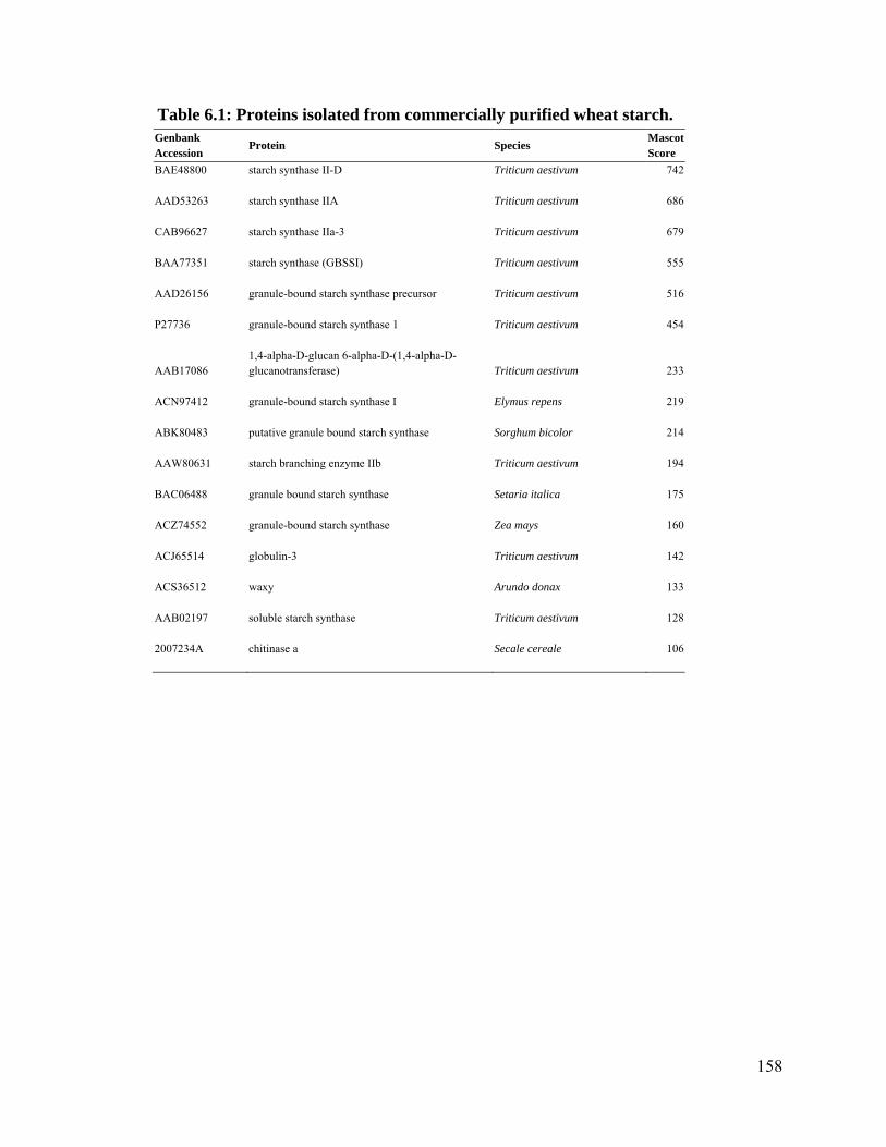
158
Table 6.1: Proteins isolated from commercially purified wheat starch. Genbank Accession
Protein Species Mascot Score
BAE48800 starch synthase II-D Triticum aestivum 742
AAD53263 starch synthase IIA Triticum aestivum 686
CAB96627 starch synthase IIa-3 Triticum aestivum 679
BAA77351 starch synthase (GBSSI) Triticum aestivum 555
AAD26156 granule-bound starch synthase precursor Triticum aestivum 516
P27736 granule-bound starch synthase 1 Triticum aestivum 454
AAB17086 1,4-alpha-D-glucan 6-alpha-D-(1,4-alpha-D-glucanotransferase) Triticum aestivum 233
ACN97412 granule-bound starch synthase I Elymus repens 219
ABK80483 putative granule bound starch synthase Sorghum bicolor 214
AAW80631 starch branching enzyme IIb Triticum aestivum 194
BAC06488 granule bound starch synthase Setaria italica 175
ACZ74552 granule-bound starch synthase Zea mays 160
ACJ65514 globulin-3 Triticum aestivum 142
ACS36512 waxy Arundo donax 133
AAB02197 soluble starch synthase Triticum aestivum 128
2007234A chitinase a Secale cereale 106

159
Appendix B: Curriculum Vitae
Adam Koziol Academic History Ph.D. Biochemistry and Systems Biology. University of Ottawa. 2007 - 2013. M.Sc. Biology. University of New Brunswick. Fredericton, New Brunswick, Canada. 2004-2007. B.Sc. Biology and Biotechnology (High Honours). Carleton University. Ottawa, Ontario, Canada. 1999-2003. Theses and Supervisors Koziol AG. Application of direct-sequencing peptide proteomics to the characterization of antagonistic (endogenous and exogenous) proteins in cereal grains. University of Ottawa. Supervisor: Illimar Altosaar. Koziol AG. 2007. The evolution of the chlorophyll a/b light-harvesting complex superfamily. M.Sc. Thesis. University of New Brunswick, Fredericton, New Brunswick, Canada, 100 pp. Supervisor: Dion G. Durnford. Koziol AG. 2003. The role of auxin in phase II secondary xylem development. B.Sc. Honours Thesis. Carleton University, Ottawa, Ontario, Canada. 25 pp. Supervisor: Sharon Regan. Areas of Academic Interest Food safety, mass spectrometry, bioinformatics, molecular biology, molecular evolution, antimicrobial peptides, recombinant protein expression. a. publications in refereed journals Koziol AG, Marquez BK, Huebsch MP, Smith JC and Altosaar I. (2012). The starch granule associated proteomes of commercially purified starch reference materials from rice and maize. Journal of Proteomics. 75: 993-1003. (PhD work) Koziol AG, Loit E, McNulty M, MacFarlane A, Scott FW and Altosaar I. (2012). Seed storage proteins of the globulin family are cleaved post-translationally in wheat embryos. Accepted July 5, 2012. 19 pages. BMC Research Notes. 5: 385, 10 pages, DOI: 10.1186/1756-0500-5-385. (PhD work) Koziol AG, Marquez BK, Huebsch MP, Smith JC and Altosaar I. (2012). Commercially produced rice and maize starches contain non-host proteins, as shown by mass

160
spectrometry. 14 pages. Cereal Chemistry. 89(5): 262-264, DOI: 10.1094/CCHEM-04-12-0043-N. (PhD work) Zaidi MA, El Bilali JE, Koziol AG, Styles G, Ward TL, Greenham TJ, Faiella WM, Son HH, Wan S, Taga I, and Altosaar I. (2012). Gene technology in agriculture, environment and biopharming: Beyond Bt-rice and building better breeding budgets for crops. 20 pages. Journal of Plant Biochemistry and Biotechnology, 21 (Suppl 1): S2-S9, DOI: 10.1007/s13562-012-0128-z. Koziol AG and Durnford DG. (2008). Euglena light-harvesting complexes are encoded by multifarious polyprotein mRNAs that evolve in concert. Molecular Biology and Evolution. 25: 92–100. (MSc work) Koziol AG, Borza T, Ishida KI, Keeling P, Lee RW and Durnford DG. (2007). Tracing the evolution of the light-harvesting antennae in chlorophyll a/b-containing organisms. Plant Physiology. 143: 1802-1816. (MSc work) b. Articles submitted to refereed journals Koziol AG, Reid LM, Altosaar I. (2012). Taking stock of the protein remnants on the battlefield between host and pathogen: Maize CL30-Fusarium interactome. 18 pages. Submitted to Journal of Proteomics July, 2012 JPROT-S-12-00689. (PhD work) c. Non-refereed contributions Koziol AG and Altosaar I. 2012. Molecular bio-pharming. BMI Poster Day, Ottawa, ON, May 19. Poster. (PhD work) Koziol AG. 2011. The proteome of a standard reference material: Starch granule associated proteins in rice. BMI Work in progress series. Ottawa, ON, March 2. Koziol AG and Altosaar I. 2011. The development of a puroindoline protein tethering model. BMI Seminar Symposium (1st annual), Ottawa, ON, February 18. Koziol AG Wheeler H and Altosaar I. 2008. Solaromics. Meeting of the Canadian Society of Plant Scientists (50th annual). Ottawa, ON. June 14-17. Poster. (PhD work) Koziol AG and Durnford DG. 2007. Fusion, Duplication, and Deletion: Evolution of Euglena gracilis LHC Polyprotein-coding genes. Northeast Algal Society (46th Annual), Narragansett, RI, USA, April 20 – 22. Koziol AG and Durnford DG. 2006. An Examination of the Evolution and Diversification of Light-Harvesting Complexes in Euglena gracilis. Eastern Regional Photosynthesis Conference (23rd annual), Woods Hole, MA, USA, April 21 - 23. Poster.

161
Appendix C: Reuse permissions
Figure 1.1

162

163
Figure 1.2

164
Figure 1.3

165

166

167
168
Figure 1.4
169
170
171

172
Figure 1.5

173
174

175
176
Figure 1.6

177

178

179
Figure 1.7

180
181

182

183
Figure 1.8

184