application of an acoustofluidic perfusion bioreactor for cartilage tissue engineering
TRANSCRIPT
www.rsc.org/loc
ISSN 1473-0197
Lab on a ChipMiniaturisation for chemistry, physics, biology, materials science and bioengineering
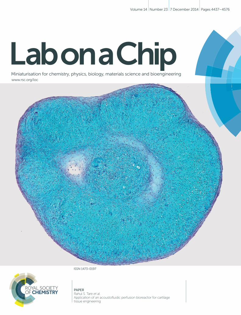
PAPERRahul S. Tare et al.Application of an acoustofluidic perfusion bioreactor for cartilage tissue engineering
Volume 14 Number 23 7 December 2014 Pages 4437–4576

Lab on a Chip
Ope
n A
cces
s A
rtic
le. P
ublis
hed
on 2
4 Se
ptem
ber
2014
. Dow
nloa
ded
on 1
8/11
/201
4 15
:38:
46.
Thi
s ar
ticle
is li
cens
ed u
nder
a C
reat
ive
Com
mon
s A
ttrib
utio
n 3.
0 U
npor
ted
Lic
ence
.
PAPER View Article OnlineView Journal | View Issue
Lab ChipThis journal is © The Royal Society of Chemistry 2014
a Centre for Human Development, Stem Cells and Regeneration, Faculty of Medicine,
University of Southampton, Southampton SO16 6YD, UK. E-mail: [email protected];
Fax: +44 2381 204221; Tel: +44 (0)2381 205257b Engineering Sciences, Faculty of Engineering and the Environment, University of
Southampton, Southampton SO17 1BJ, UKc Institute of Lightweight Design and Structural Biomechanics, Vienna University of
Technology, Gusshausstrasse 27-29 A-1040, Vienna, Austriad nCATS, Faculty of Engineering and the Environment, University of Southampton,
Southampton SO17 1BJ, UK
† Both authors contributed equally to this work.
Cite this: Lab Chip, 2014, 14, 4475
Received 15th August 2014,Accepted 24th September 2014
DOI: 10.1039/c4lc00956h
www.rsc.org/loc
Application of an acoustofluidic perfusionbioreactor for cartilage tissue engineering
Siwei Li,†a Peter Glynne-Jones,†b Orestis G. Andriotis,c Kuan Y. Ching,d
Umesh S. Jonnalagadda,b Richard O. C. Oreffo,a Martyn Hillb and Rahul S. Tare*ab
Cartilage grafts generated using conventional static tissue engineering strategies are characterised by low
cell viability, suboptimal hyaline cartilage formation and, critically, inferior mechanical competency, which
limit their application for resurfacing articular cartilage defects. To address the limitations of conventional
static cartilage bioengineering strategies and generate robust, scaffold-free neocartilage grafts of human
articular chondrocytes, the present study utilised custom-built microfluidic perfusion bioreactors with inte-
grated ultrasound standing wave traps. The system employed sweeping acoustic drive frequencies over the
range of 890 to 910 kHz and continuous perfusion of the chondrogenic culture medium at a low-shear
flow rate to promote the generation of three-dimensional agglomerates of human articular chondrocytes,
and enhance cartilage formation by cells of the agglomerates via improved mechanical stimulation and
mass transfer rates. Histological examination and assessment of micromechanical properties using
indentation-type atomic force microscopy confirmed that the neocartilage grafts were analogous to native
hyaline cartilage. Furthermore, in the ex vivo organ culture partial thickness cartilage defect model, implan-
tation of the neocartilage grafts into defects for 16 weeks resulted in the formation of hyaline cartilage-like
repair tissue that adhered to the host cartilage and contributed to significant improvements to the tissue
architecture within the defects, compared to the empty defects. The study has demonstrated the first
successful application of the acoustofluidic perfusion bioreactors to bioengineer scaffold-free neocartilage
grafts of human articular chondrocytes that have the potential for subsequent use in second generation
autologous chondrocyte implantation procedures for the repair of partial thickness cartilage defects.
1. Introduction
Osteoarthritis (OA), the most common form of arthritis inthe western world, is a degenerative joint disease that ischaracterised by the deterioration and progressive loss ofarticular cartilage. Early stages of OA are associated with thedevelopment of clefts and fissures in the articular cartilagesurface that resemble partial thickness defects or chondraldefects. Partial thickness defects are unable to heal spontane-ously because the lesions do not extend into the marrowspaces of the subchondral bone and cannot gain access to the
bone marrow skeletal stem cells to promote repair.1 It istherefore critical to repair these defects in the early stages ofOA because, if left untreated, the fibrillated lesions typicallygrow larger and deeper over time, and contribute to progres-sive articular cartilage degeneration and joint immobilisation.Currently, there are no effective pharmacological agents thatare able to promote comprehensive healing of articular carti-lage defects.
Surgical interventions for functional restoration of articu-lar cartilage defects include reparative bone marrow stimula-tion techniques such as abrasion arthroplasty, drilling,microfracture, and restorative approaches such as autolo-gous chondrocyte implantation (ACI), osteochondral auto/allografts, periosteal/perichondral grafts.2 Although theseinterventions provide symptomatic relief and improve jointfunction temporarily, to date, no technique has beencompletely successful in restoring/regenerating damagedarticular cartilage to its native state. This is because therepair tissue that is generated is often fibrocartilaginous innature, and therefore lacks the mechanical competency ofhyaline articular cartilage. Moreover, inability of the
, 2014, 14, 4475–4485 | 4475
Lab on a ChipPaper
Ope
n A
cces
s A
rtic
le. P
ublis
hed
on 2
4 Se
ptem
ber
2014
. Dow
nloa
ded
on 1
8/11
/201
4 15
:38:
46.
Thi
s ar
ticle
is li
cens
ed u
nder
a C
reat
ive
Com
mon
s A
ttrib
utio
n 3.
0 U
npor
ted
Lic
ence
.View Article Online
implanted graft/fibrocartilaginous repair tissue to integratewith the surrounding native cartilage contributes to graftfailure and further degeneration of the joint.
Attempts to improve the outcomes of cell-based transplan-tation methods, such as ACI, have therefore focused on theapplication of autologous chondrocytes seeded onto collagenscaffolds, in matrix-induced ACI, and three-dimensional (3-D)cartilaginous constructs, in second (II) generation ACI, forthe repair of articular cartilage defects.3–5 This, in turn, hasled to considerable interest in the development of effectivescaffold-free and scaffold-based tissue engineering strategiesfor generating cartilage grafts. Scaffold-free modalities havebeen employed to stimulate chondrogenic differentiation andcartilage formation via cell–cell and cell–matrix interactionsin high-density chondrospheres and multilayer articularchondrocyte sheets.6,7 In contrast to the scaffold-free modali-ties, which require cells to develop their own structure andmatrix during development, strategies harnessing scaffoldsprovide cells with an effective 3-D framework that supportstissue assembly and growth. However, the successful applica-tion of biomaterials in tissue engineering, including cartilagebioengineering, requires careful consideration of their 3-Darchitecture, biofunctionality, biocompatibility, biomechanics,degradation rates and immunogenicity of the degradationproducts.8,9
Tissue grafts generated using conventional static tissueengineering strategies are often characterised by appreciablenecrosis and suboptimal tissue formation due to poor nutri-ent mass transfer rates and oxygen diffusion in almost anygraft site with diffusion distance more than 1 mm.10 Toaddress some of the issues related to suboptimal tissue for-mation and cell viability in constructs generated using 3-Dstatic culture techniques, tissue engineering strategies haveincreasingly applied bioreactors, which provide a closelymonitored environment and, critically, biomechanical stimulisuch as hydrodynamic shear stress, hydrostatic pressure anddynamic compression for optimal tissue growth.11 Bioreac-tors have been used to culture a wide range of cells, namelystem cells, chondrocytes, osteoblasts, keratinocytes, hepato-cytes, cardiomyocytes and myofibroblasts, for a diverse arrayof applications including cartilage, bone, skin, liver and car-diovascular tissue engineering.11,12 In particular, perfusionflow bioreactors have been frequently used for cartilage tissueengineering due to their ability to enhance cartilage forma-tion by chondrocytes and mesenchymal cell populationsthrough the application of mechanical stimuli in the form offluid flow-induced hydrodynamic shear stresses, and improve-ment of mass transfer rates of metabolites and oxygen.13,14
The safe use of ultrasound and its diverse diagnostic andtherapeutic applications are widely acknowledged.15–17 Lowintensity pulsed ultrasound has been demonstrated to accel-erate the repair of damaged cartilage in a number of animalstudies. Low intensity ultrasound typically refers to fieldintensities below 1 W cm−2 that are applied either in continu-ous or burst (pulsed) mode.18,19 A variation of low intensityultrasound, referred to as low intensity diffuse ultrasound,
4476 | Lab Chip, 2014, 14, 4475–4485
involves scattering of acoustic waves generated by the trans-ducer throughout the chamber.20 In the rat model of papain-induced knee osteoarthritis, the application of ultrasoundwas shown to enhance cartilage repair in the early stage ofthe disease, and arrest further deteriorative cartilage damagein the later stage of osteoarthritis.21 Daily low intensitypulsed ultrasound was demonstrated to have a significantpositive effect on the repair of full thickness osteochondraldefects created in the patellar grooves of rabbits.22 In acanine model, low intensity pulsed ultrasound enhanced theincorporation of autologous osteochondral plugs by improv-ing the characteristics of the interface repair tissue and itsintegration with the adjacent cartilage.23
In the field of tissue engineering, ultrasound has predomi-nantly been applied in the form of low intensity ultrasoundto stimulate cells, and ultrasonic standing wave fields togenerate acoustic traps, which can spatially manipulate cells,proteins and microbeads.24 Specifically for cartilage tissueengineering, the application of low intensity ultrasound toenhance chondrogenic differentiation of bone marrowmesenchymal stem cells and chondrocytes, cultured in avariety of 3-D environments, has been documented in a num-ber of studies.25–29 However, ultrasonic cell trapping, a non-destructive and non-invasive cell manipulation technique,30
is a relatively less exploited application of ultrasound for car-tilage tissue engineering. When a fluid containing a suspen-sion of cells is exposed to an ultrasonic standing wave fieldin a chamber/trap, the acoustic radiation force arising fromthe scattering of the acoustic waves on the cells directs themotion (i.e. acoustophoresis) of cells typically to areas ofminimum pressure, referred to as pressure nodes, and facili-tates their aggregation into multicellular clusters.31–33 Fur-thermore, it is possible to drive ensembles of cells intogeometric formations, including linear clusters and planar(2-D) sheets, by appropriately shaping the resonant wave fieldwithin the ultrasonic trap, and also levitate the multicellularaggregates away from the influence of the solid substrate.34,35
The present study applied a novel approach that com-bined bioreactor technology with ultrasonic cell trapping tobioengineer 3-D, scaffold-free neocartilage grafts of humanarticular chondrocytes in custom-built microfluidic perfusionbioreactors with integrated ultrasound standing wave traps(USWT). The neocartilage grafts were then examined for theirpotential to repair partial thickness chondral defects. Thestudy has demonstrated the first successful application of theacoustofluidic perfusion bioreactor for bioengineering scaf-fold-free, hyaline cartilage-like explants of human articularchondrocytes. Following implantation into partial thicknesschondral defects, the bioengineered explants generatedhyaline cartilage-like repair tissue that integrated closelywith the surrounding host articular cartilage and contributedto significant improvements to the tissue architecture withinthe defects. The neocartilage grafts, therefore, have thepotential for application in restorative surgical procedures,such as II generation ACI, to repair early-stage articular carti-lage damage and limit further cartilage degeneration.
This journal is © The Royal Society of Chemistry 2014
Lab on a Chip Paper
Ope
n A
cces
s A
rtic
le. P
ublis
hed
on 2
4 Se
ptem
ber
2014
. Dow
nloa
ded
on 1
8/11
/201
4 15
:38:
46.
Thi
s ar
ticle
is li
cens
ed u
nder
a C
reat
ive
Com
mon
s A
ttrib
utio
n 3.
0 U
npor
ted
Lic
ence
.View Article Online
2. Materials and methods
Most chemicals and reagents were purchased fromInvitrogen/Life Technologies Ltd. (Paisley, UK) and Sigma-Aldrich(Gillingham, UK) unless specified otherwise.
2.1 Isolation of human articular chondrocytes (HACs)
Femoral head samples were obtained from 4 haematologicallynormal osteoarthritic individuals (3 female and 1 male, meanage: 80 years) following routine total hip replacement surgeryfor late-stage OA at Southampton General Hospital. Onlytissue that would have been discarded was used in thisstudy with approval of the Southampton and South WestHampshire Research Ethics Committee (ref no. 194/99/1 &210/01). HACs were isolated by sequential enzymatic diges-tion of deep-zone articular cartilage pieces dissected fromfemoral heads, as explained previously.36 In brief, cartilagepieces were sequentially digested with 500 μg ml−1 trypsin-EDTA for 30 min, 1 mg ml−1 hyaluronidase for 15 min and10 mg ml−1 collagenase B (Roche Diagnostics, Burgess Hill, UK)overnight on a rotating mixer at 37 °C. Isolated chondrocyteswere cultured to confluence in monolayer cultures in α-MEMsupplemented with 10% (v/v) FCS, 100 units ml−1 penicillin,100 μg ml−1 streptomycin and 100 μM ascorbate 2-phosphate.Cultures were maintained in humidified atmosphere at37 °C, 5% CO2 and 21% O2. Passage 1 cells were utilised forthe experiments.
2.2 Fabrication of perfusion bioreactors with integratedUSWT/acoustofluidic perfusion bioreactors and theirapplication for generation of neocartilage grafts of HACs
The acoustofluidic perfusion bioreactors were custom-builtusing commercially available rectangular glass capillaries(VitroCom 4608-100, CM Scientific, Silsden, UK) and polydi-methylsiloxane (PDMS) connectors cast from acrylic moulds(Fig. 1a). The glass capillaries functioned efficiently as highlyresonant cavities,37–39 and it was possible to autoclave themalong with the non toxic PDMS. A set of 3 resonant chamberswere fabricated from the rectangular glass capillaries (length5 cm, ID 0.8 × 8 mm2, wall thickness 0.54 mm) and a ceramicpiezoelectric transducer (PZT) (Ferroperm PZ26, Kvistgaard,Denmark; 10 × 8 × 1 mm3) was glued (Epoxy 353ND, Epotek,Billerica, MA, USA) to each chamber. Wires soldered to theelectrodes on the transducer were connected to a custom-made amplifier (LT1210 IC, RS Components, Corby, UK) thatwas driven by a signal generator (TG200, TTi, Huntingdon, UK).
The transducer generated an ultrasonic standing wavefield in the lumen of the capillary, with the upper glasssurface acting as a reflector. An impedance spectrum wasused in conjunction with a transfer impedance model topredict the resonant frequencies of the system.40,41 The half-wavelength resonance of the cavity was found at 897, 899 and902 kHz for the three devices respectively. The half-wavelength resonance had a pressure node in the centre ofthe chamber and was observed to i) promote the formation
This journal is © The Royal Society of Chemistry 2014
of a 3-D multicellular agglomerate by rapid aggregation atthe pressure node of HACs introduced into the chamber, andii) levitate the agglomerate in the lumen of the chamberabove the transducer away from the influence of the solidsubstrate (Fig. 1b). The voltage drop method was used toassess the acoustic pressure amplitude;31 a range of locationsover the transducer were examined with average acousticpressure amplitude of 17 ± 5.1 kPa V−1. The generator wasadjusted to create a voltage of 10 Vpp (peak-to-peak voltage)across the transducer (average over sweep range). In additionto the primary potential energy gradients found perpendicu-lar to the transducer, smaller forces caused by gradients inthe kinetic energy density parallel to the transducer createdlocalised trapping forces.42 These forces assisted agglomerateformation (aided by the secondary inter-particle forces) andalso held the agglomerate against the perfusion flow. It wasfound that a typical agglomerate of HACs could withstandlinear fluid velocities of up to approximately 1 mm s−1, thustrapping the levitated agglomerate against the flow of contin-uous perfusion.
Serum free chondrogenic culture medium held within areservoir was circulated around a closed loop by a peristalticpump (403U/VM2, Watson-Marlow, Falmouth, UK) andMarprene tubing (505DZ/RL, Fisher Scientific, UK; ID0.8 mm) at a rate of 1.32 ml h−1 (Fig. 1c). The serum-freechondrogenic medium was composed of α-MEM supplementedwith 10 ng ml−1 rhTGF-β3 (PeproTech, London, UK), 100 μMascorbate-2-phosphate, 10 nM dexamethasone and 1X ITSliquid supplement (10 μg ml−1 insulin, 5.5 μg ml−1 transferrinand 5 ng ml−1 selenite premix). The loop included the reso-nant chamber connected to the tubing via PDMS connectorsand a bubble trap in close proximity to the chamber to pre-vent bubbles disrupting the levitated agglomerate. A syringepump enabled excess bubbles to be extracted from thebubble trap; during the experiment this was run at a continu-ous rate of 0.02 ml h−1. The optimum CO2 concentration wasmaintained by preconditioning the chondrogenic medium ina standard CO2 incubator for 24 h to allow gaseous equilib-rium and creating a 5% CO2 atmosphere in the space abovethe culture medium in the reservoir through introduction ofthe gas via a HEPA filter with 0.22 μm pore size.
Another syringe pump was used to introduce cells into thechamber via a dedicated inlet. A suspension of HACscontaining 1 × 10⁶ cells was introduced at a rate of 1 ml min−1
with the ultrasound active. The perfusion peristaltic pumpwas activated once stable agglomerates were formed. The sys-tem was placed within a poly(methyl methacrylate)/PMMAbox covered with expanded polystyrene sheets for thermalinsulation and a custom heating controller circulated hot airto maintain the ambient temperature at 36 °C. Although acti-vation of the ultrasound was found to cause heating, at theambient temperature, the active region of the chamberreached a steady 37.0 ± 0.5 °C. The multicellular agglomerateswere cultured within the acoustofluidic perfusion bioreactorsover a period of 21 days in serum-free chondrogenic mediumto promote cartilage formation. At the end of the culture
Lab Chip, 2014, 14, 4475–4485 | 4477

Lab on a ChipPaper
Ope
n A
cces
s A
rtic
le. P
ublis
hed
on 2
4 Se
ptem
ber
2014
. Dow
nloa
ded
on 1
8/11
/201
4 15
:38:
46.
Thi
s ar
ticle
is li
cens
ed u
nder
a C
reat
ive
Com
mon
s A
ttrib
utio
n 3.
0 U
npor
ted
Lic
ence
.View Article Online
period, two grafts were labelled with Cell Tracker™ Green andEthidium homodimer-1, fixed in 4% paraformaldehyde (PFA)overnight at 4 °C and used for histological analysis; threegrafts were used for determination of biomechanical proper-ties and two grafts were harvested for implantation into par-tial thickness chondral defects.
4478 | Lab Chip, 2014, 14, 4475–4485
Fig. 1 Application of the acoustofluidic perfusion bioreactor for the generectangular glass capillary (length 5 cm, ID 0.8 × 8 mm2, wall thicknesspiezoelectric transducer (PZT), which was glued to the capillary. (b) Schemthe resonant chamber. The transducer generated an ultrasonic standingacting as a reflector. At the resonant frequency (0.897 MHz) of the cavitycentre of the chamber. When cells suspended in culture medium were incells to the pressure node, where they aggregated rapidly and eventually fof the chamber above the transducer. (c) Serum-free chondrogenic mediuclosed loop that included the resonant chamber, bubble trap and syringeelectrodes on the transducer were connected to a custom-made amplifiersound caused heating, at the ambient temperature (36 °C) of the system, th
2.3 Assessment of cell viability
Cell viability in the day 21 neocartilage grafts was examinedby ‘live-dead’ staining using a combination of CellTracker™
green/CTG CMFDA IJ5-chloromethylfluorescein diacetate)and Ethidium homodimer-1/EH-1. Harvested samples were
This journal is © The Royal Society of Chemistry 2014
ration of neocartilage grafts. (a) The bioreactor was fabricated using a0.54 mm), polydimethylsiloxane (PDMS) connectors and a ceramicatic diagram illustrating formation of the multicellular agglomerate inwave field in the lumen of the capillary, with the upper glass surface, a half-wavelength mode was established with a pressure node in thetroduced into the chamber, the acoustic radiation forces directed theormed the multicellular agglomerate, which was levitated in the lumenm held in the reservoir was circulated by a peristaltic pump around apump used to introduce cells into the chamber. Wires soldered to thethat was driven by a signal generator. Although activation of the ultra-e active region of the chamber reached a steady 37.0 ± 0.5 °C.

Lab on a Chip Paper
Ope
n A
cces
s A
rtic
le. P
ublis
hed
on 2
4 Se
ptem
ber
2014
. Dow
nloa
ded
on 1
8/11
/201
4 15
:38:
46.
Thi
s ar
ticle
is li
cens
ed u
nder
a C
reat
ive
Com
mon
s A
ttrib
utio
n 3.
0 U
npor
ted
Lic
ence
.View Article Online
washed in PBS and incubated in 1 ml of CTG (10 μg ml−1)and EH-1 (5 μg ml−1) solution for 45 min in a humidifiedatmosphere at 37 °C, 5% CO2 and 21% O2. Samples werethen fixed in 4% PFA overnight at 4 °C. Viable cells (green)and necrotic cells (red) were visualised in paraffin waxsections of the constructs by fluorescence microscopy using aZeiss Axiovert 200 inverted fluorescence microscope (CarlZeiss, Cambridge, UK) and imaged using a CCD camera withAxiovision software.
2.4 Ex vivo organ culture partial thickness cartilagedefect model
Near full-thickness articular cartilage pieces (1 × 1 cm2,2 mm thick) were dissected from healthy non load-bearingregions of human femoral heads. A partial thickness defect(~2 × 2 mm2, 1 mm deep) was created in each articular carti-lage piece with a sterile drill bit, taking extreme care to avoidfull penetration of the cartilage. One neocartilage graft wasimplanted into each defect and the neocartilage graft-hostcartilage construct was then placed on a Millipore filter insert(hydrophilic polytetrafluoroethylene/PTFE membrane, 0.4 μmpore size, 6-well plate configuration) and cultured at air–liquid interface in a humidified atmosphere at 37 °C, 5%CO2 and 21% O2 for 16 weeks. Pieces of articular cartilagewith empty defects cultured similarly for 16 weeks served ascontrols. The samples were harvested at 16 weeks, fixed in 4%PFA overnight at 4 °C and processed for histological analysis.
2.5 Processing, embedding and section cutting
PFA-fixed samples were processed through graded ethanol(50–100%) and histoclear (100%) prior to embedding inparaffin wax. Sequential sections (7 μm) were cut on themicrotome and mounted on glass slides for histological andimmunohistochemical staining. Images were captured withOlympus dotSlide virtual microscopy system (Olympus,Southend-on-Sea, UK).
2.6 Alcian blue and Sirius red staining
Sections were stained with Alcian blue 8GX (5 mg ml−1 in 1%(v/v) glacial acetic acid) and Sirius red F3B (10 mg ml−1 insaturated picric acid) following nuclear staining withWeigert's haematoxylin.
2.7 Immunohistochemistry
After quenching endogenous peroxidase activity with 3% (v/v)H2O2 and blocking with 10 mg ml−1 BSA in PBS, sectionswere incubated with relevant primary antisera (diluted appro-priately using 10 mg ml−1 BSA in PBS) at 4 °C overnight. Thiswas followed by hour-long incubation each with the appropriatebiotinylated secondary antibody and ExtrAvidin®-Peroxidase.Visualisation of the immune complex was undertakenusing the avidin–biotin method linked to peroxidase andAEC (3-amino-9-ethylcarbazole), resulting in a reddish brownreaction product. Negative controls (omission of the primary
This journal is © The Royal Society of Chemistry 2014
antisera) were included in all immunohistochemistry proce-dures. No staining was observed in the sections used as nega-tive controls. Sections were counter-stained with Alcian blue.The anti-SOX9 (AB5535, Millipore, Watford, UK) antibody wasused at dilution of 1 : 150 following the antigen retrieval pro-cedure, which involved heating sections in 0.01 M citratebuffer (pH 6.0) for 5 minutes before the application of thestandard immunohistochemistry procedure. Sections weretreated with type I hyaluronidase at 37 °C for 20 min inorder to unmask the collagen fibres and render them acces-sible for immunostaining with the antibodies against thethree types of collagen. The anti-collagen type I antibody(LF68 from Dr Larry Fisher, NIH, USA), anti-collagen type IIand anti-collagen type X antibodies (cat. no. 234187 and234196, both sourced from Millipore, Watford, UK) wereused at dilutions of 1 : 1000, 1 : 500 and 1 : 100, respectively.
2.8 Indentation-type Atomic Force Microscopy (IT-AFM)
As described previously,43 cantilever-based IT-AFM with amicrometre-sized spherical tip was used to measure themicroscale elastic moduli of day 21 neocartilage graftsand full-thickness articular cartilage pieces, dissected fromfemoral heads of patients (2 female and 1 male, average age:77 years) who had suffered a fracture in the neck of femur.In brief, hard borosilicate glass spheres (diameter: 10 μm;02715-AB, SPI Supplies, West Chester, PA, USA) were gluedonto tipless rectangular silicon AFM cantilevers (springconstant: 6.11 N m−1; type All In One-TL, BudgetSensors, Sofia,Bulgaria) for probing the samples. Force volume maps ofload–displacement curves (16 × 16 i.e. 256 load–displacementcurves) were recorded in a regular grid over the sample surface.Each individual set of data consisted of load–displacementcurves recorded at a rate of half full loading cycles per secondin a sample area of 10 μm × 10 μm. A maximum cantileverdeflection of 200 nm coupled with a maximum applied loadof 1.2 μN was used for the measurement of all samples. Thesamples were fixed on a glass slide and submerged in PBSduring the measurements, which were performed at roomtemperature using a MFP-3D AFM (Oxford Systems, AsylumResearch, Santa Barbara, CA, USA). Values for microscaleelastic moduli of the samples were calculated from theunloading curves obtained from IT-AFM, ensuring thatthe displacement data did not contain irreversible (i.e.plastic) deformation. Statistical analysis was performed usingMann–Whitney U test. Results were considered significant ifthe probability of occurrence by random chance alone wasless than 5% (i.e. p < 0.05).
3. Results3.1 Examination of day 21 grafts for assessment of cellviability, cartilage formation and micromechanical properties
3-D membrane-like constructs (measuring ~2.5 × 2.5 mm2,0.2 mm thick) were harvested from the acoustofluidic perfu-sion bioreactors following the 21 day culture period. The con-structs were labelled with Cell Tracker™ Green and Ethidium
Lab Chip, 2014, 14, 4475–4485 | 4479
Lab on a ChipPaper
Ope
n A
cces
s A
rtic
le. P
ublis
hed
on 2
4 Se
ptem
ber
2014
. Dow
nloa
ded
on 1
8/11
/201
4 15
:38:
46.
Thi
s ar
ticle
is li
cens
ed u
nder
a C
reat
ive
Com
mon
s A
ttrib
utio
n 3.
0 U
npor
ted
Lic
ence
.View Article Online
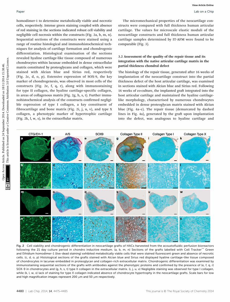
homodimer-1 to determine metabolically viable and necroticcells, respectively. Intense green staining coupled with absenceof red staining in the sections indicated robust cell viability andnegligible cell necrosis within the constructs (Fig. 2a, b, m, n).Sequential sections of the constructs were stained using arange of routine histological and immunohistochemical tech-niques for analysis of cartilage formation and chondrogenicdifferentiation. Histological examination of the sectionsrevealed hyaline cartilage-like tissue composed of numerouschondrocytes within lacunae embedded in dense extracellularmatrix constituted by proteoglycans and collagen, which werestained with Alcian blue and Sirius red, respectively(Fig. 2c, d, o, p). Extensive expression of SOX-9, the keymarker of chondrogenesis, was observed in most cells of theconstructs (Fig. 2e, f, q, r), along with immunostainingfor type II collagen, the hyaline cartilage-specific collagen,in areas of collagenous matrix (Fig. 2g, h, s, t). Further immu-nohistochemical analysis of the constructs confirmed negligi-ble expression of type I collagen, a key constituent offibrocartilage and bone matrix (Fig. 2i, j, u, v), and type Xcollagen, a phenotypic marker of hypertrophic cartilage(Fig. 2k, l, w, x), in the extracellular matrix.
4480 | Lab Chip, 2014, 14, 4475–4485
Fig. 2 Cell viability and chondrogenic differentiation in neocartilage grafollowing the 21 day culture period in chondro inductive medium. (a,and Ethidium homodimer-1 (live-dead staining) exhibited metabolically viacells. (c, d, o, p) Histological sections of the grafts stained with Alcianof chondrocytes in lacunae embedded in proteoglycan and collagen-richimmunostaining sequential sections of the grafts with antibodies againstSOX-9 in chondrocytes and (g, h, s, t) type II collagen in the extracellularwhile (k, l, w, x) lack of staining for type X collagen indicated absence of cand high magnification images represent 200 μm and 50 μm respectively.
The micromechanical properties of the neocartilage con-structs were compared with full thickness human articularcartilage. The values for microscale elastic moduli of theneocartilage constructs and full thickness human articularcartilage samples determined by IT-AFM were found to becomparable (Fig. 3).
3.2 Assessment of the quality of the repair tissue and itsintegration with the native articular cartilage matrix in thepartial thickness chondral defect
The histology of the repair tissue, generated after 16 weeks ofimplantation of the neocartilage construct into the partialthickness defect of the host articular cartilage, was examinedin sections stained with Alcian blue and Sirius red. Following16 weeks of co-culture, the implanted graft integrated into thehost articular cartilage and maintained the hyaline cartilage-like morphology, characterised by numerous chondrocytesembedded in dense proteoglycan matrix stained with Alcianblue (Fig. 4a–c). The repair tissue (demarcated by dashedlines in Fig. 4a), generated by the graft upon implantationinto the defect, was analogous to hyaline cartilage and
This journal is © The Royal Society of Chemistry 2014
fts of HACs harvested from the acoustofluidic perfusion bioreactorsb, m, n) Sections of the grafts labelled with Cell Tracker™ Greenble cells that were stained fluorescent green and absence of necroticblue and Sirius red displayed hyaline cartilage-like tissue composedextracellular matrix. Chondrogenic differentiation was examined bythe phenotypic proteins and confirmed by the presence of (e, f, q, r)matrix. (i, j, u, v) Negligible staining was observed for type I collagen,hondrocyte hypertrophy in the neocartilage grafts. Scale bars for low

Fig. 3 Micromechanical properties of human articular cartilage andneocartilage grafts generated using the acoustofluidic perfusionbioreactors were determined by IT-AFM. The difference betweenvalues for microscale elastic moduli of full thickness human articularcartilage samples and day 21 neocartilage grafts was not statisticallysignificant. Values are expressed as mean ± SD, n = 3 in each group.
Lab on a Chip Paper
Ope
n A
cces
s A
rtic
le. P
ublis
hed
on 2
4 Se
ptem
ber
2014
. Dow
nloa
ded
on 1
8/11
/201
4 15
:38:
46.
Thi
s ar
ticle
is li
cens
ed u
nder
a C
reat
ive
Com
mon
s A
ttrib
utio
n 3.
0 U
npor
ted
Lic
ence
.View Article Online
demonstrated the presence of chondrocytes and proteoglycan-rich extracellular matrix. Moreover, the repair tissue exhibitedclose continuous attachment with the host cartilage and nogaps were observed between the newly synthesized cartilageand host articular cartilage along the boundary of the defect(demarcated by dashed line in Fig. 4d). Representative imageof Alcian blue and Sirius red-stained section of the host articu-lar cartilage with partial thickness defect after the 16 weekculture period without the neocartilage implant demonstratedabsence of cartilage regeneration (Fig. 4e).
4. Discussion
The present study represents the first successful application ofthe novel microfluidic perfusion bioreactors with integratedUSWT to bioengineer robust, 3-D, scaffold-free neocartilagegrafts of HACs that are analogous to native hyaline cartilage.Additionally, in an ex vivo organ culture model, the study hasdemonstrated the potential of the neocartilage grafts to medi-ate repair of partial thickness chondral defects by generationof hyaline cartilage-like repair tissue. A scaffold-free tissueengineering approach was employed by the present study,instead of a scaffold-based approach, for the ex vivo genera-tion of 3-D cartilage constructs. This is because, in compari-son to the natural extracellular matrix, which aids cartilageregeneration by providing crucial cues to chondrocytes, someartificial scaffold materials may impair tissue formation anddefect regeneration due to their unpredictable degradationrates and immunogenicity of the degradation products.44
While the pellet culture technique is an effective ex vivoscaffold-free strategy to stimulate chondrogenic redifferentiationof dedifferentiated chondrocytes and promote cartilage for-mation in a high cell density 3-D microenvironment,45 it haslimited clinical application because conventional static pelletculture is associated with lack of mechanical stimulation,inefficient oxygen diffusion and suboptimal metabolite mass
This journal is © The Royal Society of Chemistry 2014
transfer rates, which adversely affect the scale-up, quality(i.e. formation of fibrous versus hyaline cartilage) and biome-chanical properties of the chondrospheres. This was demon-strated in our previous study, where attempts to scale-up thesize of constructs by conventional static pellet culture using1 × 106 HACs, resulted in the formation of chondrospheresthat were characterised by the development of necrotic coresand suboptimal hyaline cartilage.46 Mass transport limita-tions and oxygen diffusion gradients in the macroscopicpellets have been recognised as significant obstacles torobust chondrogenesis, thereby limiting the prospects ofmacroscopic cartilaginous pellets/chondrospheres in therepair of chondral defects.47
The scaffold-free grafts bioengineered in the acoustofluidicperfusion bioreactors using 1 × 10⁶ HACs exhibited distincthyaline cartilage-like tissue, which was characterised by therobust expression of chondrogenic markers, namely SOX-9,type II collagen and proteoglycans, coupled with negligibleexpression of collagen types I and X. Previous studies havedemonstrated induction of type II collagen and proteoglycansby human articular chondrocytes in 3-D alginate culture inresponse to low intensity ultrasound, and suppression ofchondrocyte hypertrophy by inhibition of expression of type Xcollagen due to low intensity ultrasound treatment.48,49
Although the exact mechanism by which ultrasound pro-motes the expression of chondrogenic markers remains to befully elucidated, it has been suggested that excitation ofmicrobubbles or acoustic streaming produced by the ultra-sound can modulate mechanoreceptor-mediated transmem-brane signalling mechanisms (involving protein kinase C) forthe regulation of Aggrecan gene expression and stimulationof subsequent proteoglycan synthesis.50–52 Moreover, bothcell shape and cytoskeletal organisation were shown to beessential for initiation of Sox-9 expression and maintenanceof the differentiated chondrocyte phenotype in 2-D aggregatesof chick wing bud mesenchymal cells, which were generatedand levitated using an USWT.53
To enhance mass transfer and mechanical stimulation,the current system employed two strategies, namely continu-ous perfusion of the culture medium at rates considered low-shear and sweeping acoustic drive frequencies over the rangeof 890 to 910 kHz, at a sweep rate of 50 Hz. The sweep rate of50 Hz reflected the maximum value available from the signalgenerator that was used in the study. The application of fluidshear from the perfusion system in our device compensatedfor the reduced acoustically-induced forces exerted on thecells by the ultrasonic waves. Mechanical stimulationconveyed by the flow of the culture medium i.e. fluidflow-induced shear, has been acknowledged as a crucial bio-mechanical stimulus, which enhances chondrocyte functionand ex vivo cartilage formation by directly influencing cellmetabolism and extracellular matrix synthesis.13 Moreover,chondrocytes respond positively to fluid convection, whichhas been shown to promote the transport of molecules andfurther improve cartilage formation due to enhanced masstransfer rates of metabolites.54
Lab Chip, 2014, 14, 4475–4485 | 4481
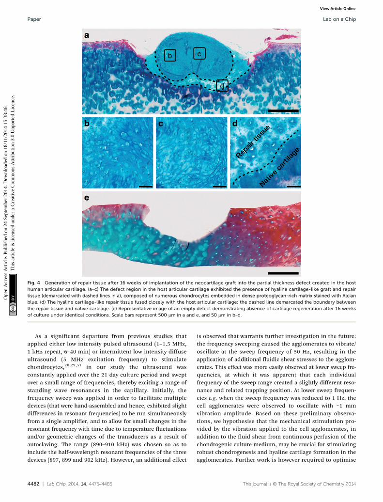
Fig. 4 Generation of repair tissue after 16 weeks of implantation of the neocartilage graft into the partial thickness defect created in the hosthuman articular cartilage. (a–c) The defect region in the host articular cartilage exhibited the presence of hyaline cartilage-like graft and repairtissue (demarcated with dashed lines in a), composed of numerous chondrocytes embedded in dense proteoglycan-rich matrix stained with Alcianblue. (d) The hyaline cartilage-like repair tissue fused closely with the host articular cartilage; the dashed line demarcated the boundary betweenthe repair tissue and native cartilage. (e) Representative image of an empty defect demonstrating absence of cartilage regeneration after 16 weeksof culture under identical conditions. Scale bars represent 500 μm in a and e, and 50 μm in b–d.
Lab on a ChipPaper
Ope
n A
cces
s A
rtic
le. P
ublis
hed
on 2
4 Se
ptem
ber
2014
. Dow
nloa
ded
on 1
8/11
/201
4 15
:38:
46.
Thi
s ar
ticle
is li
cens
ed u
nder
a C
reat
ive
Com
mon
s A
ttrib
utio
n 3.
0 U
npor
ted
Lic
ence
.View Article Online
As a significant departure from previous studies thatapplied either low intensity pulsed ultrasound (1–1.5 MHz,1 kHz repeat, 6–40 min) or intermittent low intensity diffuseultrasound (5 MHz excitation frequency) to stimulatechondrocytes,20,29,51 in our study the ultrasound wasconstantly applied over the 21 day culture period and sweptover a small range of frequencies, thereby exciting a range ofstanding wave resonances in the capillary. Initially, thefrequency sweep was applied in order to facilitate multipledevices (that were hand-assembled and hence, exhibited slightdifferences in resonant frequencies) to be run simultaneouslyfrom a single amplifier, and to allow for small changes in theresonant frequency with time due to temperature fluctuationsand/or geometric changes of the transducers as a result ofautoclaving. The range (890–910 kHz) was chosen so as toinclude the half-wavelength resonant frequencies of the threedevices (897, 899 and 902 kHz). However, an additional effect
4482 | Lab Chip, 2014, 14, 4475–4485
is observed that warrants further investigation in the future:the frequency sweeping caused the agglomerates to vibrate/oscillate at the sweep frequency of 50 Hz, resulting in theapplication of additional fluidic shear stresses to the agglom-erates. This effect was more easily observed at lower sweep fre-quencies, at which it was apparent that each individualfrequency of the sweep range created a slightly different reso-nance and related trapping position. At lower sweep frequen-cies e.g. when the sweep frequency was reduced to 1 Hz, thecell agglomerates were observed to oscillate with ~1 mmvibration amplitude. Based on these preliminary observa-tions, we hypothesise that the mechanical stimulation pro-vided by the vibration applied to the cell agglomerates, inaddition to the fluid shear from continuous perfusion of thechondrogenic culture medium, may be crucial for stimulatingrobust chondrogenesis and hyaline cartilage formation in theagglomerates. Further work is however required to optimise
This journal is © The Royal Society of Chemistry 2014

Lab on a Chip Paper
Ope
n A
cces
s A
rtic
le. P
ublis
hed
on 2
4 Se
ptem
ber
2014
. Dow
nloa
ded
on 1
8/11
/201
4 15
:38:
46.
Thi
s ar
ticle
is li
cens
ed u
nder
a C
reat
ive
Com
mon
s A
ttrib
utio
n 3.
0 U
npor
ted
Lic
ence
.View Article Online
the sweep rate, quantify the mechanical forces acting on theagglomerates and examine their effects in detail on cartilageformation by the cells of the agglomerates.
When ultrasound is absorbed by a material, its mechanicalenergy is primarily converted into heat. The acoustofluidicperfusion bioreactors in the present study are custom-builtusing glass capillaries and filled with culture medium, bothof which are ‘low-loss materials’. Moreover, the capillary con-struction limits transmission of the ultrasound into thesurrounding structure, thereby resulting in a high resonantQ-factor. Hence, it is possible to generate a substantial acous-tic field without significant heating.55 The steady state tem-perature rise recorded by the thermocouple embedded withinour device was 0.8 °C. Additionally, in the current experimen-tal setup, a custom made incubator incorporating a fan andthermostatic control maintained the environmental tempera-ture at around 36 °C, thus ensuring the optimal cell culturetemperature close to 37 °C in the active region of the bioreac-tor. Prolonged exposure to ultrasound in the acoustofluidicperfusion bioreactor therefore did not adversely affect cellviability, as confirmed by the presence of metabolicallyactive viable cells and absence of necrotic cells in day 21neocartilage explants.
To determine the micromechanical properties of theneocartilage grafts, elastic moduli of the bulk material of day21 grafts were measured using an IT-AFM microtip (sphericalindenter diameter of 10 μm), and compared to the elasticmoduli of freshly isolated full-thickness human articular car-tilage pieces. Previous work has reported that the microscaleelastic modulus of human articular cartilage, determinedusing IT-AFM, is 1.3 MPa regardless of the degree of OA,while changes due to aging and/or OA are only depicted atthe nanometer scale.43 Harnessing a similar IT-AFM setup,we obtained comparable value for elastic modulus [1.34 ±0.315 MPa] of the full-thickness human articular cartilagepieces utilised in the present study. Interestingly, the elasticmodulus [0.90 ± 0.372 MPa] of day 21 neocartilage grafts wassimilar to the elastic modulus of human articular cartilage.Thus, the neocartilage constructs were not only histologicallycomparable to hyaline cartilage, but also displayed compara-ble mechanical competency as native articular cartilage.
An ex vivo organ culture partial thickness cartilage defectmodel was utilised in the present study to determine the abil-ity of the neocartilage explants to integrate with nativehuman articular cartilage and repair the defects. Integrationis defined as the absence of gaps between the surface of therepair tissue generated by the cartilage graft and the borderof the native cartilage matrix in the defect region.56 Oftenafter implantation, cartilaginous grafts do not integrate read-ily or predictably with the host tissue to form a continuousmechanically stable attachment. This largely occurs becausethe repair tissue is predominantly composed of fibrocartilage,which is deficient in proteoglycans and mechanically inferior,compared to the host hyaline cartilage.57 In the presentstudy, following implantation of the neocartilage graft intothe chondral defect and co-culture for 16 weeks ex vivo, the
This journal is © The Royal Society of Chemistry 2014
defect was filled with hyaline cartilage-like tissue characterisedby the presence of numerous chondrocytes embedded indense proteoglycan matrix. Although a discernible boundarywas visible, no gaps were observed between the edge of therepair tissue and the border of the defect in the host cartilage.This was indicative of continuous close attachment betweenthe newly synthesized hyaline cartilage-like repair tissue andthe host articular cartilage, and integration of the repair tis-sue into the surrounding native cartilage. Thus, implantationof the neocartilage graft into the chondral defect resulted inthe generation of hyaline cartilage-like repair tissue that con-tributed to significant improvements to the tissue architec-ture within the defect, in comparison to absence of cartilageregeneration in the empty defect. Further investigations willfocus on examining the biomechanical properties of therepair tissue.
We acknowledge the limitations of the ex vivo organculture partial thickness cartilage defect model used in ourstudy to reproduce the complex biological and mechanicalenvironment of the joint. Since constructs demonstrate a cer-tain threshold of function in vitro, to carry out a realisticassessment of their potential for cartilage repair, we recog-nise that they should be assayed in a large animal (e.g. lapinemodel) load-bearing environment.9 Future work will thereforeinvolve implantation of the neocartilage grafts in partialthickness chondral defects created in rabbit lateral femoralcondyles and long-term assessment of the repair tissue post-implantation.
5. Conclusions
In summary, we have successfully applied a novel tissue engi-neering approach that combines microfluidic perfusion bio-reactor technology with continuous application of ultrasoundto bioengineer scaffold-free (hence devoid of any foreignmaterial) neocartilage grafts, which are analogous to nativehyaline cartilage both histologically and biomechanically,and have the ability to repair partial thickness chondraldefects. The work therefore presents a real opportunity toderive a robust tissue-based product, which has the potentialfor subsequent use in restorative approaches, such as IIgeneration ACI, for the repair of focal partial thicknesschondral defects in early stage OA.
Acknowledgements
The authors gratefully acknowledge financial support for thework from the Faculty of Medicine Research ManagementCommittee and Wessex Medical Research innovation grantsto RT, Engineering and Physical Sciences Research Councilgrant funding to PG-J (EP/K027115/1) and MH (EP/G012075/1),Biotechnology and Biological Sciences Research Council grantfunding to RO (G0 105791/1), and studentship support to SLfrom the University of Southampton PhD scholarship. Theauthors would like to thank Mr Pierre Dole for his assistancein fabricating the device, Dr Martin Stolz (University of
Lab Chip, 2014, 14, 4475–4485 | 4483
Lab on a ChipPaper
Ope
n A
cces
s A
rtic
le. P
ublis
hed
on 2
4 Se
ptem
ber
2014
. Dow
nloa
ded
on 1
8/11
/201
4 15
:38:
46.
Thi
s ar
ticle
is li
cens
ed u
nder
a C
reat
ive
Com
mon
s A
ttrib
utio
n 3.
0 U
npor
ted
Lic
ence
.View Article Online
Southampton) and Prof. Philipp Thurner (Vienna University ofTechnology) for their help with IT-AFM, and the OrthopaedicSurgeons at Southampton General Hospital for provision offemoral head samples.
References
1 E. B. Hunziker and L. C. Rosenberg, J. Bone Jt. Surg., Am.
Vol., 1996, 78, 721–733.2 S. N. Redman, S. F. Oldfield and C. W. Archer, Eur. Cells
Mater., 2005, 9, 23–32.3 M. Brittberg, A. Lindahl, A. Nilsson, C. Ohlsson, O. Isaksson
and L. Peterson, N. Engl. J. Med., 1994, 331, 889–895.4 P. Behrens, E. M. Ehlers, K. U. Köchermann, J. Rohwedel,
M. Russlies and W. Plötz, MMW Fortschr. Med., 1999, 141,49–51.5 R. S. Tuan, Arthritis Res. Ther., 2007, 9, 109.
6 T. Schubert, S. Anders, E. Neumann, J. Scholmerich,F. Hofstadter, J. Grifka, U. Muller-Ladner, J. Libera andJ. Schedel, Int. J. Mol. Med., 2009, 23, 455–460.
7 N. Kaneshiro, M. Sato, M. Ishihara, G. Mitani, H. Sakai,
T. Kikuchi and J. Mochida, Eur. Cells Mater., 2007, 13, 87–92.8 J. P. Vacanti and R. Langer, Lancet, 1999, 354, SI32–34.
9 B. Johnstone, M. Alini, M. Cucchiarini, G. R. Dodge,D. Eglin, F. Guilak, H. Madry, A. Mata, R. L. Mauck,C. E. Semino and M. J. Stoddart, Eur. Cells Mater., 2013, 25,248–267.
10 G. F. Muschler, C. Nakamoto and L. G. Griffith, J. Bone Jt.
Surg., Am. Vol., 2004, 86-A, 1541–1558.11 N. Plunkett and F. J. O'Brien, Technol. Health Care, 2011, 19,
55–69.12 I. Martin, D. Wendt and M. Heberer, Trends Biotechnol.,
2004, 22, 80–86.13 R. G. LeBaron and K. A. Athanasiou, Biomaterials, 2000, 21,
2575–2587.14 R. M. Schulz and A. Bader, Eur. Biophys. J., 2007, 36, 539–568.
15 K. K. Shung, J. Biomech. Eng., 1985, 107, 309–314. 16 M. Hadjiargyrou, K. McLeod, J. P. Ryaby and C. Rubin, Clin.Orthop. Relat. Res., 1998, 355, S216–229.17 M. Wiklund, Lab Chip, 2012, 12, 2018–2028.
18 G. ter Haar, Prog. Biophys. Mol. Biol., 2007, 93, 111–129. 19 F. Padilla, R. Puts, L. Vico and K. Raum, Ultrasonics,2014, 54, 1125–1145.20 A. Subramanian, J. A. Turner, G. Budhiraja, S. Guha
Thakurta, N. P. Whitney and S. S. Nudurupati, Tissue Eng.,Part C, 2013, 19, 244–255.
21 M. H. Huang, H. J. Ding, C. Y. Chai, Y. F. Huang and
R. C. Yang, J. Rheumatol., 1997, 24, 1978–1984.22 S. D. Cook, S. L. Salkeld, L. S. Popich-Patron, J. P. Ryaby,
D. G. Jones and R. L. Barrack, Clin. Orthop. Relat. Res.,2001, 391, S231–243.23 S. D. Cook, S. L. Salkeld, L. P. Patron, E. S. Doughty and
D. G. Jones, Am. J. Sports Med., 2008, 36, 1733–1741.24 M. Evander and J. Nilsson, Lab Chip, 2012, 12, 4667–4676.
25 K. Ebisawa, K. Hata, K. Okada, K. Kimata, M. Ueda, S. Toriiand H. Watanabe, Tissue Eng., 2004, 10, 921–929.
4484 | Lab Chip, 2014, 14, 4475–4485
26 H. J. Lee, B. H. Choi, B. H. Min, Y. S. Son and S. R. Park,
Artif. Organs, 2006, 30, 707–715.27 S. Noriega, T. Mamedov, J. A. Turner and A. Subramanian,
Tissue Eng., 2007, 13, 611–618.28 B. H. Min, B. H. Choi and S. R. Park, Biotechnol. Bioprocess
Eng., 2007, 12, 22–31.29 G. I. Hasanova, S. E. Noriega, T. G. Mamedov, S. Guha
Thakurta, J. A. Turner and A. Subramanian, J. Tissue Eng.Regener. Med., 2011, 5, 815–822.30 W. T. Coakley, D. W. Bardsley, M. A. Grundy, F. Zamani and
D. J. Clarke, J. Chem. Technol. Biotechnol., 1989, 44, 43–62.31 J. F. Spengler, M. Jekel, K. T. Christensen, R. J. Adrian,
J. J. Hawkes and W. T. Coakley, Bioseparation, 2001, 9,329–341.32 H. Bruus, Lab Chip, 2012, 12, 1014–1021.
33 M. Wiklund, S. Radel and J. J. Hawkes, Lab Chip, 2013, 13,25–39.34 D. Bazou, G. A. Foster, J. R. Ralphs and W. T. Coakley, Mol.
Membr. Biol., 2005, 22, 229–240.35 D. Bazou, G. P. Dowthwaite, I. M. Khan, C. W. Archer,
J. R. Ralphs and W. T. Coakley, Mol. Membr. Biol., 2006, 23,195–205.
36 M. C. de Andres, K. Imagawa, K. Hashimoto, A. Gonzalez,
M. B. Goldring, H. I. Roach and R. O. Oreffo, Biochem.Biophys. Res. Commun., 2011, 407, 54–59.37 B. Hammarstrom, M. Evander, H. Barbeau, M. Bruzelius,
J. Larsson, T. Laurell and J. Nilsson, Lab Chip, 2010, 10,2251–2257.38 A. Lenshof, M. Evander, T. Laurell and J. Nilsson, Lab Chip,
2012, 12, 684–695.39 I. Gralinski, S. Raymond, T. Alan and A. Neild, J. Appl. Phys.,
2014, 115, 054505.40 P. Glynne-Jones, R. J. Boltryk and M. Hill, Lab Chip,
2012, 12, 1417–1426.41 J. Dual, P. Hahn, I. Leibacher, D. Moller and T. Schwarz, Lab
Chip, 2012, 12, 852–862.42 P. Glynne-Jones, C. E. Demore, C. Ye, Y. Qiu, S. Cochran and
M. Hill, IEEE Trans. Ultrason. Ferroelectr. Freq. Control,2012, 59, 1258–1266.43 M. Stolz, R. Gottardi, R. Raiteri, S. Miot, I. Martin, R. Imer,
U. Staufer, A. Raducanu, M. Duggelin, W. Baschong,A. U. Daniels, N. F. Friederich, A. Aszodi and U. Aebi, Nat.Nanotechnol., 2009, 4, 186–192.44 P. M. van der Kraan, P. Buma, T. van Kuppevelt and
W. B. van den Berg, Osteoarthritis Cartilage, 2002, 10,631–637.45 R. S. Tare, D. Howard, J. C. Pound, H. I. Roach and
R. O. Oreffo, Biochem. Biophys. Res. Commun., 2005, 333,609–621.46 S. Li, R. O. Oreffo, B. G. Sengers and R. S. Tare, Biotechnol.
Bioeng., 2014, 111, 1876–1885.47 B. D. Markway, G. K. Tan, G. Brooke, J. E. Hudson,
J. J. Cooper-White and M. R. Doran, Cell Transplant.,2010, 19, 29–42.48 Z. J. Zhang, J. Huckle, C. A. Francomano and R. G. Spencer,
Ultrasound Med. Biol., 2003, 29, 1645–1651.This journal is © The Royal Society of Chemistry 2014
Lab on a Chip Paper
Ope
n A
cces
s A
rtic
le. P
ublis
hed
on 2
4 Se
ptem
ber
2014
. Dow
nloa
ded
on 1
8/11
/201
4 15
:38:
46.
Thi
s ar
ticle
is li
cens
ed u
nder
a C
reat
ive
Com
mon
s A
ttrib
utio
n 3.
0 U
npor
ted
Lic
ence
.View Article Online
49 B. H. Choi, J. I. Woo, B. H. Min and S. R. Park, J. Biomed.
Mater. Res., Part A., 2006, 79, 858–864.50 K. H. Yang, J. Parvizi, S. J. Wang, D. G. Lewallen,
R. R. Kinnick, J. F. Greenleaf and M. E. Bolander, J. Orthop.Res., 1996, 14, 802–809.51 J. Parvizi, C. C. Wu, D. G. Lewallen, J. F. Greenleaf and
M. E. Bolander, J. Orthop. Res., 1999, 17, 488–494.52 M. Wiklund, R. Green and M. Ohlin, Lab Chip, 2012, 12,
2438–2451.This journal is © The Royal Society of Chemistry 2014
53 G. O. Edwards, W. T. Coakley, J. R. Ralphs and C. W. Archer,
Eur. Cells Mater., 2010, 19, 1–12.54 C. A. Heath and S. R. Magari, Biotechnol. Bioeng., 1996, 50,
430–437.55 P. Augustsson, R. Barnkob, S. T. Wereley, H. Bruus and
T. Laurell, Lab Chip, 2011, 11, 4152–4164.56 T. Ahsan and R. L. Sah, Osteoarthritis Cartilage, 1999, 7, 29–40.
57 T. M. Simon and D. W. Jackson, Sports Med. Arthrosc.,2006, 14, 146–154.
Lab Chip, 2014, 14, 4475–4485 | 4485