application note investigating fully hydrated samples ...€¦ · with esem to look at virtually...
TRANSCRIPT
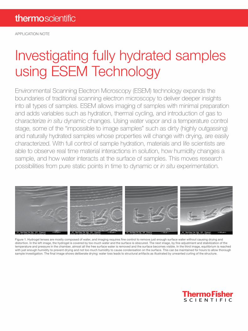
Investigating fully hydrated samples using ESEM TechnologyEnvironmental Scanning Electron Microscopy (ESEM) technology expands the boundaries of traditional scanning electron microscopy to deliver deeper insights into all types of samples. ESEM allows imaging of samples with minimal preparation and adds variables such as hydration, thermal cycling, and introduction of gas to characterize in situ dynamic changes. Using water vapor and a temperature control stage, some of the “impossible to image samples” such as dirty (highly outgassing) and naturally hydrated samples whose properties will change with drying, are easily characterized. With full control of sample hydration, materials and life scientists are able to observe real time material interactions in solution, how humidity changes a sample, and how water interacts at the surface of samples. This moves research possibilities from pure static points in time to dynamic or in situ experimentation.
APPLICATION NOTE
Figure 1. Hydrogel lenses are mostly composed of water, and imaging requires fine control to remove just enough surface water without causing drying and distortion. In the left image, the hydrogel is covered by too much water and the surface is obscured. The next image, by fine adjustment and stabilization of the temperature and pressure in the chamber, almost all the free surface water is removed and the surface becomes visible. In the third image, equilibrium is reached with just enough humidity to prevent drying and not too much humidity to cause condensation on the surface. This can be maintained for hours to allow thorough sample investigation. The final image shows deliberate drying; water loss leads to structural artifacts as illustrated by unwanted curling of the structure.

Naturally hydrated samplesWith the increasing need for higher resolution, researchers move from optical techniques to electron microscopy. For some samples, this transition will necessitate removal of water and may change sample structures. Naturally hydrated specimens (>25% “free” water content) may or may not be normally covered in water, but the internal water content is what often retains a sample’s shape, and removal of the internal water can collapse and massively alter the sample surface. For some samples this can be reversible, but for others, the structural modification could lead to defects and distortion.
Pairing ESEM with a cooling stage allows maintaining any hydration state by varying temperature and pressure. The phase diagram chart below shows the relationship to humidity level.
Figure 3. Images of a fresh flower petal show that having sufficient hydration allows imaging without damage (left), while the effects of having only slightly too little humidity of 70% (right) can lead to shrinkage, water loss and structural collapse.
Figure 4. Naturally hydrated samples like molds require relative humidity from 70-90% and are thus a bit more tolerant of humidity changes. Bread mold is shown on the left and Danish blue cheese on the right (also showing crystal spikes likely from salts).
Figure 2. The phase diagram for pure water illustrates the humidity control range available to keep wet samples wet. Samples should be cooled to keep moisture in the sample within the vacuum chamber. However freezing, which would convert the water to ice, should be avoided when the water content is high, to prevent formation of hexagonal ice crystals that can damage sample structure.
Figure 5. Tissue samples can also be kept wet enough to avoid the collapse of red blood cells, as seen in the above image of liver tissue cut across a blood vessel. This sample was cut, fixed in gluteraldehyde and rinsed, and placed in the ESEM for imaging.
Ideally, samples should be maintained above freezing to avoid the formation of crystalline ice which can rip apart delicate structures. In order to maintain a water hydration state, the imaging gas needs to be pure water vapor (not moist air or inert nitrogen) so the partial pressures of water will match known pure water partial pressure data to control hydration. In the ESEM, this is automated with a live display of humidity set point. Imaging naturally hydrated samples has never been easier.
Samples such as biological materials are naturally hydrated and traditionally all the water is removed by a substitution drying method. With ESEM, samples are imaged directly without substitution and drying. In Figure 3, examples of a flower petal compare adequate hydration with conditions not matching the sample’s needs — the latter resulting in structural collapse.
Of course it is possible to freeze hydrated samples and investigate with cryogenic conditions in SEM as well, however with ESEM, the general guideline is to avoid freezing. When water freezes, the molecules assume a hexagonal structure which is more open than liquid water and this space transformation will often damage and distort wet sample structures (i.e. samples with significant free water; generally >25%). For freezing without hexagonal ice, nitrogen slush plunge techniques should be used with cryogenic stages to achieve amorphous ice. ESEM allows viewing hydrated samples directly without freezing as shown in Figures 4 and 5.
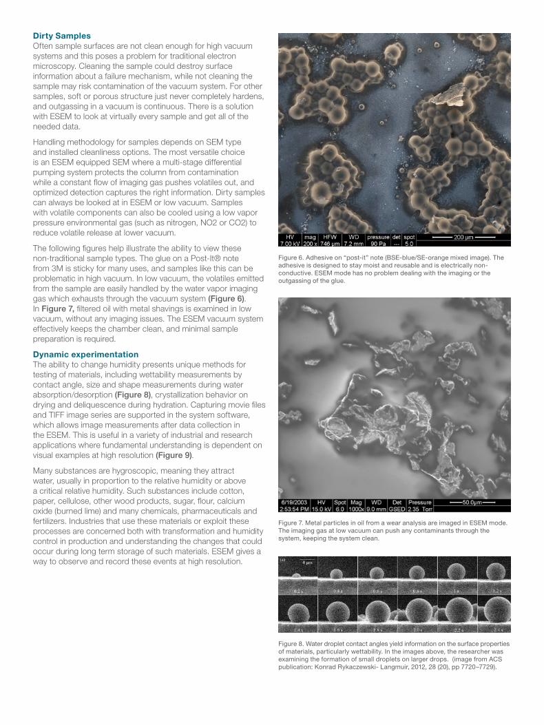
Figure 6. Adhesive on “post-it” note (BSE-blue/SE-orange mixed image). The adhesive is designed to stay moist and reusable and is electrically non-conductive. ESEM mode has no problem dealing with the imaging or the outgassing of the glue.
Figure 7. Metal particles in oil from a wear analysis are imaged in ESEM mode. The imaging gas at low vacuum can push any contaminants through the system, keeping the system clean.
Figure 8. Water droplet contact angles yield information on the surface properties of materials, particularly wettability. In the images above, the researcher was examining the formation of small droplets on larger drops. (image from ACS publication: Konrad Rykaczewski- Langmuir, 2012, 28 (20), pp 7720–7729).
Dirty SamplesOften sample surfaces are not clean enough for high vacuum systems and this poses a problem for traditional electron microscopy. Cleaning the sample could destroy surface information about a failure mechanism, while not cleaning the sample may risk contamination of the vacuum system. For other samples, soft or porous structure just never completely hardens, and outgassing in a vacuum is continuous. There is a solution with ESEM to look at virtually every sample and get all of the needed data.
Handling methodology for samples depends on SEM type and installed cleanliness options. The most versatile choice is an ESEM equipped SEM where a multi-stage differential pumping system protects the column from contamination while a constant flow of imaging gas pushes volatiles out, and optimized detection captures the right information. Dirty samples can always be looked at in ESEM or low vacuum. Samples with volatile components can also be cooled using a low vapor pressure environmental gas (such as nitrogen, NO2 or CO2) to reduce volatile release at lower vacuum.
The following figures help illustrate the ability to view these non-traditional sample types. The glue on a Post-It® note from 3M is sticky for many uses, and samples like this can be problematic in high vacuum. In low vacuum, the volatiles emitted from the sample are easily handled by the water vapor imaging gas which exhausts through the vacuum system (Figure 6). In Figure 7, filtered oil with metal shavings is examined in low vacuum, without any imaging issues. The ESEM vacuum system effectively keeps the chamber clean, and minimal sample preparation is required.
Dynamic experimentationThe ability to change humidity presents unique methods for testing of materials, including wettability measurements by contact angle, size and shape measurements during water absorption/desorption (Figure 8), crystallization behavior on drying and deliquescence during hydration. Capturing movie files and TIFF image series are supported in the system software, which allows image measurements after data collection in the ESEM. This is useful in a variety of industrial and research applications where fundamental understanding is dependent on visual examples at high resolution (Figure 9).
Many substances are hygroscopic, meaning they attract water, usually in proportion to the relative humidity or above a critical relative humidity. Such substances include cotton, paper, cellulose, other wood products, sugar, flour, calcium oxide (burned lime) and many chemicals, pharmaceuticals and fertilizers. Industries that use these materials or exploit these processes are concerned both with transformation and humidity control in production and understanding the changes that could occur during long term storage of such materials. ESEM gives a way to observe and record these events at high resolution.
For current certifications, visit FEI.com/certifications. © 2017 Thermo Fisher Scientific Inc. All rights reserved. All trademarks are the property of Thermo Fisher Scientific and its subsidiaries unless otherwise specified. AN0057-EN-11-2017
Find out more at thermofisher.com/EM-Sales
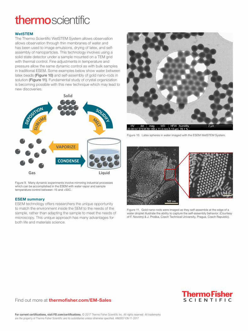
Figure 10. Latex spheres in water imaged with the ESEM WetSTEM System.
Figure 9. Many dynamic experiments involve mirroring industrial processes which can be accomplished in the ESEM with water vapor and sample temperature control between -15 and +50C.
Figure 11. Gold nano-rods were imaged as they self-assemble at the edge of a water droplet illustrate the ability to capture the self-assembly behavior. (Courtesy of F. Novotný & J. Proška, Czech Technical University, Prague, Czech Republic).
WetSTEMThe Thermo Scientific WetSTEM System allows observation allows observation through thin membranes of water and has been used to image emulsions, drying of latex, and self-assembly of nanoparticles. This technology involves using a solid state detector under a sample mounted on a TEM grid with thermal control. Fine adjustments in temperature and pressure allow the same dynamic control as with bulk samples in traditional ESEM. Some examples below show water between latex beads (Figure 10) and self-assembly of gold nano-rods in solution (Figure 11). Fundamental study of crystal organization is becoming possible with this new technique which may lead to new discoveries.
ESEM summaryESEM technology offers researchers the unique opportunity to match the environment inside the SEM to the needs of the sample, rather than adapting the sample to meet the needs of microscopy. This unique approach has many advantages for both life and materials science.