apkc enables development of zonula adherens by ......(ajs) and desmosomes – in order to establish...
TRANSCRIPT

2481Research Article
IntroductionEpithelial cells develop a set of characteristic cell-cell adhesionstructures – termed tight junctions (TJs), belt-like adherens junctions(AJs) and desmosomes – in order to establish and maintain theirapicobasal polarity (Yeaman et al., 1999). Among these structures,belt-like AJs, also called zonula adherens, represent the mostessential adhesion structures, in which E-cadherin works as anadhesion molecule and to which tangential thick actin belts, calledperijunctional actin belts, are closely associated (Tsukita et al., 1992;Yonemura et al., 1995). Owing to their crucial roles in epithelial-cell polarity, the development and regulation of belt-like AJs hasbeen an important research subject in cell biology.
The developmental process of belt-like AJs is tightly coupledwith dramatic reorganization of F-actin (Adams et al., 1998; Vaeziet al., 2002; Vasioukhin et al., 2000; Yonemura et al., 1995; Zhanget al., 2005). Initial cell-cell contacts, via cadherin, induce rapidactin polymerization and cadherin clustering, which result in theformation of spot-like AJs, to which multiple actin filaments areperpendicularly associated (hereafter referred to as radial actinfibers; see Fig. 1B) (Adams et al., 1998; Vasioukhin et al., 2000;Yonemura et al., 1995). Spot-like AJs are also formed in fibroblasts.However, the spot-like AJs observed in epithelial cells are veryunique because they subsequently become reorganized into
continuous belt-like AJs. During this process, the free ends of radialactin fibers associate with the epithelial-specific structure, thecircumferential loose cables of F-actin (hereafter referred to ascircumferential actin cables; see Fig. 1B), and finally develop intoperijunctional actin belts (Vaezi et al., 2002; Vasioukhin et al., 2000).Immunofluorescence studies have demonstrated that, as epithelialcells become polarized, the circumferential actin cables expandtowards the cell periphery with concomitant shortening of the radialactin fibers, and finally develop into perijunctional actin belts thatare closely associated with the membrane (Yonemura et al., 1995).These results suggest that the epithelium-specific F-actinreorganization is crucial for the formation of belt-like AJs. However,the underlying molecular mechanisms are largely unknown.
The perijunctional actin belts as well as circumferential actincables contain activated myosin II (Ivanov et al., 2005; Zhang etal., 2005). Furthermore, recent studies have revealed that theaddition of myosin-II inhibitors or siRNA-mediated knockdown ofnonmuscle myosin heavy chain IIA disrupt the circumferential actincables and suppress epithelial-junction development and cellpolarization (Chen and Macara, 2005; Ivanov et al., 2007; Ivanovet al., 2005; Miyake et al., 2006; Zhang et al., 2005), indicatingthat myosin-II activity is essential for epithelial-junctiondevelopment. Taken together with the observation that the
Atypical protein kinase C (aPKC) generally plays crucial rolesin the establishment of cell polarity in various biologicalcontexts. In mammalian epithelial cells, aPKC essentially workstowards the transition of primordial spot-like adherensjunctions (AJs) into continuous belt-like AJs, also called zonulaadherens, lined with perijunctional actin belts. To reveal themechanism underlying this aPKC function, we investigated thefunctional relationship between aPKC and myosin II, theessential role of which in epithelial-junction development wasrecently demonstrated. Despite its deleterious effects on junctionformation, overexpression of a dominant-negative mutant ofaPKC (aPKCλ kn) did not interfere with the initial phase ofmyosin-II activation triggered by the formation of Ca2+-switch-induced cell-cell contacts. Furthermore, cells overexpressingaPKCλ kn exhibited myosin-II-dependent asymmetricorganization of F-actin along the apicobasal axis, suggesting thataPKC contributes to junction development without affectingthe centripetal contraction of the circumferential actomyosin
cables. Time-lapse analyses using GFP-actin directly revealedthat the circumferential actomyosin cables were centrifugallyexpanded and developed into perijunctional actin belts duringepithelial polarization, and that aPKCλ kn specificallycompromised this process. Taken together, we conclude thataPKC is required for antagonizing the myosin-II-drivencentripetal contraction of the circumferential actin cables,thereby efficiently coupling the myosin-II activity with junctiondevelopment and cell polarization. The present results providenovel insights into not only the site of action of aPKC kinaseactivity but also the role of actomyosin contraction in epithelialpolarization.
Supplementary material available online athttp://jcs.biologists.org/cgi/content/full/121/15/2481/DC1
Key words: PAR, aPKC, Actin dynamics, Adherens junction,Epithelial cells, Polarity
Summary
aPKC enables development of zonula adherens byantagonizing centripetal contraction of thecircumferential actomyosin cablesMasaru Kishikawa, Atsushi Suzuki* and Shigeo OhnoDepartment of Molecular Biology, Yokohama City University Graduate School of Medical Science, 3-9 Fuku-ura, Kanazawa-ku, Yokohama,236-0004, Japan*Author for correspondence (e-mail: [email protected])
Accepted 12 May 2008Journal of Cell Science 121, 2481-2492 Published by The Company of Biologists 2008doi:10.1242/jcs.024109
Jour
nal o
f Cel
l Sci
ence
2482
circumferential actin cables exhibit a continuous flux of centripetal(retrograde) movement from the cytoplasmic periphery towards thenucleus when the cells are free from cell-cell contacts (Vaezi et al.,2002), these results suggest that myosin-II-dependent centripetalcontraction of the circumferential actin cables is crucial for belt-like AJ formation. However, there are apparently conflicting resultsthat the same myosin inhibitors suppress the junction disassembly(Ivanov et al., 2004). Furthermore, several studies havedemonstrated that the circumferential actin cables disassemble inthe vicinity of the cell-cell boundary when the cell makes contactwith other cells (Adams et al., 1996; Gloushankova et al., 1997;Krendel and Bonder, 1999; Yamada and Nelson, 2007). The authorsargued that actomyosin contraction does not act at the region ofcell-cell contact but at cell-cell-contact-free areas in order to expandthe contact regions laterally and render the contacting cells morecompact. Taken together, it still remains to be elucidated howmyosin-II-dependent contraction of the circumferential actin cablesis used for belt-like AJ formation and epithelial polarization (Megeet al., 2006).
Atypical protein kinase C (aPKC) is a crucial component of theaPKC–PAR-6–PAR-3 complex, an evolutionarily conservedsignaling complex for cell polarity (Macara, 2004; Suzuki and Ohno,2006). In epithelial cells, aPKC plays an essential role in thedevelopment of epithelium-specific junction structures, such as belt-like AJs and TJs, and thus contributes to the development ofapicobasal polarity (Knust and Bossinger, 2002; Suzuki et al., 2001).By analyzing the repolarization process during wound healing ofa mouse epithelial cell line, MTD1-A cells, we previouslydemonstrated that the kinase activity of aPKC is not required forthe formation of spot-like AJs to which aPKC itself is recruited,but is essential for their subsequent transition into belt-like AJs(Suzuki et al., 2002; Suzuki et al., 2001). We further found thatcells subjected to aPKC inhibition terminated the F-actinreorganization at an intermediate state in which the radial actin fibersremained associated with the circumferential actin cablesperpendicularly. In the present study, we examined the possibilitythat aPKC contributes to the development of epithelium-specificjunctions by regulating myosin-II activity. Our results indicate thataPKC does not affect initial myosin-II activation triggered by Ca2+-switch-induced formation of cell-cell contacts, but is required toantagonize the myosin-II-dependent centripetal contraction of thecircumferential actin cables within individual cells. By doing so,aPKC transforms the centripetal contractile force of thecircumferential actin cables towards the development of belt-likeAJs. The present results provide not only important clues for themolecular basis of how aPKC regulates the development ofepithelium-specific junctions but also a novel concept for the roleof the contractile force of the perijunctional actin belts.
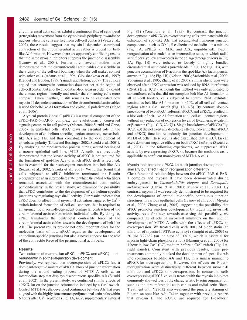
ResultsTwo isoforms of mammalian aPKC – aPKCλ and aPKCζ – actredundantly in epithelial-junction developmentPreviously, we reported that overexpression of aPKCλ kn, adominant-negative mutant of aPKCλ, blocked junction reformationduring the wound-healing process of MTD1-A cells at anintermediate step that displays discontinuous spot-like AJs (Suzukiet al., 2002). In the present study, we confirmed similar effects ofaPKCλ kn on the junction reformation induced by a Ca2+ switch.Control MTD1-A cells developed continuous belt-like AJs that werealigned with the highly concentrated perijunctional actin belts within6 hours after Ca2+ repletion (Fig. 1A, lacZ; supplementary material
Fig. S1) (Yonemura et al., 1995). By contrast, the junctiondevelopment in aPKCλ-kn-overexpressing cells terminated with theexhibition of spot-like AJs that accumulated numerous junctioncomponents – such as ZO-1, E-cadherin and occludin – in a mixture(Fig. 1A, aPKCλ kn; M.K. and A.S., unpublished). F-actinreorganization also ended at an intermediate state, in which radialactin fibers (yellow arrowheads in the enlarged merged views in Fig.1A; Fig. 1B) were tethered to loosely or tightly bundledcircumferential actin cables (arrowheads in Fig. 1A; Fig. 1B), withpunctate accumulation of F-actin on the spot-like AJs (actin puncta;arrows in Fig. 1A; Fig. 1B) (Nelson, 2003; Vasioukhin et al., 2000;Yonemura et al., 1995; Zhang et al., 2005). Similar phenotypes wereobserved after aPKC expression was reduced by RNA interference(RNAi) (Fig. 1C,D). Although this method was only applicable tosubconfluent cells that did not complete belt-like AJ formation atall cell-cell borders, cells subjected to control RNAi exhibitedcontinuous belt-like AJ formation in ~50% of all cell-cell-contactregions after a Ca2+ switch (Fig. 1D, NS). By contrast, double-knockdown of two aPKC isoforms, aPKCλ and aPKCζ, resulted ina blockade of belt-like AJ formation at all cell-cell-contact regions,without any reduction of expression levels of E-cadherin, α-cateninor β-catenin (Fig. 1C,D, λ3+ζ1). Single knockdown of aPKCλ (Fig.1C,D, λ3) did not exert any detectable effects, indicating that aPKCλand aPKCζ function redundantly for junction development inMTD1-A cells. These results also established that aPKCλ kn canexert dominant-negative effects on both aPKC isoforms (Suzuki etal., 2001). In the following experiments, we suppressed aPKCactivity by overexpressing aPKCλ kn, because this method is easilyapplicable to confluent monolayers of MTD1-A cells.
Myosin inhibitors and aPKCλ kn block junction developmentsimilarly but affect F-actin reorganization differentlyClose functional relationships between the aPKC–PAR-6–PAR-3 complex and myosin II have been demonstrated duringembryogenesis in Caenorhabditis elegans and Drosophilamelanogaster (Barros et al., 2003; Munro et al., 2004). Bycontrast, myosin II was recently demonstrated to be required forthe development of epithelium-specific continuous junctionstructures in various epithelial cells (Ivanov et al., 2005; Miyakeet al., 2006; Zhang et al., 2005), suggesting the possibility thataPKC promotes junction development by regulating myosin-IIactivity. As a first step towards assessing this possibility, wecompared the effects of myosin-II inhibitors on the junctiondevelopment of MTD1-A cells with the effects of aPKCλ-knoverexpression. We treated cells with 100 μM blebbistatin (aninhibitor of myosin-II ATPase activity) (Straight et al., 2003) or20 μM Y27632 (an inhibitor of ROCK, which is essential formyosin light-chain phosphorylation) (Narumiya et al., 2000) for1 hour in low Ca2+ (LC) medium before a Ca2+ switch (Fig. 1A,right panels). Consistent with previous results, these pre-treatments commonly blocked the development of spot-like AJsinto continuous belt-like AJs and TJs, in a similar manner toaPKCλ-kn overexpression. However, the effects on F-actinreorganization were distinctively different between myosin-IIinhibition and aPKCλ-kn overexpression. In contrast to cellsoverexpressing aPKCλ kn, cells treated with the myosin inhibitorscommonly showed loss of the characteristic F-actin organization,such as the circumferential actin cables and radial actin fibers.Treatment with Y27632 also weakened the punctate staining ofF-actin on spot-like AJs. Taken together with previous reportsthat myosin II and ROCK are required for E-cadherin
Journal of Cell Science 121 (15)
Jour
nal o
f Cel
l Sci
ence
2483aPKC function in epithelial polarization
accumulation at initial cell-cell-contact regions and radial actin-fiber formation (Shewan et al., 2005; Vaezi et al., 2002), theseresults suggest that the myosin inhibitors suppressed F-actinreorganization at an early stage of junction development, when
aPKC activity is not required. These findings indicate that, despitetheir apparently similar effects on junction formation, myosin IIfunctions independently of aPKC, at least at the initial stage ofjunction development.
Fig. 1. Comparison of the effects of aPKCλ kn and of myosin-II inhibitors on the junction development of MTD1-A cells. (A) Myosin inhibitors and aPKCλ kn, adominant-negative mutant of aPKCλ, block junction development similarly but affect actin reorganization differently. Confluent monolayers of MTD1-A cellstransformed by adenovirus vectors encoding β-galactosidase (LacZ) or aPKCλ kn were cultured in LC medium for 40 hours and then subjected to a Ca2+ switch inthe presence or absence of the myosin inhibitors blebbistatin (100 μM) or Y27632 (20 μM). The cells were fixed at 6 hours after the Ca2+ switch and stained withan anti-ZO-1 antibody (magenta in merged images) and phalloidin (green in merged images). The images shown are single confocal sections that were selected todemonstrate the ZO-1 staining most clearly. Enlargements of the boxed regions in the merged views are shown at the bottom. For cells overexpressing aPKCλ kn,two typical images are presented, in which the left and right panels show cells exhibiting tightly and loosely bundled circumferential actin cables, respectively(each represents ~40% and ~60% of aPKCλ-kn-overexpressing cells, respectively). White arrowheads, circumferential actin cables; arrows, punctate staining of F-actin on spot-like AJs; yellow arrowheads, radial actin fibers. (B) Schematic presentation of the intermediate state of F-actin organization observed in aPKCλ-kn-overexpressing cells. (C) RNAi knockdown of aPKC isoforms in MTD1-A cells. Subconfluent MTD1-A cells were transfected with the indicated siRNAoligonucleotide duplexes (NS, non-silencing siRNA; λ3, aPKCλ siRNA; ζ1, aPKCζ siRNA). Top panels: western blot analyses of total extracts of cells subjectedto the indicated RNAi. aPKCλ was specifically detected by an anti-aPKCι (human aPKCλ) antibody, whereas both aPKC isoforms (aPKCλ and aPKCζ) weresimultaneously detected by an anti-aPKC antibody (C20) that reacts with both isoforms. GAPDH was used as a loading control. The data for E-cadherin, α-cateninand β-catenin indicated no significant change in the expression levels of these AJ proteins. Images shown underneath: the indicated cells were immunostained withan anti-aPKC antibody (C20). (D) aPKC knockdown suppresses junction development after a Ca2+ switch, in a similar manner to aPKCλ-kn overexpression. Theindicated cells were subjected to a Ca2+ switch as described in A and then immunostained with an anti-ZO-1 antibody (magenta in merged images) and phalloidin(green in merged images). Scale bars: 10 μm.
Jour
nal o
f Cel
l Sci
ence

2484
aPKCλ kn does not affect Ca2+-switch-induced activation ofmyosin II in the early stage of junction formationNext, we analyzed myosin-II localization and activation inpolarizing MTD1A cells, and examined the effects of aPKCλ onthese activities. Because MTD1-A cells dominantly expressnonmuscle myosin heavy chains IIA and IIB (M.K. and A.S.,unpublished), we stained these chains using a mixture of specificantibodies that recognize these isoforms. In control cells expressinglacZ, myosin II showed weak peripheral localization in depolarizedcells (Fig. 2A). Upon Ca2+ repletion, it immediately accumulatedon loosely bundled circumferential actin cables (Fig. 2A, arrows, 0.5 hours). Because the cables contained Ser19monophosphorylated regulatory myosin light chain (p-MLC2)(Fig. 2B,C), these circumferential actin cables were considered tobe contractile actomyosin bundles. By contrast, radial actin fibers,which were strongly positive for α-actinin (Fig. 2A), were almostcompletely free from myosin II (Vaezi et al., 2002) (Fig. 2B),suggesting that radial actin fibers are not contractile structures. Inthe later stage, the circumferential actin cables progressively
developed into highly compacted perijunctional actin belts that werein close contact with cell-cell contacts and remained positive forp-MLC2 (arrowheads in Fig. 2A, 6 hours; Fig. 2C, 6 hours;supplementary material Fig. S2). Western blot analysis of p-MLC2in total cell lysate also revealed the rapid increase in the level ofp-MLC2 upon Ca2+-switch-induced cell-cell contact, although thetime courses of bulk MLC2 phosphorylation did not appear to beperfectly consistent with the immunocytochemical resultscontaining spatiotemporal information: bulk MLC2phosphorylation initially peaked at the very early stage after a Ca2+
switch (30 minutes) and showed a slight increase again at 7.5 hourswhen the cells completed the formation of belt-like AJs and TJs.Notably, aPKCλ-kn overexpression did not inhibit the accumulationof myosin II and p-MLC2 on the circumferential actin cables(arrows in Fig. 2A; Fig. 2C) or affect the level and time course ofMLC2 monophosphorylation (Fig. 2D). Nevertheless, most of thecircumferential actomyosin cables in aPKCλ-kn-overexpressingcells were not expanded or brought into close contact with theplasma membrane, even at 6 hours after the Ca2+ switch (Fig. 2A,C).
Journal of Cell Science 121 (15)
Fig. 2. Suppression of aPKC activity does notaffect cell-cell-contact-induced myosin-IIactivation. (A) MTD1-A cells transfected withadenovirus vectors encoding β-galactosidase(LacZ) or aPKCλ kn were subjected to a Ca2+
switch as described in the legend for Fig. 1,fixed at the indicated times after the Ca2+
switch, and double stained with a mixture ofantibodies against nonmuscle myosin heavychain IIA and IIB (magenta in merged images)and against α-actinin (green in mergedimages). Arrows, circumferential actin cables;arrowheads, perijunctional actin belts.(B) Control MTD1A cells were fixed at 30minutes after a Ca2+ switch and double stainedfor F-actin (phalloidin; green in mergedimages) and with antibodies against eithermyosin II, Ser19 monophosphorylated myosinlight-chain 2 (p-MLC2) or Thr18 and Ser19diphosphorylated MLC2 (pp-MLC2) (magentain merged images) as indicated at the top. Notethat radial actin fibers were free from myosinII. (C) MTD1-A cells were immunostainedwith anti-p-MLC2 antibody at the indicatedtimes after the Ca2+ switch. Pre-treatment withY27632 completely abolishes the staining,suggesting that ROCK is dominantlyresponsible for this phosphorylation. (D) Totalcell extracts of MTD1-A cells were prepared atthe indicated times after the Ca2+ switch, andthe changes in the p-MLC2 level were analyzedby western blotting. Note that overexpressionof aPKCλ kn does not affect the Ca2+-switch-induced increase in p-MLC2. Scale bars: 10μm.
Jour
nal o
f Cel
l Sci
ence

2485aPKC function in epithelial polarization
Ivanov et al. demonstrated atransient increase of Thr18 andSer19 diphosphorylated MLC2(pp-MLC2) 1 hour after Ca2+
repletion in T84 cells (Ivanov etal., 2005). However, we couldnot detect such an early increasein the level of pp-MLC2 inMTD1-A cells not onlybiochemically but alsoimmunocytochemically (Fig. 2B;supplementary material Fig. S2).This discrepancy might arisefrom cell-type difference.However, Ivanov et al. and weboth found that pp-MLC2 wasexclusively detected on thematured perijunctional actinbelts, but not on thecircumferential actin cables(supplementary material Fig. S2)(Ivanov et al., 2005), suggestingthat pp-MLC2 is involved in thefinal stage of junctionaldevelopment, when thematuration of perijunctional actinbelts proceeds, but not in theearly stage, when thecircumferential actin cablevigorously develops and starts toexpand. Consistently, thisincrease of pp-MLC2 at the finalstage of junctional developmentwas suppressed in aPKCλ-kn-overexpressing cells, in which thecompletion of perijunctional actin belts was significantly inhibited.
Collectively, the present results indicate that the effect of aPKCλkn on the development of belt-like AJs was not a result of inhibitingthe myosin-II-dependent contractile force of the circumferentialactin cables observed in the early stage of junctional development.
aPKCλ-kn-overexpressing cells exhibit myosin-II-dependentdevelopment of polarized F-actin organization and cuboidalcell shapeWe previously reported that, despite incomplete formation of belt-like AJs, aPKCλ-kn-overexpressing cells develop a polarizedcolumnar shape, in which the apical F-actin structures, such as thecircumferential actin cables and radial actin fibers, are clearlyseparated from the basal stress fibers (Suzuki et al., 2002).Consistently, vinculin exhibited two kinds of discrete localizationin aPKCλ-kn-overexpressing cells: at the apical cell-cell contacts(spot-like AJs) and at basal focal contacts (arrows and arrowheads,respectively, Fig. 3A,B). These findings were in sharp contrast tothe effects of myosin-II inhibitors, which disrupt the increase in thelateral domain and development of a polarized organization of F-actin (Zhang et al., 2005). In these cases, vinculin staining at thecell-cell and cell-substrate contacts became significantly weaker,and were observed on the same focal plane at the basal side (Fig.3A,B). Recently, Miyake et al. demonstrated that vinculinrecruitment into cadherin-catenin complexes is tension dependent(Miyake et al., 2006). Furthermore, Zhang et al. demonstrated thatmyosin-II-based contraction is involved in the increase in the height
of the lateral domain during epithelial-cell polarization (Zhang etal., 2005). Taken together, the present results suggest that aPKCλ-kn-overexpressing cells normally utilize myosin-II activity, at leastin part, to develop their polarized F-actin organization and cuboidalcell shape. This is extremely consistent with the above results thatshow that aPKCλ kn did not interfere with the myosin-II activationobserved at the early stage of junctional development. Nevertheless,cells lacking aPKC activity could not induce the transition of spot-like AJs into continuous AJs and TJs. Therefore, our resultsindicate the presence of a novel elementary process during the latestage of epithelial-junction development, in which aPKC activityis crucially required independently of myosin-II-dependentcentripetal contraction of circumferential actin cables.
Centripetal contraction of the circumferential actin cablesneeds to be antagonized for use in epithelial-junctiondevelopmentNext, we investigated what was lacking in aPKCλ-kn-overexpressing cells that was needed to promote epithelial-junctiondevelopment. While considering this aspect, we noted thatcontraction of the circumferential actomyosin cables should conflictwith expansion of the cables. Therefore, when cells becomepolarized, the myosin-II-mediated contractile force imposed on thecircumferential actin cables needs to be counterbalanced by anantagonistic centrifugal force for use in junction development andcell polarization. If this does not occur, the contraction of thecircumferential actomyosin cables would be detrimental to junctiondevelopment. In fact, a recent study demonstrated that myosin-II-
Fig. 3. Suppression of aPKC activity does not disrupt the polarized organization of vinculin-containing structures atregions of cell-cell and cell-substrate contact. (A) MTD1-A cells treated with the indicated adenovirus vectors ormyosin inhibitors were subjected to a Ca2+ switch as described in the legend for Fig. 1, and were then immunostainedwith rhodamine-phalloidin (top panels) and an anti-vinculin antibody (bottom panels) at 6 hours after the Ca2+ switch.Single confocal sections at the basal planes (basal) and apical planes 2.5 μm upward from the basal planes (apical) areshown. The signal intensity of phalloidin staining in the basal planes was enhanced to a comparable level to that in theapical planes. Note that aPKCλ kn does not disrupt the polarized distribution of vinculin into the two distinctstructures at apical cell-cell contacts (arrows) and basal cell-substrate contacts (arrowheads). By contrast, cells treatedwith myosin-II inhibitors exhibit vinculin staining in the same planes. (B) Reconstituted xz views of the cellsdemonstrated in A were presented for rhodamine-phalloidin staining. Arrowheads point to the apical and basal planesat which the confocal data were obtained. Scale bar: 10 μm.
Jour
nal o
f Cel
l Sci
ence
2486
dependent contraction of perijunctional actin belts plays a positiverole in disassembly of epithelial cell-cell junctions when cadherin-mediated cell-cell adhesions are weakened by Ca2+ depletion(Ivanov et al., 2004). This indicates the possibility that aPKCregulates this putative outward force that is required for antagonizingthe contractile force of the actomyosin cables, and thus contributesto epithelial-junction formation and polarization.
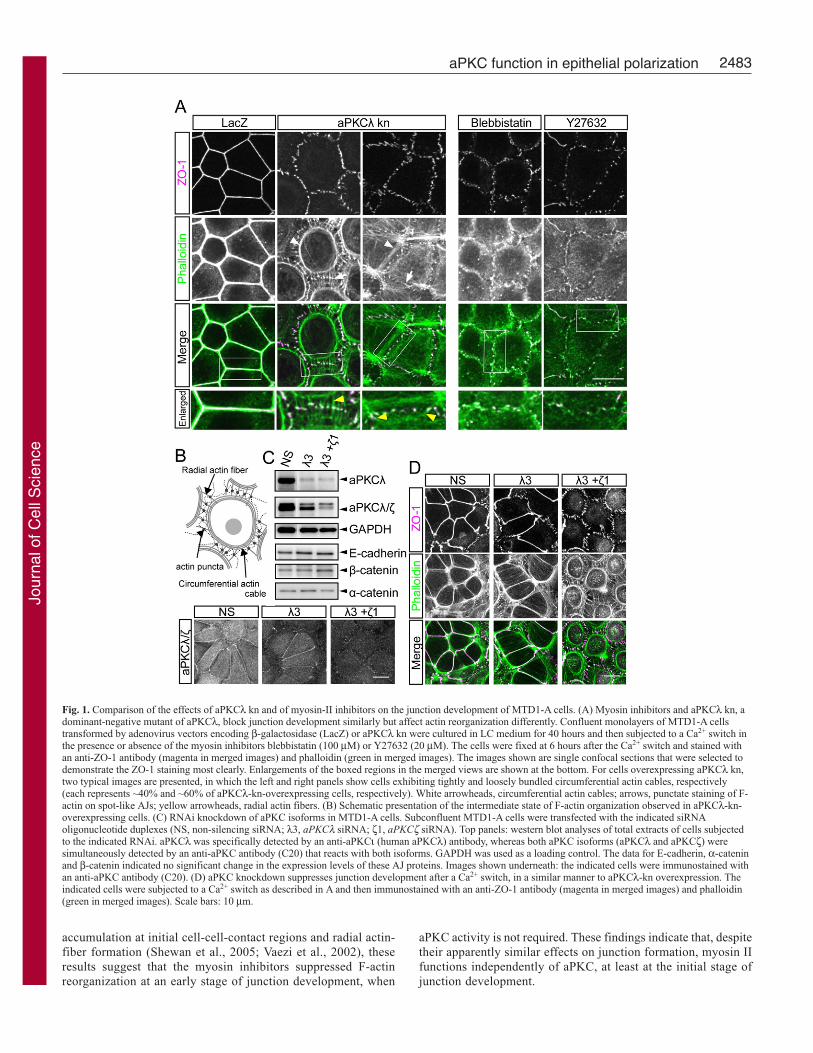
Consistent with this hypothesis, aPKCλ-kn-overexpressing cellssometimes showed hyper-shrinkage of the circumferential actincables after a Ca2+ switch (supplementary material Fig. S3). Thiseffect of aPKCλ kn was more conspicuous when junctiondevelopment was acutely induced by lysophosphatidic acid (LPA)in serum-starved MTD1-A cells. During the course of the presentstudy, we found that serum-starved MTD1-A cells could notdevelop continuous junction structures, even in the presence of Ca2+,and terminated the junction development with spot-like AJformation (Fig. 4; supplementary material Fig. S4) in a similarmanner to myosin-II inhibitors. Consistent with the hypothesis thatreduced myosin-II activity is one of the causes of the defects inserum-starved MTD1-A cells, addition of LPA, a physiologicalligand for Rho activation, acutely induced a dramatic reorganizationof F-actin, leading to the formation of circumferential actin cables,and restored normal junction development in a myosin-II-inhibitor-sensitive manner (Fig. 4; M.K. and A.S., unpublished). Again,aPKCλ kn inhibited this LPA-induced junction formation in serum-
starved MTD1-A cells without suppressing the circumferential actin-cable formation (Fig. 4). Interestingly, and probably due to the acutemyosin-II activation, most of the aPKCλ-kn-overexpressing cellsexhibited hyper-shrinkage of the circumferential actomyosin cables,which were tethered to the dot-like AJs by radial actin fibers (Fig.4). These results are consistent with the above hypothesis that aPKCactivity is required to antagonize the centripetal contraction ofactomyosin circumferential actin cables in the late phase ofepithelial-junction development.
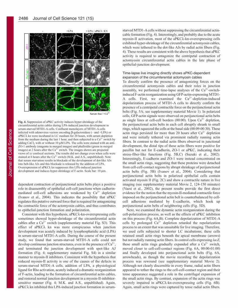
Time-lapse live imaging directly shows aPKC-dependentexpansion of the circumferential actomyosin cablesTo directly confirm the presence of antagonizing forces on thecircumferential actomyosin cables and their roles in junctionassembly, we performed time-lapse analyses of the Ca2+-switch-induced F-actin reorganization using GFP-actin-expressing MTD1-A cells. First, we examined the Ca2+-depletion-induceddepolarization process of MTD1-A cells to directly confirm thepresence of a centripetal contractile force on the perijunctional actinbelts (Fig. 5A; see supplementary material Movie 1). In polarizedcells, GFP-actin signals were observed on perijunctional actin beltsas single lines at cell-cell borders (00:00). Upon Ca2+ depletion,the perijunctional actin belts in each cell abruptly shrank to smallrings, which squeezed the cells at the basal side (00:09-00:30). Theseactin rings persisted for more than 20 hours after Ca2+ depletionand were initially tethered via prominent radially running actinfibers. In contrast to the radial actin fibers observed in junctionaldevelopment, the distal tips of these actin fibers were positive forpaxillin but not for E-cadherin, ZO-1 or aPKC, indicating theirstress-fiber-like functions (Fig. 5B,C) (Suzuki et al., 2002).Interestingly, E-cadherin and ZO-1 were instead concentrated onthe small actin rings, suggesting that these proteins were detachedfrom cell-cell-contact regions by abrupt shrinkage of perijunctionalactin belts (Fig. 5B) (Ivanov et al., 2004). Considering thatperijunctional actin belts in polarized epithelial cells containactivated myosin II (Fig. 2C) and show a contractile nature in liveimaging (see supplementary material Movie 2, 124-150 minutes)(Vaesi et al., 2002), the present results provide the first directevidence for the notion that the myosin-II-mediated contractile forceimposed on the perijunctional actin belt is counterbalanced by cell-cell adhesions mediated by E-cadherin, which hook upperijunctional actin belts of neighboring cells (Fig. 5D).
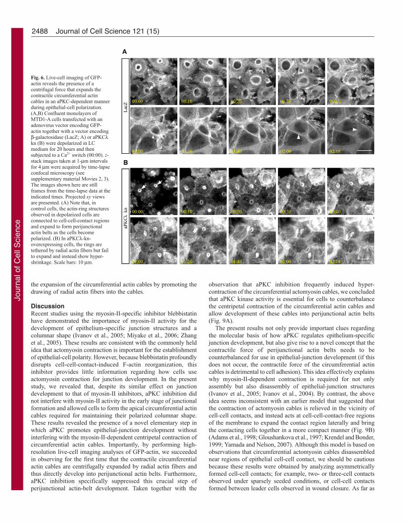
Next, we examined the dynamic actin reorganization during thecell-polarization process, as well as the effects of aPKC inhibitionon this process (Fig. 6A,B). Complete depolarization of MTD1-Acells by prolonged Ca2+ depletion retarded the repolarizationprocess to an extent that was unsuitable for live imaging. Therefore,we used cells subjected to shorter LC incubations; these cellsretained small actin rings beneath the apical membrane (arrows)but not radially running actin fibers. In control cells expressing lacZ,these small actin rings gradually expanded after a Ca2+ switch,moved closer to cell-cell-contact regions (Fig. 6A, 00:00-01:00)and finally developed into perijunctional actin belts (Fig. 6A,arrowheads), as though the movie recording the depolarizationprocess was rewound (see supplementary material Movie 2).Although not clearly discernible in every frame, radial actin fibersappeared to tether the rings to the cell-cell-contact region and theirtense appearance suggested a role in the centrifugal expansion ofthe small actin rings. By contrast, expansion of the actin rings wasseverely impaired in aPKCλ-kn-overexpressing cells (Fig. 6B).Again, small actin rings were captured by tense radial actin fibers.
Journal of Cell Science 121 (15)
Fig. 4. Suppression of aPKC activity induces hyper-shrinkage of thecircumferential actin cables during LPA-induced junction development inserum-starved MTD1-A cells. Confluent monolayers of MTD1-A cellsinfected with adenovirus vectors encoding β-galactosidase (– and +LPA) oraPKCλ kn were incubated in LC medium for 20 hours, with serum depletionfrom the medium during the last 1 hour, and then subjected to a Ca2+ switch byadding CaCl2 with or without 10 μM LPA. The cells were stained with an anti-ZO-1 antibody (magenta in merged images) and phalloidin (green in mergedimages) at 2 hours after the Ca2+ switch. The images shown are projectedviews of xy confocal sections. The results did not change even when cells werestained at 6 hours after the Ca2+ switch (M.K. and A.S., unpublished). Notethat serum starvation results in blockade of the development of dot-like AJsinto belt-like AJs and this blockade is released by the addition of LPA.Overexpression of aPKCλ kn suppresses this LPA-induced junctiondevelopment and induces hyper-shrinkage of F-actin. Scale bar: 10 μm.
Jour
nal o
f Cel
l Sci
ence
2487aPKC function in epithelial polarization
However, they did not expand and instead shrank to veryconcentrated aggregates (see supplementary material Movie 3). Inthe present experimental condition, many actin rings that formedin aPKCλ-kn-overexpressing cells finally disassembled anddisappeared.
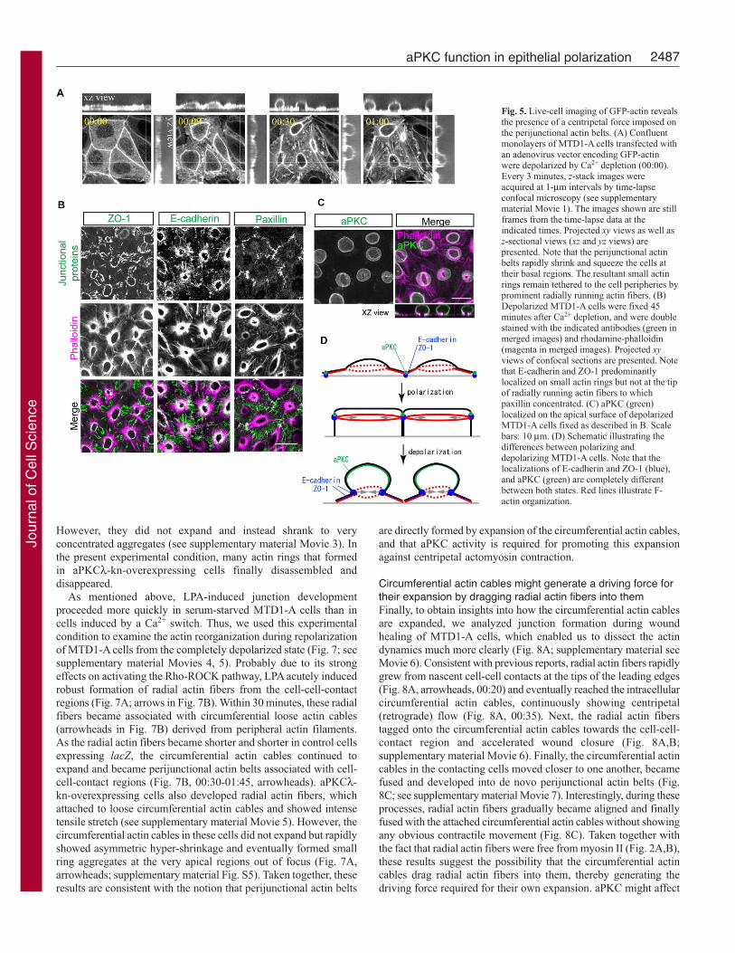
As mentioned above, LPA-induced junction developmentproceeded more quickly in serum-starved MTD1-A cells than incells induced by a Ca2+ switch. Thus, we used this experimentalcondition to examine the actin reorganization during repolarizationof MTD1-A cells from the completely depolarized state (Fig. 7; seesupplementary material Movies 4, 5). Probably due to its strongeffects on activating the Rho-ROCK pathway, LPA acutely inducedrobust formation of radial actin fibers from the cell-cell-contactregions (Fig. 7A; arrows in Fig. 7B). Within 30 minutes, these radialfibers became associated with circumferential loose actin cables(arrowheads in Fig. 7B) derived from peripheral actin filaments.As the radial actin fibers became shorter and shorter in control cellsexpressing lacZ, the circumferential actin cables continued toexpand and became perijunctional actin belts associated with cell-cell-contact regions (Fig. 7B, 00:30-01:45, arrowheads). aPKCλ-kn-overexpressing cells also developed radial actin fibers, whichattached to loose circumferential actin cables and showed intensetensile stretch (see supplementary material Movie 5). However, thecircumferential actin cables in these cells did not expand but rapidlyshowed asymmetric hyper-shrinkage and eventually formed smallring aggregates at the very apical regions out of focus (Fig. 7A,arrowheads; supplementary material Fig. S5). Taken together, theseresults are consistent with the notion that perijunctional actin belts
are directly formed by expansion of the circumferential actin cables,and that aPKC activity is required for promoting this expansionagainst centripetal actomyosin contraction.
Circumferential actin cables might generate a driving force fortheir expansion by dragging radial actin fibers into themFinally, to obtain insights into how the circumferential actin cablesare expanded, we analyzed junction formation during woundhealing of MTD1-A cells, which enabled us to dissect the actindynamics much more clearly (Fig. 8A; supplementary material seeMovie 6). Consistent with previous reports, radial actin fibers rapidlygrew from nascent cell-cell contacts at the tips of the leading edges(Fig. 8A, arrowheads, 00:20) and eventually reached the intracellularcircumferential actin cables, continuously showing centripetal(retrograde) flow (Fig. 8A, 00:35). Next, the radial actin fiberstagged onto the circumferential actin cables towards the cell-cell-contact region and accelerated wound closure (Fig. 8A,B;supplementary material Movie 6). Finally, the circumferential actincables in the contacting cells moved closer to one another, becamefused and developed into de novo perijunctional actin belts (Fig.8C; see supplementary material Movie 7). Interestingly, during theseprocesses, radial actin fibers gradually became aligned and finallyfused with the attached circumferential actin cables without showingany obvious contractile movement (Fig. 8C). Taken together withthe fact that radial actin fibers were free from myosin II (Fig. 2A,B),these results suggest the possibility that the circumferential actincables drag radial actin fibers into them, thereby generating thedriving force required for their own expansion. aPKC might affect
Fig. 5. Live-cell imaging of GFP-actin revealsthe presence of a centripetal force imposed onthe perijunctional actin belts. (A) Confluentmonolayers of MTD1-A cells transfected withan adenovirus vector encoding GFP-actinwere depolarized by Ca2+ depletion (00:00).Every 3 minutes, z-stack images wereacquired at 1-μm intervals by time-lapseconfocal microscopy (see supplementarymaterial Movie 1). The images shown are stillframes from the time-lapse data at theindicated times. Projected xy views as well asz-sectional views (xz and yz views) arepresented. Note that the perijunctional actinbelts rapidly shrink and squeeze the cells attheir basal regions. The resultant small actinrings remain tethered to the cell peripheries byprominent radially running actin fibers. (B)Depolarized MTD1-A cells were fixed 45minutes after Ca2+ depletion, and were doublestained with the indicated antibodies (green inmerged images) and rhodamine-phalloidin(magenta in merged images). Projected xyviews of confocal sections are presented. Notethat E-cadherin and ZO-1 predominantlylocalized on small actin rings but not at the tipof radially running actin fibers to whichpaxillin concentrated. (C) aPKC (green)localized on the apical surface of depolarizedMTD1-A cells fixed as described in B. Scalebars: 10 μm. (D) Schematic illustrating thedifferences between polarizing anddepolarizing MTD1-A cells. Note that thelocalizations of E-cadherin and ZO-1 (blue),and aPKC (green) are completely differentbetween both states. Red lines illustrate F-actin organization.
Jour
nal o
f Cel
l Sci
ence
2488
the expansion of the circumferential actin cables by promoting thedrawing of radial actin fibers into the cables.
DiscussionRecent studies using the myosin-II-specific inhibitor blebbistatinhave demonstrated the importance of myosin-II activity for thedevelopment of epithelium-specific junction structures and acolumnar shape (Ivanov et al., 2005; Miyake et al., 2006; Zhanget al., 2005). These results are consistent with the commonly heldidea that actomyosin contraction is important for the establishmentof epithelial-cell polarity. However, because blebbistatin profoundlydisrupts cell-cell-contact-induced F-actin reorganization, thisinhibitor provides little information regarding how cells useactomyosin contraction for junction development. In the presentstudy, we revealed that, despite its similar effect on junctiondevelopment to that of myosin-II inhibitors, aPKC inhibition didnot interfere with myosin-II activity in the early stage of junctionalformation and allowed cells to form the apical circumferential actincables required for maintaining their polarized columnar shape.These results revealed the presence of a novel elementary step inwhich aPKC promotes epithelial-junction development withoutinterfering with the myosin-II-dependent centripetal contraction ofcircumferential actin cables. Importantly, by performing high-resolution live-cell imaging analyses of GFP-actin, we succeededin observing for the first time that the contractile circumferentialactin cables are centrifugally expanded by radial actin fibers andthus directly develop into perijunctional actin belts. Furthermore,aPKC inhibition specifically suppressed this crucial step ofperijunctional actin-belt development. Taken together with the
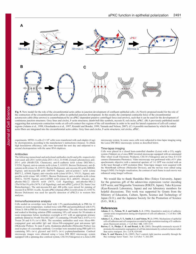
observation that aPKC inhibition frequently induced hyper-contraction of the circumferential actomyosin cables, we concludedthat aPKC kinase activity is essential for cells to counterbalancethe centripetal contraction of the circumferential actin cables andallow development of these cables into perijunctional actin belts(Fig. 9A).
The present results not only provide important clues regardingthe molecular basis of how aPKC regulates epithelium-specificjunction development, but also give rise to a novel concept that thecontractile force of perijunctional actin belts needs to becounterbalanced for use in epithelial-junction development (if thisdoes not occur, the contractile force of the circumferential actincables is detrimental to cell adhesion). This idea effectively explainswhy myosin-II-dependent contraction is required for not onlyassembly but also disassembly of epithelial-junction structures(Ivanov et al., 2005; Ivanov et al., 2004). By contrast, the aboveidea seems inconsistent with an earlier model that suggested thatthe contraction of actomyosin cables is relieved in the vicinity ofcell-cell contacts, and instead acts at cell-cell-contact-free regionsof the membrane to expand the contact region laterally and bringthe contacting cells together in a more compact manner (Fig. 9B)(Adams et al., 1998; Gloushankova et al., 1997; Krendel and Bonder,1999; Yamada and Nelson, 2007). Although this model is based onobservations that circumferential actomyosin cables disassemblednear regions of epithelial cell-cell contact, we should be cautiousbecause these results were obtained by analyzing asymmetricallyformed cell-cell contacts; for example, two- or three-cell contactsobserved under sparsely seeded conditions, or cell-cell contactsformed between leader cells observed in wound closure. As far as
Journal of Cell Science 121 (15)
Fig. 6. Live-cell imaging of GFP-actin reveals the presence of acentrifugal force that expands thecontractile circumferential actincables in an aPKC-dependent mannerduring epithelial-cell polarization.(A,B) Confluent monolayers ofMTD1-A cells transfected with anadenovirus vector encoding GFP-actin together with a vector encodingβ-galactosidase (LacZ; A) or aPKCλkn (B) were depolarized in LCmedium for 20 hours and thensubjected to a Ca2+ switch (00:00). z-stack images taken at 1-μm intervalsfor 4 μm were acquired by time-lapseconfocal microscopy (seesupplementary material Movies 2, 3).The images shown here are stillframes from the time-lapse data at theindicated times. Projected xy viewsare presented. (A) Note that, incontrol cells, the actin-ring structuresobserved in depolarized cells areconnected to cell-cell-contact regionsand expand to form perijunctionalactin belts as the cells becomepolarized. (B) In aPKCλ-kn-overexpressing cells, the rings aretethered by radial actin fibers but failto expand and instead show hyper-shrinkage. Scale bars: 10 μm.
Jour
nal o
f Cel
l Sci
ence

2489aPKC function in epithelial polarization
we could examine, it is not easily applicable to the polarizationprocess of confluent epithelial cells that develop cell-cell contactsrather symmetrically and essentially lack cell-cell-contact-freeregions of the membrane. We observed in our wound-healingexperiments that the association of radial actin fibers impaired theintegrity of the circumferential actin cables and sometimes causedcomplete disassembly of the cables in the vicinity of cell-cellcontacts, especially when their association occurred at very restrictedregions (see supplementary material Movie 8). In these cases, theends of the fragmented circumferential actin cables of each
contacting cell fused to form arc-like cables at the edgesof the contact-free surfaces and appeared to shrink tominimize the cell-cell-contact-free area in a purse-stringmanner (Krendel and Bonder, 1999). However, even inwound healing, the circumferential actin cables did notalways disassemble completely, but often directlydeveloped into strong perijunctional actin belts (Fig.7A,B: see supplementary material Movie 9). Also, wedid not observe the complete disappearance ofcircumferential actin cables during the polarizationprocess of confluent MTD1-A cells, in which multipleassociations of radial actin fibers with the circumferentialactin cables occurred almost simultaneously. Takentogether, we propose that, at least during the polarizationprocess of confluent epithelial cells, the contractile forceof the circumferential actin cables is not diminished butis counterbalanced by an antagonistic centrifugal forcetransmitted from the radial actin fibers in order to beefficiently used for the development of continuousjunction structures (Fig. 9A).
The present results also provided a novel idea thataPKC kinase activity, which is thought to be activatedupon initial cell-cell adhesion (Suzuki et al., 2002),contributes to epithelial-cell polarity by coupling thecentripetal contractile forces of the circumferentialactomyosin cables with the development of epithelium-specific junction structures. aPKC inhibition did notaffect the formation of radial actin fibers or their linkagewith the circumferential actin cables (Fig. 7; seesupplementary material Movie 5). Therefore, theactivation of aPKC appears to be required for thegeneration of an outward-pulling force imposed on thecircumferential actin cables through radial actin fibers.We have not been able to ascertain the precise natureof this force or the molecular mechanism by whichaPKC affects this force generation. However, thepresent results suggested that the entanglement anddragging of radial actin fibers into the circumferentialactin cables provided the driving force required for thecentrifugal expansion of the circumferential actomyosincables themselves (Fig. 9C). In this model, we postulatethat the entangled radial actin fibers are dragged intothe circumferential actin by the myosin-II-dependentcentripetal contraction of the circumferential actincables. Therefore, this hypothesis is based on anapparently self-contradictory idea that, by contractingcentripetally, the circumferential actomyosin cablesgenerate the centrifugal force required for their outwardexpansion. That is, the more the circumferential actincables drag radial fibers within themselves, the morethese cables are subjected to the outward force.
Although we confirmed that aPKC kn did not affect MLC2monophosphorylation (Fig. 2), aPKC might phosphorylate themyosin heavy chain and affecting its assembly (Even-Faitelsonand Ravid, 2006). We also cannot exclude the possibility thataPKC activates myosin-II activity by regulating thediphosphorylation of MLC2 (see supplementary material Fig. S2),although the upregulation of this phosphorylation appeared tooccur too late to affect circumferential actin-cable expansion. Bycontrast, there remains another possibility that aPKC regulatesactin polymerization at spot-like AJs and thereby promotes
Fig. 7. aPKCλ kn does not affect the formation of the radial actin fibers that is rapidlyinduced by LPA in serum-depleted MTD1-A cells. (A) Subconfluent MTD1-A cellsexpressing GFP-actin together with β-galactosidase (LacZ) or aPKCλ kn were depolarizedin LC medium for 48 hours and subjected to serum starvation as described in the legend forFig. 4. CaCl2 and LPA were simultaneously added to the cells at time 0, and time-lapseimages were acquired by confocal microscopy (see supplementary material Movies 4, 5).The images shown are still-frames (projected xy views of intermediate sections) from thetime-lapse data at the indicated times. (B) Enlarged views corresponding to the boxedregions in A. Scale bar: 10 μm.
Jour
nal o
f Cel
l Sci
ence

2490
effective dragging of the radial actin fibers indirectly, becauseaPKC was mainly localized at the spot-like AJs immediately afterthe initial cell-cell contacts (Fig. 9A, red circles), and becauseepithelial junction formation has been shown to depend onnucleation of actin filaments (Ivanov et al., 2005). aPKC has alsobeen shown to phosphorylate several polarity proteins, such asPAR-1 (Suzuki et al., 2004), PAR-3 (Nagai-Tamai et al., 2002)and Lgl (Yamanaka et al., 2003), during the epithelial-cellpolarization process. Therefore, we also need to examinepossibilities that aPKC indirectly affects myosin-II activity throughthe phosphorylation of these polarity proteins; especially Lgl,which has been shown to inhibit myosin-II activity in Drosophilaembryogenesis (Munro et al., 2004). Although phenomenological,the present study provides important clues to elucidate themolecular targets of aPKC that are crucially involved inepithelium-specific junction development. Future studies arerequired to clarify the molecular basis underlying this novelfunction of aPKC.
Materials and MethodsCell cultureMTD1-A cells, a polarized epithelial cell line from a mouse mammary tumor (Enamiet al., 1984; Hirose et al., 2002), were cultured in Dulbecco’s modified Eagle’s medium(DMEM) supplemented with 10% fetal bovine serum (FBS). When subjected to a Ca2+
switch, cells were incubated in LC medium containing 5% FBS and 3 μM Ca2+ formore than 40 hours, and were then switched to a normal Ca2+ (NC) growth medium
containing 1.8 mM CaCl2 (Gumbiner and Simons, 1986; Stuart et al., 1994). Atappropriate times after the Ca2+ switch, cells were processed for immunofluorescenceanalysis. For brief serum starvation, cells depolarized in LC medium were switched toserum-depleted LC medium for 1 hour, before CaCl2 was added to a final concentrationof 1.8 mM to induce the formation of cell-cell contacts. To establish MTD1-A clonesthat stably expressed GFP-actin, cells were transfected with a GFP-actin expressionvector (Clontech) in OPTI-MEM using Lipofectamine 2000 (Invitrogen) and wereselected in growth medium containing 400 μg/ml of G418 for 2 weeks.
Drug treatmentTo inhibit myosin-II activity, confluent MTD1-A cells grown in LC medium for ~40hours were pre-treated with 100 μM blebbistatin (Calbiochem), 20 μM Y27632(Calbiochem) or vehicle as a control for 1 hour, and then transferred to NC mediumcontaining the same drugs to induce cell-cell-contact formation. To induce belt-likeAJ formation in serum-starved MTD1-A cells, 10 μM LPA (Sigma) was addedsimultaneously or at 2 hours after the Ca2+ addition.
Adenovirus infectionThe adenovirus expression vectors encoding lacZ and aPKCλ kn, a dominant-negativemutant of aPKCλ, were described previously (Ebnet et al., 2001). MTD1-A cells wereseeded on coverslips at a density of 1�105 cells/cm2, cultured for 2-3 days, incubatedwith the appropriate virus solution (1�107 pfu/ml) in LC medium overnight and thensubjected to a Ca2+ switch. We confirmed that this infection procedure resulted in~100% expression of the ectopic proteins (M.K. and A.S., unpublished). For time-lapse imaging of actin dynamics, an adenovirus expression vector encoding GFP-actin(Furuyashiki et al., 2002) was simultaneously transformed with lacZ or aPKCλ kn.
RNA interferenceSynthetic RNA oligonucleotide duplexes with 3�-dTdT overhangs based on thesequences 5�-GGUUGUUUUUUGUCAUAGA-3 for mouse aPKCλ and 5�-GGAAAAGUUAGCGUGUAAU-3� for mouse aPKCζ were purchased fromAmbion. The sequence 5�-AATTCTCCGAACGTGTCACGT-3� was used for control
Journal of Cell Science 121 (15)
Fig. 8. GFP-actin dynamics during wound healing of MTD1-Acells. (A) Confluent monolayers of MTD1-A cells stablyexpressing GFP-actin were scratched with a needle (18 gauge)after a brief incubation in PBS. Time-lapse images of GFP-actin (single confocal sections) were acquired by confocalmicroscopy at an appropriate time after wounding (seesupplementary material Movie 6). The images shown are still-frames from the time-lapse data at the indicated times. Notethat rapid actin polymerization occurs at the tips of the leadingedges immediately after the initial cell-cell contacts(arrowheads), and the resultant radial actin fibers tag thecircumferential actin cables when they come into contact(00:35). (B) The images in A were subjected to kymographanalysis to monitor the changes in the distance between the twocircumferential actin cables in contacting cells along thebroken line. Arrow, indicates the time point when radial actinfibers reached the intracellular circumferential actin cables. (C)GFP-actin dynamics at the late stage of wound healing. Notethat the radial actin fibers (traced by magenta lines in thebottom panels) are aligned and fused with the correspondingcircumferential actin cables (illustrated by white lines in thebottom panels) (see supplementary material Movie 7). Scalebars: 10 μm.
Jour
nal o
f Cel
l Sci
ence
2491aPKC function in epithelial polarization
experiments. MTD1-A cells (2�106 cells) were transfected with each duplex (4 μg)by electroporation, according to the manufacturer’s instructions (Amaxa). To obtainhigh knockdown efficiency, cells were harvested the next day and subjected to asecond electroporation with the same RNA duplexes.
AntibodiesThe following monoclonal and polyclonal antibodies (mAb and pAb, respectively)were used: anti-ZO-1 mAb (clone ZO1-1A12, 33-9100; Zymed Laboratories); anti-ZO-1 pAb (MAB1520; Chemicon); anti-E-cadherin mAb (clone DECMA-1,U3254; Sigma); anti-α-catenin mAb (clone 5, 610193; Becton Dickinson); anti-β-catenin mAb (clone 14, 610153; Becton Dickinson); anti-myosin-IIA pAb (M8064;Sigma); anti-myosin-IIB pAb (M7939; Sigma); anti-α-actinin-1 mAb (cloneBM75.2, A5044; Sigma); anti-vinculin mAb (clone hVIN-1, V9131; Sigma); anti-paxillin mAb (clone 349, 610051; Becton Dickinson); anti-α-tubulin mAb (cloneDM1A, T6199; Sigma); anti-GAPDH mAb (clone 6C2, ab8245; Abcam); anti-phosho-MLC2 (Ser19) mAb (3675; Cell Signaling); anti-phosho-MLC2(Thr18/Ser19) pAb (3674; Cell Signaling); and anti-aPKCζ pAb (C20; Santa CruzBiotechnology). The anti-myosin-IIA and -IIB pAbs were mixed for staining ofmyosin II in MTD1-A cells. An anti-aPKCι (human aPKCλ) mAb (clone 23, 610176;Becton Dickinson) was used for specific detection of aPKCλ/ι in western blotanalyses.
Immunofluorescence analysisCells seeded on coverslips were fixed with 1.5% paraformaldehyde in PBS for 12minutes at room temperature, washed twice with PBS and permeabilized with 0.5%Triton X-100 in PBS for 10 minutes at room temperature. The cells were then washedand soaked in blocking solution (PBS containing 10% calf serum) for 30 minutes atroom temperature before incubation overnight at 4°C with an appropriate primaryantibody diluted in 10 mM Tris-HCl (pH 7.5) containing 150 mM NaCl, 0.01% (v/v)Tween 20 and 0.1% (w/v) BSA. The secondary antibodies used were Alexa-488-conjugated goat anti-rabbit IgG and Alexa-568-conjugated goat anti-mouse IgG(Molecular Probes). To stain F-actin, rhodamine-phalloidin (Molecular Probes) wasused in place of a secondary antibody. Coverslips were mounted using PBS (pH 8.5)containing 50% (w/v) glycerol and 0.01% (w/v) p-phenylenediamine. Confocalmicroscopy images were obtained using a Leica DM IRE2 microscopy systemequipped with a spinning-disc confocal system, CSU10 (Yokogawa) or a Zeiss LSM
microscopy system. In some cases, cells were subjected to time-lapse imaging usingthe Leica DM IRE2 microscopy system as described below.
Time-lapse imagingCells were placed in a closed heat-controlled chamber (Leica) with a CO2 supplysystem (Tokken) on a Leica IRB2 inverted microscope equipped with an automatedfilter wheel (Ludl Electronic Products), CSU10 (Yokogawa) and an Orca II CCDcamera (Hamamatsu Photonics). Video microscopy was performed with a 63� planapochromatic objective with a numerical aperture of 1.4. GFP was excited with anAr/Kr laser through a GFP excitation filter. Time-lapse images were acquired withthe MetaMorph software (Molecular Devices), and the movies were edited usingImageJ (NIH). For bright visualization, the contrast of each frame in each movie wasenhanced using ImageJ software.
We would like to thank Haruhiko Bito (Tokyo University, Japan)for the generous gift of the adenovirus expression vector encodingGFP-actin; and Shigenobu Yonemura (RIKEN, Japan), Yuko Kiyosue(Kan-Research Laboratory, Japan) and our laboratory members forhelpful discussions. This work was supported by Grants from theMinistry of Education, Culture, Sports, Science and Technology ofJapan (S.O.), and the Japanese Society for the Promotion of Science(S.O., M.K.).
ReferencesAdams, C. L., Nelson, W. J. and Smith, S. J. (1996). Quantitative analysis of cadherin-
catenin-actin reorganization during development of cell-cell adhesion. J. Cell Biol. 135,1899-1911.
Adams, C. L., Chen, Y. T., Smith, S. J. and Nelson, W. J. (1998). Mechanisms of epithelialcell-cell adhesion and cell compaction revealed by high-resolution tracking of E-cadherin-green fluorescent protein. J. Cell Biol. 142, 1105-1119.
Barros, C. S., Phelps, C. B. and Brand, A. H. (2003). Drosophila nonmuscle myosin IIpromotes the asymmetric segregation of cell fate determinants by cortical exclusion ratherthan active transport. Dev. Cell 5, 829-840.
Chen, X. and Macara, I. G. (2005). Par-3 controls tight junction assembly through theRac exchange factor Tiam1. Nat. Cell Biol. 7, 262-269.
Fig. 9. New model for the role of the circumferential actin cables in junction development of confluent epithelial cells. (A) Newly proposed model for the role ofthe contraction of the circumferential actin cables in epithelial-junction development. In this model, the centripetal contractile force of the circumferentialactomyosin cable (blue arrows) is counterbalanced by an aPKC-dependent putative centrifugal force (red arrows), such that it can be used for the development ofcontinuous junction structures. Grey lines and circles, F-actin structures; dumb-bell-like symbols, myosin II; red circles, aPKC. (B) A previously published modelsuggesting that actomyosin contraction works at cell-cell-contact-free regions of the cell membrane in order to be used for lateral expansion of cell-cell contactregions (Adams et al., 1998; Gloushankova et al., 1997; Krendel and Bonder, 1999; Yamada and Nelson, 2007). (C) A possible mechanism by which the radialactin fibers are integrated into the circumferential actin cables. Grey lines and circles, F-actin structures; red circles, aPKC.
Jour
nal o
f Cel
l Sci
ence

2492
Ebnet, K., Suzuki, A., Horikoshi, Y., Hirose, T., Meyer Zu Brickwedde, M. K., Ohno,S. and Vestweber, D. (2001). The cell polarity protein ASIP/PAR-3 directly associateswith junctional adhesion molecule (JAM). EMBO J. 20, 3738-3748.
Enami, J., Enami, S. and Koga, M. (1984). Isolation of an insulin-responsive preadiposecell line and a mammary tumor virus-producing, dome-forming epithelial cell line froma mouse mammary tumor. Dev. Growth Differ. 26, 223-234.
Even-Faitelson, L. and Ravid, S. (2006). PAK1 and aPKCzeta regulate myosin II-Bphosphorylation: a novel signaling pathway regulating filament assembly. Mol. Biol.Cell 17, 2869-2881.
Furuyashiki, T., Arakawa, Y., Takemoto-Kimura, S., Bito, H. and Narumiya, S. (2002).Multiple spatiotemporal modes of actin reorganization by NMDA receptors and voltage-gated Ca2+ channels. Proc. Natl. Acad. Sci. USA 99, 14458-14463.
Gloushankova, N. A., Alieva, N. A., Krendel, M. F., Bonder, E. M., Feder, H. H., Vasiliev,J. M. and Gelfand, I. M. (1997). Cell-cell contact changes the dynamics of lamellaractivity in nontransformed epitheliocytes but not in their ras-transformed descendants.Proc. Natl. Acad. Sci. USA 94, 879-883.
Gumbiner, B. and Simons, K. (1986). A functional assay for proteins involved inestablishing an epithelial occluding barrier: identification of a uvomorulin-likepolypeptide. J. Cell Biol. 102, 457-468.
Hirose, T., Izumi, Y., Nagashima, Y., Tamai-Nagai, Y., Kurihara, H., Sakai, T., Suzuki,Y., Yamanaka, T., Suzuki, A., Mizuno, K. et al. (2002). Involvement of ASIP/PAR-3 in the promotion of epithelial tight junction formation. J. Cell Sci. 115, 2485-2495.
Ivanov, A. I., McCall, I. C., Parkos, C. A. and Nusrat, A. (2004). Role for actin filamentturnover and a myosin II motor in cytoskeleton-driven disassembly of the epithelialapical junctional complex. Mol. Biol. Cell 15, 2639-2651.
Ivanov, A. I., Hunt, D., Utech, M., Nusrat, A. and Parkos, C. A. (2005). Differentialroles for actin polymerization and a myosin II motor in assembly of the epithelial apicaljunctional complex. Mol. Biol. Cell 16, 2636-2650.
Ivanov, A. I., Bachar, M., Babbin, B. A., Adelstein, R. S., Nusrat, A. and Parkos, C.A. (2007). A unique role for nonmuscle myosin heavy chain IIA in regulation of epithelialapical junctions. PLoS ONE 2, e658.
Knust, E. and Bossinger, O. (2002). Composition and formation of intercellular junctionsin epithelial cells. Science 298, 1955-1959.
Krendel, M. F. and Bonder, E. M. (1999). Analysis of actin filament bundle dynamicsduring contact formation in live epithelial cells. Cell Motil. Cytoskeleton 43, 296-309.
Macara, I. G. (2004). Parsing the polarity code. Nat. Rev. Mol. Cell Biol. 5, 220-231.Mege, R. M., Gavard, J. and Lambert, M. (2006). Regulation of cell-cell junctions by
the cytoskeleton. Curr. Opin. Cell Biol. 18, 541-548.Miyake, Y., Inoue, N., Nishimura, K., Kinoshita, N., Hosoya, H. and Yonemura, S.
(2006). Actomyosin tension is required for correct recruitment of adherens junctioncomponents and zonula occludens formation. Exp. Cell Res. 312, 1637-1650.
Munro, E., Nance, J. and Priess, J. R. (2004). Cortical flows powered by asymmetricalcontraction transport PAR proteins to establish and maintain anterior-posterior polarityin the early C. elegans embryo. Dev. Cell 7, 413-424.
Nagai-Tamai, Y., Mizuno, K., Hirose, T., Suzuki, A. and Ohno, S. (2002). Regulatedprotein-protein interaction between aPKC and PAR-3 plays an essential role in thepolarization of epithelial cells. Genes Cells 7, 1161-1171.
Narumiya, S., Ishizaki, T. and Uehata, M. (2000). Use and properties of ROCK-specificinhibitor Y-27632. Meth. Enzymol. 325, 273-284.
Nelson, W. J. (2003). Adaptation of core mechanisms to generate cell polarity. Nature 422,766-774.
Shewan, A. M., Maddugoda, M., Kraemer, A., Stehbens, S. J., Verma, S., Kovacs, E.M. and Yap, A. S. (2005). Myosin 2 is a key Rho kinase target necessary for the localconcentration of E-cadherin at cell-cell contacts. Mol. Biol. Cell 16, 4531-4542.
Straight, A. F., Cheung, A., Limouze, J., Chen, I., Westwood, N. J., Sellers, J. R. andMitchison, T. J. (2003). Dissecting temporal and spatial control of cytokinesis with amyosin II inhibitor. Science 299, 1743-1747.
Stuart, R. O., Sun, A., Panichas, M., Hebert, S. C., Brenner, B. M. and Nigam, S. K.(1994). Critical role for intracellular calcium in tight junction biogenesis. J. Cell Physiol.159, 423-433.
Suzuki, A. and Ohno, S. (2006). The PAR-aPKC system: lessons in polarity. J. Cell Sci.119, 979-987.
Suzuki, A., Yamanaka, T., Hirose, T., Manabe, N., Mizuno, K., Shimizu, M., Akimoto,K., Izumi, Y., Ohnishi, T. and Ohno, S. (2001). Atypical protein kinase C is involvedin the evolutionarily conserved par protein complex and plays a critical role inestablishing epithelia-specific junctional structures. J. Cell Biol. 152, 1183-1196.
Suzuki, A., Ishiyama, C., Hashiba, K., Shimizu, M., Ebnet, K. and Ohno, S. (2002).aPKC kinase activity is required for the asymmetric differentiation of the prematurejunctional complex during epithelial cell polarization. J. Cell Sci. 115, 3565-3573.
Suzuki, A., Hirata, M., Kamimura, K., Maniwa, R., Yamanaka, T., Mizuno, K.,Kishikawa, M., Hirose, H., Amano, Y., Izumi, N. et al. (2004). aPKC acts upstreamof PAR-1b in both the establishment and maintenance of mammalian epithelial polarity.Curr. Biol. 14, 1425-1435.
Tsukita, S., Nagafuchi, A. and Yonemura, S. (1992). Molecular linkage between cadherinsand actin filaments in cell-cell adherens junctions. Curr. Opin. Cell Biol. 4, 834-839.
Vaezi, A., Bauer, C., Vasioukhin, V. and Fuchs, E. (2002). Actin cable dynamics andRho/Rock orchestrate a polarized cytoskeletal architecture in the early steps of assemblinga stratified epithelium. Dev. Cell 3, 367-381.
Vasioukhin, V., Bauer, C., Yin, M. and Fuchs, E. (2000). Directed actin polymerizationis the driving force for epithelial cell-cell adhesion. Cell 100, 209-219.
Yamada, S. and Nelson, W. J. (2007). Localized zones of Rho and Rac activities driveinitiation and expansion of epithelial cell-cell adhesion. J. Cell Biol. 178, 517-527.
Yamanaka, T., Horikoshi, Y., Sugiyama, Y., Ishiyama, C., Suzuki, A., Hirose, T.,Iwamatsu, A., Shinohara, A. and Ohno, S. (2003). Mammalian Lgl forms a proteincomplex with PAR-6 and aPKC independently of PAR-3 to regulate epithelial cell polarity.Curr. Biol. 13, 734-743.
Yeaman, C., Grindstaff, K. K. and Nelson, W. J. (1999). New perspectives onmechanisms involved in generating epithelial cell polarity. Physiol. Rev. 79, 73-98.
Yonemura, S., Itoh, M., Nagafuchi, A. and Tsukita, S. (1995). Cell-to-cell adherensjunction formation and actin filament organization: similarities and differences betweennon-polarized fibroblasts and polarized epithelial cells. J. Cell Sci. 108, 127-142.
Zhang, J., Betson, M., Erasmus, J., Zeikos, K., Bailly, M., Cramer, L. P. and Braga,V. M. (2005). Actin at cell-cell junctions is composed of two dynamic and functionalpopulations. J. Cell Sci. 118, 5549-5562.
Journal of Cell Science 121 (15)
Jour
nal o
f Cel
l Sci
ence