apical hypertrophic cardiomyopathy presenting as
TRANSCRIPT

306
Journal of Kerman University of Medical Sciences 2021; 28(3): 306-310
http://jkmu.kmu.ac.ir/
Apical Hypertrophic Cardiomyopathy Presenting as Peripheral Cyanosis on Exertion and Paroxysmal Nocturnal Dyspnea: An Atypical Case Report Reza Ghasemi
1, Rasool Gharaee
2, Susan Nazari
3, Shirin Sadri
4, Fahimeh Tehrani Zadeh
5,
Mohsen Yaghubi 6
1. Department of Cardiology, Torbat Heydariyeh University of Medical Sciences, Torbat Heydariyeh, Iran
2. Mashhad University of Medical Sciences, Mashhad, Iran
3. Department of Obstetrics and Gynecology, Sina Hospital, Mashhad University of Medical Sciences, Mashhad, Iran
4. Department of Obstetrics and Gynecology, Mashhad University of Medical Sciences, Mashhad, Iran
5. Department of Critical Care, Razavi Hospital, Imam Reza International University, Mashhad, Iran
6. Department of Extra-Corporeal Circulation (ECC), Razavi Hospital, Imam Reza International University, Mashhad, Iran
Open Access
Publish Free
ABSTRACT Background: Hypertrophic cardiomyopathy (HCM) is defined by the presence of significant left ventricular hypertrophy (LVH) in the absence of secondary factors like systemic hypertension, aortic stenosis, and athlete's heart syndrome. Case presentation: A 67-year-old woman, with a complaint of severe fatigue, peripheral cyanosis on normal daily activity life, and paroxysmal nocturnal dyspnea, was admitted to Cardiac Care Unit, Razavi Hospital, Mashhad, Iran. In the primary physical examination, cardiac auscultation revealed pathologic S4 sound. Clinical investigations such as electrocardiography, chest X-ray, and echocardiography approved Apical Hypertrophic Cardiomyopathy (AHCM). Only administration of Metoprolol succinate with a short-term follow-up showed completely relieved pathologic presentation of this case. Conclusion: In this case report, the management of a patient with peripheral cyanosis on normal activity, paroxysmal nocturnal dyspnea, and AHCM was emphasized. This case showed that early diagnosis followed by medication and supportive care, can control the patient's symptoms and postpone the progression of heart failure symptoms. Keywords: Apical Hypertrophic Cardiomyopathy, Dyspnea, Echocardiography
Citation: Ghasemi R, Gharaee R, Nazari S, Sadri Sh, Tehrani Zadeh F, Yaghubi M. Apical Hypertrophic Cardiomyopathy Presenting as Peripheral Cyanosis on Exertion and Paroxysmal Nocturnal Dyspnea: An Atypical Case Report. Journal of Kerman University of Medical Sciences 2021; 28(3): 306-310. doi: 10.22062/JKMU.2021.91672
Received: 25.02. 2020 Accepted: 18.05. 2020 *Correspondence: Mohsen Yaghubi; Email: [email protected]
Published by Kerman University of Medical Sciences
10.22062/JKMU.2021.91672
Case Report
Journal of Kerman University of Medical Sciences 2021; Vol. 28, Issue 3
307
Introduction
ypertrophic cardiomyopathy (HCM) is
an autosomal dominant familial disease
due to mutations of the myocardial
sarcomeric contractile proteins that occurs in all
races worldwide (1). In many series, annual
death rates are as high as 3-6%, but recent studies
from centers in Europe and the United States
have reported substantially better prognosis with
overall mortality rates of 1% or less (2). It is
defined by the presence of significant left
ventricular hypertrophy (LVH) in the absence of
secondary factors such as systemic hypertension,
aortic stenosis, and athlete’s heart (3). Apical
hypertrophic cardiomyopathy (AHCM) is a rare
variant of HCM that it is more frequent in Japan
(4). We reported a case of AHCM presenting as
severe dyspnea on exertion and peripheral
cyanosis.
Case Report
A 67-year-old woman was admitted to the
Cardiac Care Unit, Razavi Hospital, Mashhad,
Iran. with a complaint of fatigue, peripheral
cyanosis on normal daily activity life, and
paroxysmal nocturnal dyspnea. She had no
history of hospitalization for these complaints,
but she had a history of numbness and tingling in
fingers and severe fatigue that was neglected by
the patient. There was also no reported family
history of cardiac disease. She reported
worsening of symptoms in the week before
admission. On admission, she was in stable
condition with a blood pressure of 145/84
mmHg, regular heart rate at 102 beats per
minute, respiratory rate of 26 breaths per minute,
and was afebrile. Oxygen saturation, at rest, with
finger pulse oximeter on the index finger was
82% and the patient had a cyanotic appearance
in the upper and lower extremities. In the initial
physical examination, a pathologic S4 on cardiac
auscultation was found. The lung sounds were
clear.
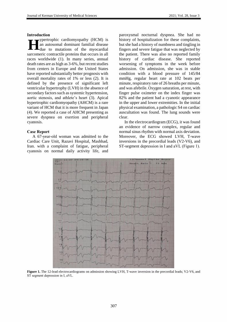
In the electrocardiogram (ECG), it was found
an evidence of narrow complex, regular and
normal sinus rhythm with normal axis deviation.
Moreover, the ECG showed LVH, T-wave
inversions in the precordial leads (V2-V6), and
ST-segment depression in І and aVL (Figure 1).
Figure 1. The 12-lead electrocardiograms on admission showing LVH, T-wave inversion in the precordial leads; V2-V6, and
ST segment depression in І, aVL.
H

Apical hypertrophic cardiomyopathy presented as an atypical case Ghasemi et al.
308
For other diagnostic investigations, the
patient was referred to the echocardiography unit
for transthoracic echocardiography. It was
performed in the Cardiac Care Unit and showed
a left atrium size of 45 mm and the apex of 21
mm. Transthoracic echocardiography in the
parasternal long-axis view in end diastole
revealed that the inter-ventricular septum and
posterior wall thickness was 14 mm. There was
also an evidence of asymmetric septal
hypertrophy (ASH) in short axis and parasternal
long-axis views. The left ventricular end
diameter was 52 mm at the end of diastole in the
parasternal long-axis view, with preserved
systolic function (ejection fraction of 55%) and
Left ventricular diastolic dysfunction. The right
ventricle was normal with a good function. The
patient did not have any evidence of left
ventricular outflow tract obstruction (LVOTO),
systolic anterior motion (SAM), or significant
mitral regurgitation. An apical four-chamber
view of the patient’s transthoracic
echocardiography is shown in Figure 2. Chest X-
ray (in PA view) showed an increased
cardiothoracic ratio. The aortic arch was dilated,
the main pulmonary artery (MPA) and the left
atrial appendage (LAA) were normal.
Considering these findings, the patient was
scheduled for a 30-day follow-up with the
prescription of Metohexal (Metoprolol
succinate) with a dose of 23.75 mg twice a day,
as a β-blocker. After this period, her symptoms
were relieved and she had no complaint of
dyspnea on exertion. Also, she had no evidence
of cyanosis in the upper and lower extremities.
Finally, the patient was suggested to return to the
clinic for adjustment of her medications, and
screening of first-degree family members, if
symptoms recurred.
Figure 2. Transthoracic echocardiographic four-chamber view of patient demonstrated apical hypertrophic cardiomyopathy.

Journal of Kerman University of Medical Sciences 2021; Vol. 28, Issue 3
309
Discussion
Cardiac diseases are significant since they are
the most common chronic diseases of the 21st
century and are regarded as the main causes of
death (5). Hypertrophic cardiomyopathy (HCM)
is a genetic cardiac disease characterized by
marked variability in morphological expression
and natural history (6). It may involve mainly the
proximal septum, or there may be diffuse LVH.
However, there are other patterns, such as apical
hypertrophy (7).
Apical Hypertrophic Cardiomyopathy
(AHCM) can be asymptomatic or present with
syncope, chest pain (symptoms similar to those
of acute coronary syndrome), palpitations, and
dyspnea (4). Another study by Ahmed et al.
showed that the most common presenting
symptoms were dyspnea with or without chest
pain (1). The main complaint and unique
presentation of our patient was a cyanotic
appearance in the upper limbs extremities that
was not a common presentation. This
presentation may be misdiagnosed with severe
respiratory diseases at the first time. Also,
paroxysmal nocturnal dyspnea in our patient is a
relatively uncommon presentation of that seen in
the progressive HCM. But our patient had no
other symptoms of advanced features of AHCM
such as syncope or pre-syncope, arrhythmia,
angina, and dizziness.
AHCM is predominantly a hereditary
disease, although it can also be present in
patients with no family history (8). Our patient
had no evidence of familial history.
In Western patients, there seems to be a
varied presentation as far as clinical and ECG
features are concerned, compared to the classic
AHC as defined in the Asian population. Hence,
in the presence of AHCM situations,
understanding the unique ECG features of
AHCM can be of assistance in the diagnostic
process of this uncommon condition (4,8). The
presence of ECG findings indicative of LVH
with giant T-wave inversions (especially in
precordial leads) and loss of septal Q-waves
should raise strong suspicions of AHCM. These
are considered pathogenic for this disorder (4, 9,
10). Our patient’s ECG demonstrated the same
features, and based on these characteristics,
additional investigations such as transthoracic
echocardiography were performed.
Numerous studies have investigated the
effect of β-blockers on HCM patients and
reported symptoms improvement (11,12). A
study by Kasirye et al. also mentioned that β-
blockers were the first drugs used for the
treatment of symptomatic HCM (4). Hence, our
patient mentioned relief of her symptoms after 1-
month use of this drug agent and it can be a
selective drug in the first step of treatment of
AHCM. On the other hand, in some studies (10-
12), it was stated that β-blockers can relieve the
symptoms in the two to five months after
administration, but our patient’s symptoms
improved after only one month and in a short-
term use of metoprolol succinate. This is a rare
case that was treated with only one agent β-
blocker in a short time. So, in other cases of
AHCM, metoprolol succinate is suggested to be
considered in the first step of treatment.
Symptoms such as severe dyspnea should be
controlled in patients diagnosed with AHCM.
The treatment process can include β-blockers,
ACEi, and calcium channel blockers. If this
approach is not successful, patients with this
condition should be referred for changes in the
medication protocol to postpone heart failure
progression due to AHCM complications.
Acknowledgments
The authors would like to thank the patient
who participated in this study. And special
thanks to all of the nurses from Cardiac Care
Unit of 9 Day hospital, Torbat Heydariyeh, Iran,
who cooperated in the study. Written informed
consent was obtained from the patient for
publication of this case report and any
accompanying images.
Conflict of interests
The authors declare that there is no conflict
of interests.

Apical hypertrophic cardiomyopathy presented as an atypical case Ghasemi et al.
310
References
1. Ahmed W, Akhtar N, Bech-Hanssen O, Al
Mahdi B, Al Otaibi T, Fadel BM.
Hypertrophic cardiomyopathy in the Saudi
Arabian population: clinical and
echocardiographic characteristics and
outcome analysis. J Saudi Heart Assoc
2014; 26(1):7-13.
2. Elliott PM, Gimeno JR, Thaman R, Shah J,
Ward D, Dickie S, et al. Historical trends in
reported survival rates in patients with
hypertrophic cardiomyopathy. Heart 2006;
92(6):785-91.
3. Younessi Heravi MA, Mojdekanlu M,
Seyed Sharifi SH, Yaghubi M. The role of
cardiovascular risk factors in involvement
of coronary arteries; A predictive model in
angiographic study. Journal of North
Khorasan University of Medical Sciences.
2014; 6(1): 199-205.
4. Kasirye Y, Manne JR, Epperla N, Bapani S,
Garcia-Montilla R. Apical hypertrophic
cardiomyopathy presenting as recurrent
unexplained syncope. Clin Med Res 2012;
10(1):26-31.
5. Younessi Heravi MA, Yaghubi M,
Joharinia S. Effect of change in patient’s
bed angles on pain after coronary
angiography according to vital signals. J
Res Med Sci 2015; 20(10):937-43.
6. Scudeler TL, Rezende PC, Oikawa FT, da
Costa LM, Hueb AC, Hueb W. A case of
mid-apical obstructive hypertrophic
cardiomyopathy treated with a transapical
myectomy approach: a case report. J Med
Case Rep 2014; 8:364.
7. Kunkala MR, Schaff HV, Nishimura RA,
Abel MD, Sorajja P, Dearani JA, et al.
Transapical approach to myectomy for
midventricular obstruction in hypertrophic
cardiomyopathy. Ann Thorac Surg 2013;
96(2):564-70.
8. Abdy NA, Valdes SO, Sorrell VL, Klewer
SE, Barber BJ. Apical hypertrophic
cardiomyopathy in an adolescent. Congenit
Heart Dis 2010; 5(2):182-7.
9. 9. Bagheri M.M, Naghibzadeh-Tahami A,
Niknafs P, Daei- parizi Z. Prolonged QT
interval in the infants of diabetic mothers.
Journal of Kerman University of Medical
Sciences, 2019; 26 (1): 120-125.
10. Eriksson MJ, Sonnenberg B, Woo A,
Rakowski P, Parker TG, Wigle ED, et al.
Long-term outcome in patients with apical
hypertrophic cardiomyopathy. J Am Coll
Cardiol 2002; 39(4):638-45.
11. Spoladore R, Maron MS, D'amato R,
Camici PG, Olivotto I. Pharmacological
treatment options for hypertrophic
cardiomyopathy: high time for evidence. Eur
Heart J 2012; 33(14):1724-33.
12. Kim SH, Kim SO, Han S, Hwang KW, Lee
CW, Nam GB, et al. Long-term comparison
of apical versus asymmetric hypertrophic
cardiomyopathy. Int Heart J 2013;
54(4):207-11.