apcccdh1 controls ctip stability during the cell...
TRANSCRIPT
Article
APC/CCdh1 controls CtIP stability during the cellcycle and in response to DNA damageLorenzo Lafranchi1,†, Harmen R de Boer2,†, Elisabeth GE de Vries2, Shao-En Ong3, Alessandro A Sartori1,*
& Marcel ATM van Vugt2,**
Abstract
Human cells have evolved elaborate mechanisms for responding toDNA damage to maintain genome stability and prevent carcino-genesis. For instance, the cell cycle can be arrested at differentstages to allow time for DNA repair. The APC/CCdh1 ubiquitin ligasemainly regulates mitotic exit but is also implicated in the DNAdamage-induced G2 arrest. However, it is currently unknownwhether APC/CCdh1 also contributes to DNA repair. Here, we showthat Cdh1 depletion causes increased levels of genomic instabilityand enhanced sensitivity to DNA-damaging agents. Using an inte-grated proteomics and bioinformatics approach, we identify CtIP, aDNA-end resection factor, as a novel APC/CCdh1 target. CtIP inter-acts with Cdh1 through a conserved KEN box, mutation of whichimpedes ubiquitylation and downregulation of CtIP both during G1
and after DNA damage in G2. Finally, we find that abrogating theCtIP–Cdh1 interaction results in delayed CtIP clearance from DNAdamage foci, increased DNA-end resection, and reduced homolo-gous recombination efficiency. Combined, our results highlight theimpact of APC/CCdh1 on the maintenance of genome integrity andshow that this is, at least partially, achieved by controlling CtIPstability in a cell cycle- and DNA damage-dependent manner.
Keywords Cdh1; cell cycle; CtIP; DNA damage; DNA double-strand break
repair
Subject Categories Cell Cycle; DNA Replication, Repair & Recombination;
Post-translational Modifications, Proteolysis & Proteomics
DOI 10.15252/embj.201489017 | Received 16 May 2014 | Revised 7 September
2014 | Accepted 30 September 2014 | Published online 27 October 2014
The EMBO Journal (2014) 33: 2860–2879
Introduction
Our genome is constantly exposed to various forms of endogenous
and exogenous insults provoking different types of DNA lesions,
which can promote tumorigenesis. To maintain genomic integrity,
the DNA damage response (DDR) activates cell cycle checkpoints to
slow cell cycle progression, thereby allowing time for appropriate
repair (Jackson & Bartek, 2009). DNA double-strand breaks (DSBs)
are the most cytotoxic lesions induced by ionizing radiation (IR)
and certain anticancer drugs. Cells have evolved two major DSB
repair mechanisms: non-homologous end-joining (NHEJ) and
homologous recombination (HR) (Lieber, 2010).
In the G0/G1 phase of the cell cycle, NHEJ is the preferred mecha-
nism for DSB repair (Lieber, 2010). In this process, DNA ends are
joined without the requirement for a homologous sequence, making
NHEJ potentially mutagenic. In contrast, cells that have entered S
phase can use the sister chromatid as a template for high-fidelity
DSB repair through HR (Aylon et al, 2004; Ferreira & Cooper, 2004;
Sonoda et al, 2006). NHEJ and HR are mutually exclusive pathways
since DNA-end resection, which generates long stretches of single-
stranded DNA (ssDNA), commits cells to HR and prevents repair by
NHEJ. Mechanisms controlling DSB repair pathway choice are
under vigorous investigation (Chapman et al, 2012). The temporal
restriction of HR repair to S/G2 is controlled both at the transcrip-
tional and post-transcriptional level. The expression of many HR
factors including Rad51, Rad54, and Brca1 is cell cycle-dependent,
being much lower in G0/G1 than in S/G2 (Gudas et al, 1996;
Yamamoto et al, 1996). In addition, cyclin-dependent kinases
(CDKs), core components of the cell cycle machinery, play an
important role in DSB repair pathway choice through phosphoryla-
tion of multiple HR components, including members of the MRN
complex as well as Brca1 and Brca2 (Esashi et al, 2005; Ayoub et al,
2009; Falck et al, 2012). CDK-mediated regulation of DSB repair
occurs mainly at the level of DNA-end resection (Aylon et al, 2004;
Ira et al, 2004; Henderson et al, 2006; Jazayeri et al, 2006; Johnson
et al, 2011). Human CtIP is essential for the initiation of DNA-end
resection, and its function in this process is controlled by various
post-translational modifications including phosphorylation, ubiqui-
tylation, and acetylation (Sartori et al, 2007; Huertas & Jackson,
2009; Kaidi et al, 2010; Steger et al, 2013).
Likewise, targeted proteolysis through the ubiquitin-proteasome
system (UPS) is a highly regulated process that allows the removal
of potentially harmful proteins, thereby restricting their activity. The
anaphase-promoting complex/cyclosome (APC/C) is an E3 ubiquitin
1 Institute of Molecular Cancer Research, University of Zurich, Zurich, Switzerland2 Department of Medical Oncology, University Medical Center Groningen, University of Groningen, Groningen, The Netherlands3 Department of Pharmacology, University of Washington, Seattle, WA, USA
*Corresponding author. Tel: +41 446353473; Fax: +41 446353484; E-mail: [email protected]**Corresponding author. Tel: +31 50 3619554; Fax: +31 50 3614862; E-mail: [email protected]†These authors contributed equally to this work
The EMBO Journal Vol 33 | No 23 | 2014 ª 2014 The Authors2860
Published online: October 27, 2014
ligase involved in cell cycle regulation and becomes activated upon
sequential binding to the Cdc20 and Cdh1 adaptor proteins
(Peters, 2006; Pesin & Orr-Weaver, 2008). Cdc20 is associated with
the APC/C during early mitosis and principally regulates mitotic
progression, whereas Cdh1 interacts with the APC/C from late
mitosis onwards until the following G1/S transition (Kramer et al,
2000; Peters, 2006). In most cases, APC/CCdh1 interacts with its
substrates through the recognition of a short consensus motif called
the KEN box (Pfleger & Kirschner, 2000; Pines, 2011). During S/G2
phase, premature APC/C activation is at least in part prevented
through CDK-mediated phosphorylation of Cdh1, which hinders
association of Cdh1 with the APC/C (Lukas et al, 1999; Kramer
et al, 2000; Miller et al, 2006).
As a first indication of APC/CCdh1 playing a role in the DDR,
CDH1�/� chicken DT40 cells failed to maintain a G2 arrest after IR
(Sudo et al, 2001). In addition, activation of the APC/CCdh1 in
response to DNA damage during G2 phase was shown to depend on
the Cdc14B phosphatase and to result in the degradation of Polo-like
kinase 1 (Plk1) (Bassermann et al, 2008; Wiebusch & Hagemeier,
2010). Upon completion of DSB repair, Cdk1 and Plk1 are reacti-
vated to allow cell cycle progression from G2 into mitosis (van Vugt
et al, 2004). Further experiments performed in Cdc14B-deficient
cells showed that those cells are unable to repair DSBs even if they
efficiently arrest in G2 (Mocciaro et al, 2010). However, direct
participation of the APC/CCdh1 in the regulation of DSB repair has
never been reported. The observation that Cdh1-depleted cells or
Cdh1�/� mice have elevated levels of DNA damage and chromo-
somal aberrations (Garcıa-Higuera et al, 2008; Sigl et al, 2009;
Delgado-Esteban et al, 2013) strengthens this notion, but direct
mechanistic insights into how the APC/CCdh1 ubiquitin ligase
connects the cell cycle machinery to DNA repair is still lacking and
the responsible substrates remain elusive.
In this study, we show that inactivation of Cdh1 results in geno-
mic instability in different human cell lines. Moreover, Cdh1-
depleted cells are hypersensitive toward DNA-damaging agents and
display reduced Rad51 foci upon IR treatment. Making use of an inte-
grated proteomics and bioinformatics approach, we identify the
DNA-end resection factor CtIP as a novel substrate of the APC/CCdh1
E3 ubiquitin ligase. CtIP–Cdh1 interaction is mediated by an evolu-
tionary conserved KEN box and is required for the downregulation
of CtIP protein levels both after mitotic exit and in late S/G2 in
response to DNA damage. U2OS cells inducibly expressing a CtIP
KEN box mutant exhibit increased DNA-end resection capacity,
which correlates with a decrease in HR and hypersensitivity to PARP
inhibition. Together, our data describe a novel regulatory role for the
APC/CCdh1 in DNA repair, at least in part by limiting CtIP-dependent
DNA-end resection activity in late S/G2 phases of the cell cycle.
Results
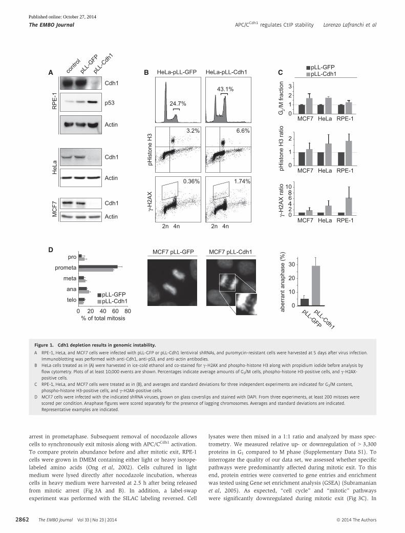
Cdh1 depletion provokes DNA damage and hypersensitivity toDSB-inducing agents
To analyze the role of APC/CCdh1 in the maintenance of genome
stability, we stably suppressed Cdh1 in non-transformed, immortal-
ized human retina pigment epithelium (hTERT-RPE-1), HeLa cervi-
cal cancer, and MCF7 breast cancer cells using lentiviral shRNAs
(Fig 1A). Sustained downregulation of Cdh1 over the course of
5 days resulted in approximately twofold increase of cells in G2/M
phase accompanied by elevated levels of c-H2AX (Fig 1A–C). Also,
Cdh1 depletion resulted in upregulation of p53 in RPE-1 and MCF7
cells (Fig 1A and Supplementary Fig S1). Combined, these results
are indicative of DNA damage accumulation in Cdh1-depleted cells,
even in the absence of genotoxic agents, which is in line with obser-
vations in other cell types (Garcıa-Higuera et al, 2008; Sigl et al,
2009; Delgado-Esteban et al, 2013; Eguren et al, 2013). We next
analyzed the distribution of cells over the various mitotic stages to
address whether the acquisition of DNA damage caused by Cdh1
downregulation translates into aberrant mitotic progression. While
Cdh1 depletion in MCF7 cells did not significantly alter the distribu-
tion of the various mitotic phases, it gave rise to a higher frequency
of bridging chromosomes in anaphase (Fig 1D). Such abnormal
chromosome segregation events are frequently observed in cells that
exhibit G2/M checkpoint or DNA repair defects (French et al, 2006;
Acilan et al, 2007; Chan et al, 2007; Laulier et al, 2011).
This prompted us to examine whether Cdh1 depletion leads to
increased sensitivity to DNA-damaging agents. Clonogenic survival
assays showed that Cdh1-depleted MCF7 cells are hypersensitive to
IR (Fig 2A). Notably, the colonies of irradiated Cdh1-depleted cells
were considerably smaller compared to control-depleted cells
(Fig 2A). In line with these results, we found that shRNA-mediated
depletion of Cdh1 reduced cell survival after treatment with doxoru-
bicin, a chemotherapeutic compound inducing DSBs (Fig 2B).
We next examined whether DSB repair mechanisms are affected
by Cdh1 depletion. Since cell cycle status significantly influences the
mode of DSB repair, we made use of the FUCCI system which allows
to specifically analyze S/G2 cells without employing synchroniza-
tion protocols (Supplementary Fig S2A) (Sakaue-Sawano et al,
2008). First, Cdh1 depletion did not affect IR-induced phosphoryla-
tion of KAP1 at S824, an early event in the DNA damage response
(Fig 2C) (White et al, 2006; Ziv et al, 2006). Interestingly, however,
Cdh1-depleted cells displayed significantly reduced numbers of
Rad51 foci at both 2 and 5 h after irradiation, without affecting
Rad51 levels (Fig 2C and D, and Supplementary Fig S2B). In
contrast, 53BP1 foci numbers remained largely unaffected by the
absence of Cdh1 (Fig 2D and Supplementary Fig S2C). Taken
together, our results demonstrate that Cdh1 ensures genome stabil-
ity, promotes survival under conditions of DNA damage, and influ-
ences the dynamics of IR-induced Rad51 foci formation, indicative
of a regulatory function of Cdh1 in HR.
Proteomic analysis of potential Cdh1 targets
We next set out to identify APC/CCdh1 substrates that contribute to
the role of Cdh1 in maintaining genome stability. To select for
potential candidates, we focused on two selection criteria. Firstly,
we screened for proteins that are downregulated when APC/CCdh1
activity is turned on after mitotic exit using quantitative mass spec-
trometry. Secondly, we analyzed the primary amino acid sequence
of each of those downregulated proteins for the presence of so-
called D-boxes and KEN boxes, through which Cdh1 recruits targets
to the APC/C (Pfleger et al, 2001; Liu et al, 2012). To identify proteins
that are degraded during the mitosis-to-G1 transition, we treated cells
with nocodazole, a reversible microtubule-depolymerizing agent that
activates the spindle assembly checkpoint, thereby causing cells to
ª 2014 The Authors The EMBO Journal Vol 33 | No 23 | 2014
Lorenzo Lafranchi et al APC/CCdh1 regulates CtIP stability The EMBO Journal
2861
Published online: October 27, 2014
arrest in prometaphase. Subsequent removal of nocodazole allows
cells to synchronously exit mitosis along with APC/CCdh1 activation.
To compare protein abundance before and after mitotic exit, RPE-1
cells were grown in DMEM containing either light or heavy isotope-
labeled amino acids (Ong et al, 2002). Cells cultured in light
medium were lysed directly after nocodazole incubation, whereas
cells in heavy medium were harvested at 2.5 h after being released
from mitotic arrest (Fig 3A and B). In addition, a label-swap
experiment was performed with the SILAC labeling reversed. Cell
lysates were then mixed in a 1:1 ratio and analyzed by mass spec-
trometry. We measured relative up- or downregulation of > 3,300
proteins in G1 compared to M phase (Supplementary Data S1). To
interrogate the quality of our data set, we assessed whether specific
pathways were predominantly affected during mitotic exit. To this
end, protein entries were converted to gene entries and enrichment
was tested using Gene set enrichment analysis (GSEA) (Subramanian
et al, 2005). As expected, “cell cycle” and “mitotic” pathways
were significantly downregulated during mitotic exit (Fig 3C). In
A
pHis
tone
H3
2n 4n 2n 4n
-H2A
X
HeLa-pLL-GFP HeLa-pLL-Cdh1
24.7%
43.1%
6.6%3.2%
1.74%0.36%
B
Cdh1
G2/M
frac
tion
Cdh1
Cdh1
Actin
Actin
pLL-G
FP
pLL-C
dh1
contr
ol
Actin
HeL
aM
CF7
RP
E-1
p53
D MCF7 pLL-GFP MCF7 pLL-Cdh1 )%( esahpana tnarreba
pro
prometa
meta
ana
telopLL-GFPpLL-Cdh1
800 20 40 60% of total mitosis
0
30
20
10
pLL-GFPpLL-Cdh1C
pLL-GFP
pLL-Cdh1
1
HeLaMCF7 RPE-10
23
HeLaMCF7 RPE-1
HeLaMCF7 RPE-1
-H2A
X ra
tiopH
isto
ne H
3 ra
tio
20
6
10
1
0
2
4
8
Figure 1. Cdh1 depletion results in genomic instability.
A RPE-1, HeLa, and MCF7 cells were infected with pLL-GFP or pLL-Cdh1 lentiviral shRNAs, and puromycin-resistant cells were harvested at 5 days after virus infection.Immunoblotting was performed with anti-Cdh1, anti-p53, and anti-actin antibodies.
B HeLa cells treated as in (A) were harvested in ice-cold ethanol and co-stained for c-H2AX and phospho-histone H3 along with propidium iodide before analysis byflow cytometry. Plots of at least 10,000 events are shown. Percentages indicate average amounts of G2/M cells, phospho-histone H3-positive cells, and c-H2AX-positive cells.
C RPE-1, HeLa, and MCF7 cells were treated as in (B), and averages and standard deviations for three independent experiments are indicated for G2/M content,phospho-histone H3-positive cells, and c-H2AX-positive cells.
D MCF7 cells were infected with the indicated shRNA viruses, grown on glass coverslips and stained with DAPI. From three experiments, at least 200 mitoses werescored per condition. Anaphase figures were scored separately for the presence of lagging chromosomes. Averages and standard deviations are indicated.Representative examples are indicated.
The EMBO Journal Vol 33 | No 23 | 2014 ª 2014 The Authors
The EMBO Journal APC/CCdh1 regulates CtIP stability Lorenzo Lafranchi et al
2862
Published online: October 27, 2014

contrast, proteins involved in translation and transcription were
upregulated, in line with observations that chromatin is
re-established after mitosis and that translational and transcriptional
processes are inactive during mitosis and need to be re-initiated
upon mitotic exit (Fig 3C) (Prescott & Bender, 1963; Bonneau &
Sonenberg, 1987).
Our data set for proteins that were considerably downregulated
after mitotic exit included various known APC/CCdh1 targets such as
p15-PAF, cyclin B1, Plk1, UbcH10, Aurora A, and Aurora B (Fig 3D
and Supplementary Fig S3A) (Pfleger et al, 2001; Littlepage &
Ruderman, 2002; Taguchi et al, 2002; Lindon & Pines, 2004; Rape &
Kirschner, 2004; Nguyen et al, 2005; Stewart & Fang, 2005; Emanuele
et al, 2011). As expected, both KEN box and D-box motifs were
significantly enriched in proteins downregulated during mitotic exit
compared to the entire proteomic data set (Fig 3E) (Liu et al, 2012).
When we applied “DNA damage response” (see Supplementary
Table S1 for list of Gene Ontology terms) as a functional criterion
for proteins downregulated during mitotic exit containing a
conserved destruction motif, we discovered various proteins
involved in the maintenance of genomic integrity including Rif1,
Smc5, Mdc1, CtIP, and Top2A (Fig 3F). DNA topoisomerase 2-alpha
was recently discovered as a novel Cdh1 substrate (Eguren et al,
2014). Interestingly, Rif1 has been reported to act as a 53BP1 effec-
tor protein, antagonizing the role of BRCA1 in promoting CtIP-
dependent resection and HR (Kumar & Cheok, 2014). However,
sequence analysis revealed that none of the predicted functional
destruction motifs in human Rif1 were evolutionary conserved,
whereas both putative KEN boxes in human CtIP were highly
conserved in vertebrates (Supplementary Fig S3). Therefore, we
decided to focus on the DNA-end resection factor CtIP as a potential
target of the APC/CCdh1 involved in the maintenance of genomic
integrity.
A
100
0
80
60
40
20
surv
ival
(%)
pLL-Cdh1pLL-GFP
untre
ated
2 G
y
B
pLL-GFPpLL-Cdh1
control
0 50 100 200 400
1
0.1
0.010 2 4 6
irradiation (Gy)
pLL-GFPpLL-Cdh1
doxorubicin (ng/ml)
surv
ivin
g fra
ctio
n (%
)
TFIIH
IR (2 Gy)
siCTRL2h 5h
Plk1
siCdh1#1- 2h 5h-
KAP1-pS824
Cdh1
Rad51
C DU2OS-FUCCI
Rad
51 fo
ci in
S/G
2 ce
ll 40
30
20
10
02h after IR 5h after IR
siCTRLsiCdh1#1
< 0.0001< 0.0001
53B
P1
foci
in S
/G2
cell 50
40
30
20
10
02h after IR 5h after IR
siCTRLsiCdh1#1
n.s.0.0035
KAP1
MCF7 MCF7
Figure 2. Cdh1 depletion sensitizes to DNA-damaging agents and affects recruitment of DNA repair components.
A MCF7 cells were infected with the indicated shRNAs and selected with puromycin. pLL-GFP- or pLL-Cdh1-infected MCF7 cells were plated in 6-well plates andsubsequently irradiated with the indicated amounts of ionizing radiation. Surviving colonies were stained, and relative amounts of colony numbers compared to non-irradiated cells are shown. Averages and standard deviations of three independent experiments are shown.
B pLL-GFP- or pLL-Cdh1-infected MCF7 cells were plated in 96-well plates and subsequently treated with the indicated amounts of doxorubicine for 4 days. Cellularviability was assessed using MTT conversion, and untreated cells were used as a reference. Averages and standard deviations of three independent experiments areshown.
C At 48 h after siRNA transfection, U2OS-FUCCI cells were irradiated (2 Gy). At 2 or 5 h after treatment, whole-cell extracts were prepared and analyzed by Westernblotting with the indicated antibodies.
D U2OS-FUCCI cells treated as in (C) were prepared for 53BP1 and Rad51 immunofluorescence. Graphs show the amounts of 53BP1 or Rad51 foci in S/G2 cells. At least120 cells from three independent experiments were counted for each condition, and data are presented as box plots with whiskers representing the minimal andmaximal values. Unpaired Student’s t-tests (two-sided) were done to compare control-depleted and Cdh1-depleted conditions. Representative images can be found inSupplementary Fig S2B and C.
ª 2014 The Authors The EMBO Journal Vol 33 | No 23 | 2014
Lorenzo Lafranchi et al APC/CCdh1 regulates CtIP stability The EMBO Journal
2863
Published online: October 27, 2014
A
-5
-4
-3
-2
-1
0
1
2
3
4
5
diffe
rent
ial p
rote
in a
bund
ance
(log
2)
protein abundance after mitotic exit
downregulation upregulation
light medium heavy mediumArg-0Lys-0
Arg-10Lys-8
mitotic shake-off, replate
harvest harvest after 2.5hforward SILAC
reverse SILACharvestharvest after 2.5h
Aurora Ap15-PAF
TK1Cyclin B
Plk1CtIP
translation (p=0.004)
transcription (p=0.010)
0
0.1
0.2
0
0.1
cell cycle phase (p=0.002)
0
-0.2
-0.1
B
mitosis (p=0.009)0
-0.2
-0.1enric
hmen
t sco
re
DE
F
Rif1
Mdc1
down-regulated‘DNA damage response’ log2
Top2A
-1.29
-1.24
-1.27CtIP
optimaldestruction
motifn=721
>2-fold downafter
mitotic exit
Gene Set:DNA Damage Response
n=93
n=255
n=5
n=40
KEN box enrichment
D box enrichment
-0.1
0.40.30.20.1
0
0.50.40.30.20.1
0
p=0.00504
p=0.00107
enric
hmen
t sco
reen
richm
ent s
core
destruction motifs
0
20
40
60
80
100
untreated
nocodazole
release 2.5h
p-H
isto
neH
3 (%
)
C
Aurora B
n=2
-1.34
-1.31Smc5
any conserved/optimal
++
n=36
+++
+
---
-
RPE-1
Figure 3. A combined proteomics/bioinformatics approach to identify potential APC/CCdh1 substrates.
A Overview of mass spectrometry analysis of changes in protein abundance during mitotic exit. RPE-1 cells were grown in “light” or “heavy” SILAC media and treatedwith nocodazole. Mitotic cells were obtained by shake-off and were directly lysed or replated in nocodazole-free medium and lysed after 2.5 h. Alternatively,treatments were swapped (“reverse SILAC”). Cell lysates were mixed 1:1 and analyzed by mass spectrometry.
B RPE-1 cells were treated and harvested in parallel to (A) and were stained for phospho-histone H3/Alexa-488.C Gene set enrichment analysis (GSEA) of SILAC results. Indicated pathways are significantly affected during mitotic exit.D Log2-transformed ratios are indicated for the > 3,300 proteins that were identified both in forward and in reverse SILAC. Negative ratios indicate downregulation
during mitotic exit. Established APC/CCdh1 targets are indicated in red.E GPS-ARM software was used to identify D-box or KEN box sequences in mass spectrometry hits. GSEA was used to assess enrichment for destruction motif-
containing proteins within downregulated proteins.F Venn diagram indicating proteins that are downregulated at least twofold, contain any destruction motif, and belong to the “DNA damage response/DNA repair”
gene set.
The EMBO Journal Vol 33 | No 23 | 2014 ª 2014 The Authors
The EMBO Journal APC/CCdh1 regulates CtIP stability Lorenzo Lafranchi et al
2864
Published online: October 27, 2014
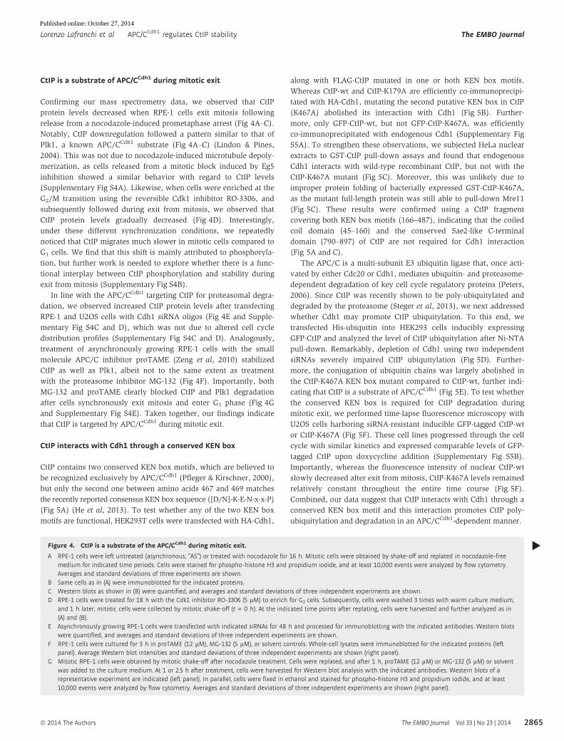
CtIP is a substrate of APC/CCdh1 during mitotic exit
Confirming our mass spectrometry data, we observed that CtIP
protein levels decreased when RPE-1 cells exit mitosis following
release from a nocodazole-induced prometaphase arrest (Fig 4A–C).
Notably, CtIP downregulation followed a pattern similar to that of
Plk1, a known APC/CCdh1 substrate (Fig 4A–C) (Lindon & Pines,
2004). This was not due to nocodazole-induced microtubule depoly-
merization, as cells released from a mitotic block induced by Eg5
inhibition showed a similar behavior with regard to CtIP levels
(Supplementary Fig S4A). Likewise, when cells were enriched at the
G2/M transition using the reversible Cdk1 inhibitor RO-3306, and
subsequently followed during exit from mitosis, we observed that
CtIP protein levels gradually decreased (Fig 4D). Interestingly,
under these different synchronization conditions, we repeatedly
noticed that CtIP migrates much slower in mitotic cells compared to
G1 cells. We find that this shift is mainly attributed to phosphoryla-
tion, but further work is needed to explore whether there is a func-
tional interplay between CtIP phosphorylation and stability during
exit from mitosis (Supplementary Fig S4B).
In line with the APC/CCdh1 targeting CtIP for proteasomal degra-
dation, we observed increased CtIP protein levels after transfecting
RPE-1 and U2OS cells with Cdh1 siRNA oligos (Fig 4E and Supple-
mentary Fig S4C and D), which was not due to altered cell cycle
distribution profiles (Supplementary Fig S4C and D). Analogously,
treatment of asynchronously growing RPE-1 cells with the small
molecule APC/C inhibitor proTAME (Zeng et al, 2010) stabilized
CtIP as well as Plk1, albeit not to the same extent as treatment
with the proteasome inhibitor MG-132 (Fig 4F). Importantly, both
MG-132 and proTAME clearly blocked CtIP and Plk1 degradation
after cells synchronously exit mitosis and enter G1 phase (Fig 4G
and Supplementary Fig S4E). Taken together, our findings indicate
that CtIP is targeted by APC/CCdh1 during mitotic exit.
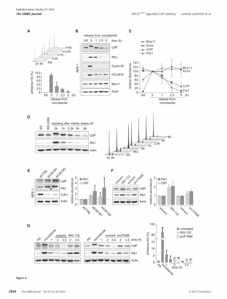
CtIP interacts with Cdh1 through a conserved KEN box
CtIP contains two conserved KEN box motifs, which are believed to
be recognized exclusively by APC/CCdh1 (Pfleger & Kirschner, 2000),
but only the second one between amino acids 467 and 469 matches
the recently reported consensus KEN box sequence ([D/N]-K-E-N-x-x-P)
(Fig 5A) (He et al, 2013). To test whether any of the two KEN box
motifs are functional, HEK293T cells were transfected with HA-Cdh1,
along with FLAG-CtIP mutated in one or both KEN box motifs.
Whereas CtIP-wt and CtIP-K179A are efficiently co-immunoprecipi-
tated with HA-Cdh1, mutating the second putative KEN box in CtIP
(K467A) abolished its interaction with Cdh1 (Fig 5B). Further-
more, only GFP-CtIP-wt, but not GFP-CtIP-K467A, was efficiently
co-immunoprecipitated with endogenous Cdh1 (Supplementary Fig
S5A). To strengthen these observations, we subjected HeLa nuclear
extracts to GST-CtIP pull-down assays and found that endogenous
Cdh1 interacts with wild-type recombinant CtIP, but not with the
CtIP-K467A mutant (Fig 5C). Moreover, this was unlikely due to
improper protein folding of bacterially expressed GST-CtIP-K467A,
as the mutant full-length protein was still able to pull-down Mre11
(Fig 5C). These results were confirmed using a CtIP fragment
covering both KEN box motifs (166–487), indicating that the coiled
coil domain (45–160) and the conserved Sae2-like C-terminal
domain (790–897) of CtIP are not required for Cdh1 interaction
(Fig 5A and C).
The APC/C is a multi-subunit E3 ubiquitin ligase that, once acti-
vated by either Cdc20 or Cdh1, mediates ubiquitin- and proteasome-
dependent degradation of key cell cycle regulatory proteins (Peters,
2006). Since CtIP was recently shown to be poly-ubiquitylated and
degraded by the proteasome (Steger et al, 2013), we next addressed
whether Cdh1 may promote CtIP ubiquitylation. To this end, we
transfected His-ubiquitin into HEK293 cells inducibly expressing
GFP-CtIP and analyzed the level of CtIP ubiquitylation after Ni-NTA
pull-down. Remarkably, depletion of Cdh1 using two independent
siRNAs severely impaired CtIP ubiquitylation (Fig 5D). Further-
more, the conjugation of ubiquitin chains was largely abolished in
the CtIP-K467A KEN box mutant compared to CtIP-wt, further indi-
cating that CtIP is a substrate of APC/CCdh1 (Fig 5E). To test whether
the conserved KEN box is required for CtIP degradation during
mitotic exit, we performed time-lapse fluorescence microscopy with
U2OS cells harboring siRNA-resistant inducible GFP-tagged CtIP-wt
or CtIP-K467A (Fig 5F). These cell lines progressed through the cell
cycle with similar kinetics and expressed comparable levels of GFP-
tagged CtIP upon doxycycline addition (Supplementary Fig S5B).
Importantly, whereas the fluorescence intensity of nuclear CtIP-wt
slowly decreased after exit from mitosis, CtIP-K467A levels remained
relatively constant throughout the entire time course (Fig 5F).
Combined, our data suggest that CtIP interacts with Cdh1 through a
conserved KEN box motif and this interaction promotes CtIP poly-
ubiquitylation and degradation in an APC/CCdh1-dependent manner.
Figure 4. CtIP is a substrate of the APC/CCdh1 during mitotic exit.
A RPE-1 cells were left untreated (asynchronous, “AS”) or treated with nocodazole for 16 h. Mitotic cells were obtained by shake-off and replated in nocodazole-freemedium for indicated time periods. Cells were stained for phospho-histone H3 and propidium iodide, and at least 10,000 events were analyzed by flow cytometry.Averages and standard deviations of three experiments are shown.
B Same cells as in (A) were immunoblotted for the indicated proteins.C Western blots as shown in (B) were quantified, and averages and standard deviations of three independent experiments are shown.D RPE-1 cells were treated for 18 h with the Cdk1 inhibitor RO-3306 (5 lM) to enrich for G2 cells. Subsequently, cells were washed 3 times with warm culture medium,
and 1 h later, mitotic cells were collected by mitotic shake-off (t = 0 h). At the indicated time points after replating, cells were harvested and further analyzed as in(A) and (B).
E Asynchronously growing RPE-1 cells were transfected with indicated siRNAs for 48 h and processed for immunoblotting with the indicated antibodies. Western blotswere quantified, and averages and standard deviations of three independent experiments are shown.
F RPE-1 cells were cultured for 3 h in proTAME (12 lM), MG-132 (5 lM), or solvent controls. Whole-cell lysates were immunoblotted for the indicated proteins (leftpanel). Average Western blot intensities and standard deviations of three independent experiments are shown (right panel).
G Mitotic RPE-1 cells were obtained by mitotic shake-off after nocodazole treatment. Cells were replated, and after 1 h, proTAME (12 lM) or MG-132 (5 lM) or solventwas added to the culture medium. At 1 or 2.5 h after treatment, cells were harvested for Western blot analysis with the indicated antibodies. Western blots of arepresentative experiment are indicated (left panel). In parallel, cells were fixed in ethanol and stained for phospho-histone H3 and propidium iodide, and at least10,000 events were analyzed by flow cytometry. Averages and standard deviations of three independent experiments are shown (right panel).
▸
ª 2014 The Authors The EMBO Journal Vol 33 | No 23 | 2014
Lorenzo Lafranchi et al APC/CCdh1 regulates CtIP stability The EMBO Journal
2865
Published online: October 27, 2014
100
Plk1
CtIP
Cyclin B1
H3-pS10
Actin
Mre11
AS 0 1 2.5 5release from nocodazole
time (h)
A B
ASt=0h
t=1ht=2.5h
t=5h
2n 4n
0AS 0 1 2.5 5
release from nocodazole
(h)
10080604020
100
0
40
8060
20
120140
AS 0 1 2.5 5 (h)release from nocodazole
Plk1CtIPActinMre11
RP
E-1
Mre11Actin
Plk1CtIP
C
E F
Plk1
CtIP
Cdh1
Actin
siCTRL
siCdh
1#1
siCdh
1#2
RP
E-1
Plk1
CtIP
Actin
solve
nt
MG-132
solve
nt
proTAME
Actin
Plk1
CtIP
AS noco
dazo
le
3.51 time (h)2 3.52solvent
AS noco
dazo
le
3.51 2 3.52solvent MG-132 proTAME
0
1
2
3
3.51 2
untreatedMG-132proTAME
Plk1CtIP
siCTRL
siCdh1#1
siCdh1#2
1
0
3
2
5
4
solvent
MG-132
solvent
proTAME
Plk1CtIP
0
5
10
15
80
AS nocodazole
time (h)
G
rela
tive
abun
danc
e
rela
tive
abun
danc
e
rela
tive
abun
danc
e
Plk1
CtIP
Actin
AS
RO
-330
6
0h 1h 2.5h 5h 8hreplating after mitotic shake-off
D
2n 4nAS
0h1h
2.5h5h
8h
RO
pHis
tone
H3
(%)
pHis
tone
H3
(%)
Figure 4.
The EMBO Journal Vol 33 | No 23 | 2014 ª 2014 The Authors
The EMBO Journal APC/CCdh1 regulates CtIP stability Lorenzo Lafranchi et al
2866
Published online: October 27, 2014

A B45-160
CtIPKEN
(467-469)KEN
(179-181)
790-897Input (10%)
FLAG-CtIP:
anti-HA IPHEK293T + HA-Cdh1
HA
Mre11
FLAG
HA-Cdh1IgG
Ponceau IgG
KK
AA
K17
9A
K46
7A
wt
K17
9A
K46
7A
wt
KK
AA
C
K46
7Aw
t
2% in
put
K46
7A
wt
Mre11Cdh1
Pon
ceau
S
166-487
150 kDa
75
50
100
GST pull-downHeLa NE
1-897
D
GFP
FK2
Cdh1
Mre11
GFP
inpu
tN
i-NT A
PD
siCTRL +- - -siCdh1 - #2 #1-
+-doxycycline + +
250 kDa
150
100
HEK293GFP-CtIP
*
His-Ub + + + +
E
F
anaphase onset
100
200
0 300400
20010050 150
250350
-50time (minutes)
rela
tive
nucl
ear G
FP le
vel
50
150
GFP-CtIP-wtGFP-CtIP-K467A
0
0’-10’ 90’ 305’
Phase
GFP
-70’
U2O
SG
FP-C
tIP-w
t
-70’ 0’-10’ 65’ 285’
Phase
GFP
U2O
SG
FP-C
tIP-K
467A
ActinCtIP
GFP-CtIP
wt K467AsiCTRL
siCtIP+ +- - - -doxycycline
CtIP
+++ ++ +--- - - -
U2OSGFP-CtIP
+-MG-132 - +
wt K467A
inpu
tN
i-NTA
PD
GFP
FK2
Mre11
GFP
250 kDa
150
75
HEK293GFP-CtIP
His-Ub + + + +
HumanDog
MouseRat
ChickenFrog
464 472
Figure 5.
ª 2014 The Authors The EMBO Journal Vol 33 | No 23 | 2014
Lorenzo Lafranchi et al APC/CCdh1 regulates CtIP stability The EMBO Journal
2867
Published online: October 27, 2014
APC/C promotes CtIP degradation in response to DSBs
Since APC/CCdh1 has been described to be activated in G2 phase after
DNA damage (Sudo et al, 2001; Bassermann et al, 2008), we decided
to investigate whether CtIP is also targeted by the APC/C under these
conditions. To this end, we synchronized U2OS cells at the G1/S
transition using a single thymidine treatment and then allowed cells
to re-enter the cell cycle. At 7 h after the release, G2-enriched cells
were irradiated at low (2 Gy) or high dose (10 Gy) and analyzed
after 2 and 5 h by Western blotting (Fig 6A). Whereas CtIP levels
only slightly decreased at 5 h after 2 Gy, CtIP downregulation in
response to 10 Gy occurred to an extent that was similar to that
of Claspin, a previously reported APC/CCdh1 substrate (Fig 6B)
(Bassermann et al, 2008). These changes did not represent post-
mitotic degradation of CtIP, as judged by comparable cell cycle
profiles (Fig 6A). Importantly, CtIP and Claspin levels were partially
restored when proTAME was added to the cells immediately after IR
(Fig 6B), indicating that the APC/C is responsible for CtIP degrada-
tion in G2-irradiated cells. Similar results were obtained after treat-
ment of G2-enriched cells with doxorubicin (Supplementary Fig S6A
and B). Interestingly, we noticed that proTAME treatment led to an
increase in RPA2 phosphorylation following IR (Fig 6B, lane 7), while
other DNA damage signaling events including Chk1 and Chk2 phos-
phorylation remained largely unaffected. In line with a requirement
for the APC/C in CtIP degradation after DNA damage in G2 cells,
ubiquitylation of endogenous CtIP in response to IR was significantly
reduced in presence of proTAME (Fig 6C).
To strengthen these findings, we monitored GFP-CtIP-wt fluores-
cent intensity following irradiation of G2 cells using live-cell imag-
ing. Consistent with our Western blot data, CtIP levels drop to
approximately 50% starting at 4 h after IR (Fig 6D and Supplemen-
tary Fig S6C). Also in this case, the observed CtIP downregulation
was not due to post-mitotic degradation, since none of the analyzed
cells entered mitosis during the entire time-lapse experiments, most
likely due to a strong G2-arrest produced by 10 Gy (data not
shown). In line with our previous observation, GFP-CtIP-wt levels
in cells co-treated with proTAME remained constant throughout the
analyzed time frame, supporting our hypothesis that the APC/C is
required for CtIP downregulation in response to DNA damage in G2
cells (Fig 6D and Supplementary Fig S6C). Altogether, these data
suggest that CtIP is degraded after DSB induction in G2 and that this
regulatory mechanism is dependent on APC/C activity.
CtIP–Cdh1 interaction is required for CtIP downregulation afterDNA damage and clearance of IR-induced CtIP foci
Having established that CtIP is degraded in an APC/C-dependent
manner in response to DNA damage, we wanted to test whether this
requires physical interaction between Cdh1 and CtIP. Quantitative
live-cell imaging revealed that a functional KEN box is crucial for
IR-induced downregulation of CtIP, since GFP-CtIP-K467A levels
remained stable throughout the entire time course (Fig 7A and B).
Strikingly, we observed that CtIP-K467A IR-induced foci (IRIF)
persisted, whereas CtIP-wt IRIF were resolved over time, indicating
that APC/CCdh1 facilitates the spatiotemporal release of CtIP from
damaged chromatin (Fig 7C). Analysis of GFP-CtIP localization in
response to laser micro-irradiation further confirmed that both
CtIP-wt and CtIP-K467A are efficiently recruited to microlaser-
generated DSB tracks, and overlap with c-H2AX-decoratedchromatin (Supplementary Fig S7A).
To further investigate the role of Cdh1–CtIP interaction in
response to DNA damage, we isolated monoclonal cell lines,
inducibly expressing siRNA-resistant GFP-CtIP-wt and GFP-CtIP-
K467A. Importantly, expression of GFP-CtIP-wt rescued DNA-end
resection defects caused by CtIP depletion, as judged by the
restoration of RPA2 phosphorylation at S4/S8 in response to IR
treatment (Supplementary Fig S7B) (Sartori et al, 2007). The DNA-
end resection capacity of CtIP was previously shown to be
required for the maintenance of IR-induced G2 arrest (Kousholt
et al, 2012). In line with this report, depletion of CtIP did not
interfere with checkpoint initiation, but did result in defective
checkpoint maintenance (Supplementary Fig S7C). Notably,
expression of either GFP-CtIP-wt or GFP-CtIP-K467A rescued the
checkpoint maintenance defect (Supplementary Fig S7C), suggest-
ing that CtIP-K467A is proficient in DNA-end resection. In addi-
tion, we did not find any significant defects in initiation and
maintenance of the IR-induced G2 checkpoint upon Cdh1 depletion
(Supplementary Fig S7D).
In agreement with CtIP-K467A mutant cells being checkpoint
proficient, irradiation resulted in robust activation of ATM and
ATR, as assessed by Chk2 and Chk1 phosphorylation, respec-
tively (Fig 7D). Moreover, both CtIP-wt and CtIP-K467A were
able to promote RPA2 phosphorylation, with the KEN
box mutant being slightly more efficient than the wt, whereas
expression of the resection-deficient CtIP-T847A mutant largely
Figure 5. CtIP interacts with Cdh1 through a conserved KEN box.
A Schematic representation of human CtIP protein with its coil-coiled domain (45–160), Sae2-like domain (790–897), and putative KEN boxes (179–181 and 467–469).Conservation of KEN box at 467 is shown for the indicated species.
B HEK293T cells were transfected with HA-Cdh1 in combination with indicated FLAG-CtIP plasmids. Cells lysates were used for anti-HA immunoprecipitations. Westernblotting was performed with the indicated antibodies for whole-cell lysate or immunoprecipitations.
C HeLa nuclear extract (NE) was incubated with GST fusion proteins with the indicated full-length (1–897) CtIP variants or CtIP fragment (166–487) variants. GST pull-downs were immunoblotted for Mre11 and Cdh1.
D HEK293 Flp-In T-REx cells were induced to express GFP-CtIP-wt using doxycycline and were transfected with His-tagged ubiquitin (“His-Ub”) along with controlsiRNA or Cdh1 siRNA. Before lysis, cells were treated with proteasome inhibitor MG-132 for 4 h. Cell lysates were used for Ni-NTA precipitations. Total cell lysates(“input”) and Ni-NTA pull-downs (“PD”) were immunoblotted for the indicated proteins.
E HEK293 Flp-In T-REx cells were induced to express GFP-CtIP-wt or GFP-CtIP-K467A and transfected with His-tagged ubiquitin (“His-Ub”), and treated withproteasome inhibitor MG-132 for 4 h. Cell lysates were subsequently used for Ni-NTA pull-down. Total cell lysates (“input”) and Ni-NTA pull-downs (“PD”) wereimmunoblotted for the indicated proteins.
F U2OS Flp-In T-REx cells were induced to express GFP-CtIP-wt or GFP-CtIP-K467A and were transfected with control siRNA or siCtIP. Cell lysates were processed forimmunoblotting for CtIP and actin (upper right panel). U2OS cells were then imaged every 5 min for GFP expression or phase contrast using live-cell microscopy.Representative stills from live-cell imaging are shown, in which anaphase onset was used as a reference time point (left panels). Quantifications of the averagenuclear GFP signal from time-lapse movies are indicated for GFP-CtIP-wt (n = 12) and GFP-CtIP-K467A (n = 18) (lower right panel).
◂
The EMBO Journal Vol 33 | No 23 | 2014 ª 2014 The Authors
The EMBO Journal APC/CCdh1 regulates CtIP stability Lorenzo Lafranchi et al
2868
Published online: October 27, 2014

A
C
1008060
160140120
4020
00’ 750’500’250’125’ 375’ 625’
(time after 10 Gy irradiation)
U2OS-GFP-CtIP-wt + proTAME (n=12)
rela
tive
GFP
leve
l
0’ 750’500’250’125’ 375’ 625’(time after 10 Gy irradiation)
1008060
160140120
4020
0
U2OS-GFP-CtIP-wt + DMSO (n=9)
rela
tive
GFP
leve
l
U2OS (G2-enriched)
-time after 2 Gy (h): 5
- - -
52
proTAME:
CtIP
Claspin- -
-time after 10 Gy (h): 552- - -- - -
Chk2
Chk1
+ +
Chk2-pT68
Chk1-pS345
RPA2
RPA2-pS4/S8
*1 2 3 654 7
HEK293 (G2-enriched)
CtIP
- - -proTAME: +
250 kDa
-time after 10 Gy (h): 55--RO-3306: - -+
-MG-132: ++ +
Inpu
tC
tIP-IP
HA150 kDa
HA-Ub: ++ ++
ATM-pS1981
ATMCtIP
150 kDa
1 2 3 4
B
2n 4n 2n 4n 2n 4n 2n 4n
2n 4n 2n 4n 2n 4n
2Gy
+PT
10Gy
+PT
1 2
5
3
6
4
7
untr.
D
2n 4n 2n 4n
+IR
+RO+MG
+MG+PT
+IR+MG
2n 4n 2n 4n
1 2
3 4
18h thymidine 7h
+/- IR (10 or 2Gy)+/- proTAME (PT)
5h
1 2 36
samples5
47
Figure 6. CtIP is degraded in response to DNA damage in G2 phase in an APC/C-dependent manner.
A U2OS cells were synchronized using a single thymidine block with 2 mM thymidine for 18 h. Seven hours after release, cells enriched in G2 phase were either fixeddirectly (lane 1) or exposed to low (2 Gy) or high dose (10 Gy) of IR and harvested at the indicated time points following irradiation. Where indicated, cells weretreated with the APC inhibitor proTAME (20 lM) immediately after irradiation. Cells were stained with DAPI and analyzed by flow cytometry.
B Cells were treated as in (A) and lysed in RIPA buffer for immunoblotting with the indicated antibodies. The anti-RPA2 blot was reprobed with anti-pRPA2-S4/S8antibody. The arrow indicates leftover signals of the unmodified RPA2 protein. The asterisk indicates hyperphosphorylated RPA2.
C HEK293 cells were transfected with HA-tagged ubiquitin (“HA-Ub”). Thirty hours post-transfection, cells were synchronized using a single thymidine block with 2 mMthymidine for 18 h. Five hours after the release, cells enriched in late S/G2 phase were either lysed directly (lane 1), or further incubated for 5 h in the presence ofCDK1 inhibitor RO-3306 (9 lM) to keep cells in G2 (lane 2), or irradiated with 10 Gy in the absence (lane 3) or presence (lane 4) of the APC inhibitor proTAME (20 lM)and lysed after 5 h. Where indicated, cells were treated with MG-132 (20 lM) for 5 h before lysis. Samples were then further processed for immunoprecipitationusing a polyclonal anti-CtIP antibody as indicated in Materials and Methods. Cell cycle profiles of corresponding samples are indicated on the right.
D U2OS Flp-In T-REx cells were induced to express GFP-CtIP-wt and transfected with CtIP siRNA. Cells were subsequently synchronized by thymidine incubation for24 h and released from thymidine for 8 h. At 8 h after release, cells were treated with DMSO or proTAME (12 lM), subsequently irradiated with 10 Gy, and imagedusing fluorescence time-lapse microscopy. Representative stills of GFP and DIC movies are presented in Supplementary Fig S6C. Averages and standard deviations oftotal nuclear GFP intensity are indicated from 9 and 12 movies for DMSO-treated and proTAME-treated cells, respectively.
ª 2014 The Authors The EMBO Journal Vol 33 | No 23 | 2014
Lorenzo Lafranchi et al APC/CCdh1 regulates CtIP stability The EMBO Journal
2869
Published online: October 27, 2014

abolished RPA2 phosphorylation (Fig 7D) (Huertas & Jackson,
2009). These data suggest that CtIP–Cdh1 interaction is not
required for DNA-end resection, but may control the proper
timing of resection.
The CtIP–Cdh1 interaction facilitates homology-directed repair
The above findings prompted us to test whether CtIP–Cdh1 interac-
tion plays a role in DSB repair. To this end, we analyzed the repair
U2O
S-G
FP-C
tIP-K
467A
U2O
S-G
FP-C
tIP-w
tt = 0’ t = 750’ t = 200’ t = 400’
1008060
160140120
4020
0
180
0’ 750’500’250’125’ 375’ 625’(time after 10 Gy irradiation)
1008060
160140120
4020
0
180
0’ 750’500’250’125’ 375’ 625’(time after 10 Gy irradiation)
BA
t = 0’ t = 750’ t = 200’ t = 400’
C
100
02040
120140160
6080
180
GFP
-CtIP
foci
(%)
GFP-CtIP-wt
0’ 750’500’250’125’ 375’ 625’
100
02040
120140160
6080
180
GFP
-CtIP
foci
(%)
GFP-CtIP-K467A
0’ 750’500’250’125’ 375’ 625’(time after 10 Gy irradiation)
U2OS-GFP-CtIP-wt (n=12)
U2OS-GFP-CtIP-K467A (n=12)
rela
tive
GFP
leve
lre
lativ
e G
FP le
vel
IR (10 Gy): - ++
time after IR (h): - 1 4
- ++- 1 4
- ++- 1 4
K467A T847Awt
+ siCtIPU2OSGFP-CtIP
RPA2 *
RPA2-pS4/S8
Chk1
Chk1-pS345
CtIP
Chk2Chk2-pT68
TFIIHCyclin B1
D
1 2 3 4 5 6 7 8 9
Figure 7. CtIP–Cdh1 interaction is not required for CtIP recruitment to sites of DNA damage and initiation of DNA-end resection.
A U2OS Flp-In T-REx cells were induced to express GFP-CtIP-wt or GFP-CtIP-K467A and transfected with CtIP siRNA. Cells were subsequently synchronized bythymidine incubation for 24 h and released for 8 h. GFP-CtIP-wt and GFP-CtIP-K467A were expressed to similar degree, and expression of GFP-CtIP-wt or GFP-CtIP-K467A did not alter cell cycle progression as judged by flow cytometry (Supplementary Fig S5B). Cells were then irradiated with 10 Gy and imaged using fluorescencetime-lapse microscopy. Representative stills are indicated for GFP and DIC images.
B Quantifications of time-lapse movies from (A). Averages and standard deviations of total nuclear GFP intensity are indicated from 12 movies for each cell line.C In the same experiment as described in (A), numbers of GFP-CtIP foci per nucleus were counted and plotted as a percentage of GFP-CtIP foci at the start of imaging.
Averages and standard deviations of 15 movies per cell line are indicated.D U2OS Flp-In T-REx clones stably expressing doxycycline-inducible GFP-CtIP-wt, GFP-CtIP-T847A, and GFP-CtIP-K467A were transfected with siCtIP. At 24 h post-
transfection, cells were cultivated in the absence or presence of doxycycline. At 48 h post-transfection, cells were mock-treated or harvested at the indicated timepoints following irradiation (10 Gy). Whole-cell lysates were immunoblotted using the indicated antibodies.
The EMBO Journal Vol 33 | No 23 | 2014 ª 2014 The Authors
The EMBO Journal APC/CCdh1 regulates CtIP stability Lorenzo Lafranchi et al
2870
Published online: October 27, 2014
of I-SceI-induced DSBs in HEK293 cell lines containing two different
reporters measuring HR (DR-GFP) and total NHEJ (EJ5-GFP). As
shown previously, CtIP depletion interfered with HR, and led to a
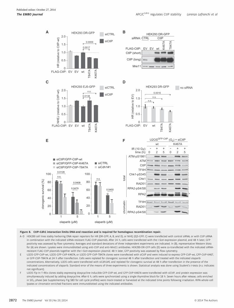
slight increase in total NHEJ (Fig 8A and B) (Bennardo et al, 2008).
Importantly, these effects could be rescued by expression of siRNA-
resistant FLAG-CtIP-wt (Fig 8A and B). Interestingly, expression of
the K467A mutant did not rescue HR, but caused a similar decrease
as compared to the DNA-end resection-defective T847A mutant
(Fig 8A and B). Surprisingly, unlike T847A, we observed that
expression of K467A does not lead to a significant increase in
total NHEJ (Fig 8C). The observation that CtIP-K467A impaired
homology-directed repair but does not result in compensation
through NHEJ, suggested a dominant negative effect of the KEN box
mutant. To test this hypothesis, FLAG-CtIP constructs were transfected
into HEK293 DR-GFP cells without siRNA-mediated depletion of
endogenous CtIP. Indeed, expression of CtIP-K467A caused a reduc-
tion in HR, whereas expression of CtIP-wt or CtIP-T847A did not
significantly alter HR (Fig 8D). Taken together, these results suggest
that abolishing the interaction between CtIP and Cdh1 does not inter-
fere with the initiation of DNA-end resection, which would otherwise
increase NHEJ efficiency. Instead, after resection has been initiated,
the CtIP–Cdh1 interaction appears to be required for the proper
execution of downstream HR events.
Defective HR was previously shown to result in increased
sensitivity to PARP inhibition (Bryant et al, 2005; Farmer et al,
2005). In line with a defect in HR, we observed that cells inducibly
expressing GFP-CtIP-K467A showed elevated sensitivity to the PARP
inhibitor olaparib, although not as pronounced as in GFP-CtIP-
T847A mutant cells (Fig 8E). Notably, depletion of Cdh1 also
resulted in an elevated sensitivity to PARP inhibition (Fig 8E).
However, GFP-CtIP-K467A-expressing cells did not display hypersen-
sitivity to IR or doxorubicin treatment, indicating that the inability of
Cdh1 to interact with CtIP cannot explain all phenotypic responses
associated with Cdh1 loss in combination with DNA damage
(Supplementary Fig S8A–C).
Besides decreased DNA-end resection capacity, also excessive or
temporally unrestricted resection may potentially impair HR. Since
we have observed that CtIP is targeted by the APC/C for proteaso-
mal degradation after DNA damage in G2, we monitored DNA-end
resection in wt and K467A mutant cells that had been synchronized
in G2 prior to IR (Fig 8F and Supplementary Fig S8D). Clearly,
expression of GFP-CtIP-K467A resulted in elevated levels of RPA2
phosphorylation at S4/S8, indicative of hyper-resection. This
matched our earlier observation of elevated levels of RPA2 phos-
phorylation upon proTAME treatment in irradiated cells (Fig 6B).
Remarkably, increased RPA2 phosphorylation in GFP-CtIP-K467A-
expressing cells coincided with lower amounts of Rad51 being
recruited to damaged chromatin (Fig 8F). Combined, these data
suggest that CtIP–Cdh1 interaction is involved in limiting DSB resec-
tion, which probably allows correct assembly of Rad51–ssDNA
nucleoprotein filaments, to facilitate HR.
Discussion
The response to DSBs is tightly regulated during the cell cycle. As a
consequence, deregulated cell cycle control may lead to aberrant DSB
repair and ensuing genomic instability. An example thereof is
provided by the APC/CCdh1 cell cycle regulator, as genetic inactivation
of Cdh1 in either mouse embryonic fibroblasts or primary human
cells has been shown to cause elevated levels of DNA damage and
chromosomal instability. We could recapitulate these findings in
Cdh1-depleted human cell lines of different origins. Moreover, we
were able to extend these findings and demonstrate that depletion of
Cdh1 results in hypersensitivity to DSB-inducing agents and nega-
tively affects Rad51 IRIF formation. Concerning potential APC/C
substrates responsible for these effects, Rhp54 (the fission yeast ortho-
log of Rad54) and Rad17 were shown to be degraded by the APC/C
(Trickey et al, 2008; Zhang et al, 2010). However, when Rad54 was
investigated in other species, no APC/C-dependent degradation was
observed, and the degradation of Rad17 by the APC/C appeared to be
UV-induced and appeared to control checkpoint duration rather than
DNA repair. Finally, Cdh1 was reported to control the duration of the
G2 cell cycle arrest in response to DSBs by targeting Polo-like kinase 1
(Plk1) for proteolytic degradation (Wasch & Engelbert, 2005; Basser-
mann et al, 2008; Engelbert et al, 2008). So far, it remained elusive,
however, whether the APC/CCdh1 also contributes to the regulation of
DSB repair.
Using a proteomics analysis of mitotic exit combined with bio-
informatics analysis of the presence of KEN and D-box motifs, we
have identified a number of candidate APC/CCdh1 substrates. Several
of the putative Cdh1 targets play key roles in the regulation of DSB
repair, including Rif1, MDC1, SMC5, and CtIP. Detailed in silico
analysis of multiple protein sequences for the conservation of puta-
tive KEN and D-box motifs guided us to focus on CtIP as a previ-
ously unrecognized APC/CCdh1 substrate. Human CtIP contains two
conserved KEN box motifs, but only the second KEN box strongly
matches the consensus sequence recently proposed by Barford and
colleagues (He et al, 2013) and is required for Cdh1–CtIP interac-
tion. In addition to being targeted by the APC/CCdh1 for proteasomal
degradation in G1, we discovered that CtIP protein levels are
controlled by the APC/CCdh1 prior to mitotic entry in response to
DSBs. Concerning the activation of the APC/CCdh1 in response to
DNA damage in G2 cells, we noted that APC/CCdh1 activation is
achieved most efficiently after high levels of DNA damage. This
implies that especially under conditions provoking high amounts of
DNA damage, such as after chemotherapy or radiotherapy, the
APC/CCdh1 may acquire new functions, which under these circum-
stances may determine cell fate and genomic integrity.
Due to its crucial role in initiating DNA-end resection, CtIP is
essential for homology-directed repair of DSBs (Sartori et al, 2007;
Bennardo et al, 2008). DNA-end resection dictates the choice
between HR and NHEJ and is thus proposed to be tightly regulated
during the cell cycle (Ferretti et al, 2013). For instance, CtIP phos-
phorylation at T847 by cyclin-dependent kinases represents a key
step toward the commencement of DNA-end resection and, conse-
quently, a CtIP-T847A mutant abrogates HR (Huertas & Jackson,
2009). Here, we show that a CtIP KEN box mutant (K467A) compro-
mises HR to a similar extent as the T847A mutant, indicating that
the interaction between Cdh1 and CtIP facilitates HR. NHEJ requires
only very limited DSB processing and is therefore not suitable for
repairing DSBs which have undergone extensive resection. In other
words, NHEJ can only compensate for HR in cells that are defective
in DNA-end resection (Shibata et al, 2011). This is in line with our
results showing that CtIP-T847A results in higher levels of NHEJ. In
contrast, we find that CtIP-K467A does not lead to a concomitant
ª 2014 The Authors The EMBO Journal Vol 33 | No 23 | 2014
Lorenzo Lafranchi et al APC/CCdh1 regulates CtIP stability The EMBO Journal
2871
Published online: October 27, 2014
CtIP
IR (10 Gy): - +time (h): 2
+5
- +2
+5
U2OSGFP-CtIP (G2) + siCtIP
tota
lch
rom
atin
wt
RAD51
RPA2 *
RPA2-pS4/S8
TFIIH
RAD51
RPA2-pS4/S8
Chk1Chk1-pS345
K467A
00ATM-pS1981
ATM
TFIIH
A HEK293 DR-GFP
HR
(rel
ativ
e to
CtIP
-wt)
1.0
1.5
2.0
0
0.5
0.0009
0.0017
FLAG-CtIP: wt
T847
A
K46
7AEV EV
HEK293 EJ5-GFP
NH
EJ
(rel
ativ
e to
CtIP
-wt)
1.0
1.5
2.0
0
0.5
n.s.
0.0138
wt
T847
A
K46
7AEV EV
C
siCTRL
siCtIP
Dno siRNAHEK293 DR-GFP
HR
(rel
ativ
e to
EV
)
1.0
1.5
2.0
0
0.5
0.0016
FLAG-CtIP: wt
T847
A
K46
7AEV
n.s.n.s.
FLAG-CtIP:
siCTRL
siCtIP
n.s.
0.0128
0.0479
0
20
40
60
80
100
1 5
siCtIP/GFP-CtIP-wt
siCtIP/GFP-CtIP-T847AsiCtIP/GFP-CtIP-K467A
0
20
40
60
80
100
siCTRLsiCdh1#1
n.s.
0.0450
1 5
clon
ogen
ic s
urvi
val (
%)
olaparib ( M) olaparib ( M)
E F
CtIP (long)
EVFLAG-CtIP:
CtIPsiRNA: CTRL
EVCtIP (short)
HEK293 DR-GFP
Mre11
wt K46
7A
T847
A
B
0.0408
Figure 8. CtIP–Cdh1 interaction limits DNA-end resection and is required for homologous recombination repair.
A–D HEK293 cell lines stably harboring DNA repair reporters for HR (DR-GFP; A, B, and D), or NHEJ (EJ5-GFP; C) were transfected with control siRNA, or with CtIP siRNAin combination with the indicated siRNA-resistant FLAG-CtIP plasmids. After 24 h, cells were transfected with the I-SceI-expression plasmid, and 48 h later, GFPpositivity was assessed by flow cytometry. Averages and standard deviations of three independent experiments are indicated. In (B), representative Western blotsfor (A) are shown. Lysates were immunoblotted using anti-CtIP and anti-Mre11 antibodies. HEK293-DR-GFP cells (D) were co-transfected with the indicated siRNA-resistant FLAG-CtIP plasmids together with the I-SceI-expression plasmid. 48 h later, GFP positivity was assessed by flow cytometry.
E U2OS-GFP-CtIP-wt, U2OS-GFP-CtIP-K467A, or U2OS-GFP-CtIP-T847A clones were transfected with siCtIP and were induced to express GFP-CtIP-wt, GFP-CtIP-K467,or GFP-CtIP-T847A at 24 h after transfection. Cells were replated for clonogenic survival 48 h after transfection and treated with the indicated olaparibconcentrations. Alternatively, U2OS cells were transfected with siCdh1#1 and replated for clonogenic survival at 48 h after transfection in the presence of theindicated concentrations of olaparib. Standard error of the means of three experiments is shown. Statistical analysis was done using Student’s t-tests (n.s. indicatesnot significant).
F U2OS Flp-In T-REx clones stably expressing doxycycline-inducible GFP-CtIP-wt, and GFP-CtIP-K467A were transfected with siCtIP, and protein expression wassimultaneously induced by adding doxycycline. After 6 h, cells were synchronized using a single thymidine block for 18 h. Seven hours after release, cells enrichedin S/G2 phase (see Supplementary Fig S8D for cell cycle profiles) were mock-treated or harvested at the indicated time points following irradiation. RIPA whole-celllysates or chromatin-enriched fractions were immunoblotted using the indicated antibodies.
The EMBO Journal Vol 33 | No 23 | 2014 ª 2014 The Authors
The EMBO Journal APC/CCdh1 regulates CtIP stability Lorenzo Lafranchi et al
2872
Published online: October 27, 2014

increase in NHEJ, suggesting that resection has occurred in those
cells. In fact, CtIP-K467A-expressing cells irradiated in G2 display
even heightened levels of DNA-end resection compare to CtIP-wt
cells, but are partially impaired in promoting efficient Rad51 recruit-
ment to damaged chromatin, which is similar to what we observed
in Cdh1-depleted cells. Moreover, reduced HR efficiency of K467A
mutant cells is in line with our data of decreased survival upon
PARP inhibition.
Combined, our data support a model in which APC/CCdh1 activity
is involved in negatively regulating the stability of CtIP both after
mitotic exit in unperturbed cells and after DNA damage in G2
(Fig 9). Moreover, we speculate that the APC/CCdh1 is required at a
late stage within the HR process, after initiation of resection has
occurred and NHEJ is no longer an option for DSB repair. One possi-
bility is that APC/CCdh1 mediates clearance of CtIP IRIF through
ubiquitin-mediated degradation, thereby limiting resection to
amounts of ssDNA that can be handled by the downstream recombi-
nation machinery. A similar mechanism has been recently reported
by Choi et al for the regulation of nuclear PTEN, in which Cdh1
promotes the removal of PTEN from chromatin during mitotic exit
(Choi et al, 2014).
Our observations that Rad51 IRIF are decreased in Cdh1-depleted
G2 cells, that CtIP-K467 IRIF persist much longer, and that Rad51
loading onto damaged chromatin is compromised in G2-enriched
cells expressing the CtIP KEN box mutant are in line with a role for
APC/CCdh1-dependent CtIP degradation in controlling HR. In its role
of keeping DNA-end resection in check, the APC/CCdh1 may play a
similar function in G1 and G2. In response to DNA breaks in G1 cells,
the end resection machinery cannot be activated due to lack of CDK
activity. In this context, APC/CCdh1-mediated degradation of CtIP
may serve as a backup mechanism to prevent unscheduled end
resection. In G2 cells, on the other hand, end resection is required for
error-free DSB repair by HR. Here, APC/CCdh1-mediated degradation
of CtIP, after initial resection has been performed, may be required
to limit end resection to levels that optimally facilitate HR repair.
Materials and Methods
Cell culture
hTERT-immortalized retinal pigment epithelium (RPE-1), U2OS,
U2OS-FUCCI, and HEK293T cells were grown in DMEM (Gibco, Life
Technologies). MCF7 cells were cultured in RPMI (Gibco, Life Tech-
nologies). HeLa cells were cultured in DMEM/Ham’s F12 (1:1)
medium (Gibco, Life Technologies). All culture media were supple-
mented with 10% fetal calf serum (FCS), 100 units/ml penicillin,
and 100 lg/ml streptomycin. U2OS and HEK293 Flp-In T-REx cells
were grown in DMEM supplemented with 10% Tet system approved
FCS, 100 U/ml penicillin, 100 mg/ml streptomycin, 125 lg/ml
hygromycin B, and 12.5 lg/ml blasticidin S.
IR was given using a CIS international/IBL 637 irradiator
equipped with a cesium137 source (dose rate: 0.01083 Gy/s), or
using a Faxitron X-ray device. For serum starvation experiments,
RPE-1 cells were initially plated in medium containing 10% FCS and
were washed with phosphate-buffered saline (PBS) at 24 h after
plating and subsequently cultured without serum for another 24 h.
After serum starvation, serum was added to a final concentration of
20%. At the time of serum addition, bromodeoxyuridine (BrdU) was
added to a final concentration of 10 lM to measure replication
onset. If indicated, cells were treated with 5 lM of the proteasome
inhibitor MG-132 (Sigma-Aldrich, St. Louis, MO), 250 nM of the
microtubule polymerization inhibitor nocodazole (Sigma-Aldrich),
5 lM of the Eg5 inhibitor S-trityl-L-cysteine (STLC, Sigma-Aldrich),
5 lM of the Cdk1 inhibitor RO-3306 (Axon Medchem, Groningen,
the Netherlands), or with the APC/C inhibitor proTAME (Zeng et al,
2010) at a final concentration of 12 or 20 lM. ProTAME was kindly
provided by Randy King, Harvard Medical School, Boston MA, or
obtained from Boston Biochem.
Generation of stable GFP-CtIP cell lines
The Flp-In T-REx system (Invitrogen, Life Technologies) was used to
generate cell lines stably expressing different siRNA-resistant
GFP-CtIP constructs in an inducible manner. The GFP-CtIP-containing
pcDNA5/FRT/TO vector and the Flp recombinase expression plas-
mid pOG44 were mixed in a 1:9 ratio and transfected into Flp-In
T-REx 293 (Invitrogen, Life Technologies) and Flp-In T-REx U2OS
(a kind gift of Daniel Durocher, University of Toronto) cells using
APC/C
Cdh1pp
p
CtIP
APC/CCdh1
G1
M
G2
S
unperturbed cell cycle
CtIP
resection
APC/CCdh1
CtIPUb
UbUb
limitation of DNA-endresection
CtIPUb
UbUb
HR
NHEJ
Figure 9. Model of cell cycle- and DNA damage-dependent regulation ofCtIP by the APC/CCdh1.The central area represents unperturbed cell cycle, when CtIP is degraded aftermitotic exit by the APC/CCdh1. Cells in G0/G1 lack CDK activity, which precludesphosphorylation and consequent activation of CtIP. Post-mitotic CtIPdegradation by the APC/CCdh1 E3 ubiquitin ligase may contribute to preventunscheduled DNA-end resection in G0/G1 phase. In response to DSBs in S/G2
phase, CtIP promotes DNA-end resection to facilitate HR repair. In response tohigh levels of DSBs, CtIP is initially recruited to DSBs to resect DNA ends andpromote HR repair. In a late response to high levels of DNA damage, theAPC/CCdh1 promotes ubiquitin-dependent proteolysis of CtIP. Downregulation ofCtIP by the APC/CCdh1 promotes its clearance from DSBs and prevents excessiveDNA-end resection, a prerogative for proper homology-directed repair.
ª 2014 The Authors The EMBO Journal Vol 33 | No 23 | 2014
Lorenzo Lafranchi et al APC/CCdh1 regulates CtIP stability The EMBO Journal
2873
Published online: October 27, 2014

FuGENE 6 transfection reagent (Promega) at 60% confluency. After
6 h, medium was exchanged to fresh DMEM and cells were incu-
bated at 30°C (6% CO2). Two days later, cells were replated at
different dilutions in 10-cm plates. After 24 h, the medium was
supplemented with 250 lg/ml hygromycin B and 12.5 lg/ml
blasticidin S. The medium was replaced every 2–3 days, and cells
were selected for approximately 2 weeks. Resistant colonies were
picked and further characterized as single clones or pooled to gener-
ate bulk cultures. All cell lines were screened for inducible GFP-CtIP
expression by both immunofluorescence microscopy and immuno-
blotting. To induce expression of GFP-CtIP, cells were treated with
0.5 or 1 lg/ml doxycycline (Dox) for 24 h as indicated.
Immunoprecipitation and GST pull-down
For immunoprecipitation and glutathione S-transferase (GST) pull-
down assays, cells were lysed in NP-40 extraction buffer (50 mM
Tris–HCl, pH 7.5, 120 mM NaCl, 1 mM EDTA, 6 mM EGTA, 15 mM
sodium pyrophosphate, 1% NP-40), supplemented with phospha-
tase inhibitors (20 mM NaF, 1 mM sodium orthovanadate) and
protease inhibitors (1 mM benzamidine and 0.1 mM phenylmethyl-
sulfonyl fluoride (PMSF)), and clarified by centrifugation at
20,000 g. HeLa nuclear extracts (HNE) were purchased from Ipracell
(Belgium). Generation of the GST–CtIP constructs was described
previously (Sartori et al, 2007). GST fusion plasmids were grown in
BL21 RIL (CodonPlus) Escherichia coli (Stratagene), and recombi-
nant proteins were expressed by incubating the bacteria for 24 h at
16°C after the addition of 100 lM IPTG. After centrifugation, the
bacterial pellet was resuspended in cold PBS, supplemented with
1% Triton X-100 and protease inhibitors (1 mM PMSF, 1 mM benz-
amidine, and Roche protease inhibitor cocktail). After sonication
and centrifugation, GST-tagged proteins were purified from soluble
extracts using Glutathione Sepharose 4 Fast Flow beads (GE
Healthcare). GST fusion proteins bound to glutathione beads were
mixed with 1 mg of HeLa nuclear extract and incubated for 1 h at
4°C in 1 ml of TEN100 buffer (20 mM Tris–HCl (pH 7.4), 0.1 mM
EDTA, and 100 mM NaCl). Beads were then washed three times
with NTEN500 buffer (0.5% NP-40, 0.1 mM EDTA, 20 mM Tris–
HCl (pH 7.4), and 500 mM NaCl) and once with TEN100 buffer.
Recovered complexes were boiled in SDS sample buffer and
analyzed by SDS–PAGE followed by immunoblotting.
Immunoprecipitating antibodies were added to the cell lysates
and incubated overnight at 4°C. After 2 h incubation with protein A
or protein G beads, precipitated immunocomplexes were washed
four times with lysis buffer or three times with TNE buffer (50 mM
Tris–HCl (pH 7.4), 100 mM NaCl, 0.1 mM EDTA) containing 1%
Triton X-100 and once with TNE buffer, boiled in SDS sample
buffer, and loaded on an SDS–polyacrylamide gel. Proteins were
analyzed by immunoblotting as described below.
In vivo ubiquitylation assays
HEK293 Flp-In T-REx GFP-CtIP cells were transfected with His-
ubiquitin using the FuGENE 6 transfection reagent (Promega), and
after 24 h, GFP-CtIP expression was induced with 1 lg/ml Dox.
After 24 h, cells were treated for 4 h with 20 lM MG-132 and then
washed and scraped in 500 ll of ice-cold PBS. 2% of the cell suspen-
sion was used for direct Western blot analysis. The remaining
cells were lysed in “buffer A” (6 M guanidine–HCl, 0.1 M Na2HPO4/
NaH2PO4, pH 8.0, 10 mM imidazole), and lysates were incubated
with Ni2+-NTA agarose beads for 3 h under rotation at room temper-
ature. The beads were washed two times with buffer A, two times
with “buffer A/TI” (1 volume buffer A: 3 volume buffer “TI”
(25 mM Tris–HCl, pH 6.8, and 20 mM imidazole)), and two times
with buffer TI. Bound proteins were eluted by boiling the beads in
2× SDS sample buffer supplemented with 250 mM imidazole and
analyzed by immunoblotting. In case of siRNA treatment, cells were
first transfected with the indicated siRNA and after 24 h transfected
with His-ubiquitin using the FuGENE6 transfection reagent
(Promega). At the same time, GFP-CtIP expression was induced with
1 lg/ml Dox, and after 24 h, samples were processed as described
above.
To analyze ubiquitylation of endogenous CtIP, HEK293 cells
were transfected with HA-ubiquitin using the FuGENE 6 transfection
reagent (Promega) and enriched in S/G2 phase of the cell cycle by
releasing them from a single thymidine block. After treatment, cells
were lysed in (5 mM Tris–HCl (pH 7.5), 5 mM DTT, 1% SDS) and
boiled for 5 min (El-Shemerly et al, 2005). After sonication, samples
were clarified by centrifugation and diluted 4 times with NP-40
buffer supplemented with phosphatase inhibitors (20 mM NaF,
1 mM sodium orthovanadate), protease inhibitors (1 mM benzami-
dine and 0.1 mM phenylmethylsulfonyl fluoride (PMSF)), and the
deubiquitinases inhibitor N-ethylmaleimide (NEM, 20 mM). Immu-
noprecipitation was performed overnight at 4°C, using a polyclonal
rabbit antibody (612L, raised against CtIP N-terminus, a kind gift of
Prof. Richard Baer, Columbia University). After 2 h incubation with
protein A beads, precipitated immunocomplexes were washed three
times with NTEN500 buffer and once with TEN100 buffer, boiled in
SDS sample buffer, and loaded on an SDS–polyacrylamide gel. After
transfer, membranes were incubated for 30 min at 4°C in denaturing
buffer (6 M guanidine–HCl, 20 mM Tris–HCl (pH 7.5), 1 mM PMSF,
and 5 lM b-mercaptoethanol) as described in Penengo et al (2006).
After extensive washing with TBS-Tween buffer, membranes were
incubated with the appropriate antibody and further processed as
described below.
Immunoblotting
If not specified otherwise, cell extracts were prepared in Laemmli
buffer (4% SDS, 20% glycerol, 120 mM Tris–HCl pH 6.8). If indi-
cated, cells were lysed in RIPA buffer (50 mM Tris–HCl, pH 7.5, 1%
NP-40, 0.25% sodium deoxycholate, 150 mM NaCl, 1 mM EDTA,
and 0.1% SDS) supplemented with phosphatase and protease inhibi-
tors. Proteins were resolved by SDS–PAGE and transferred to nitro-
cellulose. Immunoblots were performed using the appropriate
antibodies, and proteins were visualized using the ECL detection
system (Amersham). Primary antibodies used in this study are listed
in Supplementary Table S2. The anti-Claspin antibody was a kind
gift of Dr. Raimundo Freire, University of Tenerife) and was
described previously (Semple et al, 2007).
When indicated, a Triton X-100-insoluble (chromatin-enriched)
fraction was isolated as described in Pena-Diaz et al (2012). Briefly,
cells were rinsed twice in cold PBS and incubated for 5 min on ice
in pre-extraction buffer (25 mM HEPES (pH 7.9), 50 mM NaCl,
1 mM EDTA, 3 mM MgCl2, 300 mM sucrose, 0.5% Triton X-100,
and protease inhibitors). After buffer removal and rinsing in PBS,
The EMBO Journal Vol 33 | No 23 | 2014 ª 2014 The Authors
The EMBO Journal APC/CCdh1 regulates CtIP stability Lorenzo Lafranchi et al
2874
Published online: October 27, 2014
adherent cellular material was harvested by scraping it into
Laemmli buffer. The chromatin-enriched fraction was then heat
denatured, sonicated, and analyzed by immunoblotting.
HR and NHEJ DNA repair assays
DSB repair efficiency by HR or NHEJ was measured in DR-GFP or
EJ5-GFP HEK293 cell lines as described previously (Bennardo et al,
2008). Briefly, 0.6 × 10⁶ cells were plated in 6-well plates (poly-L-
lysine coated) and, after 24 h, cells were transfected with siRNA
oligos (40 nM). The next day, 0.24 × 10⁶ cells were reseeded in
12-well plates. At 48 h after siRNA transfection, cells were either
mock-transfected or transfected with 0.6 lg I-SceI expression plas-
mid (pCBASce) in combination with 0.2 lg of the appropriate
FLAG-tagged CtIP constructs (pcDNA3) using 1.6 ll of JetPrime
(Polyplus). At 4 h after plasmids transfection, media were replaced
and a second transfection with siRNA oligos (15 nM) was
performed. Alternatively, cells were only transfected with 0.6 lgI-SceI expression plasmid (pCBASce) in combination with 0.2 lg of
the appropriate FLAG-tagged CtIP constructs (pcDNA3) using 1.6 llof JetPrime (Polyplus). At 48 h after I-SceI transfection, cells were
analyzed for GFP expression by flow cytometry on a CyAn ADP 9
(Dako).
Laser micro-irradiation
Laser micro-irradiation to generate DSBs in a defined nuclear region
was performed as described previously (Lukas et al, 2003; Bekker-
Jensen et al, 2006). Briefly, 24 h before irradiation, culture medium
was supplemented with 10 lM BrdU. Around 100 cells were micro-
irradiated at room temperature (a procedure lasting around 10 min)
using the MMI CELLCUT system containing a UVA laser of 355 nm
(Molecular Machines and Industries, Zurich, Switzerland). The laser
intensity was set to 50% energy output, and each cell was generally
exposed to the laser beam for < 300 ms (Meerang et al, 2011).
DNA plasmids and RNA interference
Plasmids were transfected by using either the standard calcium
phosphate method or FuGENE 6 (Promega) according to manufac-
turer’s instructions. The epitope-tagged expression vectors for
human CtIP have been described previously (Yu et al, 2006; Sartori
et al, 2007). The HA-tagged expression vector for human Cdh1 was
described previously and was purchased from Addgene (plasmid
#11596) (Pfleger et al, 2001). The pcDNA3.1-6×His-ubiquitin plas-
mid was a kind gift of Matthias Peter (ETH Zurich, Switzerland),
whereas the HA-ubiquitin plasmid was described previously and
purchased from Addgene (plasmid #18712) (Kamitani et al, 1997).
All CtIP point mutants were introduced by site-directed mutagenesis
using Expand Long Template PCR System (Roche) and confirmed
by sequencing. shRNA interference for Cdh1 in RPE-1, MCF7, or
HeLa cells was performed using lentiviral infection with control
pLL3.7-GFP (targeting sequence: 50-GGCATCAAGGTGAACTTCA-30)or pLL3.7-Cdh1 (targeting sequence: 50-GGATTAACGAGAATGAGAA-30; provided by r, University of Freiburg, Germany) (Engelbert
et al, 2008). To this end, HEK293T cells were transfected with the
pLL3.7 plasmids along with the packaging plasmids pCMV-VSVG,
pCMV-dR8.2 (provided by Robert Weinberg, MIT, Cambridge, MA),
and pAdVAntage (Promega) in a 4:3:1:0.5 ratio. Virus-containing
supernatant was harvested at 24 and 48 h after transfection, filtered
through a 0.45-lM syringe filter, and used to infect target cells,
which were subsequently selected with 1 lg/ml puromycin. Analy-
sis of stable Cdh1-depleted cells was done at 5 days post-infection.
For transient siRNA experiments, RPE-1, MCF7, HeLa, or U2OS
cells were plated in 6-well plates and transfected with the indicated
amounts of siRNA oligos (40 nM final concentration of oligos) using
Oligofectamine or Lipofectamine RNAiMAX using manufacturer’s
guidelines (Invitrogen, Life Technologies). In brief, medium was
replaced with Opti-MEM (Gibco, Life Technologies) prior to incuba-
tion with siRNAs and Oligofectamine for 4 h. Thereafter, medium
containing FCS (10% final concentration) was added and cells were
analyzed at 48 or 72 h after transfection. siRNA oligos targeting
Cdh1 #1 (50-GGATTAACGAGAATGAGAAdTdT-30), Cdh1 #2 (50-AATGAGAAGTCTCCCAGTCAGdTdT-30), CtIP (50-GCUAAACAGGAACGAAUCTTdTdT-30) (Sartori et al, 2007), or luciferase (50-CGUACGCGGAAUACUUCGAdTdT-30) were purchased from Ambion (Life Tech-
nologies) or Microsynth (Balgach, Switzerland). In addition, “medium
GC duplex” control siRNA (Cat. No: 12935-300) was purchased from
Ambion (Life Technologies).
SILAC and mass spectrometry
For SILAC experiments, RPE-1 cells were cultured in ready-to-use
light and heavy DMEM media, containing light or heavy arginine
and lysine (“R0K0 DMEM” with 12C6-L-arginine and 12C6-L-lysine or
“R10K8 DMEM” with 13C6-L-arginine and 13C6-L-lysine, respec-
tively) and supplemented with dialyzed FBS. Media were obtained
from Silantes (Munich, Germany). SILAC labeling was performed as
previously described by Ong and Mann (Ong et al, 2002). Briefly,
RPE-1 cells were cultured in normal media or complete heavy
DMEM medium containing 10% dialyzed FBS for at least 10 cell
doublings (5 passages) to allow full incorporation of both labeled
amino acids within the proteome. Cells were subsequently treated
with nocodazole for 16 h (250 ng/ml) and collected by mitotic
shake-off. Cells were washed three times in pre-warmed PBS and
replated in the absence of nocodazole. Immediately after replating
or 2.5 h after replating, cells were harvested by trypsinization, snap-
frozen in liquid nitrogen, and resuspended in lysis buffer (6 M urea
and 2 M thio-urea). A label-swap replicate was also performed
where SILAC states and harvesting conditions were reversed
(Fig 3A). To determine protein concentration in lysates, 10 ll lysatewas added to 150 ll of 660 nM protein assay reagent (Pierce). Equal
amounts of protein from R0K0 and R10K8 were mixed, reduced with
1 mM DTT, and alkylated with 8 mM iodoacetamide. Proteins were
separated on SDS–PAGE 4–12% gradient gels and visualized by
staining by Coomassie staining. Gel lanes were divided into eight
slices and proteins digested with an in-gel digestion protocol with
trypsin. Peptides were desalted on C18 StageTips and loaded on a
10-cm-long 360 lm O.D. by 75 lm I.D. column packed with 3 lmReproSil-Pur C18 AQ 3beads (Dr. Maisch, Germany) with an Agilent
1100 nano-flow pump and autosampler. A 60 min gradient from 3
to 35% acetonitrile containing 1% formic acid at 200 nl/min was
applied to elute peptides for analysis in the LTQ-Orbitrap-Velos
(Thermo, Bremen) in a Top5 CID data-dependent acquisition mode.
Peptide identification and quantification was performed using
MaxQuant v.1.1.1.14 with the IPI human database ver. 3.70 with
ª 2014 The Authors The EMBO Journal Vol 33 | No 23 | 2014
Lorenzo Lafranchi et al APC/CCdh1 regulates CtIP stability The EMBO Journal
2875
Published online: October 27, 2014

variable modifications of oxidized methionine and acetylated
protein N-termini. Cysteines were carbamidomethylated. Peptide
and protein FDR was set at 1%.
Flow cytometry
Cells were harvested at the indicated time points after treatment and
fixed in ice-cold 70% ethanol. Cells were stained with rabbit anti-
phospho-histone H3 antibody, mouse anti-MPM-2, and/or mouse
anti-c-H2AX (details of used antibodies are in Supplementary Table
S2), subsequently stained with Alexa488-conjugated and Alexa647-
conjugated secondary antibodies (Molecular Probes, 1:300), and
counterstained with propidium iodide/RNase (Sigma). Samples
were analyzed on a FACS-Calibur (Becton Dickinson) equipped with
Cell Quest software. Per sample, a minimum of 104 events was
analyzed and indicated results show averages and standard devia-
tions of three independent experiments.
Immunofluorescence microscopy
MCF7, MEFs, or HeLa cells were cultured on glass coverslips and, if
indicated, were irradiated at 24 h after plating. After treatment, cells
were fixed in formaldehyde (3.7% in PBS) for 15 min at room
temperature. After washing with PBS, cells were permeabilized with
Triton X-100 (0.1% in PBS) for 5 min at room temperature. After
extensive washing, cells were stained with mouse anti-Cdh1, mouse
anti-p53 diluted in PBS, 0.05%Tween-20, 2.5%BSA for 16 h at 4°C.
After extensive washing, cells were stained with Alexa488-,
Alexa568-, or Alexa647-conjugated secondary antibodies for 30 min
at room temperature and counterstained with DAPI. Images were
obtained using a Leica DM6000B microscope, equipped with 63×
immersion lens (PL-S-APO, numerical aperture: 1.30) and Xenon
light source using LAS-AF software (Leica).
Alternatively, U2OS-FUCCI cells were grown on glass coverslips
and, at different time points after treatment, fixed directly in 4%
formaldehyde (w/v) in PBS for 15 min as described previously
(Eid et al, 2010). After incubation with rabbit anti-Rad51 or rabbit
anti-53BP1 and appropriate secondary antibodies the coverslips
were mounted and sealed with Vectashield (Vector Laboratories)
containing DAPI. Images were acquired on a Leica DMRB
fluorescence microscope.
For live-cell microscopy, stable U2OS cells were plated in cham-
bered coverglass 8-well plates (LabTek-II, Nunc). At 24 h before
imaging, GFP-CtIP expression was induced by adding doxycycline to
a final concentration of 1 lg/ml. GFP and DIC images were obtained
every 5 min on a DeltaVision Elite microscope, equipped with a
CoolSNAP HQ2 camera, 40× immersion objective (U-APO 340,
numerical aperture: 1.35). In the Z-plane, 12 images were acquired
at 0.5-lm interval, which were subsequently deconvolved using
SoftWorx 5.5 software (Applied Precision). Nuclear fluorescence
intensity was quantified using ImageJ software.
Clonogenic and short-term survival assays
HeLa or MCF7 cells were cultured in 6-well plates. One day after
plating, cells were irradiated with 1, 2, or 4 Gy. When surviving
colonies were approximately 50 cells in size, cells were fixed
and stained using methanol/ acidic acid/water in a 5:2:3 ratio,
supplemented with 0.01% Coomassie brilliant blue. Surviving frac-
tions were calculated using the plating efficiencies with non-
irradiated conditions as a reference. Shown averages are from three
experiments, with three replicates each. Alternatively, clonal U2OS
cells expressing GFP-CtIP variants were transfected with siRNAs
and, if indicated, were treated with doxycycline 24 h later to induce
expression of GFP-CtIP. At 48 h after transfection, cells were trypsi-
nized and replated in 6-well plates. During replating, indicated doses
of doxorubicin or the PARP inhibitor olaparib (Axon Medchem,
Groningen, the Netherlands) were added. To test whether Cdh1-
depleted cells also are more sensitive to PARP inhibition, U2OS cells
were transfected with siCdh1#1, and after 48 h, cells were replated
in the presence of olaparib. Statistical testing was done using the
Student’s t-test.
For short-term survival assays, 2,000 cells were plated in 96-wells
plates and treated at 24 h after plating. At 4 days after treatment
initiation, 20 ll of 3-(4,5-dimethylthiazol-2-yl)-2,5-diphenyltetrazolium
bromide (MTT) was added to the culture medium to a final concen-
tration of 5 mg/ml for 3 h. Formazan crystals were dissolved in
DMSO, and absorbance was analyzed at 520 nM using a Bio-Rad
benchmark III microtiter spectrophotometer. Survival was calculated
as a percentage of untreated cells.
Gene set enrichment analysis (GSEA) and destructionmotif analysis
GSEA was performed with GSEA 2.0 (Broad Institute, Cambridge,
MA) (Mootha et al, 2003; Subramanian et al, 2005). A significance
threshold was set at a nominal P-value of 0.05 and a false discovery
rate (FDR) of 0.30. In our analysis, we transformed protein names
from SILAC MS analysis to HUGO gene symbols and tested enrich-
ment in Kyoto Encyclopedia of Genes and Genomes (KEGG), Gene
Map Annotator and Pathway Profiler (GenMAPP) (Dahlquist et al,
2002; Kanehisa et al, 2012), and the BioCarta database (http://
www.biocarta.com). Within the set of identified proteins from the
SILAC MS analysis, we used GPS-ARM (Liu et al, 2012) to identify
proteins with a D-box or a KEN box using a threshold setting of
“high” for D-boxes and “medium” for KEN boxes. Subsequently,
GSEA was used to test for enrichment of KEN box or D-box contain-
ing proteins in the downregulated fraction. FDR rates of 0.25 were
used in combination with 10,000 permutations. Conservation of
KEN or D-boxes was done using Clustal Omega. In order to identify
proteins related to the DNA damage response and DNA damage
repair, a combined list of genes was compiled by merging the
following Gene Ontology gene sets (see also Supplementary Table
S1): GO:0006302 (“double-strand break repair); GO:0006974 (“cellu-
lar response to DNA damage stimulus”); GO:0000077 (“DNA
damage checkpoint”); and GO:0000724 (“double-strand repair via
homologous recombination”). In total, 255 genes were included in
this combined gene set (Supplementary Table S1), which in Fig 3 is
referred to as “DNA damage response”.
Supplementary information for this article is available online:
http://emboj.embopress.org
AcknowledgementsWe thank Dr. Rudolf Fehrmann, Dr. Małgorzata Krajewska, and Anne
Margriet Heijink for technical help and Dr. Floris Foijer for help with
The EMBO Journal Vol 33 | No 23 | 2014 ª 2014 The Authors
The EMBO Journal APC/CCdh1 regulates CtIP stability Lorenzo Lafranchi et al
2876
Published online: October 27, 2014

live-cell microscopy. We are grateful to Dr. Jeremy Stark, Dr. Robert Wein-
berg, Dr. Ralph Waesch, Dr. Stefano Ferrari, Dr. Randy King, Dr. Richard Baer,
Dr. Raimundo Freire, and Dr. Matthias Peter for providing reagents. We
thank Chris Soon Heng Tan and Rob Wolthuis for helpful discussions. This
research was supported by an NWO-VIDI grant (016.136.334) and Dutch
Cancer Society grant (RUG-2011-5093) to M.A.T.M.v.V, an ERC Advanced
Grant to E.G.E.d.V (ERC-2011-293445). Alessandro A. Sartori is supported by
the Vontobel-Stiftung. Lorenzo Lafranchi is supported by grants of the Swiss
National Science Foundation (31003A_135507 to A.A.S) and the Promedica
Stiftung (to A.A.S).
Author contributionsLL and HRdB performed experiments, analyzed the data, and edited the
manuscript. SEO performed mass spectrometry analyses. EGEdV edited the
manuscript. AAS and MATMvV directed and supervised the studies, analyzed
the data, and wrote the manuscript.
Conflict of interestThe authors declare that they have no conflict of interest.
References
Acilan C, Potter DM, Saunders WS (2007) DNA repair pathways involved in
anaphase bridge formation. Genes Chromosom Cancer 46: 522 – 531
Aylon Y, Liefshitz B, Kupiec M (2004) The CDK regulates repair of
double-strand breaks by homologous recombination during the cell cycle.
EMBO J 23: 4868 – 4875
Ayoub N, Rajendra E, Su X, Jeyasekharan AD, Mahen R, Venkitaraman AR
(2009) The carboxyl terminus of Brca2 links the disassembly of Rad51
complexes to mitotic entry. Curr Biol 19: 1075 – 1085
Bassermann F, Frescas D, Guardavaccaro D, Busino L, Peschiaroli A, Pagano M
(2008) The Cdc14B-Cdh1-Plk1 axis controls the G2 DNA-damage-response
checkpoint. Cell 134: 256 – 267
Bekker-Jensen S, Lukas C, Kitagawa R, Melander F, Kastan MB, Bartek J,
Lukas J (2006) Spatial organization of the mammalian genome
surveillance machinery in response to DNA strand breaks. J Cell Biol
173: 195 – 206
Bennardo N, Cheng A, Huang N, Stark JM (2008) Alternative-NHEJ is a
mechanistically distinct pathway of mammalian chromosome break
repair. PLoS Genet 4: e1000110
Bonneau AM, Sonenberg N (1987) Involvement of the 24-kDa cap-binding
protein in regulation of protein synthesis in mitosis. J Biol Chem 262:
11134 – 11139
Bryant HE, Schultz N, Thomas HD, Parker KM, Flower D, Lopez E, Kyle S,
Meuth M, Curtin NJ, Helleday T (2005) Specific killing of BRCA2-deficient
tumours with inhibitors of poly(ADP-ribose) polymerase. Nature 434:
913 – 917
Chan K-L, North PS, Hickson ID (2007) BLM is required for faithful
chromosome segregation and its localization defines a class of ultrafine
anaphase bridges. EMBO J 26: 3397 – 3409
Chapman JR, Taylor MRG, Boulton SJ (2012) Playing the end game: DNA
double-strand break repair pathway choice. Mol Cell 47: 497 – 510
Choi BH, Pagano M, Huang C, Dai W (2014) Cdh1, a substrate-recruiting
component of anaphase-promoting complex/cyclosome (APC/C) ubiquitin
E3 ligase, specifically interacts with phosphatase and tensin homolog
(PTEN) and promotes its removal from chromatin. J Biol Chem 289:
17951 – 17959
Dahlquist KD, Salomonis N, Vranizan K, Lawlor SC, Conklin BR (2002)
GenMAPP, a new tool for viewing and analyzing microarray data on
biological pathways. Nat Genet 31: 19 – 20
Delgado-Esteban M, García-Higuera I, Maestre C, Moreno S, Almeida A (2013)
APC/C-Cdh1 coordinates neurogenesis and cortical size during
development. Nat Commun 4: 2879
Eguren M, Porlan E, Manchado E, García-Higuera I, Cañamero M, Fariñas I,
Malumbres M (2013) The APC/C cofactor Cdh1 prevents replicative stress
and p53-dependent cell death in neural progenitors. Nat Commun 4:
2880
Eguren M, Álvarez-Fernández M, García F, López-Contreras AJ, Fujimitsu K,
Yaguchi H, Luque-García JL, Fernandez-Capetillo O, Muñoz J, Yamano H,
Malumbres M (2014) A synthetic lethal interaction between APC/C and
topoisomerase poisons uncovered by proteomic screens. Cell Rep 6: 670 – 683
Eid W, Steger M, El-Shemerly M, Ferretti LP, Peña-Diaz J, König C, Valtorta E,
Sartori AA, Ferrari S (2010) DNA end resection by CtIP and exonuclease 1
prevents genomic instability. EMBO Rep 11: 962 – 968
El-Shemerly M, Janscak P, Hess D, Jiricny J, Ferrari S (2005) Degradation of
human exonuclease 1b upon DNA synthesis inhibition. Cancer Res 65:
3604 – 3609
Emanuele MJ, Ciccia A, Elia AEH, Elledge SJ (2011) Proliferating cell nuclear
antigen (PCNA)-associated KIAA0101/PAF15 protein is a cell
cycle-regulated anaphase-promoting complex/cyclosome substrate. Proc
Natl Acad Sci USA 108: 9845 – 9850
Engelbert D, Schnerch D, Baumgarten A, Wäsch R (2008) The ubiquitin ligase
APC(Cdh1) is required to maintain genome integrity in primary human
cells. Oncogene 27: 907 – 917
Esashi F, Christ N, Gannon J, Liu Y, Hunt T, Jasin M, West SC (2005)
CDK-dependent phosphorylation of BRCA2 as a regulatory mechanism for
recombinational repair. Nature 434: 598 – 604
Falck J, Forment JV, Coates J, Mistrik M, Lukas J, Bartek J, Jackson SP (2012)
CDK targeting of NBS1 promotes DNA-end resection, replication restart
and homologous recombination. EMBO Rep 13: 561 – 568
Farmer H, McCabe N, Lord CJ, Tutt ANJ, Johnson DA, Richardson TB,
Santarosa M, Dillon KJ, Hickson I, Knights C, Martin NMB, Jackson SP,
Smith GCM, Ashworth A (2005) Targeting the DNA repair defect in
BRCA mutant cells as a therapeutic strategy. Nature 434: 917 – 921
Ferreira MG, Cooper JP (2004) Two modes of DNA double-strand break repair
are reciprocally regulated through the fission yeast cell cycle. Genes Dev
18: 2249 – 2254
Ferretti LP, Lafranchi L, Sartori AA (2013) Controlling DNA-end resection: a
new task for CDKs. Front Genet 4: 99
French JD, Dunn J, Smart CE, Manning N, Brown MA (2006) Disruption of
BRCA1 function results in telomere lengthening and increased anaphase
bridge formation in immortalized cell lines. Genes Chromosom Cancer 45:
277 – 289
García-Higuera I, Manchado E, Dubus P, Cañamero M, Méndez J, Moreno S,
Malumbres M (2008) Genomic stability and tumour suppression by the
APC/C cofactor Cdh1. Nat Cell Biol 10: 802 – 811
Gudas JM, Li T, Nguyen H, Jensen D, Rauscher FJ, Cowan KH (1996) Cell cycle
regulation of BRCA1 messenger RNA in human breast epithelial cells. Cell
Growth Differ 7: 717 – 723
He J, Chao WCH, Zhang Z, Yang J, Cronin N, Barford D (2013) Insights into
degron recognition by APC/C coactivators from the structure of an
Acm1-Cdh1 complex. Mol Cell 50: 649 – 660
Henderson KA, Kee K, Maleki S, Santini PA, Keeney S (2006) Cyclin-dependent
kinase directly regulates initiation of meiotic recombination. Cell 125:
1321 – 1332
ª 2014 The Authors The EMBO Journal Vol 33 | No 23 | 2014
Lorenzo Lafranchi et al APC/CCdh1 regulates CtIP stability The EMBO Journal
2877
Published online: October 27, 2014
Huertas P, Jackson SP (2009) Human CtIP mediates cell cycle control of
DNA end resection and double strand break repair. J Biol Chem 284:
9558 – 9565
Ira G, Pellicioli A, Balijja A, Wang X, Fiorani S, Carotenuto W, Liberi G, Bressan
D, Wan L, Hollingsworth NM, Haber JE, Foiani M (2004) DNA end
resection, homologous recombination and DNA damage checkpoint
activation require CDK1. Nature 431: 1011 – 1017
Jackson SP, Bartek J (2009) The DNA-damage response in human biology and
disease. Nature 461: 1071 – 1078
Jazayeri A, Falck J, Lukas C, Bartek J, Smith GCM, Lukas J, Jackson SP (2006)
ATM- and cell cycle-dependent regulation of ATR in response to DNA
double-strand breaks. Nat Cell Biol 8: 37 – 45
Johnson N, Li Y-C, Walton ZE, Cheng KA, Li D, Rodig SJ, Moreau LA, Unitt C,
Bronson RT, Thomas HD, Newell DR, D’Andrea AD, Curtin NJ, Wong K-K,
Shapiro GI (2011) Compromised CDK1 activity sensitizes BRCA-proficient
cancers to PARP inhibition. Nat Med 17: 875 – 882
Kaidi A, Weinert BT, Choudhary C, Jackson SP (2010) Human SIRT6
promotes DNA end resection through CtIP deacetylation. Science 329:
1348 – 1353
Kamitani T, Kito K, Nguyen HP, Yeh ET (1997) Characterization of NEDD8, a
developmentally down-regulated ubiquitin-like protein. J Biol Chem 272:
28557 – 28562
Kanehisa M, Goto S, Sato Y, Furumichi M, Tanabe M (2012) KEGG for
integration and interpretation of large-scale molecular data sets. Nucleic
Acids Res 40: D109 –D114
Kousholt AN, Fugger K, Hoffmann S, Larsen BD, Menzel T, Sartori AA, Sørensen
CS (2012) CtIP-dependent DNA resection is required for DNA damage
checkpoint maintenance but not initiation. J Cell Biol 197: 869 – 876
Kramer ER, Scheuringer N, Podtelejnikov AV, Mann M, Peters JM (2000)
Mitotic regulation of the APC activator proteins CDC20 and CDH1. Mol
Biol Cell 11: 1555 – 1569
Kumar R, Cheok CF (2014) RIF1: a novel regulatory factor for DNA replication
and DNA damage response signaling. DNA Repair 15: 54 – 59
Laulier C, Cheng A, Stark JM (2011) The relative efficiency of
homology-directed repair has distinct effects on proper anaphase
chromosome separation. Nucleic Acids Res 39: 5935 – 5944
Lieber MR (2010) The mechanism of double-strand DNA break repair by the
nonhomologous DNA end-joining pathway. Annu Rev Biochem 79:
181 – 211
Lindon C, Pines J (2004) Ordered proteolysis in anaphase inactivates Plk1 to
contribute to proper mitotic exit in human cells. J Cell Biol 164: 233 – 241
Littlepage LE, Ruderman JV (2002) Identification of a new APC/C recognition
domain, the A box, which is required for the Cdh1-dependent destruction
of the kinase Aurora-A during mitotic exit. Genes Dev 16: 2274 – 2285
Liu Z, Yuan F, Ren J, Cao J, Zhou Y, Yang Q, Xue Y (2012) GPS-ARM:
computational analysis of the APC/C recognition motif by predicting
D-boxes and KEN-boxes. PLoS ONE 7: e34370
Lukas C, Sørensen CS, Kramer E, Santoni-Rugiu E, Lindeneg C, Peters JM,
Bartek J, Lukas J (1999) Accumulation of cyclin B1 requires E2F and
cyclin-A-dependent rearrangement of the anaphase-promoting complex.
Nature 401: 815 – 818
Lukas C, Falck J, Bartkova J, Bartek J, Lukas J (2003) Distinct spatiotemporal
dynamics of mammalian checkpoint regulators induced by DNA damage.
Nat Cell Biol 5: 255 – 260
Meerang M, Ritz D, Paliwal S, Garajova Z, Bosshard M, Mailand N, Janscak P,
Hübscher U, Meyer H, Ramadan K (2011) The ubiquitin-selective
segregase VCP/p97 orchestrates the response to DNA double-strand
breaks. Nat Cell Biol 13: 1376 – 1382
Miller JJ, Summers MK, Hansen DV, Nachury MV, Lehman NL, Loktev A, Jackson
PK (2006) Emi1 stably binds and inhibits the anaphase-promoting complex/
cyclosome as a pseudosubstrate inhibitor. Genes Dev 20: 2410 – 2420
Mocciaro A, Berdougo E, Zeng K, Black E, Vagnarelli P, Earnshaw W, Gillespie
D, Jallepalli P, Schiebel E (2010) Vertebrate cells genetically deficient for
Cdc14A or Cdc14B retain DNA damage checkpoint proficiency but are
impaired in DNA repair. J Cell Biol 189: 631 – 639
Mootha VK, Lepage P, Miller K, Bunkenborg J, Reich M, Hjerrild M, Delmonte
T, Villeneuve A, Sladek R, Xu F, Mitchell GA, Morin C, Mann M, Hudson TJ,
Robinson B, Rioux JD, Lander ES (2003) Identification of a gene causing
human cytochrome c oxidase deficiency by integrative genomics. Proc
Natl Acad Sci USA 100: 605 – 610
Nguyen HG, Chinnappan D, Urano T, Ravid K (2005) Mechanism of Aurora-B
degradation and its dependency on intact KEN and A-boxes: identification
of an aneuploidy-promoting property. Mol Cell Biol 25: 4977 – 4992
Ong S-E, Blagoev B, Kratchmarova I, Kristensen DB, Steen H, Pandey A, Mann
M (2002) Stable isotope labeling by amino acids in cell culture, SILAC, as a
simple and accurate approach to expression proteomics. Mol Cell
Proteomics 1: 376 – 386
Peña-Diaz J, Bregenhorn S, Ghodgaonkar M, Follonier C, Artola-Borán M,
Castor D, Lopes M, Sartori AA, Jiricny J (2012) Noncanonical mismatch
repair as a source of genomic instability in human cells. Mol Cell 47:
669 – 680
Penengo L, Mapelli M, Murachelli AG, Confalonieri S, Magri L, Musacchio A, Di
Fiore PP, Polo S, Schneider TR (2006) Crystal structure of the ubiquitin
binding domains of rabex-5 reveals two modes of interaction with
ubiquitin. Cell 124: 1183 – 1195
Pesin JA, Orr-Weaver TL (2008) Regulation of APC/C activators in mitosis and
meiosis. Annu Rev Cell Dev Biol 24: 475 – 499
Peters J-M (2006) The anaphase promoting complex/cyclosome: a machine
designed to destroy. Nat Rev Mol Cell Biol 7: 644 – 656
Pfleger CM, Kirschner MW (2000) The KEN box: an APC recognition signal
distinct from the D box targeted by Cdh1. Genes Dev 14: 655 – 665
Pfleger CM, Lee E, Kirschner MW (2001) Substrate recognition by the Cdc20
and Cdh1 components of the anaphase-promoting complex. Genes Dev 15:
2396 – 2407
Pines J (2011) Cubism and the cell cycle: the many faces of the APC/C. Nat
Rev Mol Cell Biol 12: 427 – 438
Prescott DM, Bender MA (1963) Autoradiographic study of chromatid
distribution of labeled DNA in two types of mammalian cells in vitro. Exp
Cell Res 29: 430 – 442
Rape M, Kirschner MW (2004) Autonomous regulation of the
anaphase-promoting complex couples mitosis to S-phase entry. Nature
432: 588 – 595
Sakaue-Sawano A, Kurokawa H, Morimura T, Hanyu A, Hama H, Osawa H,
Kashiwagi S, Fukami K, Miyata T, Miyoshi H, Imamura T, Ogawa M, Masai
H, Miyawaki A (2008) Visualizing spatiotemporal dynamics of multicellular
cell-cycle progression. Cell 132: 487 – 498
Sartori AA, Lukas C, Coates J, Mistrik M, Fu S, Bartek J, Baer R, Lukas J,
Jackson SP (2007) Human CtIP promotes DNA end resection. Nature 450:
509 – 514
Semple JI, Smits VAJ, Fernaud J-R, Mamely I, Freire R (2007) Cleavage and
degradation of Claspin during apoptosis by caspases and the proteasome.
Cell Death Differ 14: 1433 – 1442
Shibata A, Conrad S, Birraux J, Geuting V, Barton O, Ismail A, Kakarougkas A,
Meek K, Taucher-Scholz G, Löbrich M, Jeggo PA (2011) Factors determining
DNA double-strand break repair pathway choice in G2 phase. EMBO J 30:
1079 – 1092
The EMBO Journal Vol 33 | No 23 | 2014 ª 2014 The Authors
The EMBO Journal APC/CCdh1 regulates CtIP stability Lorenzo Lafranchi et al
2878
Published online: October 27, 2014

Sigl R, Wandke C, Rauch V, Kirk J, Hunt T, Geley S (2009) Loss of the
mammalian APC/C activator FZR1 shortens G1 and lengthens S phase but
has little effect on exit from mitosis. J Cell Sci 122: 4208 – 4217
Sonoda E, Hochegger H, Saberi A, Taniguchi Y, Takeda S (2006) Differential
usage of non-homologous end-joining and homologous recombination in
double strand break repair. DNA Repair 5: 1021 – 1029
Steger M, Murina O, Hühn D, Ferretti LP, Walser R, Hänggi K, Lafranchi L,
Neugebauer C, Paliwal S, Janscak P, Gerrits B, Del Sal G, Zerbe O, Sartori
AA (2013) Prolyl isomerase PIN1 regulates DNA double-strand break repair
by counteracting DNA end resection. Mol Cell 50: 333 – 343
Stewart S, Fang G (2005) Destruction box-dependent degradation of aurora B
is mediated by the anaphase-promoting complex/cyclosome and Cdh1.
Cancer Res 65: 8730 – 8735
Subramanian A, Tamayo P, Mootha VK, Mukherjee S, Ebert BL, Gillette MA,
Paulovich A, Pomeroy SL, Golub TR, Lander ES, Mesirov JP (2005) Gene set
enrichment analysis: a knowledge-based approach for interpreting
genome-wide expression profiles. Proc Natl Acad Sci USA 102:
15545 – 15550
Sudo T, Ota Y, Kotani S, Nakao M, Takami Y, Takeda S, Saya H (2001) Activation
of Cdh1-dependent APC is required for G1 cell cycle arrest and DNA
damage-induced G2 checkpoint in vertebrate cells. EMBO J 20: 6499 – 6508
Taguchi SI, Honda K, Sugiura K, Yamaguchi A, Furukawa K, Urano T (2002)
Degradation of human Aurora-A protein kinase is mediated by hCdh1.
FEBS Lett 519: 59 – 65
Trickey M, Grimaldi M, Yamano H (2008) The anaphase-promoting complex/
cyclosome controls repair and recombination by ubiquitylating Rhp54 in
fission yeast. Mol Cell Biol 28: 3905 – 3916
van Vugt MATM, Bràs A, Medema RH (2004) Polo-like kinase-1 controls
recovery from a G2 DNA damage-induced arrest in mammalian cells. Mol
Cell 15: 799 – 811
Wäsch R, Engelbert D (2005) Anaphase-promoting complex-dependent
proteolysis of cell cycle regulators and genomic instability of cancer cells.
Oncogene 24: 1 – 10
White DE, Negorev D, Peng H, Ivanov AV, Maul GG, Rauscher FJ (2006)
KAP1, a novel substrate for PIKK family members, colocalizes with
numerous damage response factors at DNA lesions. Cancer Res 66:
11594 – 11599
Wiebusch L, Hagemeier C (2010) p53- and p21-dependent premature APC/
C-Cdh1 activation in G2 is part of the long-term response to genotoxic
stress. Oncogene 29: 3477 – 3489
Yamamoto A, Taki T, Yagi H, Habu T, Yoshida K, Yoshimura Y, Yamamoto K,
Matsushiro A, Nishimune Y, Morita T (1996) Cell cycle-dependent
expression of the mouse Rad51 gene in proliferating cells. Mol Gen Genet
251: 1 – 12
Yu X, Fu S, Lai M, Baer R, Chen J (2006) BRCA1 ubiquitinates its
phosphorylation-dependent binding partner CtIP. Genes Dev 20:
1721 – 1726
Zeng X, Sigoillot F, Gaur S, Choi S, Pfaff KL, Oh D-C, Hathaway N, Dimova
N, Cuny GD, King RW (2010) Pharmacologic inhibition of the
anaphase-promoting complex induces a spindle checkpoint-dependent
mitotic arrest in the absence of spindle damage. Cancer Cell 18:
382 – 395
Zhang L, Park C-H, Wu J, Kim H, Liu W, Fujita T, Balasubramani M, Schreiber
EM, Wang X-F, Wan Y (2010) Proteolysis of Rad17 by Cdh1/APC regulates
checkpoint termination and recovery from genotoxic stress. EMBO J 29:
1726 – 1737
Ziv Y, Bielopolski D, Galanty Y, Lukas C, Taya Y, Schultz DC, Lukas J,
Bekker-Jensen S, Bartek J, Shiloh Y (2006) Chromatin relaxation in
response to DNA double-strand breaks is modulated by a novel ATM- and
KAP-1 dependent pathway. Nat Cell Biol 8: 870 – 876
ª 2014 The Authors The EMBO Journal Vol 33 | No 23 | 2014
Lorenzo Lafranchi et al APC/CCdh1 regulates CtIP stability The EMBO Journal
2879
Published online: October 27, 2014