aortic valve sparring root replacement david vs yacoub
TRANSCRIPT

Aortic valve-sparing operationsDicky A.Wartono,MD
Aortic Surgery

• Conventional treatment of patients with aortic root pathology—using a composite tube graft• lifelong anticoagulation,
• risk of thromboembolism and bleeding, • effects of cerebral microemboli,
• the continuous burden to the left ventricle owing to increasing aortic outflow resistance.
• the psychologic drawback of heart valve noise,Interactive CardioVascular and Thoracic Surgery 13 (2011) 189-197 J Thorac Cardiovasc Surg 2008;116:990-996

• Option for children and young adults with aortic sinus +/- ascending aneurysm• preserve N restore valve competence
• Avoids problems of valve prostheses
Valve Sparing Root Replacement
Valve Sparing Aortic Root Replacement: Technical Tips and PitfallsDuke E. Cameron, MD
Division of Cardiac Surgery The Johns Hopkins Hospital Baltimore, Maryland
Adult Cardiac Skills Course AATS 2012

valve-preserving techniques :•remodeling technique (by Yacoub and colleagues)•reimplantation technique (David and Feindel)

Dacron graft is anchored to the aortoventricular junction. The native
aortic valve is then resuspended within the vascular graft.
Dacron graft is tailored to conform the shape of the 3 aortic sinuses and then anastomosed to the aortic root.

Valve sparing aortic replacement – root remodelingOperative Techniques in Thoracic and Cardiovascular Surgery 2005;10(4):246–258

The diseased aortic sinuses are excised down to the aortic annulus, which is always healthy and can hold sutures securely even in patients with acute dissection.
choosing an appropriately sized Dacron tube, passing horizontal mattress sutures just above the top of each commissure and stretching the three commissures in a vertical direction while observing the position of the cusps.

choosing graft size and take a graft of approximately 1 to 2 mm smaller than the diameter of the aorto-ventricular junction.
Three longitudinal cuts are made in one of the ends of the graft. The length of these longitudinal cuts should be approximately three fourths of the diameter of the graft. The ends are rounded (B)

suture are passed from the inside to the outside of the graft immediately ahovr the end of the longitudinal cuts, and then from the inside to the outside of the rtminants of the aortic.
The Dacron graft is then sutured to the remnants of the aortic sinuses along the aortic annulus. It is safer to start at the commissural level and to sew toward the central portion of the sinus to prevent maldistrihution of the tailored graft along the aortic. annulus. The graft should lie inside the remnants of the aortic sinuses.
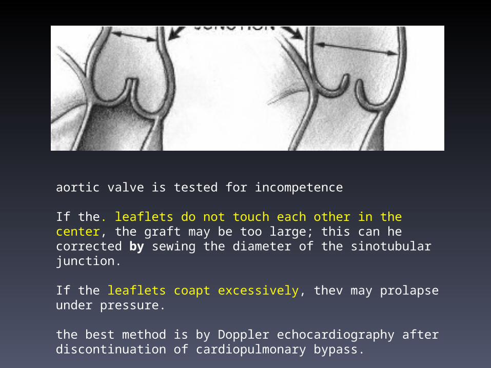
aortic valve is tested for incompetence
If the. leaflets do not touch each other in the center, the graft may be too large; this can he corrected by sewing the diameter of the sinotubular junction.
If the leaflets coapt excessively, thev may prolapse under pressure.
the best method is by Doppler echocardiography after discontinuation of cardiopulmonary bypass.

The right and left coronary arteries are then reimplanted into their respective sinuses by making an opening in the graft and suturing the remnants of the sinus wall around each cornnary artery.


Valve-sparing aortic root replacement: the inclusion (David) techniqueOperative Techniques in Thoracic and Cardiovascular Surgery 2005;10(4):246–258 David TE, Feindel CM: An aortic valve-sparing operati(in for patients with aortic incompetence and aneurysm of the asrending aorta. J Thorac Cardiovasc Surg 103:617-622, 1992

For the correct sizing of the appropriate Dacron graft, commissures must be pulled up to create a virtual cylinder with cusp coaptation of 30–50%.

Horizontal mattress sutures without pledges are placed in one lane underneath the sinuses for later fixation of the graft to the aortic root.
When knots of the horizontal mattress are
gently tied, the graft must be pushed down and held
in position by the assistant.A pivotal step of the
reimplantation procedure is the posi- tioning of the commissures high enough into the Dacron tube by pulling on the stay sutures, reshaping a correct geometry of the valve.

Valve implantation: slightly pulling on both the commissure and the vascular graft before stitching the sutures through the graft. the graft should extend roughly by half of its maximum length at this segment. Insertion of saline allows a first judgment of leakproofness of the valve.
After completion of the reimplantation, the valve must appear in means of the geometry like a ‘Mercedes star’, but indi- vidual differences in length of cusps are possible.
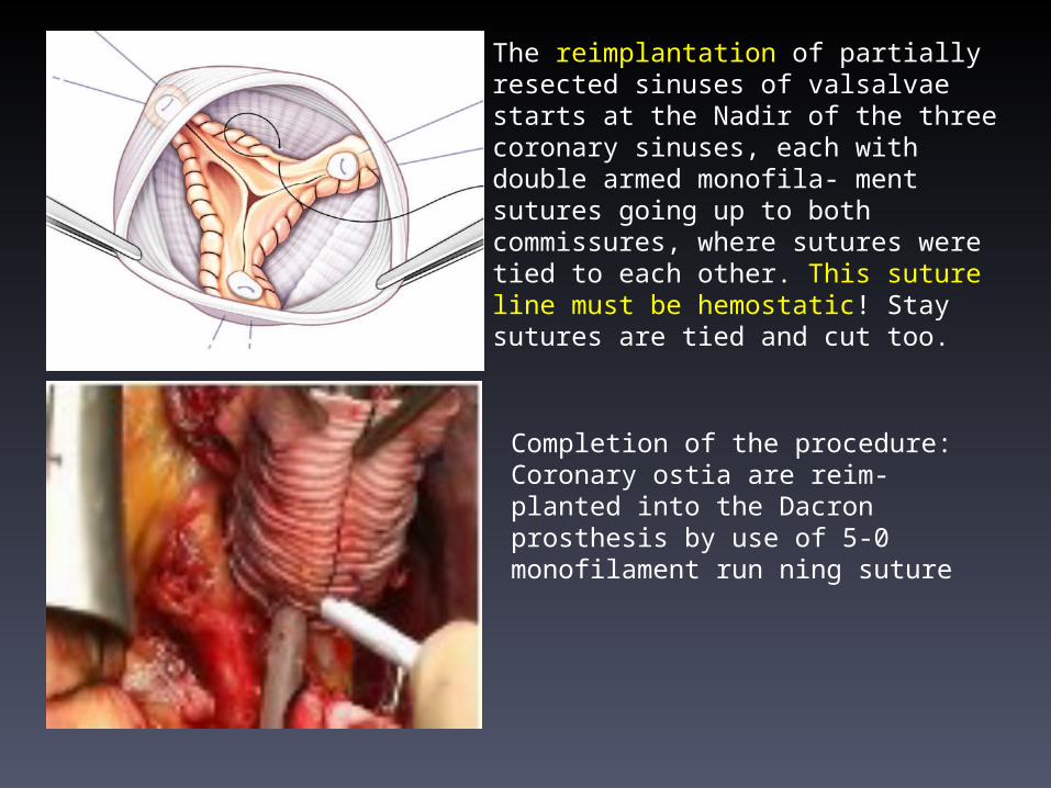
Completion of the procedure: Coronary ostia are reim- planted into the Dacron prosthesis by use of 5-0 monofilament run ning suture
The reimplantation of partially resected sinuses of valsalvae starts at the Nadir of the three coronary sinuses, each with double armed monofila- ment sutures going up to both commissures, where sutures were tied to each other. This suture line must be hemostatic! Stay sutures are tied and cut too.

Relationship between height of resuspension of the reimplanted valve and occurrence of postoperative aortic insufficiency.
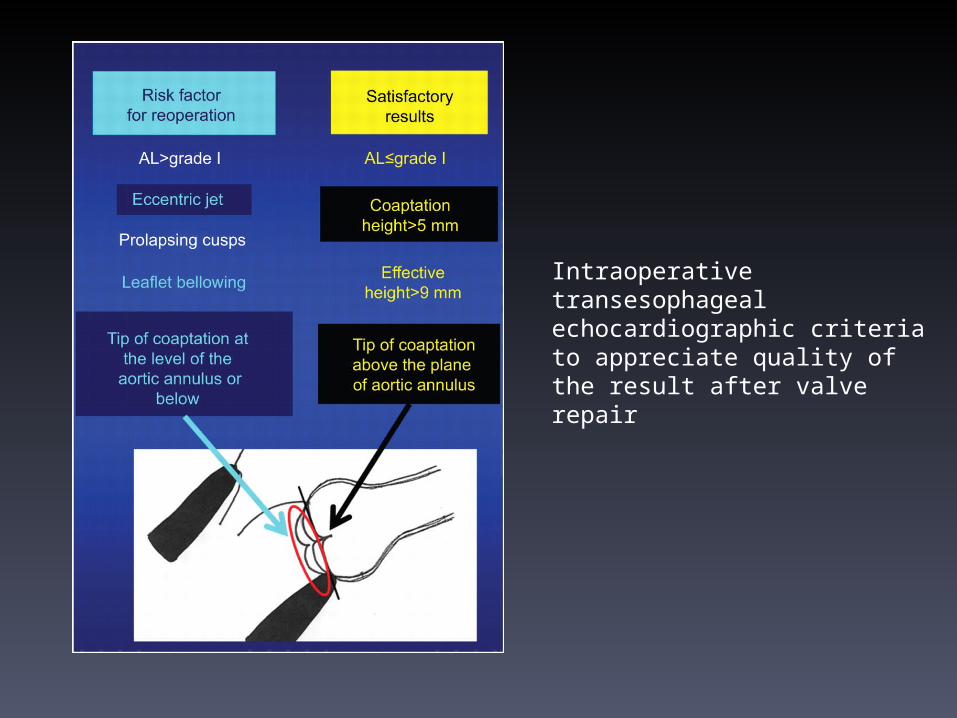
Intraoperative transesophageal echocardiographic criteria to appreciate quality of the result after valve repair

- Optimal STJ diameter (+3-4mm)- Free leaflet length (+3-4mm)=graft diameter- Height of L-NC commissure - BSA
-aortic annulus should not exceed - the length of the free margin of the leaflets - twice the height of the leaflets
• If in doubt, go with larger graft• If in severe doubt, use a 30mm graft•Never apologize for a Bentall 
Selection of Graft Size
Valve Sparing Aortic Root Replacement: Technical Tips and PitfallsDuke E. Cameron, MDDivision of Cardiac Surgery The Johns Hopkins Hospital Baltimore, MarylandAdult Cardiac Skills Course AATS 2012

Valve Sparing Aortic Root Replacement: Technical Tips and PitfallsDuke E. Cameron, MDDivision of Cardiac Surgery The Johns Hopkins Hospital Baltimore, MarylandAdult Cardiac Skills Course AATS 2012
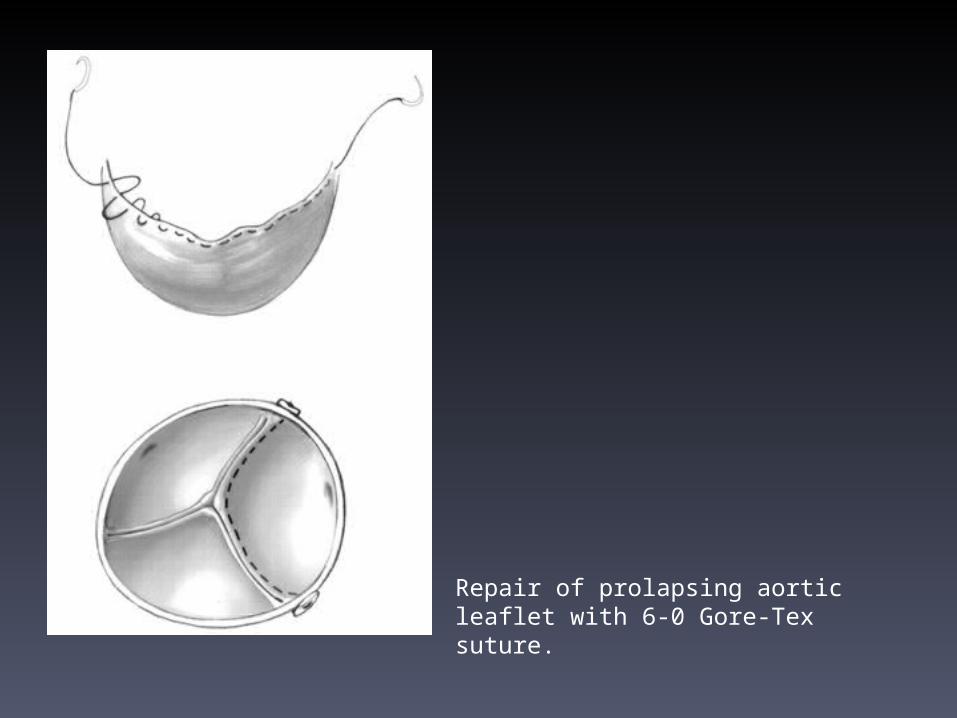
Repair of prolapsing aortic leaflet with 6-0 Gore-Tex suture.

Diagram of aortic valve lesions and corresponding re- pair teechniques. Aortic. regurgitation with decreased cusp moobilitydity.
Iliagram of aortic. valve lesions and correcspondingrt.pair techniques. Aortic reguritation with increased cusp mobility
Diagram of aortic. valve lesions and rorrrsponding repair technic1ue.s. Aortic re- gurgitation with normal cusp mobility.

Most important features of valve-preserving root replacement

In 2003, Miller introduced a classification to the Tirone David's
• David-I is the original reimplantation procedure using a cylindrical tube graft, •David-II is the original Yacoub remodeling procedure, •David-III is the remodeling procedure with an external narrowing annuloplasty strip, •David-IV is reimplantation using a 4-mm larger graft size with plication of the graft circumferentially at the sinotubular junction (STJ) above the tops of the commissures, and •David-V is reimplantation using an even larger graft size, which is ‘necked down’ at both the bottom and the top ends to create graft pseudosinuses
Miller DC
. Valve-sparing aortic root replacement in patients with the Marfan syndrome. J Thorac Cardiovasc Surg 2003;125:773-778

• whether the reimplantation (David) technique or the remodeling (Yacoub) technique provides the optimum event free survival
• 14 papers provided the best evidence • total of 1338 patients (Yacoub technique in
606 and David technique in 732) • 13 centres were included
Interactive CardioVascular and Thoracic Surgery 13 (2011) 189-197 J Thorac Cardiovasc Surg 2008;116:990-996

• Early mortality ranged from – 0% to 6.9% for the Yacoub technique and – 0–6% for the David technique.
• In the largest available series reported by David et al. in 2010 – 1.6% in the Yacoub group and – 1.7% in the David group.
• acute type A dissection ,the overall early mortality was 17%
AATS meting 2012. ctsnet.org2012Interactive CardioVascular and
Thoracic Surgery 13 (2011) jtcs.ctsnetjournals.org tcvs.2008
J Thorac Cardiovasc Surg 2008;116:990-996
Circulation. 2002;106[suppl I]:I-229-I-233

Clinical bottom line• The results for both techniques were almost
comparable. • Bicuspid
• favour of the David technique rather than the Yacoub technique in pathologies such as • Marfan syndrome, • acute type A aortic dissection • excessive annular dilatation
AATS meting 2012. ctsnet.org2012Interactive CardioVascular and
Thoracic Surgery 13 (2011) jtcs.ctsnetjournals.org tcvs.2008
J Thorac Cardiovasc Surg 2008;116:990-996
Circulation. 2002;106[suppl I]:I-229-I-233
• less freedom from AR in the Yacoub than the David
• not been associated with thromboembolic complications, the risk of valve endocarditis
• Predictor of (early) failure– Patient selection– Cusp repair– Coronary reimplantation
AATS meting 2012. ctsnet.org2012Interactive CardioVascular and Thoracic Surgery 13 (2011)
jtcs.ctsnetjournals.org tcvs.2008J Thorac Cardiovasc Surg 2008;116:990-996
Circulation. 2002;106[suppl I]:I-229-I-233.

Left to right: remodeling, modified remodeling, sinus prosthesis, modified sinus prosthesis, reimplantation, and modified reimplantation.
In vitro hydrodynamics, cusp-bending deformation, and root distensibility for different types of aortic valve–sparing operations: Remodeling, sinus prosthesis, and reimplantationArmin Erasmi, MD, Hans-H. Sievers, MD * , Michael Scharfschwerdt, Thorsten Eckel, Martin Misfeld, MD, PhD
Department of Cardiac Surgery, University Clinic of Schleswig-Holstein, Campus Luebeck, Luebeck, Germany.


Thank you

• Reimplantation of the aortic valve in a rigid tube leads to a nonphysiologic movement of the valve leaflets similar to that observed for stented bioprotheses, exposing the leaflets to
increased bending stresses and thus to the risk of premature failure. They maintain that this operation is adequate to avoid secondary dilatation of the aortic root and still to preserve or restore aortic valve function.
• Remodeling of the aortic root preserves some distensibility, with the propensity to reduce aortic outflow resistance and thus to lessen the load on the ventricle. It further allows for creation of a pseudosinus, allowing nearly normal opening and closing characteristics of the aortic valve and enhancing its durability [11, 12]. David and Feindel 8 have pointed out that, in extensive root dilatation, not only the sinuses of Valsalva are dilated but also the fibrous portions of the root inferior to the valve insertion line (ie, fibrous trigone and membranous septum). To correct the root also at this level, they have proposed mobilization of the root, anchoring a Dacron graft to the aortoventricular junction, and reimplantation of the aortic valve within the vascular graft.
• Nevertheless, debate still exists about which technique to apply to different pathologic conditions and, more generally, what there is to gain from using valve-sparing techniques compared with the standard composite prostheses. We therefore reviewed our 10-year experience with both types of valve-preserving techniques.

• Operative TechniqueAfter median sternotomy, standard cardiopulmonary bypass was initiated with a membrane oxygenator (Hollow Fiber Oxygenator, Spiral Gold, Baxter, Puerto Rico) using antegrade crystalloid or blood cardioplegia. Profound hypothermia (15° to 18°C) was used when circulatory arrest was necessary. The side of arterial cannulation was the femoral artery in 32 patients, the right subclavian artery in 5, a combination in 6, and the ascending aorta in a nondissected area in 121. Venous cannulation was performed through the right femoral vein in 9 patients and through the right atrium in the rest.
• The operative technique of the remodeling technique (group A) and the reimplantation technique (group B) has been described in detail [4, 5]. Briefly, the ascending aorta was transected 3 mm above the commissures. The sinuses of Valsalva were excised, leaving a 2-mm rim attached to the crown-shaped annulus. If the dissection affected the aortic root, gelatin-resorcin-formaldehyde glue (GRF; Cardial, Saint E'tienne, France) was used to readapt the dissected layers of the aortic wall before the sinuses were excised. The size of the tube used was determined by the distance between the straightened commissures giving a macroscopic picture of appropriate cusps coaptation and the diameter of the base of the aortic annulus, measured by means of a Hegar dilator [16].
• According to the adopted procedure, a trimmed or straight Hemashild Gold tube (Meadox Medicals, Oakland, NY), made with Dacron (DuPont, Wilmington, DE), was used to replace the excised sinuses. In some patients, one or more of the sinuses were macroscopically intact without any changes of the underlying pathology; only one sinus was replaced in 13 of these patients, and in 5 patients only two of the three sinuses were replaced using the surgical principles of the remodeling technique. In some patients of the remodeling group, we used our own technique of individual replacement of each sinus with a single piece of Dacron tube and additional replacement of the ascending aorta.
• In most of the patients, the distal anastomosis was performed in an open fashion using circulatory arrest

reimplantation technique caused significantly higher pressure gradients
Bending deformation indices for all valve-sparing techniques were more than twice those of the native aortic root and increased in relation to the degree the root was fixed with synthetic noncompliant material.
Aortic root distensibility, expressed as diastolic-to-systolic change of area, decreased in all surgical procedures compared with that of the native aortic root

Schematic drawing of measured aortic valve opening and closing characteristics of 3 distinct phases: a-b, rapid valve opening; b-c, slow systolic closure; and c-d, rapid valve closing movement. RVOT indicates rapid valve opening time; D1, maximal leaflet displacement; RVCT, rapid valve closing time; ET, ejection time; SCD, slow closing displacement; and D2, leaflet displacement before rapid valve closing.

Diagram of cyclic changes in dimensions derived from mean values of measured data at base, sinus, and commissural levels. Note reduced distensibility in group A at all levels of aortic root
Opening and Closing Characteristics of the Aortic Valve After Different Types of Valve-Preserving SurgeryRainer G. Leyh, MD; Claudia Schmidtke, MD; Hans-Hinrich Sievers, PhD, FETCS; Magdi H. Yacoub, PhD, FRCS
From the Departments of Cardiac Surgery, Medical University of Lübeck, Lübeck, Germany (R.G.L., C.S., H.-H.S.), and the National Heart and Lung Institute at the Imperial College of Science, Technology, and Medicine, London, UK (M.H.Y.).

EVOA is constant in both compliant and stiff roots
Aortic valve regional stresses in the normal aortic root and clinical valve-sparing models. *P = .0001 and {dagger}P = .0005 indicate significant difference as compared with the normal root model. b, Schematics of altered leaflet stress patterns in the graft models as compared with normal.
RE-CREATION OF SINUSES IS IMPORTANT FOR SPARING THE AORTIC VALVE: A FINITE ELEMENT STUDYK. Jane Grande-Allen, PhDa, Richard P. Cochran, MDb, Per G. Reinhall, PhDc, Karyn S. Kunzelman, PhDb
From the Department of Biomedical Engineering, Cleveland Clinic Foundation, Cleveland, Ohioa; Division of Cardiothoracic Surgery, University of Wisconsin, Madison, Wisb; and Department of Mechanical Engineering, University of Washington, Seattle, Wash.c

Both techniques are widely used in the case of aortic root aneurysms associated with non-diseased aortic valves. The classic remodeling technique allows a good anatomical reconstruction of the sinuses of Valsalva but has a higher incidence of residual valve regurgitation. the classic reimplantation technique permits more stable results through annulus stabilization but completely abolishes the sinuses of Valsalva
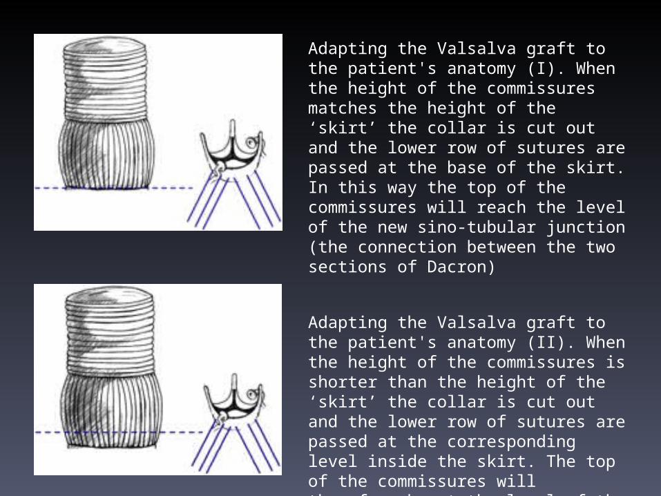
Adapting the Valsalva graft to the patient's anatomy (I). When the height of the commissures matches the height of the ‘skirt’ the collar is cut out and the lower row of sutures are passed at the base of the skirt. In this way the top of the commissures will reach the level of the new sino-tubular junction (the connection between the two sections of Dacron)
Adapting the Valsalva graft to the patient's anatomy (II). When the height of the commissures is shorter than the height of the ‘skirt’ the collar is cut out and the lower row of sutures are passed at the corresponding level inside the skirt. The top of the commissures will therefore be at the level of the new sino-tubular junction.

Adapting the Valsalva graft to the patient's anatomy (III). In the rare cases when the height of the commissures is longer than the height of the ‘skirt’ the collar can be utilized to secure the lower row of sutures, to increase the length of the reconstructed root and to consent that the top of the commissures reaches the level of the new sino-tubular junction.

Because of its particular shape, the use of the Valsalva graft simplifies the surgical procedure by offering:•Decreased tension during coronary artery suturing•Decreased tension after graft pressurization•Increased anatomical adaptability•Decreased potential for suture bleeding and pseudoaneurysm formation•Easier access to the coronary anastomosis at the end of procedure


Remodeling of the aortic root combined to an expansible aortic ring annuloplasty

Standardized and physiological approach to aortic valve repair according to each phenotype of ascending aorta. (Reproduced from Ref. [17] with permission from Elsevier.)
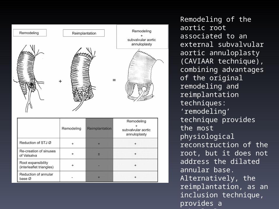
Remodeling of the aortic root associated to an external subvalvular aortic annuloplasty (CAVIAAR technique), combining advantages of the original remodeling and reimplantation techniques: ‘remodeling’ technique provides the most physiological reconstruction of the root, but it does not address the dilated annular base. Alternatively, the reimplantation, as an inclusion technique, provides a subvalvular annuloplasty to the detriment of valve dynamics [6, 7, 9,10,11,12,13,14,15,16]. (Reproduced from Ref. [17] with permission from Elsevier.)

Criteria for the choice of the subvalvular aortic ring and Valsalva graft.

First step of valve repair: alignment of adjacent cusp free edges
Placement of the subvalvular expansible aortic ring (A) and final aspect of the aortic root (B). (Reproduced from Ref. [17] with permission from Elsevier.)
Placement of the five anchoring subvalvular ‘U’
stitches

Aortic root aneurysm for bicuspid valves: remodeling of the aortic root, resuspension of cusp effective height and subvalvular aortic annuloplasty.

• One hundred and eighty-seven patients underwent remodeling associated with a subvalvular aortic ring annuloplasty (14 centers, 24 surgeons). Three strategies for cusp repair were evaluated: Group 1: gross visual estimation (74 patients), Group 2: alignment of cusp free edges (62 patients), Group 3: two-steps approach associating alignment of cusp free edges with effective height resuspension (51 patients). A composite outcome was defined as recurrence of aortic insufficiency ≧grade 2 and/or reoperation. Operative mortality was 3.2% (20). Treatment of cusp lesion was most frequently performed in Group 3 (70.6%, vs. 20.3% Group 1 and 30.6% Group 2, P≪0.001). Nine patients required reoperation during follow-up [24 months (12–45)], from Group 1 (6) and Group 2 (3). At one year, no patients in Group 3 presented with composite outcome events (vs. 28.1% Group 1, 15% Group 2, P≪0.001). Residual aortic insufficiency and tricuspid anatomy were independent risk factors for composite outcome in Groups 1 and 2. Annulus diameter, Marfan syndrome and cusp repair had no effect on aortic insufficiency recurrence or reoperation [

Conclusions: In vitro the various aortic valve–sparing operations differed charac- teristically in their ability to spare valve function, none of them completely meeting native valve behavior. The remodeling techniques exhibited valve dynamics closest to those of the native aortic root. The more the aortic valve is fixed with noncom- pliant prosthetic material, the more the native root dynamics are impaired.