antitelomerase therapy provokes alt and mitochondrial adaptive
TRANSCRIPT

Antitelomerase Therapy ProvokesALT and Mitochondrial AdaptiveMechanisms in CancerJian Hu,1,4,5,6 Soyoon Sarah Hwang,1,4,5 Marc Liesa,8 Boyi Gan,3,4,5,6 Ergun Sahin,4,5,6 Mariela Jaskelioff,4,5,6
Zhihu Ding,4,5,6 Haoqiang Ying,1,4,5,6 Adam T. Boutin,1,4,5,6 Hailei Zhang,4,5 Shawn Johnson,5 Elena Ivanova,4,5
Maria Kost-Alimova,1,4,5 Alexei Protopopov,1,4,5 Yaoqi Alan Wang,1,4,5 Orian S. Shirihai,8 Lynda Chin,2,4,5,7
and Ronald A. DePinho1,4,5,6,*1Department of Cancer Biology2Department of Genomic Medicine3Department of Experimental Radiation Oncology
University of Texas MD Anderson Cancer Center, Houston, TX 77030, USA4Belfer Institute for Applied Cancer Science5Department of Medical Oncology
Dana-Farber Cancer Institute, Boston, MA 02115, USA6Department of Genetics and Medicine7Department of Dermatology
Harvard Medical School, Boston, MA 02115, USA8Department of Medicine, Boston University School of Medicine, Boston, MA 02118, USA
*Correspondence: [email protected] 10.1016/j.cell.2011.12.028
SUMMARY
To assess telomerase as a cancer therapeutic targetand determine adaptive mechanisms to telomeraseinhibition, we modeled telomerase reactivation andsubsequent extinction in T cell lymphomas arisingin Atm�/� mice engineered with an inducible telo-merase reverse transcriptase allele. Telomerase re-activation in the setting of telomere dysfunctionenabled full malignant progression with alleviationof telomere dysfunction-induced checkpoints. Thesecancers possessed copy number alterations target-ing key loci in human T cell lymphomagenesis.Upon telomerase extinction, tumor growth eventuallyslowed with reinstatement of telomere dysfunction-induced checkpoints, yet growth subsequentlyresumed as tumors acquired alternative lengtheningof telomeres (ALT) and aberrant transcriptionalnetworks centering on mitochondrial biology andoxidative defense. ALT+ tumors acquired amplifica-tion/overexpression of PGC-1b, a master regulatorof mitochondrial biogenesis and function, and theyshowed marked sensitivity to PGC-1b or SOD2knockdown. Genetic modeling of telomerase extinc-tion reveals vulnerabilities that motivate coincidentalinhibition of mitochondrial maintenance and oxida-tive defense mechanisms to enhance antitelomerasecancer therapy.
INTRODUCTION
Telomeres are nucleoprotein complexes at chromosome ends
that function to maintain chromosomal integrity. Genetic models
have defined their critical roles in cancer (Artandi and DePinho,
2010), aging, and degenerative diseases (Sahin and DePinho,
2010). Telomeres are synthesized by telomerase consisting of
a reverse transcriptase catalytic subunit (TERT) and an RNA
template subunit (TERC) (Feng et al., 1995; Nakamura et al.,
1997). As normal or premalignant cells divide, the end-replica-
tion problem of conventional DNA polymerases, coupled with
low or absent telomerase activity, results in loss of telomere
sequences and eventual telomere uncapping, which activates
cellular checkpoints similar to those provoked by DNA double-
stranded-breaks (DSBs) (Harley and Sherwood, 1997). Like clas-
sical DSBs, telomere dysfunction has been shown to induce p53
and associated cellular responses, such as senescence and/or
apoptosis (Chin et al., 1999; d’Adda di Fagagna et al., 2003; Karl-
seder et al., 1999; Takai et al., 2003; van Steensel et al., 1998).
Upon mutational inactivation of p53, continued cell cycling and
survival of cells with telomere dysfunction provide a procarcino-
genic mutator mechanism characterized by translocations and
regional amplifications and deletions (Artandi et al., 2000; Chin
et al., 1999; O’Hagan et al., 2002). At the same time, persistent
telomere dysfunction and associated rampant chromosomal
instability (even in p53 null cells) compromises cellular viability
(Begus-Nahrmann et al., 2009; Chin et al., 1999) and constrains
full malignant progression of such cancers (Artandi et al., 2000;
Chang et al., 2003; Gonzalez-Suarez et al., 2000; Rudolph
et al., 2001).
Cell 148, 651–663, February 17, 2012 ª2012 Elsevier Inc. 651

The occurrence of telomere erosion and importance of telo-
merase-mediated telomere maintenance in fully established
cancers are evidenced by typically short telomeres relative to
normal tissues and robust telomerase activity in most human
cancers (Shay and Wright, 2006). This profile of shorter telo-
meres and telomerase activity in cancer has motivated the clin-
ical development of telomerase inhibitors including a 13-mer
antisense oligonucleotide in a number of cancer types (Agrawal
et al., 2012; Shay and Wright, 2006). At present, uncertainty
surrounds whether antitelomerase therapy will be hampered by
the potential lag time needed for telomere erosion-associated
tumor cell killing, and/or whether re-entry into telomere-based
crisis will engender genomic instability that may allow for emer-
gence of adaptive responses and resistance mechanisms, such
as alternative lengthening of telomeres (ALT) mechanism which
enables telomere maintenance via homologous recombination
(Cesare and Reddel, 2010). Notably, in mTERC�/� mice, p53
deficiency alleviates tumor suppression imparted by telomere
dysfunction (Chin et al., 1999) and, in transformed cells with
intact p53-dependent DNA damage checkpoint, there is activa-
tion of ALT (Chang et al., 2003).
The study of cancer pathogenesis and specific role of telo-
meres therein have been enabled by the use of genetically engi-
neered mouse models of cancer, which provide an in vivo
assessment of the complex adaptive responses to targeted
cancer treatments. Here, we exploited the experimental merits
of mice to model and study more precisely telomere crisis, telo-
merase reactivation and telomerase extinction in cancer devel-
opment, progression and treatment in the in vivo setting. To
that end, we engineered an inducible TERT knockin allele and
studied telomere dynamics in mice mutant for Atm, which
develop T cell lymphomas with high penetrance (Xu et al.,
1996). The Atmmutant model was selected to study telomerase
activation and its extinction as this model retains a robust
p53-mediated checkpoint response upon telomere uncapping
which strongly suppresses emergent tumors (Qi et al., 2003;
Wong et al., 2003). Our study provides in vivo genetic evidence
that telomerase reactivation facilitates the progression of spon-
taneous arising tumors experiencing telomere dysfunction and,
conversely, that telomerase extinction in established cancers
activates ALT and other adaptive mechanisms, illuminating
potential therapeutic combinations.
RESULTS
Telomerase Reactivation Promotes Tumorigenesisby Stabilizing Telomeres and Alleviating TelomereDysfunction-Induced CheckpointsWe generated mice harboring an Atm null allele (Xu et al., 1996)
and a 4-Hydroxytamoxifen (4-OHT)-inducible Telomerase
Reverse Transcriptase-Estrogen Receptor (TERT-ERT2) fusion
knockin allele (Jaskelioff et al., 2011). In the absence of 4-OHT,
mice homozygous for TERTER (designated TERTER/ER) are telo-
merase activity deficient and sustain same cytogenetic and
cellular phenotypes of conventional TERT or TERC knockout
model (Jaskelioff et al., 2011). Upon 4-OHT treatment, TERT-
ER protein activity can be restored to levels comparable to the
native TERT protein (Jaskelioff et al., 2011). To generate
652 Cell 148, 651–663, February 17, 2012 ª2012 Elsevier Inc.
lymphoma-prone mice with telomere dysfunction, Atm+/�
TERT+/ER mice were intercrossed to produce first generation
(G1) Atm�/�TERTER/ER and Atm+/�TERTER/ER mice. G1
Atm+/�TERTER/ER mice were then serially intercrossed to
successive generations in the absence of 4-OHT, culminating
in G3 andG4Atm�/�TERTER/ER experimental cohorts (Figure S1A
available online). For brevity, the Atm�/� TERT+/+ or +/ER, G1
Atm�/� TERTER/ER, and G3-4 Atm�/�TERTER/ER mice are desig-
nated G0, G1, and G3-4, respectively.
G0 and G1 mice developed T cell lymphomas at similar laten-
cies and penetrance (Figure S1B), which is consistent with
adequate telomere reserves to maintain capping in G1 mice
and avoid activated DNA damage checkpoints. In contrast,
G3-4 lymphomas emerge with a longer latency and lower pene-
trance relative to G1 controls (Figure 1A), consistent with
previous work (Maser et al., 2007; Qi et al., 2003; Wong et al.,
2003). Next, to test the impact of telomerase reactivation on
lymphoma kinetics in this model, continuous-release 4-OHT
tablets were implanted subcutaneously at 18 weeks, an age
when lymphomas in Atm�/� mice with intact telomeres start to
emerge. 4-OHT-treated G3-4 mice developed overt lymphomas
more rapidly and with higher penetrance than, not only vehicle-
treated G3-4 mice, but also G1 mice (Figure 1A). In contrast, there
were no differences in tumor latency and penetrance in the
4-OHT- and vehicle-treated G1 mice (Figure 1A). On the histo-
pathological level, vehicle-treated G3-4 tumors were smaller in
size and displayed less aggressive malignant features relative
to 4-OHT-treated G3-4 tumors as well as telomere intact G0
and G1 tumors (Figures 1B–1D; data not shown). Finally,
4-OHT treatment itself does not impact on lymphomagenesis
as reflected by similar survival curves of G3-4 Atm�/�
TERTER/ER mTR�/� mice treated with either vehicle or 4-OHT
(Figure 1A), supporting the view that acceleration of 4-OHT on
G3-4 Atm�/�TERTER/ER tumors stems from telomerase reactiva-
tion and not from nontelomere activities of 4-OHT.
The capacity to regulate endogenous telomerase activity
in vivo prompted serial analyses of malignant properties and
cellular checkpoint responses as functional readouts of telomere
status during lymphomagenesis and progression in G3-4 mice.
Quantitation of DNA damage foci by anti-gH2AX and -53BP1
staining showed markedly increased foci in vehicle-treated
G3-4 tumor cells and a dramatic decrease in such foci in
4-OHT-treated G3-4 tumor cells (Figures 2A and 2B; Figure S2).
Correspondingly, vehicle-treated G3-4 tumor cells exhibited
decreased proliferation (Ki67) and increased apoptosis (cleaved
caspase 3), senescence (SA-b-Gal), and p53 signaling (p53
phosphorylation and p21 expression), which are all alleviated
by telomerase reactivation (Figures 2C–2F).
Telomerase Reactivation in Spontaneous Tumorswith Unstable Genomes Promotes AggressiveMalignant PropertiesA higher percentage of G3-4-4-OHT mice exhibited widespread
tumor cell infiltration in spleen, kidney, liver, lung, and bone
marrow relative to vehicle-treated G3-4 mice as well as 4-OHT-
and vehicle-treated G1 mice (Figures 3A and 3B). The more
aggressive nature of 4-OHT-treated G3-4 lymphomaswas partic-
ularly evident from infiltration into brain (4/16) which was not

Figure 1. Telomerase Reactivation in Late-
Generation TERTER/ER Atm�/� Mice
Promotes T Cell Lymphomagenesis
(A) Kaplan-Meier curves (Log-rank test) of T cell
thymic lymphoma-free survival for G1 and G3-4
TERTER/ER Atm�/� (mTR�/�) mice treated with
4-OHT or vehicle.
(B) Representative T cell thymic lymphoma of G3-4
TERTER/ER Atm�/� mice treated with 4-OHT or
vehicle. Scale bars represent 1 cm.
(C) Tumor sizes of G1 and G3-4 TERTER/ER Atm�/�
lymphomas treated with 4-OHT or vehicle (t test,
and error bars indicate SD).
(D) Representative images of H&E-stained G3-4
TERTER/ER Atm�/� lymphomas treated with 4-OHT
or vehicle. Scale bars represent 100 mm.
See also Figure S1.
observed in G1 necropsies (n = 16) (Figures 3A and 3B), demon-
strating that the malignant progression of initiated tumors is
enabled by somatic reactivation of telomerase. This acquisition
of more aggressive tendencies may stem in part from increased
genome instability during the formative stages of tumor develop-
ment prior to telomerase activation. Indeed, telomere dysfunc-
tion-induced bridge-fusion-breakage events can generate
non-reciprocal translocations as well as regional amplifications
and deletions at sites of breakage that, under biological selec-
tion, can result in cancer-promoting copy number changes (Ar-
tandi et al., 2000; Maser et al., 2007; O’Hagan et al., 2002). To
examine this possibility, we compared cytogenetic and genomic
profiles of 4-OHT-treated G3-4 tumors and 4-OHT-treated G1
tumors, the latter without a period of telomere-driven crisis.
Consistent with previous work (Maser et al., 2007), spectral
karyotype (SKY) analysis showed a 3-fold increase in chromo-
somal rearrangements including nonreciprocal translocations
in 4-OHT-treated G3-4 tumors (n = 3) relative to 4-OHT-treated
G1 tumors (n = 3) (Figures 3C and 3D, 25.8 versus 8.8 total
NRTs per metaphase, respectively; p < 0.0001). The baseline
of 8.8 NRTs per metaphase is consistent with previous work
that ATM deficient tumors experience increased DNA double-
stranded breaks, which promote chromosomal rearrangements
as well (Xu et al., 1996).
Examination of copy number alterations (CNAs) in 18 G3-4-4-
OHT, 10 G1-4-OHT, and 5 G3-4-VEH tumors (Figure 3E; Figures
S3A and S3B) revealed novel copy number gains on chromo-
somes 12, 17, and 18 and copy number losses on chromosome
14 in G3-4-4-OHT tumors but not G1-4-OHT tumors, consistent
with increased genomic instability in late-generation tumors (Fig-
ure 3E; Figure S3A). Notably, G3-4-4-OHT and G3-4-VEH tumors
show comparable levels of clonal CNAs, consistent with the view
that wholesale genomic events driving lymphomagenesis occurs
early in cancer development and that telomerase reactivation
Cell 148, 651–663,
does not discernibly alter the scope of
clonal genomic events in these cancers
(Figure 3E; Figure S3B). Along these lines,
we assessedwhether the observed CNAs
in G3-4-4-OHT mice might contribute
to the lymphomagenesis by examining
whether murine and human lymphomas sustain orthologous
CNA events than might be expected by chance as conducted
previously (Maser et al., 2007). Array-CGH profiles of G3-4-4-
OHT tumors identified 4,928 genes resident within regions of
genomic gain and 2,297 genes resident within regions of
genomic loss with the use of Segment Gain or Loss (SGOL) algo-
rithm (Wiedemeyer et al., 2010; Extended Experimental Proce-
dures). Comparison of G3-4-4-OHT tumors to human T-ALLs
(Maser et al., 2007) showed that 565 of 4,928 amplified genes
(11.5%) and 300 of 2,297 deleted genes (13%) are targeted
for copy number alteration in both species (Table S1), which
are significantly higher than those expected by chance (n =
10,000; p = 3e-04 and 2e-04 for amplification and deletion,
respectively) (Figure S3C). These cross-species shared copy
number altered genes include several known tumor suppressors
and oncogenes implicated in T-ALL biology such as Crebbp,
Ikaros, Abl, Notch1, Myc, and PTEN (Figure 3E). These data
and previous work (Maser et al., 2007; O’Hagan et al., 2002)
suggest that telomere dysfunction provides a mechanism that
promotes structural genome alterations of cancer-relevant loci,
which not only drive primary tumorigenesis but also confer addi-
tional malignant properties such as enhanced invasiveness.
Genetic Extinction of Telomerase Inhibits CancerGrowth and Leads to Eventual Tumor Re-EmergenceThe TERT-ER system affords exploration of the impact of genetic
extinction of telomerase activity in established cancers that,
similar to human cancers, had evolved to first experience telo-
mere dysfunction, and then subsequently acquire telomerase
activity. In particular, this genetic model system enables assess-
ment of tumor biological impact and potential adaptive mecha-
nisms of antitelomerase therapy following reacquisition of
telomere dysfunction. Freshly harvested 4-OHT-treated G3-4
tumor cells mice were passaged directly through SCID mice
February 17, 2012 ª2012 Elsevier Inc. 653

Figure 2. Telomerase Reactivation Alleviates Telomere Dysfunction and Checkpoints in Late-Generation TERTER/ER Atm�/� Lymphomas
(A) Representative immunohistochemistry staining of g-H2AX and 53BP1 in G3-4 TERTER/ER Atm�/� thymus and lymphomas treated with 4-OHT or vehicle.
(B) Quantification of (A) (n = 5, t test).
(C) Representative immunohistochemistry staining of cleaved Caspase 3 and Ki67 in G3-4 TERTER/ER Atm�/� thymus and lymphomas treated with 4-OHT or
vehicle.
(D) Quantification of (C) (n = 5, t test).
(E) Representative beta-gal staining of G3-4 TERTER/ER Atm�/� thymus and lymphomas treated with 4-OHT or vehicle (n = 3).
(F) Immunoblotting of phosphorylated p53 (S15) and p21 in G3-4 TERTER/ER Atm�/� thymus and lymphomas treated with 4-OHT or vehicle.
Error bars indicate SD and scale bars represent 100 mm.
See also Figure S2.
preimplanted with vehicle or 4-OHT tablets (Figure S4A). Of 15
primary tumors lines (ten mice per each passage of treatment;
107 cells per intraperitoneal injection), 11 lines generated xeno-
graft tumors. In first and second passages (P1 and P2), these
lines showed no differences in the two treatment arms in terms
of penetrance and latency (Figure 4A; P1 and P2). However, at
third passage (P3), 9 of 11 lines showed lower penetrance and
longer latency in the vehicle-treated arm relative to the 4-OHT-
treated arm, a profile consistent with the view that multiple
rounds of cell division are needed to achieve sufficient telomere
erosion and re-entry into crisis. Six of these nine lines showed
complete loss of tumor formation in the vehicle-treated arm,
whereas three lines yielded escaping tumors in several vehicle-
treated recipient mice (Figure 4A; P3). When these escaping P3
654 Cell 148, 651–663, February 17, 2012 ª2012 Elsevier Inc.
tumors were passaged to P4, they reacquired aggressive malig-
nant properties approaching those of matched 4-OHT controls
(Figure 4A; P4), suggesting acquisition of resistant mechanisms
to counteract telomerase deficiency. Moreover, 2 of the original
11 tumor lines did not show any attenuation of tumor growth in
the vehicle-treated arm compared with the 4-OHT-treated arm
(data not shown), suggesting early acquisition of such resistant
mechanisms (see below).
To assess how G3-4-4-OHT tumors respond to telomerase
extinction on molecular level, we audited p53 signaling, DNA
damage foci and apoptosis in serial passages of the two
treatment arms. In 4-OHT-treated P1 to P4 and vehicle-treated
P1 tumor cells, p53 signaling, DNA damage foci and
apoptosis levels were similar to those of parental tumor cells

Figure 3. Telomerase Reactivation Promotes Invasiveness of Late-Generation TERTER/ER Atm�/� Lymphomas
(A) Representative images of infiltrated organs (lung, kidney, liver, and brain) in G3-4 TERTER/ER Atm�/� mice treated with 4-OHT. Scale bars represent 100 mm.
(B) Quantification of infiltrating incidence of G1 and G3-4 TERTER/ER Atm�/� mice treated with 4-OHT or vehicle.
(C) Representative spectral karyotype (SKY) images from metaphases of G1 and G3-4 TERTER/ER Atm�/� lymphomas treated with 4-OHT.
(D) Quantification of nonreciprocal translocations (NRTs) detected by SKY in G1 (n = 3) and G3-4 (n = 3) TERTER/ER Atm�/� lymphomas treated with 4-OHT
(p < 0.0001, t test).
(E) Recurrence plot of CNAs defined by array-CGH for 18 G3-4-4-OHT lymphomas. The x axis shows the physical location of each chromosome. The percentage
of tumors harboring gains (dark red, log2 R 0.3), amplifications (bright red, log2 R 0.6), losses (green, log2 % �0.3), and deletions (dark green, log2 % �0.6) for
each locus is depicted. Locations of physiologically relevant CNAs of TCR loci and some known cancer genes are indicated with asterisks.
See also Figure S3 and Table S1.
Cell 148, 651–663, February 17, 2012 ª2012 Elsevier Inc. 655

Figure 4. Telomerase Depletion in Late-Generation 4-OHT-TERTER/ER Atm�/� Lymphomas Leads to Cell Death and Resistance
(A) Kaplan-Meier curves (Log-rank test) of lymphoma-free survival of 4OHT- or vehicle- treatedmice serially transplantedwithG3–4 TERTER/ER Atm�/� lymphomas.
(B) Quantification of cleaved Caspase 3 positive cells in serially transplanted lymphomas treated with 4OHT or vehicle (n = 3–5, t test).
(C) Immunoblotting of phosphorylated p53 (S15) and p21 in serially transplanted lymphomas treated with 4OHT or vehicle.
(D) Quantification of 53BP1 foci positive cells in serially transplanted lymphomas treated with 4OHT or vehicle (n = 3–5, t test).
(E) Quantification of g-H2AX foci positive cells in serially transplanted lymphomas treated with 4OHT or vehicle (n = 3–5, t test).
Error bars indicate SD (*p < 0.01 relative to any other individual samples in the same group). See also Figure S4.
(P0) (Figures 4B–4E). In contrast, vehicle-treated P2 tumor cells
showed significantly higher levels of p53 signaling, DNA damage
foci and apoptosis, and all of these processes were reduced in
the resistant vehicle-treated P3-4 tumor cells (Figures 4B–4E),
raising the possibility that these resistant tumorsmay have stabi-
lized their telomeres to alleviate these checkpoints. Intriguingly,
while DNA damage foci were reduced in the resistant tumors
compared with the vehicle-treated P2 tumors, these foci remain
higher than telomerase+ (4-OHT-treated) tumors (Figures 4D
and 4E), consistent with residual genotoxic stress in the resistant
tumors. However, despite ongoing DNA damage signaling,
apoptosis was not observed, which is consistent with compro-
mised p53 signaling in the resistant tumors (Figure S4B).
Emergence of ALT in Tumors following Extinctionof TelomeraseTo elucidate adaptive mechanisms following telomerase extinc-
tion, we cataloged telomere lengths in serially passaged tumors
in the two treatment arms. In passaged 4-OHT-treated P1 to P4
tumor cells, telomere lengths remained unchanged from those
in parental tumor cells, while vehicle-treated P1 and P2 tumor
cells showed progressive telomere shortening (Figure 5A).
However, vehicle-treated P3-4 tumor cells showed a sharp
increase in average and maximal telomere lengths that ex-
ceeded those in matched 4-OHT controls (Figure 5A); however,
these vehicle-treated P3-4 tumor cells tumor cells showed more
telomere signal-free ends by telomere-FISH (Figures 5B and 5C),
a picture consistent with higher heterogeneity in the distribution
of telomere lengths in resistant tumors. In addition, costaining of
promyelocytic leukemia (PML) bodies and telomeres showed an
increase in ALT-associated PML bodies (APBs) (Figures 5D and
656 Cell 148, 651–663, February 17, 2012 ª2012 Elsevier Inc.
5E), and telomere-FISH showed more extrachromosomal telo-
mere fragments in the vehicle-treated P3-4 tumors cells relative
to the matched 4-OHT controls (Figures 5F and 5G). Finally,
vehicle-treated P3-4 tumor cells showed an increased telomere
Sister Chromatid Exchange (tSCE) rate than matched 4-OHT
controls (Figures 5H and 5I). These collective data provide clear
evidence of ALT in vehicle-treated P3-4 tumor cells that have
resumed robust tumor growth in vivo. In addition, the two tumor
lines, which did not show any attenuation of tumor growth in the
vehicle-treated arm, also showed ALT features including APBs
and extra-chromosomal telomere fragments (Figures S5A and
S5B), consistent with the early acquisition of ALT.
ALT Tumors Show Upregulation of a Master Regulatorof Mitochondrial Biogenesis/Functionand Oxidative DefenseSerial loss of telomere function and genome destabilization in
this genetically defined system provided an unprecedented
opportunity to discern on a genome scale the associated adap-
tive molecular events in these ALT+ tumor cells via integrated
transcriptomic and aCGH profile analyses. Cluster analysis of
transcriptomes of 4-OHT-treated P4 tumors (n = 3), and parental
4-OHT-treated tumor revealed no significant differences (Fig-
ure 6A), consistent with genomic stability brought about by
continuous telomerase activity. In contrast, vehicle-treated
ALT+ P4 tumor (n = 5) expression profiles showed significant
differences relative to the 4-OHT-treated telomerase-positive
P4 tumors, 891 upregulated and 1,345 downregulated genes in
ALT+ tumors with 1.5-fold change and p < 0.01 (t test; Figure 6A;
Table S2). Pathway (IPA) analysis of the ALT-specific transcrip-
tome showed strong representation of networks centering on

mitochondrial biology and oxidative stress regulation (Fig-
ure S6A). Consistent with the known role of DNA recombination
in ALT (Fan et al., 2009; Zhong et al., 2007), MRE11 and FANCA
were also upregulated in these ALT+ tumors (Figures S6B and
S6C). With regard to mitochondria and oxidative pathways,
Q-PCR verified aberrant expression of several key genes in these
networks including upregulation of a master regulator of mito-
chondrial biology and oxidative defense, PGC-1b, and its targets
such as NRF2, SOD2, and Catalase, among others (Figure 6B).
As noted, telomere dysfunction-induced DNA double-strand
breakage process and biological selection can lead to function-
ally relevant CNAs, prompting us to identify potential genetic
events that may inform how ALT+ cells cope with telomere-
based crisis. To that end, we performed array-based compara-
tive genomic hybridization (aCGH) analysis of ALT+ versus
telomerase+ P4 tumors from the two treatment arms. While seri-
ally transplanted telomerase+ tumors showed minimal CNA
differences relative to the parental tumor, ALT+ tumors acquired
numerous CNAs (see below). Strikingly, integrated transcrip-
tomic and copy number analysis revealed PGC-1b as the only
gene in the mitochondrial function and oxidative stress regula-
tion pathways showing both upregulated expression and copy
number gain in the ALT+ tumors relative to telomerase+ tumors
(Table S3); these genomic data raised the possibility that PGC-
1b might be a major driver of the adaptive response to telomere
dysfunction.
To ask whether PGC-1b amplification/overexpression is
a common event in our ALT+ tumors, we profiled eight indepen-
dent telomerase+ tumors and five independent ALT+ tumors,
and found that three of five ALT+ tumors (60%) show amplifica-
tion of a large region of chromosome 18, which includes PGC-1b
gene, whereas none of the telomerase+ tumors (n = 8) sustain
such genomic alterations (Figure 6C). To reinforce the potential
role of PGC-1b upregulation as a possible adaptive response
to ALT, we examined PGC-1b expression levels in ALT+ tumors
without PGC-1b amplification, and established that PGC-1b
expression is significantly higher in ALT+ tumors than their cor-
responding telomerase+ tumors (Figure S6D).
ALT+ Tumors Have Higher Levels of MitochondrialDysfunction and Reactive Oxygen Species Relative toTelomerase+ TumorsPGC-1a and PGC-1b are master regulators of mitochondrial
biogenesis and function, play integral roles in the regulation of
genes governing reactive oxygen species (ROS) defense, and
are downregulated in normal tissues experiencing telomere
dysfunction as a result of activated p53-mediated repression
(Sahin et al., 2011). This framework, coupled with above
genomic observations, prompted us to hypothesize that telome-
rase extinction and ensuing telomere dysfunction repress the
mitochondrial biogenesis and function in cancer cells, and
the adaptive response to such telomere-based crises targets
the p53-PGC axis in an effort to restore the mitochondrial func-
tion. To assess this possibility, we assessed PGC network
expression, mitochondrial mass and mitochondrial function in
passaged 4-OHT-treated and vehicle-treated P1-P4 tumor cells.
Mitochondria DNA content were maintained in the telomerase+
arm but sharply reduced in the vehicle-treated P2 tumor cells
yet significantly restored in the ALT+ P3-4 tumor cells (Figure 6D).
Correspondingly, in contrast to the telomerase+ arm or early
passage of vehicle-treated tumor cells, vehicle-treated P2 tumor
cells showed reduced expression of PGC-1b and its major
targets NRF-1, ERRa, PPARa, and TFAM which are critical for
mitochondrial biogenesis and function (Figure 6E; note: PGC-
1a is not expressed in these murine T cell lymphomas). These
expression patterns were significantly normalized in the ALT+
P3-4 tumor cells (Figure 6E).
With regard to mitochondrial function, mitochondrial respira-
tion assays (normalized for mtDNA content) showed that
vehicle-treated P2 tumor cells have dramatically impaired mito-
chondrial respiration relative to telomerase+ cells (Figure 6F).
ALT+ P3-4 tumor cells show substantial improvement in mito-
chondrial respiration, although function remains somewhat
compromised relative to those of telomerase+ cells (Figure 6F;
Figure S6E). Finally, the capacity of PGC-1b to regulate oxidative
defense genes (St-Pierre et al., 2006) and the integral role of
mitochondria in ROS control (Murphy, 2009) prompted examina-
tion of intracellular ROS levels in ALT+ and telomerase+ tumor
cells. Using the fluorescent dye DCF-DA protocol, FACS analysis
showed significantly increased ROS levels in vehicle-treated P2
tumor cells relative to 4-OHT-treated P0 to P4 and vehicle-
treated P1 tumor cells (Figure 6G; Figure S6F). In ALT+ tumor
cells, ROS levels are lower than vehicle-treated P2 tumor cells,
albeit higher than telomerase+ tumor cell controls (Figure 6G;
Figure S6F). Thus, telomere dysfunction in cancer cells is
commonly associated with repression of PGC1b and its network
resulting in mitochondrial mass decline and the adaptive
responses to telomere dysfunction appear to involve not only
activation of ALT telomere maintenancemechanism but also up-
regulation of genetic pathways promoting partial restoration of
mitochondrial mass and function. Importantly, however, relative
to telomerase+ cells, ALT+ tumor cells still have modestly
elevated mitochondrial dysfunction and ROS which may reflect
ongoing genotoxic stress relating to inefficiencies of telomere
maintenance via ALT as reflected by moderately increased
DNA damage foci above baseline in these tumors (see Figures
4D and 4E above).
ALT+ Tumors Show Increased Sensitivity to Inhibition ofPGC-1b or SOD2Because relatively functional mitochondria and moderate levels
of ROS are required for cancer cell survival (Weinberg et al.,
2010), we asked whether shRNA-mediated knockdown of
PGC-1b would have a negative or differential impact on telome-
rase+ and ALT+ tumor cells. Multiple independent shRNAs
achieved PGC-1b knockdown in both telomerase+ and ALT+
tumor cells (Figure 7A). Consistent with the pivotal role of PGC
coactivators in mitochondrial function and ROS regulation,
both telomerase+ and ALT+ tumor cells showed significantly
reduced mitochondrial numbers, compromised mitochondrial
function, and increased ROS after PGC-1b knockdown (Fig-
ures 7B–7D). Notably, ALT+ tumor cells showed greater compro-
mise in all three categories relative to telomerase+ tumor cells
(Figures 7B–7D), raising the possibility that ALT+ tumor cells
may show greater sensitivity to PGC-1b inhibition with increased
mitochondrial dysfunction and ROS toxicity. Correspondingly,
Cell 148, 651–663, February 17, 2012 ª2012 Elsevier Inc. 657

Figure 5. TERTER/ER Atm�/� Lymphomas Developed ALT in the Absence of Telomerase
(A) Telomere lengths of serially transplanted lymphomas treated with 4OHT or vehicle measured by telomere-FISH coupled laser scanning cytometry (n = 3–5).
(B) Telomere signal-free ends were measured by telomere-FISH in serially transplanted lymphomas treated with 4OHT or vehicle.
658 Cell 148, 651–663, February 17, 2012 ª2012 Elsevier Inc.
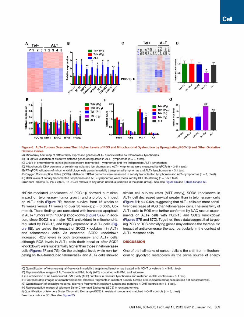
Figure 6. ALT+ Tumors Overcome Their Higher Levels of ROS and Mitochondrial Dysfunction by Upregulating PGC-1b and Other Oxidative
Defense Genes
(A) Microarray heat map of differentially expressed genes in ALT+ tumors relative to telomerase+ lymphomas.
(B) RT-qPCR validation of oxidative defense genes upregulated in ALT+ lymphomas (n = 3, t test).
(C) CNVs of chromosome 18 in eight independent telomerase+ lymphomas and five independent ALT+ lymphomas.
(D) Mitochondria DNA contents of serially transplanted lymphomas and ALT+ lymphomas were measured by qPCR (n = 3–5, t test).
(E) RT-qPCR validation of mitochondrial biogenesis genes in serially transplanted lymphomas and ALT+ lymphomas (n = 3, t test).
(F) Oxygen Consumption Rates (OCRs) relative to mtDNA contents were measured in serially transplanted lymphomas and ALT+ lymphomas (n = 3, t test).
(G) ROS levels of serially transplanted lymphomas and ALT+ lymphomas were measured by DCFDA staining (n = 3-5, t test).
Error bars indicate SD (*p < 0.001, **p < 0.01 relative to any other individual samples in the same group). See also Figure S6 and Tables S2 and S3.
shRNA-mediated knockdown of PGC-1b showed a minimal
impact on telomerase+ tumor growth and a profound impact
on ALT+ cells (Figure 7E; median survival from 15 weeks to
19 weeks versus 17 weeks to over 30 weeks; p = 0.0065, Cox
model). These findings are consistent with increased apoptosis
in ALT+ tumors with PGC-1b knockdown (Figure S7A). In addi-
tion, since SOD2 is a major ROS antioxidant in mitochondria,
regulated by PGC-1b, and highly expressed in ALT+ cells (Fig-
ure 6B), we tested the impact of SOD2 knockdown in ALT+
and telomerase+ cells. As expected, SOD2 knockdown
increased ROS levels in both telomerase+ and ALT+ cells,
although ROS levels in ALT+ cells (both basal or after SOD2
knockdown) were substantially higher than those in telomerase+
cells (Figures 7F and 7G). On the biological level, while non-tar-
geting shRNA-transduced telomerase+ and ALT+ cells showed
(C) Quantification of telomere signal-free ends in serially transplanted lymphoma
(D) Representative images of ALT-associated PML body (APB) costained with P
(E) Quantification of ALT-associated PML Body (APB) numbers in resistant lymp
(F) Representative images of extrachromosomal telomere fragments in resistant
(G) Quantification of extrachromosomal telomere fragments in resistant tumors a
(H) Representative images of telomere Sister Chromatid Exchange (tSCE) in resi
(I) Quantification of telomere Sister Chromatid Exchange (tSCE) in resistant tumo
Error bars indicate SD. See also Figure S5.
similar cell survival rates (MTT assay), SOD2 knockdown in
ALT+ cell decreased survival greater than in telomerase+ cells
(Figure 7H; p = 0.02), suggesting that ALT+ cells are more sensi-
tive to increase of ROS than telomerase+ cells. The sensitivity of
ALT+ cells to ROS was further confirmed by NAC rescue exper-
iments on ALT+ cells with PGC-1b and SOD2 knockdown
(Figures S7B and S7C). Together, these data suggest that target-
ing PGC or ROS detoxifying genes may enhance the therapeutic
impact of antitelomerase therapy, particularly in the context of
ALT+-resistant cells.
DISCUSSION
One of the hallmarks of cancer cells is the shift from mitochon-
drial to glycolytic metabolism as the prime source of energy
s treated with 4OHT or vehicle (n = 3–5, t test).
ML and telomere.
homas and matched 4-OHT controls (n = 5, t test).
tumors. Circled area indicates metaphase spread not separated well.
nd matched 4-OHT controls (n = 5, t test).
stant tumors.
rs and matched 4-OHT controls (n = 5, t test).
Cell 148, 651–663, February 17, 2012 ª2012 Elsevier Inc. 659

Figure 7. Inhibition of PGC-1b or SOD2 Differentially Kills ALT+ Tumors over Telomerase+ Tumors
(A) PGC-1b expression levels in telomerase+ and ALT+ tumor cells with PGC-1b shRNAs or control shRNA (shLuc) (n = 3, t test).
(B) Mitochondria DNA contents in telomerase+ and ALT+ tumor cells with PGC-1b shRNAs or control shRNA (shLuc) (n = 3, t test).
(C) Basal oxygen consumption rates (OCR) in telomerase+ and ALT+ tumor cells with PGC-1b shRNAs or control shRNA (shLuc) (n = 3, t test).
(D) ROS levels in telomerase+ and ALT+ tumor cells with PGC-1b shRNAs or control shRNA (shLuc) (n = 3, t test).
(E) Kaplan-Meier curves (Cox model test, p = 0.0065) of lymphoma-free survival of mice transplanted with telomerase+ and ALT+ tumor cells with PGC-1b
shRNAs (shPGC-1b-1+ shPGC-1b-2) or control shRNA (shLuc).
(F) SOD2 expression levels in telomerase+ and ALT+ tumor cells with SOD2 shRNAs or control shRNA (shLuc) (n = 3, t test).
(G) ROS levels in telomerase+ and ALT+ tumor cells with SOD2 shRNAs or control shRNA (shLuc) (n = 3, t test).
(H) Relative cells numbers of telomerase+ and ALT+ tumor cells with SOD2 shRNAs or control shRNA (shLuc) (n = 3, two-way ANOVA, p = 0.02).
(I) Model of regulation of apoptosis, senescence and mitochondrial function in telomerase+, ALT+, and telomere-dysfunctional cells.
Error bars indicate SD (*p < 0.0001, **p < 0.001, ***p < 0.02, unless otherwise stated). See also Figure S7.
and anabolic support (Vander Heiden et al., 2009; Warburg,
1956). While this bioenergetic shift conveys decreased reliance
on mitochondria as a major energy source for cancer cells, our
genetic studies indicate a continued vital role in the maintenance
of mitochondrial function in cancer. This view aligns withmultiple
lines of evidence suggesting that mitochondrial competence is
indeed important for cancer cell viability (Jose et al., 2011; Kop-
penol et al., 2011) and efficient oncogene-mediated transforma-
tion (Gao et al., 2009; Weinberg et al., 2010; Wise et al., 2008;
Yuneva et al., 2007). More specifically, our study uncovers a crit-
660 Cell 148, 651–663, February 17, 2012 ª2012 Elsevier Inc.
ical role of robust mitochondrial function in cancer cells, particu-
larly for those cancer cells with ALT-maintained telomeres.
Why might ALT+ cancer cells exhibit exquisite sensitivity to
inhibition of PGC expression or antioxidant defense? We specu-
late that continued DNA damage signaling at ALT-maintained
telomeres perhaps due to altered chromatin (Heaphy et al.,
2011) or inefficient capping via this mechanism could lead to
mitochondrial damage. Mitochondria are the major intracellular
source of ROS in mammalian cells, and dysfunctional mitochon-
dria produce more ROS, which at excessively high levels is

known to be detrimental to cell survival (Lin and Beal, 2006;
Murphy, 2009; St-Pierre et al., 2006). In this context, we propose
that the pressure to maintain mitochondrial function reflects the
need tomaintain tolerable intracellular ROS levels. This oxidative
defense mechanism may be particularly important for telomeres
as ROS are particularly injurious toward the G-rich sequences of
telomeres (Hall et al., 1996; Oexle and Zwirner, 1997; Retel et al.,
1993), thereby causing increased telomere dysfunction in the
absence of efficient telomerase-mediated repair. Correspond-
ingly, our combined genomic and functional studies establish
that ALT+ cells strive to maintain adequate mitochondrial and
oxidative defense functions and show exquisite sensitivity to
loss of such function. On the molecular level, this work also
establishes that the intimate PGC-directed link between telo-
meres and mitochondria identified recently in normal tissues
(Sahin et al., 2011) is operative in cancer cells (Figure 7I).
About 15% of human cancers maintain their telomeres in the
absence of telomerase activity by the ALTmechanism. The exis-
tence of a telomerase-independent telomere maintenance
mechanism was first identified in yeast with telomerase defi-
ciency, and this mechanism was demonstrated to be dependent
onRAD52, a gene involved in homologous recombination (Lund-
blad and Blackburn, 1993). Subsequent studies established that
the ALT mechanism in human cancer cells requires certain DNA
recombination proteins, including the MRN complex (MRE11,
RAD50, and NBS1) (Jiang et al., 2005; Zhong et al., 2007),
SMC5-SMC6 complex (Potts and Yu, 2007), flap endonuclease
1 (FEN1) (Saharia and Stewart, 2009), MUS81 (Zeng et al.,
2009), Fanconi anemia group D2 (FANCD2), and Fanconi anemia
group A (FANCA) (Fan et al., 2009). In our model, MRE11 and
FANCA genes are indeed upregulated in ALT+ cells relative to
telomerase+ cells (Figures S4B and S4C). These observations
provide genetic evidence in mammalian cells of the importance
of these factors for DNA recombination-dependent ALT mecha-
nism in this cancer model.
Our studies also highlight that, while ALT+ cells suppress
apoptosis and senescence as efficiently as telomerase+ cells,
these cells do not fully restore mitochondrial function which we
speculate may relate to ongoing genotoxic stress partly associ-
ated with less efficient telomere capping by ALT-mediated
telomere maintenance (Cesare et al., 2009) as well as p53-inde-
pendent repression of PGC (Sahin et al., 2011). In ALT+ cells, the
importance of this mitochondrial maintenance program is under-
scored by the upregulated PGC network signaling on both the
genomic (copy number) and transcriptional levels (Figure 7I).
That the PGC network is a vital regulator in our ALT+-resistant
tumors is further substantiated by our analysis of published
transcriptomic data comparing human ALT+ with telomerase+
osteosarcomas (Lafferty-Whyte et al., 2009). Specifically,
comparison of three key network genes (PGC-1a, NRF2, and
SOD2; PGC-1b data is not available in this data set) in ten
ALT+ and eight telomerase+ osteosarcomas showed that five
of ten ALT+ osteosarcomas have significantly higher levels of
one or multiple of PGC-1a, NRF2, and SOD2 genes relative to
the basal levels, which is more prominent than telomerase+
tumors (Figure S6G) (p = 0.04), suggesting that upregulation of
mitochondrial function and ROS defense pathway genes is
important in other ALT+ cancer types. Thus, our genetic
modeling of telomerase extinction in cancer increases our
understanding of how tumor cells might respond and adapt to
telomerase inhibition and illuminates a clinical path hypothesis
utilizing combination regimens targeting telomerase and PGC-
mediated adaptive mechanisms in cancer.
EXPERIMENTAL PROCEDURES
Mice
Atm, Terc, and TERT-ER mice described previously (Jaskelioff et al., 2011;
Maser et al., 2007; Wong et al., 2003) were interbred and backcrossed to
high grade of C57BL/6 (over 95%). The mating strategy to obtain experimental
cohorts G0, G1, and G3-4 is shown in Figure S1A. 4-OHT time release pellets
(5 mg; Innovative Research of America) were inserted subcutaneously at age
of 18 weeks to reach steady-state blood levels of 1 ng ml-1 4-OHT for 60
consecutive days.
Histology, Tumor Characterization, and Sample Preparation
Antibodies used for IHC include anti-Ki67 (Dako), anti-53BP1 (Bethyl Labs),
anti-gH2AX (Bethyl Labs), and anti-Cleaved Caspase 3 (Cell Signaling). For
FACS analysis, cells were immunostained with CD4, CD8, and CD3 antibodies
(eBioscience) and analyzed on a BD FACSCanto II (BD Biosciences).
Cytogenetic Analysis
For metaphase preparation, metaphases were obtained from colcemid-
treated cells incubated in 105 mM KCl hypotonic buffer for 15 min before fixa-
tion in 3:1 methanol–acetic acid. Spectral karyotyping (SKY) was done using
the SkyPaint Kit and SkyView analytical software (Applied Spectral Imaging)
according to manufacturer’s protocols. For telomere-FISH (fluorescent
in situ hybridization), metaphase spreads were applied with telomere-specific
T2AG3-FITC PNA (peptide nucleic acid) probes and centromere-specific
Cent-Cy3 or Cent-pacific blue PNA probes, and counter-stained with DAPI
(for microscopy) or TOTO3 (for laser scanning). Laser scanning cytometry
quantification was performed with an iCys Research Imaging Cytometer
(Compucyte) as described earlier (Jaskelioff et al., 2011; Sahin et al., 2011).
Telomere Sister Chromatid Exchange Assay
Telomere Co-FISH staining was performed as previously described (Potts and
Yu, 2007). Details are described in Extended Experimental Procedures.
RT-qPCR, Mitochondria DNA Content Measurement,
and Western Blot
RT-qPCR primers are described in Table S4. Mitochondrial DNA content was
measured by the relative levels of CoxI versus beta globin by qPCR. CoxI and
beta globin primers are described in Table S4. Antibodies used for Western
blotting are anti-phospho p53 (ser15, Cell Signaling), anti-p21 (Santa Cruz
Biotechnology), anti-Vinculin (Sigma), and anti-Mre11 (BD Biosciences).
shRNA Knockdown
Lentiviral based shRNA constructs targeting mouse PGC-1b and SOD2 were
ordered from The Dana-Farber/Harvard Cancer Center DNA Resource Core.
Verified shRNA sequences were cloned into inducible lentiviral constructs
PLKO-TRC-901 (IPTG inducible; Broad Institute) and PLKO-Tet (Doxycycline
inducible; Novartis).
Pathway Analysis
Eight hundred ninety-one upregulated and 1,345 downregulated genes with
1.5-fold change and p < 0.01 in ALT+ tumors relative to telomerase+ tumors
were applied with Knowledge-based Pathway (IPA) analysis. The significantly
represented pathways were compared with the pathways obtained from the
similar analysis on ten ALT+ and eight telomerase+ osteosarcomas (Lafferty-
Whyte et al., 2009). Overlapped pathways are listed.
Array-CGH Profiling and Analyses
Array-CGH, SGOL score analysis, homologous mapping, and permutation
analysis are described in Extended Experimental Procedures.
Cell 148, 651–663, February 17, 2012 ª2012 Elsevier Inc. 661

Mitochondrial Oxygen Consumption Measurements in Live Cells
Oxygen consumption rates (OCRs) were measured using the Seahorse XF24
instrument (Seahorse Biosciences). Basal mitochondrial respiration were
measured at four time points, and respiration nonlinked to mitochondrial
ATP synthesis were measured at four time points after adding 1 mM oligomy-
cin. Nonmitochondrial OCRs were obtained by adding 5 mM antimycin A, and
uncoupled respiration was obtained by adding 1 mM FCCP.
ROS Measurement
For determination of ROS levels, tumor cells were stained with 5 mM CM-H2
DFCDA (Invitrogen) at 37�C for 30 min, followed by FACS analysis on a BD
FACSCanto II (BD Biosciences).
COX Model Test
Effects of shPGC-1b knockdown on Tel+ and ALT+ cells were tested by Cox
analysis. Details are described in Extended Experimental Procedures.
ACCESSION NUMBERS
Microarray (GSE35044) and aCGH (GSE35045) data were deposited in GEO
(http://www.ncbi.nlm.nih.gov/geo/) under the accession numbers indicated.
SUPPLEMENTAL INFORMATION
Supplemental Information includes Extended Experimental Procedures, seven
figures, and four tables and can be found with this article online at doi:10.1016/
j.cell.2011.12.028.
ACKNOWLEDGMENTS
We thank S. Jiang, R. Narurkar, and E. Fletcher-Sananikone for excellent
mouse husbandry and care, and all members of DePinho and Chin labs for
helpful discussion. We also thank Yingchui Liu for the Cox model analysis.
J.H. and B.G. were supported by the leukemia and lymphoma society fellow-
ship. A.T.B. is supported by the Helen Hay Whitney fellowship. M.L. is
supported by the Fundacion Ramon Areces fellowship. O.S.S. is supported
by R01 (R01-DK074778 and R01-DK56690) grants from NIH. L.C. and
R.A.D. are supported by R01 (R01CA84628) and U01 (U01CA141508) grants
from NIH. R.A.D. is an Ellison Foundation for Medical Research Senior Scholar
and an American Cancer Society Research Professor.
Received: August 27, 2011
Revised: November 8, 2011
Accepted: December 30, 2011
Published: February 16, 2012
REFERENCES
Agrawal, A., Dang, S., and Gabrani, R. (2012). Recent patents on anti-telome-
rase cancer therapy. Recent Patents Anticancer. Drug Discov. 7, 102–117.
Artandi, S.E., and DePinho, R.A. (2010). Telomeres and telomerase in cancer.
Carcinogenesis 31, 9–18.
Artandi, S.E., Chang, S., Lee, S.L., Alson, S., Gottlieb, G.J., Chin, L., and De-
Pinho, R.A. (2000). Telomere dysfunction promotes non-reciprocal transloca-
tions and epithelial cancers in mice. Nature 406, 641–645.
Begus-Nahrmann, Y., Lechel, A., Obenauf, A.C., Nalapareddy, K., Peit, E.,
Hoffmann, E., Schlaudraff, F., Liss, B., Schirmacher, P., Kestler, H., et al.
(2009). p53 deletion impairs clearance of chromosomal-instable stem cells
in aging telomere-dysfunctional mice. Nat. Genet. 41, 1138–1143.
Cesare, A.J., and Reddel, R.R. (2010). Alternative lengthening of telomeres:
models, mechanisms and implications. Nat. Rev. Genet. 11, 319–330.
Cesare, A.J., Kaul, Z., Cohen, S.B., Napier, C.E., Pickett, H.A., Neumann, A.A.,
and Reddel, R.R. (2009). Spontaneous occurrence of telomeric DNA damage
response in the absence of chromosome fusions. Nat. Struct. Mol. Biol. 16,
1244–1251.
662 Cell 148, 651–663, February 17, 2012 ª2012 Elsevier Inc.
Chang, S., Khoo, C.M., Naylor, M.L., Maser, R.S., and DePinho, R.A. (2003).
Telomere-based crisis: functional differences between telomerase activation
and ALT in tumor progression. Genes Dev. 17, 88–100.
Chin, L., Artandi, S.E., Shen, Q., Tam, A., Lee, S.L., Gottlieb, G.J., Greider,
C.W., and DePinho, R.A. (1999). p53 deficiency rescues the adverse effects
of telomere loss and cooperates with telomere dysfunction to accelerate carci-
nogenesis. Cell 97, 527–538.
d’Adda di Fagagna, F., Reaper, P.M., Clay-Farrace, L., Fiegler, H., Carr, P.,
Von Zglinicki, T., Saretzki, G., Carter, N.P., and Jackson, S.P. (2003). A DNA
damage checkpoint response in telomere-initiated senescence. Nature 426,
194–198.
Fan, Q., Zhang, F., Barrett, B., Ren, K., and Andreassen, P.R. (2009). A role for
monoubiquitinated FANCD2 at telomeres in ALT cells. Nucleic Acids Res. 37,
1740–1754.
Feng, J., Funk, W.D., Wang, S.S., Weinrich, S.L., Avilion, A.A., Chiu, C.P.,
Adams, R.R., Chang, E., Allsopp, R.C., Yu, J., et al. (1995). The RNA compo-
nent of human telomerase. Science 269, 1236–1241.
Gao, P., Tchernyshyov, I., Chang, T.C., Lee, Y.S., Kita, K., Ochi, T., Zeller, K.I.,
De Marzo, A.M., Van Eyk, J.E., Mendell, J.T., and Dang, C.V. (2009). c-Myc
suppression of miR-23a/b enhances mitochondrial glutaminase expression
and glutamine metabolism. Nature 458, 762–765.
Gonzalez-Suarez, E., Samper, E., Flores, J.M., and Blasco, M.A. (2000). Telo-
merase-deficient mice with short telomeres are resistant to skin tumorigen-
esis. Nat. Genet. 26, 114–117.
Hall, D.B., Holmlin, R.E., and Barton, J.K. (1996). Oxidative DNA damage
through long-range electron transfer. Nature 382, 731–735.
Harley, C.B., and Sherwood, S.W. (1997). Telomerase, checkpoints and
cancer. Cancer Surv. 29, 263–284.
Heaphy, C.M., de Wilde, R.F., Jiao, Y., Klein, A.P., Edil, B.H., Shi, C., Bette-
gowda, C., Rodriguez, F.J., Eberhart, C.G., Hebbar, S., et al. (2011). Altered
telomeres in tumors with ATRX and DAXX mutations. Science 333, 425.
Jaskelioff, M., Muller, F.L., Paik, J.H., Thomas, E., Jiang, S., Adams, A.C., Sa-
hin, E., Kost-Alimova, M., Protopopov, A., Cadinanos, J., et al. (2011). Telome-
rase reactivation reverses tissue degeneration in aged telomerase-deficient
mice. Nature 469, 102–106.
Jiang, W.Q., Zhong, Z.H., Henson, J.D., Neumann, A.A., Chang, A.C., and
Reddel, R.R. (2005). Suppression of alternative lengthening of telomeres by
Sp100-mediated sequestration of the MRE11/RAD50/NBS1 complex. Mol.
Cell. Biol. 25, 2708–2721.
Jose, C., Bellance, N., and Rossignol, R. (2011). Choosing between glycolysis
and oxidative phosphorylation: a tumor’s dilemma? Biochim. Biophys. Acta
1807, 552–561.
Karlseder, J., Broccoli, D., Dai, Y., Hardy, S., and de Lange, T. (1999). p53- and
ATM-dependent apoptosis induced by telomeres lacking TRF2. Science 283,
1321–1325.
Koppenol, W.H., Bounds, P.L., and Dang, C.V. (2011). Otto Warburg’s
contributions to current concepts of cancer metabolism. Nat. Rev. Cancer
11, 325–337.
Lafferty-Whyte, K., Cairney, C.J., Will, M.B., Serakinci, N., Daidone, M.G., Zaf-
faroni, N., Bilsland, A., and Keith, W.N. (2009). A gene expression signature
classifying telomerase and ALT immortalization reveals an hTERT regulatory
network and suggests a mesenchymal stem cell origin for ALT. Oncogene
28, 3765–3774.
Lin, M.T., and Beal, M.F. (2006). Mitochondrial dysfunction and oxidative
stress in neurodegenerative diseases. Nature 443, 787–795.
Lundblad, V., and Blackburn, E.H. (1993). An alternative pathway for yeast
telomere maintenance rescues est1- senescence. Cell 73, 347–360.
Maser, R.S., Choudhury, B., Campbell, P.J., Feng, B., Wong, K.K., Protopo-
pov, A., O’Neil, J., Gutierrez, A., Ivanova, E., Perna, I., et al. (2007). Chromoso-
mally unstable mouse tumours have genomic alterations similar to diverse
human cancers. Nature 447, 966–971.
Murphy, M.P. (2009). How mitochondria produce reactive oxygen species.
Biochem. J. 417, 1–13.

Nakamura, T.M., Morin, G.B., Chapman, K.B., Weinrich, S.L., Andrews, W.H.,
Lingner, J., Harley, C.B., and Cech, T.R. (1997). Telomerase catalytic subunit
homologs from fission yeast and human. Science 277, 955–959.
O’Hagan, R.C., Chang, S., Maser, R.S., Mohan, R., Artandi, S.E., Chin, L., and
DePinho, R.A. (2002). Telomere dysfunction provokes regional amplification
and deletion in cancer genomes. Cancer Cell 2, 149–155.
Oexle, K., and Zwirner, A. (1997). Advanced telomere shortening in respiratory
chain disorders. Hum. Mol. Genet. 6, 905–908.
Potts, P.R., and Yu, H. (2007). The SMC5/6 complexmaintains telomere length
in ALT cancer cells through SUMOylation of telomere-binding proteins. Nat.
Struct. Mol. Biol. 14, 581–590.
Qi, L., Strong, M.A., Karim, B.O., Armanios, M., Huso, D.L., and Greider, C.W.
(2003). Short telomeres and ataxia-telangiectasia mutated deficiency cooper-
atively increase telomere dysfunction and suppress tumorigenesis. Cancer
Res. 63, 8188–8196.
Retel, J., Hoebee, B., Braun, J.E., Lutgerink, J.T., van den Akker, E., Wana-
marta, A.H., Joenje, H., and Lafleur, M.V. (1993). Mutational specificity of
oxidative DNA damage. Mutat. Res. 299, 165–182.
Rudolph, K.L., Millard, M., Bosenberg, M.W., and DePinho, R.A. (2001). Telo-
mere dysfunction and evolution of intestinal carcinoma in mice and humans.
Nat. Genet. 28, 155–159.
Saharia, A., and Stewart, S.A. (2009). FEN1 contributes to telomere stability in
ALT-positive tumor cells. Oncogene 28, 1162–1167.
Sahin, E., and DePinho, R.A. (2010). Linking functional decline of telomeres,
mitochondria and stem cells during ageing. Nature 464, 520–528.
Sahin, E., Colla, S., Liesa, M., Moslehi, J., Muller, F.L., Guo, M., Cooper, M.,
Kotton, D., Fabian, A.J., Walkey, C., et al. (2011). Telomere dysfunction
induces metabolic and mitochondrial compromise. Nature 470, 359–365.
Shay, J.W., andWright, W.E. (2006). Telomerase therapeutics for cancer: chal-
lenges and new directions. Nat. Rev. Drug Discov. 5, 577–584.
St-Pierre, J., Drori, S., Uldry, M., Silvaggi, J.M., Rhee, J., Jager, S., Handschin,
C., Zheng, K., Lin, J., Yang, W., et al. (2006). Suppression of reactive oxygen
species and neurodegeneration by the PGC-1 transcriptional coactivators.
Cell 127, 397–408.
Takai, H., Smogorzewska, A., and de Lange, T. (2003). DNA damage foci at
dysfunctional telomeres. Curr. Biol. 13, 1549–1556.
van Steensel, B., Smogorzewska, A., and de Lange, T. (1998). TRF2 protects
human telomeres from end-to-end fusions. Cell 92, 401–413.
Vander Heiden, M.G., Cantley, L.C., and Thompson, C.B. (2009). Under-
standing the Warburg effect: the metabolic requirements of cell proliferation.
Science 324, 1029–1033.
Warburg, O. (1956). On the origin of cancer cells. Science 123, 309–314.
Weinberg, F., Hamanaka, R., Wheaton, W.W., Weinberg, S., Joseph, J., Lo-
pez, M., Kalyanaraman, B., Mutlu, G.M., Budinger, G.R., and Chandel, N.S.
(2010). Mitochondrial metabolism and ROS generation are essential for
Kras-mediated tumorigenicity. Proc. Natl. Acad. Sci. USA 107, 8788–8793.
Wiedemeyer, W.R., Dunn, I.F., Quayle, S.N., Zhang, J., Chheda, M.G., Dunn,
G.P., Zhuang, L., Rosenbluh, J., Chen, S., Xiao, Y., et al. (2010). Pattern of reti-
noblastoma pathway inactivation dictates response to CDK4/6 inhibition in
GBM. Proc. Natl. Acad. Sci. USA 107, 11501–11506.
Wise, D.R., DeBerardinis, R.J., Mancuso, A., Sayed, N., Zhang, X.Y., Pfeiffer,
H.K., Nissim, I., Daikhin, E., Yudkoff, M., McMahon, S.B., and Thompson, C.B.
(2008). Myc regulates a transcriptional program that stimulates mitochondrial
glutaminolysis and leads to glutamine addiction. Proc. Natl. Acad. Sci. USA
105, 18782–18787.
Wong, K.K., Maser, R.S., Bachoo, R.M., Menon, J., Carrasco, D.R., Gu, Y., Alt,
F.W., and DePinho, R.A. (2003). Telomere dysfunction and Atm deficiency
compromises organ homeostasis and accelerates ageing. Nature 421,
643–648.
Xu, Y., Ashley, T., Brainerd, E.E., Bronson, R.T., Meyn, M.S., and Baltimore, D.
(1996). Targeted disruption of ATM leads to growth retardation, chromosomal
fragmentation during meiosis, immune defects, and thymic lymphoma. Genes
Dev. 10, 2411–2422.
Yuneva, M., Zamboni, N., Oefner, P., Sachidanandam, R., and Lazebnik, Y.
(2007). Deficiency in glutamine but not glucose induces MYC-dependent
apoptosis in human cells. J. Cell Biol. 178, 93–105.
Zeng, S., Xiang, T., Pandita, T.K., Gonzalez-Suarez, I., Gonzalo, S., Harris,
C.C., and Yang, Q. (2009). Telomere recombination requires theMUS81 endo-
nuclease. Nat. Cell Biol. 11, 616–623.
Zhong, Z.H., Jiang, W.Q., Cesare, A.J., Neumann, A.A., Wadhwa, R., and Red-
del, R.R. (2007). Disruption of telomere maintenance by depletion of the
MRE11/RAD50/NBS1 complex in cells that use alternative lengthening of telo-
meres. J. Biol. Chem. 282, 29314–29322.
Cell 148, 651–663, February 17, 2012 ª2012 Elsevier Inc. 663