antimycobacterial activity of some medicinal plants
TRANSCRIPT
Anti-mycobacterial activity and acute toxicity of Erythrina
abyssinica, Cryptolepis sanguinolenta and Solanum incanum
BUNALEMA LYDIA (BSC., ZOO/CHEM)
2007/HD11/9328U
A dissertation submitted to the School of Graduate Studies in partial fulfillment of the
requirements for the award of Master of Science Degree in Pharmacology, Makerere
University Kampala, Uganda
October 2010
i
DECLARATION
I Bunalema Lydia declare that this dissertation has never been submitted to any other University or
Academic Institution for purposes of getting an academic award. All the information in this dissertation is
based on my observations.
Signature....................……...Date......................................................................................
Supervisors:
1. Dr. Paul Waako, MBChB MSc. PhD
Associate Professor
Department of Pharmacology and Therapeutics
Makerere University, College of Health Sciences
Signature....................……...Date......................................................................................
2. Dr. John R.S. Tabuti, BSc. MSc. PhD
Associate Professor
Institute of Environment and Natural Resources,
Makerere University
Signature....................……...Date......................................................................................

ii
DEDICATION
I would like to dedicate this piece of work to my mum and dad George and Sarah Matovu and to
my husband Dr. J. Wasswa.
iii
ACKNOWLEDGEMENT
I would like to significantly appreciate the inputs and advise of my supervisors Dr. Paul Waako
and Dr. J.R.S. Tabuti. I appreciate the help of my colleague Mr. Claude Kirimuhuzya who
tirelessly worked towards the success and accomplishment of this work; I also extend my sincere
gratitude towards my brother Mr. A. Lubega from the Department of Pharmacology and
Therapeutics for helping me with the toxicity tests. Lastly, I thank Dr. John Wasswa for his
inputs, for the emotional and spiritual encouragement that he gave me. I would like to thank Vic
Recs project for funding this research.
iv
TABLE OF CONTENTS
DECLARATION ........................................................................................................................ i
DEDICATION ........................................................................................................................... ii
ACKNOWLEDGEMENT ......................................................................................................... iii
TABLE OF CONTENTS .......................................................................................................... iv
LIST OF TABLES ................................................................................................................... vii
LIST OF FIGURES ................................................................................................................ viii
LIST OF ABBREVIATIONS.................................................................................................... ix
ABSTRACT ...............................................................................................................................x
CHAPTER ONE .........................................................................................................................1
INTRODUCTION ...................................................................................................................1
1.1 Background ..............................................................................................................1
1.1.1 Current status of Tuberculosis ................................................................................1
1.2 Statement of the problem ..........................................................................................3
1.3 Objectives .................................................................................................................4
1.4 Justification of the study .................................................................................................4
1.5 Significance of the study............................................................................................5
CHAPTER TWO ........................................................................................................................6
LITERATURE REVIEW ........................................................................................................6
2.1 Etiology of tuberculosis ............................................................................................6
2.2 Epidemiology ............................................................................................................7
2.3 Treatment of Tuberculosis ........................................................................................7
2.4 Challenges in the control of TB .................................................................................9
2.5 In-vitro assays for evaluation of anti tubercular activity ........................................... 10
2.6 Role of Natural products in drug development ......................................................... 11
2.7 Ethnobotany and pharmacology of Erythrina abyssinica, Cryptolepis sanguinolenta
and Solanum incanum ........................................................................................................ 12
2.8 Toxicity studies ............................................................................................................ 14
CHAPTER THREE ................................................................................................................... 16
MATERIALS AND METHODS ........................................................................................... 16
v
3.1 Study design ............................................................................................................ 16
3.2 Selection criteria ...................................................................................................... 16
3.3 Plant collection and identification ............................................................................ 16
3.4 Drying and pulverizing ................................................................................................. 17
3.5 Extract preparation ....................................................................................................... 17
3.6 Mycobacterial tests .................................................................................................. 18
3.6.1 Growth media ............................................................................................................ 18
3.6.2 Preparation of inoculum for drug sensitivity testing ............................................. 19
3.6.3 Bioassay protocol for susceptibility tests .............................................................. 19
3.6.3.1 Preparation of the drugs/ extracts ....................................................................... 19
3.6.3.2 Preparation of biodiscs ........................................................................................... 20
3.6.3.3 Procedure ............................................................................................................... 20
3.6.4 Determination of the Minimum Inhibitory Concentration (MIC) ......................... 21
3.6.4.1 Preparation of the medium (Middle brook 7H9) ................................................... 21
Procedure ........................................................................................................................... 21
3.7 Acute toxicity tests .................................................................................................. 22
3.8 Qualitative phytochemical testing ............................................................................ 22
3.9 Data analysis ........................................................................................................... 23
3.10 Ethical considerations .......................................................................................... 23
CHAPTER FOUR ..................................................................................................................... 25
RESULTS ............................................................................................................................. 25
4.1 Yields from extractions ................................................................................................ 25
4.2 Anti-mycobacterial activity .......................................................................................... 25
4.3 Minimum Inhibitory Concentration of E. abyssinica and C. sanguinolenta .............. 27
4.4 Acute toxicity ............................................................................................................... 28
4.5 Phytochemical analysis ........................................................................................... 31
CHAPTER FIVE ...................................................................................................................... 32
DISCUSSION ........................................................................................................................... 32
5.1 Anti-mycobacterial activity ..................................................................................... 32
5.2 Acute toxicity ............................................................................................................... 34
vi
5.3 Phytochemical testing................................................................................................... 35
5.4 Limitations .............................................................................................................. 36
5.5 Conclusions ............................................................................................................. 37
5.6 Recommendations ................................................................................................... 37
REFERENCES ......................................................................................................................... 38
vii
LIST OF TABLES
Table 1: List of the medicinal plants used in the bioassay with their voucher numbers 17
Table 2: The percentage yield of crude extracts from E. abyssinica, S. incanum and C.
sanguinolenta using three solvents 25
Table 3: The antimycobacterial activity of ether, methanol, chloroform and Total crude
extracts of E. abyssinica, S. incanum and C. sanguinolenta against the rifampicin
resistant strain (TMC), pan sensitive (H37RV) and M. avium(MA) strains by the
disc diffusion method. 26
Table 4: The minimum inhibitory concetration of the methanol, chloroform and Total
crude extracts of E. abyssinica and C. sanguinolenta using the microbroth
dilution method. 27
Table 5: Behavioral changes observed during acute toxicity studies of the extracts
from E. abyssinica and C. sanguinolenta in the Mus musculus mice. 28
Table 6: The effect of increased dose of the extract of C. sanguinolenta and E. abyssinica
total crude extracts on the survival of white albino mice 29
Table 7: Compounds present in C. sanguinolenta and E. abyssinica crude extracts 31
viii
LIST OF FIGURES
Graph 1 A plot of probit against log dose for C. sanguinolenta 30
Graph 2 A plot of probit against log dose for E. abyssinica 31
ix
LIST OF ABBREVIATIONS
TB Tuberculosis
WHO World Health Organization
CDC Center for Disease Control
XDR Extensively Drug Resistance
MDR Multi Drug Resistance
HIV Human Immune Virus
AIDS Acquired Immune Deficiency Syndrome
MIC Minimum Inhibitory Concentration
JCRC Joint Clinical Research Center
DMSO Dimethyl sulfoxide
x
ABSTRACT
Tuberculosis (TB) kills approximately two million people annually. Efforts to treat the disease
have been made much more difficult due to development of drug resistant TB strains (MDR and
XDR TB) and co-infection with HIV AIDS. There is an urgent need therefore, to search for and
develop new, inexpensive and effective anti-TB drugs. Extracts from three plants, Solanum
incanum, Cryptolepis sanguinolenta and Erythrina abyssinica used in traditional medicine to
treat TB symptoms were screened for anti-mycobacterial properties against a Rifampicin
resistant, pan sensitive and Mycobacteria avium strains. In addition, the acute toxicity profile and
phytochemistry of the active extracts were studied.
The chloroform extract of E. abyssinica was the most active on M. avium wild strain and the
rifampicin resistant strain (MIC= 0.3 and 0.39 mg/ml respectively). Against the pansensitive
strain the methanol total crude extract was most active (MIC= 0.2 mg/ml). C. sanguinolenta total
crude methanol extract was also active against the three strains of mycobacteria; however S.
incanum did not show activity on any of the strains. Toxicity studies showed that the two plants
had an LD50 of more than 500mg/kg body weight and thus considered to be safe.
E. abyssinica extracts and C. sanguinolenta total crude extract contained alkaloids, terpenoids,
tannins and flavones. Saponins and phenols were not detected in all extracts.
C. sanguinolenta and E. abyssinica have potential to be developed into new anti-TB drugs. The
results have also validated traditional knowledge from the local people regarding the use of these
species to treat TB.
1
CHAPTER ONE
INTRODUCTION
1.1 Background
1.1.1 Current status of Tuberculosis
Tuberculosis (TB) is one of the leading causes of morbidity and mortality globally. The global
mortality rate stands at two million deaths per year with one third of the world’s population
infected with the bacilli (Centre for Disease Control (CDC), 2005; Sanjay, 2004; World Health
Organisation (WHO), 2007). It is estimated that 9.2 million new cases are diagnosed every year.
According to the World Health Organization (WHO), the incidence of tuberculosis in African
countries more than doubled between 1990 and 2005 and is taking an upward trend.
(WHO,2008). According to Chaisson and Martinson (2008), Africa carries 29% of the world’s
disease burden and 34% of the world’s total death rate. Uganda ranks 15th
among the world’s 22
countries with a high tuberculosis burden; with an estimated incidence of 355 cases per 100,000
people per year and mortality of 84 deaths per 100,000 people per year (WHO, 2008).
The emergence of drug resistant strains of Mycobacterium tuberculosis, is one of the major
reasons contributing to the rise in global incidence of tuberculosis since 1980. Multi Drug
Resistant (MDR) forms are defined as M. tuberculosis strains resistant to atleast Rifampicin and
Isoniazid: the first line drugs used in treatment of tuberculosis. Extensively Drug Resistant TB
(XDR) is tuberculosis caused by strains resistant to first line drugs, to fluoroquinolones and at
least one of three injectable second-line drugs; capreomycin, kanamycin, and amikacin (Lawn
and Wilkison, 2006).
2
According to Zignol et al., (2006), more than 420,000 TB cases world wide are due to MDR
strains of M. tuberculosis; 40,000 of these occur in Africa. In Uganda, 0.7% of all new cases of
tuberculosis are multi drug resistant (WHO, 2008). MDR TB treatment requires the use of
second-line drugs (SLDs) that are less effective, more toxic, and more costly than the first-line
isoniazid and rifampin-based regimens (CDC, 2005). This has made efforts to control
tuberculosis much more difficult. The situation is made worse by co-infection with HIV among
TB patients. Autopsy studies have shown that 50% of the 40 million HIV-infected individuals
die of tuberculosis (Corbett et al., 2003). Treatment of TB patients co-infected with HIV/AIDS
has been associated with treatment failure, relapses, acquired drug resistance in addition to drug
interactions that increase the risk of toxicity (Peloquin et al., 1996; Chan and Iseman, 2002).
1.1.2 Natural products in drug discovery and development
Natural products have continued to provide new and important leads in the drug discovery
process (Balunas and Kinghorn, 2005). Natural products or their semi synthetic derivatives have
indeed provided novel drug leads for tuberculosis therapy (Shu, 1998). Examples of such
compounds include Streptomycin and Kanamycin from Streptomyces griseus and capreomycin
isolated from S.capreolus (Copp, 2003; Shu, 1998). Rifampicin is a semi-synthetic drug that has
been derived from Rifamycin a product of Amycolatopsis mediterranei (Tribuddharat and
Fennewald, 1999).
The plant kingdom can be looked at as an important source of new drugs for the treatment of
tuberculosis because of its enormous chemical diversity (Gautam et al., 2007). Several drugs
have been derived from medicinal plants and some of these include quinine from cinchona tree,
3
codeine and morphine from Papaver somniferum and the artemisinin derivatives
from Artemisia annua (Chin et al., 2006).
In 2007, an ethno botanical survey to identify plants used to treat tuberculosis in the Lake
Victoria basin was carried out (Okemo, et al., unpublished). Medicinal plant species were
reportedly used by traditional practitioners to treat TB. However, in Uganda though the plants
have been reported to be widely used by traditional healers in TB treatment, their efficacy
against Mycobacterium tuberculosis and safety have not been scientifically validated. This study
investigated the efficacy and toxicity of Solanum incanum, Cryptolepis sanguinolenta and
Erythrina abyssinica against Mycobacterium tuberculosis.
1.2 Statement of the problem
Among infectious diseases, tuberculosis is one of the leading killers of adults in the world today.
The incidence of tuberculosis is exacerbated by the emergence of drug-resistant strains (MDR
and XDR) and HIV co-infection (Furin, 2007). Available treatment regimens are lengthy and
complex, inviting problems of non-adherence and inadequate response. In the case of MDR TB,
second line drugs used are more toxic and expensive while XDR TB is virtually untreatable.
HIV/AIDS patients presenting with tuberculosis stand a risk of drug adverse reactions as a result
of possible drug-drug interactions. On the other hand a number of traditional medicinal plants
have been reported to treat tuberculosis, however their efficacy and safety remains unknown.
This study was conducted to determine the efficacy and safety of some of the plants that have
been suggested in treatment of TB symptoms by traditional healers.
4
1.3 Objectives
1.3.1 General objective
To determine the anti-mycobacterial activity and safety of extracts from the traditional medicinal
plants Erythrina abyssinica, Cryptolepis sanguinolenta and Solanum incanum.
1.3.2 Specific Objectives
i) To determine the anti-mycobacterial activity of Erythrina abyssinica, Cryptolepis
sanguinolenta and Solanum incanum.
ii) To determine the acute toxicity profile of crude extracts found to possess anti-
mycobacterial activity in mice.
iii) To determine the compounds found in some of the crude extracts which have anti-
mycobacterial activity.
1.4 Justification of the study
Trends in the incidence of tuberculosis together with the development of multi-drug and
extensively drug resistant strains of tuberculosis raises the need to intensify the search for more
efficient drugs to combat this disease (CDC Report, 2005; Corbett et al., 2003). There are
widespread claims by some traditional healers that TB can be treated using herbs. However these
claims have no scientific justification mainly because, Uganda has one of the least published
literature on plants screened for anti-mycobacterial activity (Kirimuhuzya et al., 2009). The
situation is further complicated by an increase in loss of biodiversity within the country (Bumpi
and Kayondo, 2009). The results of this study will go along way to authenticate the claims by
5
traditional healers and will as well enrich the databases on plants with anti-mycobacterial activity
that can be used in drug discovery.
1.5 Significance of the study
This study will provide information on the safety and efficacy of E. abyssinica, C. sanguinolenta
and S. incanum. This information could lead into promotion or discourage further use of the
plants. Research elsewhere in the world has been done on anti-mycobacterial agents derived
from natural products especially plants, however little scientific work has been done on such
plants in Uganda. This study adds knowledge and identifies plants that could be sources of lead
compounds for new tuberculosis drug development. The study was therefore aimed at answering
the following questions:-
i) Does Erythrina abyssinica, Cryptolepis sanguinolenta and Solanum incanum have
antimycobacterial activity?
ii) If any of the crude extracts of the two plants is active, what is their acute toxicity profile
in mice?
iii) What are the classes of compounds contained in Erythrina abyssinica, Cryptolepsis
sanguinoleta and Solanum incanum ?
6
CHAPTER TWO
LITERATURE REVIEW
2.1 Etiology of tuberculosis
Tuberculosis (TB) is caused by different tubercle bacilli from the genus Mycobacterium (Sensi
and Grassi, 2003). The genus contains over 50 species including Mycobacterium tuberculosis,
the most common pathogen for humans, M. africanum common in West Africa and M. bovis
that causes infection in animals but can also infect humans (Gautam et al., 2007; Adeniyi et al.,
2004). Other members of Mycobacteia tuberculosis complex include M. microti, M. pinnipedii
and M. bovis. Further more M. avium, M. scrofulaceum and M. intracellurae are potentially
pathogenic species that have been reported to cause opportunistic infections in HIV/AIDS
patients. Other species, M. chelonae, M. vaccae and M. fortuitumare are important causes of
cutaneous, pulmonary and nosocomial infections (Newton et al., 2000; Adeniyi et al., 2004).
Mycobacteria are non motile rod shaped obligate aerobes that stain weakly with dyes (Bruton et
al., 2007). They are characterized as acid fast bacteria because once stained by dyes or stains,
they will remain stained after treatment with acidified organic material (Madison, 2001). One of
the major characteristics of mycobacteria is that it is slow growing and may remain inactive in
the host for a long period. This characteristic contributes to its virulence (Tripathi et al., 2004).
Secondly, the cell wall of mycobacteria is 60% lipophilic containing lipids like mycolic acids
and wax D; this hinders antibacterial agents from penetrating the cell (Some mycobacteria reside
in macrophages thus adding yet another penetration barrier for the anti TB agents) (Tripathi et
al., 2004).
7
2.2 Epidemiology
Tuberculosis is one of the leading causes of illness and mortality worldwide; it has a mortality
rate and incidence rate of approximately 2 million deaths per year and 9.2 million new cases
respectively (CDC, 2005; WHO, 2007). WHO (2004) estimates show that one third of the
world’s population is infected with the mycobacterium that causes TB and that 5,000 people die
daily from TB.
According to Adeniyi et al.,(2004), 95% of the world’s TB burden is in developing countries.
Sub-Saharan Africa has the highest incidence rate of 29% of the world’s burden and 34% of the
world’s total death (Chaisson and Martinson, 2008). Between 1990 and 2005, the average
incidence of TB in Africa more than doubled from 149 to 343 per 100,000 population and is still
rising at a rate of 3-4% annually (WHO, 2007).The high incidence rate, morbidity and mortality
of TB in Africa has led WHO to declare TB an emergence in the continent. HIV AIDS,
resistance of TB to first line drugs, poverty and overcrowding are some of the reasons that
explain the increase in Africa (Tripathi et al., 2004).
Uganda ranks 15th among the world’s 22 countries with the highest TB burden (WHO, 2007). It
has approximately 112,000 new TB cases per year, an estimated incidence of 355 cases per
100,000 populatios and mortality of 84 deaths per 100,000 pop/year.
2.3 Treatment of Tuberculosis
Effective treatment of TB involves targeting the multiple populations of bacteria that reside in
the host (Bryrne et al., 2007). The World Health Organisation in its new strategy to stop TB
recommends short course chemotherapy as a standardized TB treatment regimen. Each regimen
8
has an initial phase of about two months as the intensive phase followed by a choice of several
options for the continuation phase of either four or six months (American Thoracic Society et al.,
2003). For new TB cases, treatment involves use of isoniazid (INH), rifampicin (RMP) and
pyrazinamide (PZA) plus either streptomycin or ethambutol for the first two months followed by
four months of isoniazid and rifampicin (Fattorini et al., 2007). However, in cases where acid
fast bacilli smear and culture are negative, a regimen with isoniazid, streptomycin, rifampicin
and pyrazinamide has been recommended (Tripathi et al., 2004).
The lengthy therapy results in poor patient compliance and the emergency of drug resistance
(Bryrne et al., 2007). In order to improve adherence and cure rates WHO recommends the use of
Directly Observed Treatment shortcourse (DOTs) in which patients are observed ingesting their
medicine by a trained personnel (American thoracic society et al., 2003). Several studies have
confirmed that DOTs has significantly improved adherence and treatment completion (Jasmer et
al., 2004; Nahid et al., 2006).
Treatment of patients with MDR-TB is much more difficult and relies extensively on second-line
drugs that include fluoroquinolones, ethionamide, the aminoglycosides capreomycin,
cycloserine, para-aminosalicylic acid, and clofazimine. These agents have poorer activity than
the first-line drugs and greater tendency to cause adverse reactions. Fluoroquinolones such as
moxifloxacin and levofloxacin have considerable activity against M. tuberculosis and are
preferred in the treatment of all MDR-TB cases, unless resistance to this class is also
demonstrated as the case is in Extensively Drug Resistant TB strains (XDR). XDR-TB is that
which is resistant to any fluoroquinolone, and at least one of three injectable second-line drugs

9
(capreomycin, kanamycin, and amikacin), in addition to MDR-TB (Nachega and Chaisson 2003;
Furin, 2007).
Previously discovered chemical structures in the curtailment of TB include; thiolactoycin and
tryptanthin which have demonstrated significant activity against MDR TB. However lack of in-
vivo toxicity profile has greatly hindered their use in humans (Tripathi et al., 2004).
2.4 Challenges in the control of TB
One of the major challenges in TB control is the development of resistance that is Multi Drug
Resistant TB (MDR TB) and Extensively Drug Resistant TB (XDR TB). MDR TB is TB that is
resistant to at least rifampicin and isoniazid while XDR TB is TB that is resistant to all the first
line drugs, any flouroquinolone and one of the injectable second line drugs (Fattorini et al.,
2007). The annual global burden of MDR TB ranges from 300,000-600,000 cases according to
CDC (2006) and 40,000 of which occur in Africa (Zignol et al., 2006). In Uganda, 0.7% of all
new cases are multi drug resistant TB cases (WHO, 2008).
MDR TB treatment requires the use of second-line drugs (SLDs) that are less effective, more
toxic, and costly than first-line isoniazid- and rifampin-based regimens (CDC, 2005). In addition,
therapy should be based on individual drug susceptibility testing including residual first line and
second line drugs (Fattorini et al., 2007). If MDR TB is not well managed, XDR TB will
eventually develop which poses a much more powerful hindrance for management of TB since it
is virtually untreatable (CDC, 2006).
10
2.5 In-vitro assays for evaluation of anti tubercular activity
Anti-mycobacterial activity of plant extracts is usually done by culturing mycobacteria in
ranging types of Agar and broth based media (Newton et al., 2000). These methods are: Agar
well/disc diffusion method, Macro and micro dilution method and Micro plate alamar blue assay.
The Agar well/disc diffusion method is one of the most commonly used methods (Newton et al.,
2000). In this method, wells or micro discs are impregnated with the drug extract and placed on
to the inoculated medium; zones of inhibition are then measured after a period of incubation
(Parish and Stoker, 1998). The major disadvantage with such a method is that hydrophilic
compounds may not diffuse thus can be missed owing to the fact that mycobacteria cell wall is
lipophilic (Gautam et al., 2007; Connel and Nikaido, 1994). Secondly, these are non quantitative
methods but only indicative of whether there is activity or not (Newton et al., 2000).
In the macro and micro dilution method known concentrations of the extracts are tested on the
bacteria in agar media (Pauli et al., 2005). The method allows for quantification and
determination of minimum Inhibitory concentration (Gautam et al., 2007). The medium is
supplemented with oleic acid, albumin, dextrose and catalase (OADC supplement Difco) (Pauli
et al., 2005). The major disadvantage with the method is that it requires at least 18 days to
visibly detect growth of the colonies (Gautam et al., 2007).
Micro broth dilution method is used in the determination of Minimum Inhibitory Concentration.
In this method serial dilutions of the extract are placed in different test tubes containing
inoculated broth (Suffredin et al., 2004). Quantification is done by visualizing turbidity in the
test tube. This poses a disadvantage of misinterpretation due to the tendency of mycobacteria to
clump and also crude extracts may impart some turbidity to the medium (Gautam et al., 2007).
11
The use of a redox indicator dye (alamar blue) makes this test not only rapid but also sensitive
(Gautam et al., 2007). Micro plate alamar blue assay (MABA) can be read visually without
necessarily using instrumentation (Franzblau et al., 1998).The reduced form of the dye can also
be quantified calorimetrically by measuring absorbance at 570nm (Pauli et al., 2005)
2.6 Role of Natural products in drug development
The ability of plants to synthesize aromatic substances and secondary metabolites has been of
importance in drug development (Cowan, 1999). Some of the most important compounds of
plants with medicinal uses are alkaloids, tannins, flavonoids, sterols and phenols (Edeoga et al.,
2005). These compounds can be extracted from plants by use of different solvents ranging from
alcohols, chloroform, ether, hexane and water. Alcohols and water solvents extract the polar
components of the plant while hexane and ether extract the non polar compounds (Cowan, 1999).
Natural products or their semi synthetic derivatives have in recent years provided novel drug
leads in tuberculosis chemotherapy (Shu, 1998). Examples of such compounds include
Streptomycin and Kanamycin from Streptomyces griseus (Copp, 2003), Capreomycin isolated
from S.capreolus (Shu, 1998). Rifampicin is a semi synthetic drug that has been derived from
rifamycin a product of Amycolatopsis mediterranei that has been in natural environments for a
long time (Tribuddharat and Fennewald, 1999).
Like wise the plant kingdom continues to provide new and important leads against various
pharmacological targets (Balunas and Kinghorn, 2005). Several drugs have been derived from
medicinal plants. These include quinine from cinchona tree, codeine and morphine from Papaver
somniferum and artemether, artemisinin, an antimalarial agent isolated from Artemisia annua
12
(Chin et al., 2006). Owing to the plant kingdom’s enormous chemical diversity, it can be looked
at as an important source of new TB agents (Gautam, et al., 2007).
A number of anti-mycobacterial compounds have been isolated from plants used in traditional
medicine though they have not so far yielded comparable potency to those from micro organisms
(Newton et al., 2000). According to Gautam et al., (2007), 70% of 365 plant species in India
have shown anti-mycobacterial activity and chemical derivatives like Indicanine B and C, have
been isolated from Erythrina variegate and E. indica (Waffo, et al., 2000).
2.7 Ethnobotany and pharmacology of Erythrina abyssinica, Cryptolepis sanguinolenta
and Solanum incanum
The study species in this project were Erythrina abyssinica, Cryptolepis sanguinolenta and
Solanum incanum. These species were mentioned by traditional healers in Uganda to be used in
the treatment of tuberculosis symptoms and chest related infections according to an ethno
botanical survey that was done by Okemo et al., (un published). The berry, stem bark and Root
bark of S. incanum, E. abyssinica and C. sanguinolenta respectively were mentioned to be the
parts most used (Okemo, et al., un published).
Previous studies on E. abyssinica have shown that the stem bark as well as the root bark of the
plant have weak antibacterial activity against Bacillus subtillis and Staphylococcus aureus
(Mageresi et al., 2008; Hamill et al., 2003). Antifungal studies on the methanol and chloroform
extracts of the stem bark and leaf of E. abyssinica showed activity against Candida albicans, the
leaf chloroform extract had the highest activity (Hamill et al., 2003). However a study done by
Hamza et al., (2006) indicated that the methanol extract of E. abyssinica showed no activity
against Candida sp and he attributed this to the use of different methodologies and solvents.
13
The genus Erythrina produces an enormous array of alkaloids, phenolics and chalcones (Copp,
2003). One of the structures that have been elucidated from E. gibbosa includes an isoflavonoid
compound called Erythrabyssin II that occurs in the root bark of the plant. This compound was
found to have weak antibacterial activity against S. aureus and B. subtillis while its derivatives
have significant activity against mycobacteria (Copp, 2003; Waffo et al., 2000). Additionally, E.
indica contains indicanine B which also inhibits growth of M. smegmatis with an MIC of
18.5µg/ml.
Anti plasmodial activity of E. abyssinica has also been evaluated in a study done by Yenesew et
al., (2004). The ethyl acetate extract of the stem bark of the plant was found to be active against
the chloroquine-sensitive (D6) and chloroquine-resistant (W2) strains of Plasmodium
falciparum. A chalcone and a flavonone (5-deoxyabyssinin (II)) were responsible for the activity.
Hygroscopic crystals isolated from the fruit of S. incanum by Mbaya and Mohammed (1976)
showed antibacterial activity against both Gram-positive and Gram-negative bacteria. In
addition, the crystals were also active against Candida sp but even more active against
dermatophytes. The methanol extract of the leaf were shown to be active against C. glabrata and
C. tropicalis (Hamza et al., 2006). The extracts of S. incanum and S. nigrum have been found to
show cytotoxicity effects against human hepatoma cells. The compound solamargin has been
isolated from S. nigrum and it is responsible for the above activity (Al- Fatimi et al., 2007).
The climbing liana C. sanguinolenta has long been used in treatment of malaria by traditional
healers in Africa. Two alkaloids responsible for the anti malarial effect have been isolated and
identified as cryptolepine and isocryptolepine from the root bark of the plant. Cryptolepine had
14
IC50 in the range of 0.2 to 0.6 MSC while isocryptolepine had IC50 of about 0.8 MSC
(Frappier et al., 1995).
Cryptolepine, has been evaluated for anti-mycobacterial activity against fast growing
mycobacteria namely M. fortuitum, M. smegmatis, M. phlei, M. abcessus and M. aurium using
the micro dilution method. The compound was found to be active on all the above mycobacteria
(Gibbons et al., 2003). However studies on the slow growing M. tuberculosis, the leading cause
of TB in humans has not been done. This is one of the objectives of this study.
The same alkaloid has been evaluated for its anticancer effects and according to Ansha and
Goonderman (2002) cryptolepine was cytotoxic to V79 cells and a number of cancer cell lines.
The antibacterial and antifungal activities of C. sanguinolenta have been demonstrated in a study
by Silva et al., (1999).
2.8 Toxicity studies
Toxicity tests are carried out on animals in order to determine how toxic a chemical is and also to
determine the starting dose in humans. Animals such as mice, rats, guinea pigs, rabbits, dogs,
cats or monkeys (laboratory animals) are used. The animal tests that may be performed include
acute, sub-acute and chronic toxicity tests as well as some special tests for effects like
carcinogenicity and teratogenicity, as described by Ghosh (1984).
Acute toxicity studies are those carried out within 24 hours with single dose administration of the
drug to two species, one rodent (mice or rats) and one non rodent (rabbits). It is aimed at
establishing the therapeutic index (LD50/ED50). The higher the index the safer the compound
(Gosh 1984). A chemical is considered to be extremely toxic if it has LD50 of 1mg/kg and
practically non toxic if it has an LD50 of 1500mg/ kg and above (Gosh 1984).
15
Sub acute tests are tests in which animals are dosed daily for two to three weeks. The animals
that are preferably used in such a test are rats and dogs. The starting dose should be around the
expected therapeutic levels and this should be increased stepwise every two to three days until
toxic signs are observed. The purpose of this test is to determine the maximum tolerated dose
and nature of toxic reactions (Gosh 1984).
Chronic tests are those in which animals are dosed daily for a period of six months. Two species
are used one rodent (rat) and another non rodent usually a dog or monkey. During the course of
the tests several parameters are measured and these include; body weight, food intake,
haematology, renal function pulse rate and blood pressure. At the end of the test all animals are
sacrificed and autopsy performed (Gosh 1984).
Results of the tests in animals can then be extrapolated onto human beings. However, man is
generally six times as sensitive as the dog, and ten times as sensitive as the rat to the toxic effects
of drugs (Ghosh 1984). There are also metabolic variations and all these have to be put into
account (Ghosh, 1984). Tests for cytotoxicity may be done using lower organisms such as brine
shrimp, mosquito larvae or cell lines. Special tests may also be done to determine teratogenic
effects of the drug and also whether the chemical is carcinogenic or not (Gosh 1984).

16
CHAPTER THREE
MATERIALS AND METHODS
3.1 Study design
This study was conducted using an experimental design. Three plant species were studied that is
Erythrina abyssinica (Lam) (Eggirikiti in Luganda), Cryptolepis sanguinolenta (Kafulu in
Luganda) and Solanum incanum (Ruiz & Pav) (Entengotengo in Luganda). The stem bark of E.
abyssinica and fruits of S. incanum were collected from Mukono district while the root bark of
C. sanguinolenta were collected from Kayunga district. Crude extracts were then prepared and
tested in-vitro on three strains of mycobacteria. Susceptibility tests and minimum inhibitory
concetration for the active extracts were determined. Acute toxicity tests for the most active
extracts were also performed.
3.2 Selection criteria
Study plants were selected following three main criteria: these three plants have been mentioned
by four traditional healers in the districts of Mukono and Kayunga to be used in the treatment of
TB. Secondly the plants have never been worked on for their anti-mycobacterial activity in
Uganda. Thirdly other species from the same genera elsewhere in the world have shown
significant activity against mycobacteria.
3.3 Plant collection and identification
Fruits of S. incanum, stem bark of E. abyssinica and root bark of C. sanguinolenta were
harvested from Mukono and Kayunga districts. To ensure quality of collected material, only
plants judged as mature were harvested during a rainy season. A Parataxonomist identified the
17
collected specimens; voucher specimens were prepared, scientifically identified and kept in the
Makerere University herbarium.
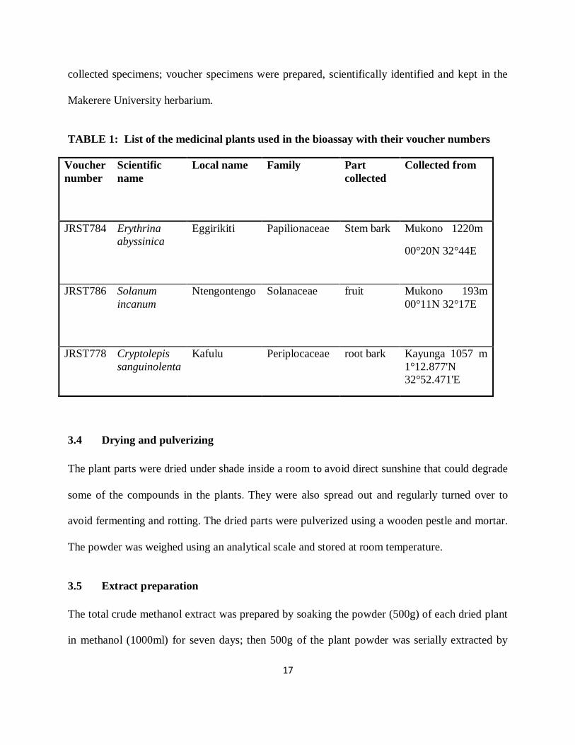
TABLE 1: List of the medicinal plants used in the bioassay with their voucher numbers
Voucher
number
Scientific
name
Local name Family Part
collected
Collected from
JRST784 Erythrina
abyssinica
Eggirikiti Papilionaceae Stem bark Mukono 1220m
00°20N 32°44E
JRST786 Solanum
incanum
Ntengontengo Solanaceae fruit Mukono 193m
00°11N 32°17E
JRST778 Cryptolepis
sanguinolenta
Kafulu Periplocaceae root bark Kayunga 1057 m
1°12.877'N
32°52.471'E
3.4 Drying and pulverizing
The plant parts were dried under shade inside a room to avoid direct sunshine that could degrade
some of the compounds in the plants. They were also spread out and regularly turned over to
avoid fermenting and rotting. The dried parts were pulverized using a wooden pestle and mortar.
The powder was weighed using an analytical scale and stored at room temperature.
3.5 Extract preparation
The total crude methanol extract was prepared by soaking the powder (500g) of each dried plant
in methanol (1000ml) for seven days; then 500g of the plant powder was serially extracted by
18
soaking in ether (1000ml), followed by chloroform (1000ml) and lastly methanol (1000ml) in the
order of increasing polarity of the solvents for three days each with occasional shaking.
Whatman’s filter paper no 1 was used for filtering to obtain the crude extract. The crude solution
was then concentrated to a minimum volume by a rotary evaporator (Büchi Labortechnik AG,
Switzerland) at 40°c and reduced pressure. The concentrated crude extracts were allowed to
evaporate to constant weight at room temperature. The drying, extraction and concentration
processes were done in the Phytochemistry laboratory in the Department of Pharmacology and
Therapeutics Makerere University College of Health Sciences.
3.6 Mycobacterial tests
These tests were done in a level three level safety laboratory at Joint Clinical Research Center
(JCRC) located in Mengo Kampala. The three preserved strains of Mycobacteria used were
obtained from JCRC. They included a rifampicin- resistant strain (TMC 331strain) as a good
indicator of MDR, a fully susceptible strain (H37Rv) as a control and Mycobacterium avium
(MA) a wild strain from a Ugandan patient to represent mycobacteria other than tuberculosis
strains (MOTT).
3.6.1 Growth media
Middle brook 7H10 agar (Becton Dickinson Company (DifcoTM
), 7 Loveton Circle, Sparks,
Maryland, USA; Lot No. 8175150) supplemented with oleic acid-albumin- catalase (OADC)
(Becton Dickinson Company Lot 8136781) was used for reviving and culturing the mycobacteria
for sensitivity testing. It was prepared by adding dehydrated medium (19.0g) to purified water
(900ml) containing glycerol (l5.0ml). The mixture was stirred well to dissolve and afterwards
19
autoclaved at 121οC for 10 minutes. Oleic acid-albumin catalase (100ml) was aseptically added
to the medium after cooling to 45οC. No adjustment for PH was made.
3.6.2 Preparation of inoculum for drug sensitivity testing
Preserved strains of mycobacteria were revived on Middle brook 7H10 agar, prior to anti
tuberculosis susceptibility testing. Cells were scraped from freshly growing colonies (three
weeks old) on Middle brook 7H10 plates and introduced into saline (10ml). Bacterial
suspensions with 0.5 McFarland standard turbidity equivalents to 108 CFU were prepared by
dilution with saline. The mixture was vortexed for 30 seconds in a glass bottle containing glass
beads and the particles allowed to settle (Parish and Stroker, 1998).
3.6.3 Bioassay protocol for susceptibility tests
The disc diffusion method was used to determine susceptibility as described by Parish and
Stroker (1998).
3.6.3.1 Preparation of the drugs/ extracts
The dried crude extracts (1 g) were each dissolved in analytical grade methanol (20ml) to give a
concentration of 50 mg/ml. .Extracts were sterilized using 0.2 m single use filters. For
rifampicin, a stock solution of 5.0 mg/ml was prepared by dissolving 0.1g in 10ml of methanol.
A stock solution of 2.5mg/ml of isoniazid was prepared by dissolving 0.1 g in 20ml of distilled
water.
20
3.6.3.2 Preparation of biodiscs
A concentration of 20 g for each of the drugs and extracts was used per disc, for the general
susceptibility tests so that for the extracts each disc contained 10mg of the extract; 0.05mg/disc
of isoniazid and 0.1mg/disc for rifampicin. The discs were left in a hood to dry for 24 hours.
3.6.3.3 Procedure
The culture medium was sterile Middle brook 7H10 agar placed in 90mm diameter Petri dishes
with quadrants. In each quadrant of the Petri dish, 5.0 ml of the medium was put. The solidified
medium in the quadrants was inoculated using a swab. A rifampicin impregnated disc was placed
in the first quadrant with a concentration of 0.1mg/ml. In the second quadrant isoniazid
impregnated disc containing 0.05mg/ml was placed. The third quadrant had an extract
impregnated disc containing 10mg. Finally the fourth quadrant contained a blank disc as a
negative control.
All the tests for the extracts and the three strains of mycobacteria were done in triplicate. The
Petri dishes were then left in the hood overnight to allow diffusion of the extracts and drug and
then sealed with a carbon dioxide-permeable tape. These were then incubated at 37°C in a carbon
dioxide incubator for four weeks. The sensitivity of M. tuberculosis and M. avium to the extracts
and the drug was determined by measuring the zones of inhibition surrounding the disc using a
millimeter scale.
21
3.6.4 Determination of the Minimum Inhibitory Concentration (MIC)
Microtitre plate method was used in the determination of MIC. Serial dilutions of the
drugs/extracts were used to determine the Minimum Inhibitory Concentration of the drug or
extract, using Middle brook 7H9 as the medium (Parish and Stroker, 1998).
3.6.4.1 Preparation of the medium (Middle brook 7H9)
The powder (4.7g) was suspended in purified water (900ml) containing glycerol (2ml) and
autoclaved at 121°C for 10 minutes. OADC enrichment (100mls) (Becton Dickinson Company,
Lot 8136781) was aseptically added to the medium when cool for enrichment. The medium was
from Becton Dickinson Microbiology Systems of Becton Dickinson Company (DifcoTM
), 7
Loveton Circle, Sparks, Maryland, USA; Lot No. 5123072.
Procedure
The procedure followed was that described by Parish and Stroker (1998) with some
modifications. Middle brook 7H9 broth (100µl) was dispensed into all the wells of a sterile 96-
well microtitre plate. In the first column, rifampicin (100 µl) at a concentration of 50mg/ml was
added using a pipettor. The drug was mixed well by sucking up and down six times using a
pipetter. 10 fold dilutions were made up to column 10 by pipetting 100 µl from column 1 and
adding it to column 2 and then taking 100 µl from column 2 into column 3. The procedure was
repeated up to column 10. From column 10, 100 µl were discarded instead of placing it into
column 11. With a pipettor 5 µl of bacteria (104-10
5CFU/ml) were dispensed in columns 1 to 11.
Plate 12 was left blank as a sterility control. The procedure was repeated for the remaining
drug/extracts in rows 2-5. The plates were incubated at 37°C for three weeks. The MIC tests for
22
the three strains of mycobacteria were done in duplicate. The lowest concentration with no
visible turbidity was taken to be the minimum inhibitory concentration.
3.7 Acute toxicity tests
Acute toxicity tests on the most active extracts were carried out on white albino mice, Mus
musculus as described by Ghosh (1984) with a few modifications. Total crude extracts of C.
sanguinolenta and E. abyssinica were worked on. A pilot study was carried out on pairs of mice
of both sexes aged four weeks, after fasting them overnight. Widely separated doses of
50,200,500, 800 and 1000 mg/kg were orally administered using a gastro intestinal tube to the
mice to determine approximate lethal and non lethal dose ranges. From the pilot study the
approximate LD50 was found to be at 800mg/kg.
The actual study involved selecting five different groups with five mice in each. These were
orally administered with 700,750,800,850 and 900mg/kg body weight as a single dose. Mice
(Mus musculus) of both sexes, for each concentration were used and they were fasted overnight
before giving them the extracts orally using a gastro intestinal tube. The control group was given
DMSO which was the solvent used to dissolve the extracts. Observations were made and
recorded after 24 hours. LD50 was then determined through plot of a graph. The tests were done
from the animal house in the Department of Pharmacology and Therapeutics.
3.8 Qualitative phytochemical testing
In the most active extracts qualitative tests for Terpenoids, Tanins, flavones and alkaloids were
carried out as described by Edeoga et al., (2005).
23
Terpenoids were tested for by adding chloroform (1ml) to the extract (1ml) and then an equal
volume of concetrated sulphuric acid was added. Formation of a bluish red coloration indicated
presence of terpenoids.
Tannins were tested for by boiling the dried powdered extract (0.5g) with water (20ml) in a test
tube. 2ml of 0.1M FeCl3 were added. Formation of a blue black coloration indicated presence of
tannins.
Flavones were tested for by adding ammonium solution (5ml) to 1ml of aqueous filtrate of the
extract followed by addition of sulphuric acid (2ml). A yellow coloration indicated presence of
flavones.
Alkaloids were tested for by mixing 50g of the powder with 250ml of 1% sulphuric acid. It was
allowed to stand and then filtered. 10mls of the filtrate was shaken and added to Meyer’s reagent.
Formation of a white precipitate indicated presence of alkaloids.
3.9 Data analysis
The numerical data from the replicated investigations is presented in form of tables and
histograms. Statistical analysis involved use of the statistics computer program, Graph pad prism
version 5.0. For toxicity studies, probit was plotted against log dose while the standard deviation
was calculated for the concetrations.
3.10 Ethical considerations
Ethical approval was sought from the Research and Ethics Committee of the Faculty of Medicine
and the Uganda National Council for Science and Technology (NS 141). Protection of the
investigators was ensured by carrying out the work in collaboration with, and under the guidance
24
of the Mycobacteriology laboratory staff at the Joint Clinical Research Centre, Mengo in
Kampala, who had the necessary expertise in handling M. tuberculosis. Additionally, the
necessary protective wears including respirators and gloves as well as safety cabinets were used,
to minimize the risk of exposure to M. tuberculosis.
Guidelines for the handling of Laboratory animals were followed. Animals were sacrificed under
general anesthesia (Ghosh, 1984).
25
CHAPTER FOUR
RESULTS
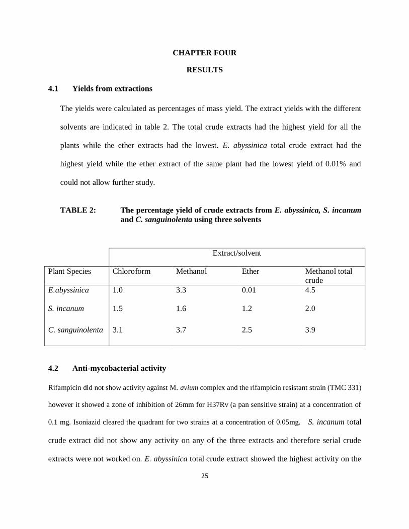
4.1 Yields from extractions
The yields were calculated as percentages of mass yield. The extract yields with the different
solvents are indicated in table 2. The total crude extracts had the highest yield for all the
plants while the ether extracts had the lowest. E. abyssinica total crude extract had the
highest yield while the ether extract of the same plant had the lowest yield of 0.01% and
could not allow further study.
TABLE 2: The percentage yield of crude extracts from E. abyssinica, S. incanum
and C. sanguinolenta using three solvents
4.2 Anti-mycobacterial activity
Rifampicin did not show activity against M. avium complex and the rifampicin resistant strain (TMC 331)
however it showed a zone of inhibition of 26mm for H37Rv (a pan sensitive strain) at a concentration of
0.1 mg. Isoniazid cleared the quadrant for two strains at a concentration of 0.05mg. S. incanum total
crude extract did not show any activity on any of the three extracts and therefore serial crude
extracts were not worked on. E. abyssinica total crude extract showed the highest activity on the
Extract/solvent
Plant Species Chloroform Methanol Ether Methanol total
crude
E.abyssinica 1.0 3.3 0.01 4.5
S. incanum 1.5 1.6 1.2 2.0
C. sanguinolenta 3.1 3.7 2.5 3.9
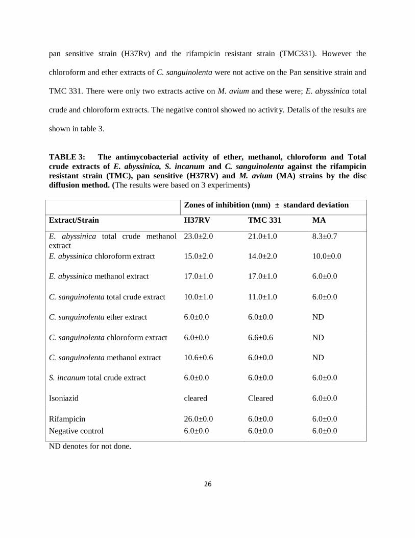
26
pan sensitive strain (H37Rv) and the rifampicin resistant strain (TMC331). However the
chloroform and ether extracts of C. sanguinolenta were not active on the Pan sensitive strain and
TMC 331. There were only two extracts active on M. avium and these were; E. abyssinica total
crude and chloroform extracts. The negative control showed no activity. Details of the results are
shown in table 3.
TABLE 3: The antimycobacterial activity of ether, methanol, chloroform and Total
crude extracts of E. abyssinica, S. incanum and C. sanguinolenta against the rifampicin
resistant strain (TMC), pan sensitive (H37RV) and M. avium (MA) strains by the disc
diffusion method. (The results were based on 3 experiments)
ND denotes for not done.
Zones of inhibition (mm) ± standard deviation
Extract/Strain H37RV TMC 331 MA
E. abyssinica total crude methanol
extract
23.0±2.0 21.0±1.0 8.3±0.7
E. abyssinica chloroform extract 15.0±2.0 14.0±2.0 10.0±0.0
E. abyssinica methanol extract 17.0±1.0 17.0±1.0 6.0±0.0
C. sanguinolenta total crude extract 10.0±1.0 11.0±1.0 6.0±0.0
C. sanguinolenta ether extract 6.0±0.0 6.0±0.0 ND
C. sanguinolenta chloroform extract 6.0±0.0 6.6±0.6 ND
C. sanguinolenta methanol extract 10.6±0.6 6.0±0.0 ND
S. incanum total crude extract 6.0±0.0 6.0±0.0 6.0±0.0
Isoniazid cleared Cleared 6.0±0.0
Rifampicin
26.0±0.0
6.0±0.0
6.0±0.0
Negative control 6.0±0.0 6.0±0.0 6.0±0.0
27
The serial extracts for C. sanguinolenta (ether, chloroform and methanol) were not tested on M.
avium because it’s total crude methanol extract was not active on the strain.
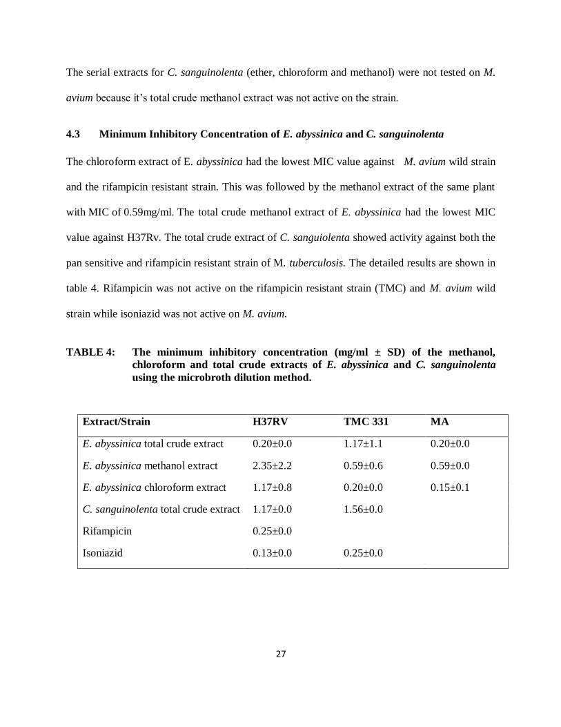
4.3 Minimum Inhibitory Concentration of E. abyssinica and C. sanguinolenta
The chloroform extract of E. abyssinica had the lowest MIC value against M. avium wild strain
and the rifampicin resistant strain. This was followed by the methanol extract of the same plant
with MIC of 0.59mg/ml. The total crude methanol extract of E. abyssinica had the lowest MIC
value against H37Rv. The total crude extract of C. sanguiolenta showed activity against both the
pan sensitive and rifampicin resistant strain of M. tuberculosis. The detailed results are shown in
table 4. Rifampicin was not active on the rifampicin resistant strain (TMC) and M. avium wild
strain while isoniazid was not active on M. avium.
TABLE 4: The minimum inhibitory concentration (mg/ml ± SD) of the methanol,
chloroform and total crude extracts of E. abyssinica and C. sanguinolenta
using the microbroth dilution method.
Extract/Strain H37RV TMC 331 MA
E. abyssinica total crude extract 0.20±0.0 1.17±1.1 0.20±0.0
E. abyssinica methanol extract 2.35±2.2 0.59±0.6 0.59±0.0
E. abyssinica chloroform extract 1.17±0.8 0.20±0.0 0.15±0.1
C. sanguinolenta total crude extract 1.17±0.0 1.56±0.0
Rifampicin 0.25±0.0
Isoniazid 0.13±0.0 0.25±0.0
28
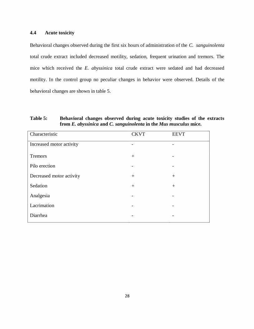
4.4 Acute toxicity
Behavioral changes observed during the first six hours of administration of the C. sanguinolenta
total crude extract included decreased motility, sedation, frequent urination and tremors. The
mice which received the E. abyssinica total crude extract were sedated and had decreased
motility. In the control group no peculiar changes in behavior were observed. Details of the
behavioral changes are shown in table 5.
Table 5: Behavioral changes observed during acute toxicity studies of the extracts
from E. abyssinica and C. sanguinolenta in the Mus musculus mice.
Characteristic CKVT EEVT
Increased motor activity - -
Tremors + -
Pilo erection - -
Decreased motor activity + +
Sedation + +
Analgesia - -
Lacrimation - -
Diarrhea - -
29
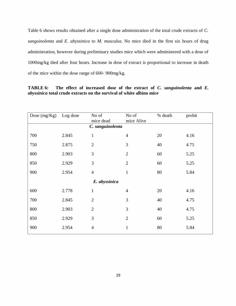
Table 6 shows results obtained after a single dose administration of the total crude extracts of C.
sanguinolenta and E. abyssinica to M. musculus. No mice died in the first six hours of drug
administration, however during preliminary studies mice which were administered with a dose of
1000mg/kg died after four hours. Increase in dose of extract is proportional to increase in death
of the mice within the dose range of 600- 900mg/kg.
TABLE 6: The effect of increased dose of the extract of C. sanguinolenta and E.
abyssinica total crude extracts on the survival of white albino mice
Dose (mg/Kg) Log dose No of
mice dead
No of
mice Alive
% death probit
C. sanguinolenta
700 2.845 1 4 20 4.16
750 2.875 2 3 40 4.75
800 2.903 3 2 60 5.25
850 2.929 3 2 60 5.25
900 2.954 4 1 80 5.84
E. abyssinica
600 2.778 1 4 20 4.16
700 2.845 2 3 40 4.75
800 2.903 2 3 40 4.75
850 2.929 3 2 60 5.25
900 2.954 4 1 80 5.84

30
From graph 1 which is a plot of the probit against log dose administered, the LD50 (Probit 5) of
the C. sanguinolenta methanol crude extract was found to correspond to a log dose of 2.88 which
is 758.6 mg/kg body weight.
2.7 2.8 2.9 3.0 3.1 3.23.5
4.0
4.5
5.0
5.5
6.0
6.5
probit
Graph 1 A plot of probit agnaist log dose for C. sanguinolenta
log dose
From graph 2 which is a plot of probit against log dose administered, the LD50 of E. abyssinica
crude extract was found to correspond to a log dose of 2.89 which is 776.2mg/kg body weight.
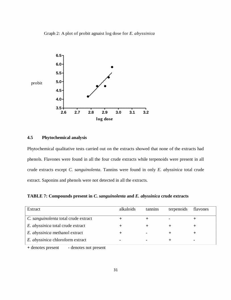
31
2.6 2.7 2.8 2.9 3.0 3.1 3.23.5
4.0
4.5
5.0
5.5
6.0
6.5
probit
Graph 2: A plot of probit agnaist log dose for E. abyssinica
log dose
4.5 Phytochemical analysis
Phytochemical qualitative tests carried out on the extracts showed that none of the extracts had
phenols. Flavones were found in all the four crude extracts while terpenoids were present in all
crude extracts except C. sanguinolenta. Tannins were found in only E. abyssinica total crude
extract. Saponins and phenols were not detected in all the extracts.
TABLE 7: Compounds present in C. sanguinolenta and E. abyssinica crude extracts
+ denotes present - denotes not present
Extract alkaloids tannins terpenoids flavones
C. sanguinolenta total crude extract + + - +
E. abyssinica total crude extract + + + +
E. abyssinica methanol extract + - + +
E. abyssinica chloroform extract - - + -
32
CHAPTER FIVE
DISCUSSION
5.1 Anti-mycobacterial activity
The study showed that extracts from two of the three plants had anti-mycobacterial activity. The
activity was found in total crude, chloroform and methanol extracts of E. abyssinica and C.
sanguinolenta. All the extracts were active against the rifampicin resistant strain of M.
tuberculosis. The methanol and chloroform extracts of E. abyssinica were actually more active
on the rifampicin resistant strain than the Pan sensitive strain of M. tuberculosis. This is
important because rifampicin resistance is a good indication of multi drug resistant TB and so the
extracts may also be active on MDR TB.
The potency of the extracts against the pan sensitive strain as compared to the standard drugs
used was low. This may be attributed to the fact that isoniazid and rifampicin were in a pure state
as compared to the extract. The wild strain of M. avium that was isolated from a Ugandan patient
seemed to be resistant to both isoniazid and rifampicin and so the impure plant extracts possess
an advantage over the two pure drugs.
Previous studies have reported several plants with anti-mycobacterial activity (Waffo et al.,
2000; Newton et al., 2000; Pauli et al., 2005; Gautam et al., 2007; Kirimuhuzya et al., 2009).
Studies had mostly reported activity in the plant families of Asteraceae, Lamiaceae, Fabaceae
and Apiaceae among others (Gautam et al., 2007). The plants in this study that showed activity
belong to the families of Fabaceae and Periplocaceae. Periplocaceae is one of the families that
have least been researched on for anti-mycobacterial activity (Gautam et al., 2007; Pauli et al.,
2005).
33
The findings in this study report a lower activity compared to above mentioned studies. This may
attributed to the fact that different in-vitro methods are used and also because in some of the
studies the pure compounds were tested.
Previous studies done on the anti-mycobacterial activity of the renowned anti-malarial climbing
liana C. sanguinolenta by Gibbons et al., (2003) are in agreement with the findings of this study
though activity was on fast growing non virulent mycobacteria. This is therefore a confirmation
that the plant extract besides being active on the fast growing mycobacteria strains, is also active
on the slow growing virulent strains of mycobacteria which were used in this study. Their study
was on an alkaloid (cryptolepine) isolated from the plant. This explains why the minimum
inhibitory concentrations of C. sanguinolenta in this study are much lower compared to that
reported by Gibbons et al., (2003).
The results of anti-mycobacterial activity for E. abyssinica are comparable to findings by Waffo
et al., (2000) on E. indica, a species from the same genus. In their study Indicanine B, an
isoflavonoid from E. indica showed anti-mycobacterial activity against M. smegmatis with an
MIC value of 18.5µg/ml. This is a higher activity as compared to the MIC value for E.
abyssinica. This could be because the pure compound was tested against M. smegmatis, a rapidly
growing a virulent saprophytic mycobacterium. This is however the first study to report activity
of E. abyssinica on different mycobacteria. Further studies are needed to isolate and identify the
anti-mycobacterial compounds in E. abyssinica.
34
5.2 Acute toxicity
In this study the total crude extracts from E. abyssinica and C. sanguinolenta affected behavior
of white albino mice. The plant extracts had LD50s within the safe range according to Gosh
(1984). Behavioral changes that were made included sedation and decreased motor activity for
both the extracts at doses higher than 500mg/kg body weight. The observations could have been
CNS-related rather than enzymatic due to the decreased motility. C. sanguinolenta treated mice
showed signs of tremors and frequent urination at very high doses (1000mg/kg). This suggests
that the plant could have some diuretic effects however more research is needed to confirm this.
Acute toxicity studies on C. sanguinolenta in this study were contrary to what was done by
Ansha et al., (2009) where the LD50 was found to be about 3000mg/kg. The difference in the
values could be attributed to the fact that they used rats which are bigger animals compared to
the mice used in this study. Further more water extracts were tested in their study while the
methanol extract was used in this study and lastly tragacanth was used as the solvent during their
study while DimethylSulphoxide was the solvent used in this study.
However they agree that there was prolonged sleep in the animals and also that there could be
some CNS disturbances. A study done by Ansha et al., (2008) concluded that C. sanguinolenta
could synergize with hypno-sedatives or other CNS depressants and therefore caution needs to
be taken in the concomitant administration of the plant with other CNS depressants.
Ansha et al., (2009) also agrees that the extract is safe for use at doses less than 500mg/kg. In
vitro studies have reported cytotoxicity at the molecular level. However this may not be reflected
in vivo (Ansha and Gooderham, 2002).
35
This is the first study that has been done concerning E. abyssinica toxicity. However in a study
that was done on E. americana by Garín-Aguilar et al., (2000), similar observations were made.
5.3 Phytochemical testing
Phytochemical tests of the four extracts that were active on mycobacteria showed presence of
alkaloids, terpenoids, tannins and flavones. The total crude extract of E. abyssinica contained all
the compounds while the methanol extract contained alkaloids, terpenoids and flavones. This
could be the possible explanation for the high activity of the E. abyssinica extracts on
mycobacteria.
The chloroform extract of E. abyssinica contained only terpenoids which could explain why the
extract was the most active on M. avium and the Rifampicin resistant strain. According to Copp
(2003), secondary metabolites of terpenoids origin lead the number of natural products with
reported anti-mycobacterial activity due to their lipophilic nature and therefore ability to
penetrate the mycobacterial cell wall.
The genus Erythrina has long been known to contain a vast array of alkaloids (Copp 2003). E.
abyssinica has been found to contain flavonones by Yenesew et al., (2004). This is in agreement
with the findings of this study. However on the contrary, saponins were found to be present in
the plant in a study done by Kareru et al., (2008). The difference could be attributed to the
different methods of preparation of the extracts and methods used in the detection of the
compounds.
C. sanguinolenta total crude extract contained tannins, alkaloids and flavones. Presence of
alkaloids in C. sanguinolenta is in agreement with a study done by Gibbons et al., (2003) in

36
which an alkaloid cryptolepine was identified and isolated. Though saponins and flavones have
been identified in C. sanguinolenta total crude extract, the compound which could have caused
the anti-mycobacterial activity could have been the cryptolepine alkaloid but more studies are
needed to confirm this. On the contrary, tannins and flavonoids were not identified in the root
bark of C. sanguinolenta in a study done by Tona et al., (1998). This may be attributed to the
different methods of preparation of the crude extracts.
The group of compounds that could have caused sedation and decreased motility in the mice for
both extracts could have been alkaloids as portrayed in a study done by Dos Santos et al., (2005).
In their study done on rats, an alkaloid that was identified as isoquercitrin increased sleeping in
the barbiturate- and diazepam-induced sleeping time. In another study done by Garín-Aguilar et
al., (2000) on E. americana, alkaloid fractions were tested on rats and there was decreased
aggressive behavior.
5.4 Limitations
One species of rodents was used in acute toxicity studies however the results may not be true for
other species. Additionally, acute toxicity measures lethality within 24 hours and there is no
information on the long term effects of the plants.
There were also problems associated with dissolving the total crude extracts because they
contain both the polar and non polar compounds and this is precisely why DMSO had to be used
as the solvent in acute toxicity tests. The yield for the ether extract of E. abyssinica was so small
that it could not be worked on.
Water extract was not used because it is cumbersome to deal with.
37
5.5 Conclusions
The results of this study have further shown that there is potential to develop new compounds
against multi drug resistant TB from Erythrina abyssinica and Cryptolepis sanguinolenta. This
therefore verifies their use in the treatment of tuberculosis by traditional practitioners and further
emphasizes that there is a strong positive correlation between the anti-mycobacterial activity
results and traditional knowledge on plants used for TB and TB-related diseases in
ethnomedicine.
The plant extracts were found to be relatively safe for use as far as lethality is concerned
however more studies on the toxicity of the plants are needed before declaring them completely
safe for use in humans.
The plant extracts contained alkaloids, tannins, terpenoids and flavones.
5.6 Recommendations
The active constituents of the plants should be isolated, identified and characterised. These
should then be tested on the resistant strains of mycobacteria. Further research on the toxicity of
E. abyssinica should be ventured into since this was practically the most active of the plants.
C. sanguinolenta and E. abyssinica should be conserved by communities where they are found.
Efforts should also be made to cultivate the two plants species.
38
REFERENCES
Adeniyi, B.A., Groves, M.J. and Gangadharam, P.R.J. (2004). In-vitro antimycobacterial activity
of three species of Cola plant extracts. Phytotherapy research, 18, 414-418
Al-Fatimi, M., Wurster, M., Schr¨oder, G. and Lindequist, U. (2007). Antioxidant, antimicrobial
and cytotoxic activities of selected medicinal plants from Yemen. Journal of
Ethnopharmacology, 111,657–666
American Thoracic Society, Center for Disease Control, and Infectious Diseases Society of
America. (2003). Treatment of Tuberculosis. Morbidity and Mortality Weekly Report, 52, 1-77
Ansha C. and Gooderham, N.J. (2002). The popular herbal antimalarial extract Cryptoleps
sanguinolenta is potentially cytotoxic. Toxicological sciences, 70, 245- 251
Ansha, C., Mfoafo, E.A., Woode, E., Opoku-Okrah, C. and Owiredu, W.K.B.A. (2008).
Toxicological evaluation of the anti-malarial herb Cryptolepis sanguinolenta in rodents. Journal
of pharmacology and toxicology, 5, 335-343
Ansha, C., Otsyina, R., Duwiejua, M., Woode, E., Aboagye, F.A. and Aning, K.G. (2009). Toxicological assessment of Cryptolepis sanguinolenta for possible use in veterinary medicine.
Journal of Veterinary Medicine and Animal Health, 1, 011-016
Balunas, M.J. and Kinghorn, A.D. (2005). Drug discovery from medicinal plants. Life science
Journal, 78, 431-441
Bruton, L.L., Lazo, J.S. and Parker, K.L. (2007).The Pharmacological basis of therapeutics (ed
11th)Mc Graw-Hill medical publishing division New York
Bryrne, S.T., Denkin, S.M., Peihua G., Nuermberger, E. and Zhang, Y. (2007). Activity of
ketoconazole against Mycobacterium tuberculosis in-vitro and in the mouse model. Journal of
Medical Microbiolology. 56, 1047-1051
Bumpi, J. and Kayondo, K. H. (2009). The impact of land use change on biodiversity loss in
Uganda. IOP Conference Series: Earth and Environmental Science, 6, 312-322
Centre for Disease Control(CDC) (2005). Worldwide emergence of Mycobacterium tuberculosis
with extensive resistance to second-line drugs. Morbidity and Mortality Weekly Report, 55,
250-253
Centre for Disease Control (CDC) (2006). Extensively drug resistant tuberculosis. Morbidity
and Mortality Weekly Report, 56, 250-253
Chan, E.D. and Iseman, M.D. (2002). Current medical treatment for Tuberculosis. British
Medical Journal, 325, 1282-1286

39
Chaisson, R.E. and Martinson, N.A. (2008). Tuberculosis in Africa-combating an HIV-driven
crisis. New English Journal of Medicine, 358, 1089–1092
Chin, Y.W., Balunas, M.J., Chai, H.B and Kinghorn, A.D. (2006). Drug discovery from natural
sources. AAPS Journal, 8, E239-E253.
Copp, B. R. (2003). Antimycobacterial natural products. Natural products report, 20, 535-557
Corbett, E.L., Watt, C.J., Walker, N., Maher D., Williams, B.G., Raviglione, M.C. and Dye, C.
(2003). The growing burden of tuberculosis. Global trends and interactions with the HIV
Epidemic. Archies of internal medicine, 163, 1009-10021
Connell, N.D. and Nikaido, H. (1994). Membrane permeability and transport. American society
of microbiology, 333-352
Cowan, M.M. (1999). Plant products as antimicrobial agents. Clinical Microbiology reviews, 12,
564-582
Dos Santos Jr, J.G., Blanco, M.M., Do Monte, F.H.M., Russi, M. and Lanziotti, V.M.N.B.
(2005). Sedative and anticonvulsant effects of hydroalcoholic extract of Equisetum arvense.
Fitoterapia, 76, 508-513.
Edeoga, H.O., Okwu, D.E. and Mbabie, B.O. (2005). Phytochemical constituents of some
Nigerian medicinal plants. African Journal of Biotechnology 4, 685-688
Fattorini, L. Migliori, G.B. and Antonio, C. (2007). Extensively drug-resistant (XDR)
tuberculosis: an old and new threat. Annali dell'Istituto superiore di sanità, 43, 317-319
Franzblau, S.G., Witzig, R.S., McLaughlin, J.C., Torres, P., Madico, G., Hernandez, A., Degnan,
M.T., Cook, M.B., Quenzer, V.K., Ferguson, R.M. and Gilman, R.H. (1998). Rapid, low-
technology MIC determination with clinical Mycobacterium tuberculosis isolates by using the
microplate alamar blue assay. Journal of Clinical Microbiology, 36, 362– 366
Frappier, F., Trigalo, F., Bodo, B., Pousset, J., Grellier, P., Ramiaramanana, L., Millerioux , V.,
Deharo , E. and Schrével, J. (1995). Antimalarial activity of Cryptolepine and Isocryptolepine,
Alkaloids Isolated from Cryptolepis sanguinolenta. phytotherapy research, 10, 317-321
Furin, J.J. (2007). The clinical management of drug-resistant tuberculosis. Current opinion in
pulmonary medicine, 13, 212–217
Garín-Aguilar, M.E., Luna, J.E.R., Soto-Hernández, M., Toro, G.V. and Vázquez, M.M. (2000).
Effect of crude extracts of Erythrina americana on aggressive behavior in rats. Journal of
Ethnopharmacology, 69, 189-196
Gautam, R., Saklani, A. and Jachak, S.M. (2007). Indian medicinal plants as a source of
antimycobacterial agents. Journal of Ethnopharmacology, 110, 200-234
40
Ghosh, M.N. (1984). Fundamentals of Experimental Pharmacology. 2nd Edition 1-11; 153-
190.Scientific Book Agency, Calcutta.
Gibbons, S., Fallah, F. and Wright, C.W. (2003). Cryptolepine Hydrochloride: A potent
antimycobacterial alkaloid derived from Cryptolepis sanguinolenta. Phytotherapy research, 17,
434-436
Hamill, F.A., Apio, S., Mubiru, N.K., Bukenya-Ziraba, R., Mosango, M., Maganyi,O.W and
Soejarto, D.D.(2003). Traditional herbal drugs of southern Uganda 11: Literature analysis and
antimicrobial assays. Journal of Ethno pharmacology, 84, 57-78.
Hamza, O.J.M., Bout-van den, V.B., Mecky, I.N., Matee, M.J., Frans H.M., Selemani, O.,
Mbwambo, Z. H., Van der Ven, A.J. and Verweij P.E. (2006). Antifungal activity of some
Tanzanian plants used traditionally for the treatment of fungal infections. Journal of
Ethnopharmacology 108,124-132
Jasmer, R.M., Seaman, C.B., Gonzalez, L.C., Kawamura L.M., Osmond, D.H. and Daley, C.L.
(2004). Tuberculosis treatment outcomes: Directly Observed Therapy compared with Self-
Administered Therapy. American Journal of Respiratory and Critical Care Medicine, 170, 561-
566.
Kareru, P.G., Keriko, J.M., Gachanja, A.N. and Kenji, G.M. (2008). Direct detection of
triterpenoid saponins in medicinal plants. African Journal of Traditional, Complementary and
Alternative Medicines, 5, 56-60.
Kirimuhuzya, C., Waako, P., Joloba, M. and Odyek, O. (2009). The anti-mycobacterial activity
of Lantana camara a plant traditionally used to treat symptoms of tuberculosis in south western
Uganda. African Health Sciences, 9, 40-45.
Lawn, S.D. and Wilkinson, R. (2006). Extensively Drug Resistant tuberculosis. British Medical
Journal, 55, 301-305.
Madison, B. (2001). Application of stains in clinical microbiology. Biotechnical Histochemistry,
76, 119–125.
Mageresi, S.M., Pieters, L., Ngassapa, O.D., Apers, S., Vingerhoets, R., Cos, P., Burgher, D.A
and Vlietinck, A.J. (2008) Screening of some Tanzanian medicinal plants from Bunda district for
antibacterial, antifungal and antiviral activities. Journal of Ethno Pharmacology, 119, 58-66
Mbaya, V.B. and Muhammed, S.I. (1976). Antibiotic action of Solanum incanum. Antimicrobial
agents Chemotherapy, 9, 920-924.
Nachega, J. B. and Chaisson, R. E. (2003). Tuberculosis drug resistance: A global threat. Clinical
infectious diseases, 36, s24-s30
41
Nahid, P., Madhukar, P. and Hopewell, P.C. (2006). Advances in the diagnosis and treatment of
tuberculosis. American Thoracic Society, 3,103-110
Newton, S.M., Lau, C. and Wright, C.W. (2000). A review of antimycobacterial natural
products. Phytotherapy Research, 14, 303-322
Okemo, P., Orodho, J. A., Tabuti, J. B., Otieno, N., Magadula, J.J and Kirimuhuzya C.
(unpublished). Ethnobotany, bioassay, and active compounds isolation of medicinal plants used
for the treatment of tuberculosis in Lake Victoria basin; the socio-economic and cultural
dimensions of improvement of management of tuberculosis in lake victoria region. Report on the
first phase of the vicres project:
Parish, T. and Stroker, N.G. (1998). Mycobacteria Protocols: Methods in molecular Biology.
(vol 101) (eds,) Humana Press, Totowa, NJ.
Pauli, G.F., Case, R.J., Inuli, T., Wang, Y., Cho, S., Fischer, N.H. and Franzblau, S.G. (2005).
New perspectives on natural products in TB drug research. Life sciences, 78, 485-494.
Peloquin, C.A., Nitta, A.T., Burman, W.J., Brudney K.F. and Miranda-Massari, J.R. (1996).Low
antituberculosis drug concentrations in patients with AIDS. Pharmacotherapy, 30, 919-925.
Sanjay, M. J. (2004). An important source for anti-tubercular drugs. Natural Products, 5, (1)
Shu, Y.Z. (1998). Recent natural products based drug development: A Pharmaceutical Industry
perspective. Journal of Natural products, 61, 1053-1071
Sensi, P. and Grassi, G.G.(2003). Chemotherapeutic agents. Medicinal chemistry and drug
discovery, 5, 821-824
Silva, O., Duarte, A., Cabrita, J., Pimentel, M., Diniz, A. and Gomes, E. (1996). Antimicrobial
activity of Guinea-Bissau traditional remedies. Journal of Ethnopharmacology, 50, 55-59
Suffredini, I.B., Sander, H.S., Goncalves, A.G., Reis, A.O., Gales, A.C., Varella, A.D. and
Younes, R.N. (2004). Screening of antibacterial extracts from plants native to Brazilian Amazon
Rain Forest and Atlantic forest. Brazilian journal of Medical and Biological research, 37,379-384
Tona, L., Kambu, K., Ngimbi, N., Cimanga,
K. and Vlietinck, A. J. ( 1998). Antiamoebic and
phytochemical screening of some Congolese medicinal plants. Journal of Ethnopharmacology,
61, 57-61.
Tribuddharat, C. and Fennewald, M. (1999). Integron-mediated rifampin resistance in
Pseudomonas aeruginosa. Antimicrobial Agents Chemotherapy, 43, 960-962
Tripathi, R.P., Neetu, T., Dwivedi, N. and Tiwari, V.K. (2004). Fighting tuberculosis: An old
disease with new challenges. Medicinal Research Reviews, 25, 93-131.
42
Waffo, A. K., Azebaze, G. A., Nkengfack, A. E., Fomum, Z. T., Meyer, M., Bodo, B. and
Heerden, F.R., (2000). Indicanines B and C, two isoflavonoids derivatives from the root bark of
Erythrina indica .Phytochemistry 53,981-985.
World Health Organisation report. (2004). Global tuberculosis control - surveillance, planning
and financing. WHO Geneva
World Health Organisation report. (2007). Global tuberculosis control - surveillance, planning
and financing. WHO Geneva.
World Health Organisation Report. (2008). Anti tuberculosis drug resistance in the World. WHO
Geneva
Yenesew, A., Induli, M., Derese, S., Midiwo, J., Heydenreich, M., Peter, M. G., Akala, H.,
Wangui, J., Liyala, P. and Waters, N.C. (2004). Anti-plasmodial flavonoids from the stem bark
of Erythrina abyssinica. Phytochemistry, 65, 3029–3032
Zignol, M., Hosseini, M.S., Wright, A., Lambregts, C., Weezenbeek, V., Nunn, P., Watt, C.J.,
Williams, B.G. and Dye, C. (2006). Global incidence of multidrug-resistant tuberculosis. Journal
of Infectious Diseases, 194, 479–485