antimicrobial research: novel bioknowledge and · pdf fileare easy to characterize and have...
TRANSCRIPT

Characteristics of antibacterial peptides produced by bacteria
Y. M. Álvarez Cisneros1, F. J Fernández1 and E. Ponce Alquicira1 1 Departamento de Biotecnología, Universidad Autónoma Metropolitana-Iztapalapa. Av. San Rafael Atlixco Nº 186,
Colonia Vicentina, Mexico, D.F., 09340
Antimicrobial peptides (AMP) are defined as small amphipathic molecules capable of interacting with the cell membranes of sensitive cells. Bacterial AMP are classified into two groups: those that are synthesized enzymatically (nonribosomal peptide synthetases, or NRPS) and those that are synthesized in ribosomes (ribosomal peptide synthetases, RPS). NRPS are produced as secondary metabolites (e.g. the antibiotics bacitracin and vancomycin) and are synthesized by one or more peptide synthetases. On the other hand, RPS produced by bacteria are called bacteriocins, which are defined as thermostable bioactive peptides with helicoidal structure, with bacteriostatic or bactericidal extracellular effects. RPS can be produced by Gram-positive (nisin, pediocin, enterocin, subtilin, coagulin, thuricin 17, entomycin 9, cerein, etc.) and Gram-negative (microcins, vibriocins, klebicins etc.) bacteria. This chapter will review aspects of stability, classification, synthesis, regulation, classification, mechanisms of action and applications of the antibacterial peptides, both NRPS and RPS, produced by bacteria.
Keywords: antibacterial peptides; antibiotic; bacteria; bacteriocins; NRPS; RPS.
1. Introduction
Food-transmitted diseases and the high percentage of hospital-reported bacterial infections are considered by the World Health Organization as an international health problem [1], primarily since the microorganisms arise from the inadequate use of antibiotics and food preservation methods, creating favorable conditions for the appearance, propagation and persistence of resistant microorganisms. One option for fighting this problem is the use of antibacterial peptides (AMP) as substitutes for antibiotics or natural preservatives, as they have been shown to have high activity against Gram positive and negative microorganisms [2], fungi, viruses and parasites [3]. AMP are defined as small, positively charged amphipathic molecules (possessing hydrophobic and hydrophilic regions) with a molecular weight <10 kDa, a varying composition of amino acids (from six to 100 amino acids) and capable of interacting with the plasma membranes of sensitive cells. They are produced by bacteria, archaea and eukaryotes (plants, birds, fish, insects, amphibians and mammals) as a defense mechanism [2, 4-5]. Within this extensive group of antimicrobial peptides, those produced by bacteria are of great interest because they are easy to characterize and have many applications in the food and pharmaceutical industries. AMP produced by bacteria are classified into two groups: those that are synthesized enzymatically (nonribosomal peptide synthetases, or NRPS) and those that are synthesized in ribosomes (ribosomal peptide synthetases, RPS) [6]. Some bacteria have been reported, such as Bacillus sp., to have the capacity to produce peptides by both pathways [2, 5].
2. Enzymatically synthesized peptides
Enzymatically synthesized peptides, or nonribosomal peptides, are produced as secondary metabolites and are synthesized by one or more nonribosomal peptide synthetases; the genes necessary for the production of a given peptide are usually organized into operons. NRPS genes have a dimeric or trimeric structure of identical, cyclical and/or branched sequences with N-methyl and N-formyl groups, or are glycosylated, acetylated, halogenated or hydroxylated [7-9]. Nonribosomal peptides are a highly diverse family of natural products with an extremely wide range of biological and pharmacological activities: they can be used as antibiotics, siderophores (iron chelators), cytostatics (drugs used in chemotherapy), immunosuppressants and pigments. NRPS with antimicrobial activity against Gram positive bacteria are the antibiotics bacitracin, tyrocidine, gramicidin and vancomycin, whose mechanism of action is to inhibit the synthesis of the bacterial cell wall, disrupting the transpeptidase function [9]. NRPS are formed by different structures, which distinguish them from RPS, such as nonprotein amino acids (ornithine, dihydroxyphenylglycine, 4-methyl-L-threonine); variable structures (macrocyclic, branched macrocyclic, structurally identical dimers and trimers); may contain heterocyclic rings (thiazoline, oxazoline or thiazole, for example); in addition to the aforementioned incorporation of N-methyl and N-formyl groups, glycosylation, or the insertion of acetate, propionate and sometimes fatty acid units [9].
Antimicrobial research: Novel bioknowledge and educational programs (A. Méndez-Vilas, Ed.)
35
_____________________________________________________________________________

2.1 Nonribosomal peptide synthesis
Nonribosomal peptides are synthesized by multifunctional modular enzymes known as nonribosomal peptide synthetases (NRPS), each one of these complexes grouped into operons (which are formed by genes coding for one or more modules). Each module is responsible for the incorporation of a single amino acid in the polypeptide structure, and since the incorporated amino acid requires a specific module, the nonribosomal peptide synthetases are large enzymes formed by one or more modules. Figure 1 shows the grouping of three operons for the synthesis of bacitracin: each operon is transcribed and translated to form three nonribosomal synthetases, and each of the enzymatic complexes has a different molecular weight, since it contains a different number of modules (M). In addition, each module comprises three main domains: adenylation (A), thiol (T) or peptide-carrying protein (PCP) and condensation (C), which work together to form peptides with the successive addition of amino acids in each module [8-10]. Figure 1 shows the distribution of different operons that code for the synthesis of bacitracin.
Figure 1. Organization of the genetic determinants implicated in the synthesis of the decapeptide bacitracin. a) Bacitracin is encoded in three different sized operons (bacA, bacB and bacC); b) Following transcription and translation, a total of twelve modules are obtained, distributed in three NRPS (BacA, BacB and BacC), used to synthesize the nonribosomal peptide. Adapted from Schwarzer et al. [8] Once the NRPS genes are transcribed and translated, an NRPS system with the inactive T-domain (apo-NRPS) is obtained, which must undergo a series of steps (activation, initiation, elongation, termination and modifications) to obtain the active peptide. Figure 2 shows an example of a three-step biosynthetic pathway of a nonribosomal peptide: 1) NRPS activation (priming) via transference of the thiol (-SH) group from CoA to the T-domain mediated by a phosphopantetheinyl transferase (PPTase), which produces a holo-NRPS capable of synthesizing a nonribosomal peptide; 2) The amino acids are activated in the A-domain through a reaction with ATP to form the aminoacyl-AMP intermediate, which is captured by the thiol group of the T-domain. 3 and 4) The first step in a NRPS is known as the initiation module and can be typically subdivided into an A-domain and a T-domain. There follows a series of elongation modules that also contain A and T-domains but which have a condensation (C) domain at the beginning of each module. The function of the C-domain is to catalyze the formation of successive peptide bonds between the first amino acid (initiation) and the adjacent amino acids (elongation) to form the peptidyl-thioester intermediate anchored to the adjoining T-domain. Each module incorporates a single amino acid, so as many modules are required as there are amino acids in the final peptide product; 5) The final module contains an additional thioesterase (TE) domain that catalyzes hydrolysis or cyclization to release the peptide from the NRPS. The enzymatic complex can be regenerated to begin a new cycle of synthesis; 6) The synthesized nonribosomal peptide may have post-synthesis modifications in additional domains in the synthesis modules, such as: epimerization (E-domain), N-methylation (NMT-domain), cyclization (Cy-domain), oxidation (Ox-domain) and enzymatic modification (glycosyltransferases, halogens and hydroxylases) [8-9]. The indiscriminate use of these compounds has caused the appearance of resistant microorganisms, including methicillin-resistant Staphylococcus aureus and nosocomial strains of vancomycin-resistant Enterococcus sp. [11]. Furthermore, their spectrum of bacterial inhibition is limited, since Gram negative bacteria are intrinsically resistant to these compounds. The current focus of attention, therefore, is on antimicrobial peptides of ribosomal origin, with broader antimicrobial spectra, a wider range of applications and less possibility of producing resistance in sensitive microorganisms [2, 12].
b)
Antimicrobial research: Novel bioknowledge and educational programs (A. Méndez-Vilas, Ed.)
36
_____________________________________________________________________________

Figure 2. Biosynthetic pathway of nonribosomal peptides. Adapted from Schwarzer et al. [8]; Winn et al. [9]
3. Ribosomally synthesized peptides
Antimicrobial peptides synthesized in ribosomes and produced by bacteria are called bacteriocins [2, 5, 13-16]. Bacteriocins are defined as simple or complex thermostable bioactive peptides of helical structure which exert extracellular bacteriostatic or bactericidal effects, inhibiting the taxonomic growth of nearby bacteria. In addition, they are active at wide pH values, water-soluble and impart neither flavor nor aroma [4, 17]. Nevertheless, some studies have shown the existence of bacteriocins that prevent the proliferation of other Gram positive bacteria, not so closely related taxonomically with the producing strain, and also some species of Gram negative bacteria [18]. Given their protein origin, bacteriocins are inactivated, at least, by a proteolytic enzyme of pancreatic (trypsin and alpha-chymotrypsin) and gastric (pepsin) origin, a characteristic that makes these bacterial metabolites safe substances for use as food biopreservatives (since they would be inactivated during their passage through the gastrointestinal tract). Additionally, other bacteriocins have been found that are sensitive to non-proteolytic enzymes, such as leconocin S, which is inactivated by an amylase [19]. Another characteristic of these compounds is their thermal stability at different temperatures, as used in milk pasteurization (63°C, 30 min). Thermal stability may be due to the formation of small globular structures and the presence of strongly hydrophobic regions and the formation of stable cross-links [20]. Bacteriocins are classified into two major groups, depending on whether they are produced by Gram-positive or Gram-negative bacteria [2, 21], and have great potential to be used in the food industry as natural preservatives for the control of pathogenic and deteriorating microorganisms [13, 17, 22-23]; in addition, some authors have recently proposed them as good substitutes for antibiotics [5, 12, 15, 24]. Antimicrobial peptides produced by Gram-positive bacteria differ from those synthesized by Gram-negative bacteria in ecological and evolutionary aspects: the biosynthesis of bacteriocins of Gram-positive bacteria is self-regulating and is a non-lethal event, has a broader spectrum of antimicrobial activity, a regulated mechanism of peptide release, gene clusters are generally in the genome and include genes that code for structural proteins and proteins responsible for post-translational modifications, regulation, immunity and transportation across the membrane. In Gram-negative bacteria, however, bacteriocin production is a lethal event, peptide release is controlled by regulating mechanisms (such as an SOS regulator) and proteins responsible for cellular lysis, encoded in the bacteriocin operon [2].
(holo-NRPS)
Antimicrobial research: Novel bioknowledge and educational programs (A. Méndez-Vilas, Ed.)
37
_____________________________________________________________________________

3.1 Bacteriocins produced by Gram-negative bacteria
3.1.1 Enterobacteria
The first bacteriocin discovered was colicin V, produced by Escherichia coli and originally described as “principle V” (now known as microcin V). Ribosomally synthesized antimicrobial compounds produced by this group of bacteria are classified according to their molecular weight as follows: i) colicins, proteins with a weight exceeding 10 kDa, and ii) microcins, peptides with a molecular weight less than 10 kDa [2, 6, 13, 21]. In this chapter, only the microcins will be described. Microcins are defined as bacteriocins produced by enterobacteria, mainly by Escherichia coli and Klebsiella pneumoniae [6], and share some characteristics with the peptides produced by Gram-positive bacteria, such as thermal stability, resistance to protease treatment, high hydrophobicity and resistance to extreme pH conditions [21]. Microcins are classified into two groups based on weight, presence of disulfide bridges and post-translational modification status [6, 15, 25, 26]. Class I comprises microcins with molecular weights below 5 kDa, generally with post-translational modifications. Class II microcins have a molecular weight between 5 and 10 kDa, do not have post-translational modifications and exert their antimicrobial action through the disruption of the membrane potential. Class II microcins are, in turn, divided into two classes: class IIa, require three different genes to synthesize and assemble a functional peptide, while class IIb are linear peptides with or without post-translational modifications at the carbonyl terminal and chromosome-coded. Table 1 shows the classification and mechanisms of action of bacteriocins produced by Gram-negative bacteria. Table 1. Classification of microcins produced by Gram-negative bacteria [15- 21].
Microcins have a diversity of mechanisms of action, including pore formation, DNase and RNase activity, and the inhibition of protein synthesis and DNA replication. The spectrum of antimicrobial activity they exhibit includes microorganisms from the Enterobacteriaceae family, such as Escherichia, Salmonella, Shigella, Citrobacter, Klebsiella and Enterobacter [6, 13]. Figure 3 shows the different mechanisms of actions of microcins. Class I microcins can kill target cells by interfering with DNA, RNA and protein synthesis, using porins and iron-siderophore receptors as well as peptide transporters in the inner membrane. For example, B17 passes through the outer membrane via a porin (OmpF) and across the inner membrane via a peptide transporter immersed in the membrane (SbmA). Once in the cytoplasm, the microcin inhibits DNA gyrase, thereby interfering with DNA replication. The microcin MJ25 is recognized by the iron siderophore receptor FhuA on the outer membrane and requires the TonB and SbmA transporters in the inner membrane to enter the cell, where it inhibits transcription by blocking RNA polymerase. Another case is that of C7/C51, which uses the OmpF
Classification Characteristics Microcins Molecular weight (Da)
Producing strain
Mechanism of action
Class I
Low molecular weight peptides (< 5 kDa), post-translationally
modified
B17 3094 E. coli Inhibition of DNA gyrase
C7/C51 1177 E. coli Inhibition of Asp-tRNA
synthase
D93 <1000 E. coli Inhibition of DNA gyrase
J25 2107 E. coli Inhibition of RNA polymerase
Class II Larger (5–10 kDa) peptides, with or without post-translational modifications
Class IIa
Required more than one genes to synthesize
and assemble functional peptides
L 8884 E. coli Membrane potential disruption
V 8741 E. coli Membrane potential disruption
N/24 7274 E. coli Pore formation and membrane potential
disruption
Class IIb
Linear peptides with post-translational
modifications or not at C-terminal
E-492 7886 K. pneumoniae Pore-forming
M 7284 E. coli Pore-forming
H47 4865 E. coli Pore-forming
Antimicrobial research: Novel bioknowledge and educational programs (A. Méndez-Vilas, Ed.)
38
_____________________________________________________________________________

porin and the YejABEF transporter to enter the cell where it is processed by a cytoplasmic aminopeptidase to form a modified aspartyl-adenylate which inhibits aspartyl-tRNA synthetase, thus blocking protein synthesis. On the other hand, class II microcins must first be recognized by iron-siderophore receptors (FepA, CirA or Fiu), which enable them to cross the outer membrane and form pores on the inner membrane [2, 6, 24].
Figure 3. Mechanism of action of class I and class II microcins. GlcNac=N-acetylglucosamine; MurNac= N-acetylmuramic acid.
Adapted from Gillor et al. [21]; Cotter et al. [24] The majority of microcins are plasmid-encoded peptides and the genes responsible for their biosynthesis are grouped into operons that include: (i) a structural gene, encoding the peptide precursor; (ii) an auto-immune gene, that protects the producing strain against its own antimicrobial peptide; (iii) genes encoding the microcin export system, and (iv) several genes coding for auxiliary proteins or modification enzymes [15, 26]. All microcins (with the exception of microcin C) have a leader peptide, which is eliminated at a specific cleavage site (usually a double glycine or glycine-alanine) and which is recognized by the Type 1 ABC (ATP-binding cassette) transport proteins. To date, the roles of leader peptides have not been fully clarified, but they may contribute to maturation, secretion, protection of the precursor against degradation, or serve to keep the precursor inactive inside the host cells during biosynthesis [6].
3.1.2 Other genera of Gram-negative bacteria
Although the majority of antimicrobial peptides of Gram-negative bacteria are produced by strains of Escherichia coli, some authors have focused their lines of research on other bacteria, finding bacteriocins produced by Pseudomonas, such as Psvp10 with a molecular mass of 2.6 kDa, stable to heat, to proteases and pH, and which exhibit extensive antimicrobial activity against Enterococcus faecalis, Salmonella Typhimurium, Shigella flexneri, Streptococcus mutans and Streptococcus sobrinus [27, 28, 29]. Other notable bacteriocins are: vibriocins, from the genus Vibrio; klebicins and pneumocins, from Klebsiella pneumoniae; enterocoliticin, from Yersinia enterocolitica; BC1 and BC2, from Vibrio vulnificus; IW1, from Vibrio cholera; and BLS, from Aeromonas hydrophila [2].
3.2 Bacteriocins of Lactic Acid Bacteria
Lactic acid bacteria (LAB) are Gram-positive bacteria used in products that have been consumed by man for centuries. These bacteria play an important role in the dairy product industry; they are used in the preparation and preservation of fermented products, being hygienically and toxicologically safe. Often, some types of LAB produce desirable textures and flavors and inhibit the development of pathogenic bacteria and contaminants. These bacteria are used in the dairy industry as starter or probiotic cultures. The genera that comprise this group of bacteria are: Lactobacillus, Lactococcus, Leuconostoc, Carnobacterium, Pediococcus, Enterococcus and Bifidobacterium. LAB bacteriocins are the most promising for developing applications [30], being categorized as GRAS (Generally Recognized as Safe); the majority of these bacteriocins have shown some resistance to heat, being able to tolerate treatments equivalent to pasteurization. Resistance to temperature, however, depends on the purification state of the bacteriocin, decreasing in purified and partially purified bacteriocins [31]. Nisin is the only bacteriocin approved for commercial use as a food preserver by the WHO, the European Union and the US Food and Drug Administration [12] and as an antimicrobial to treat bovine mastitis [15]. This bacteriocin is
Antimicrobial research: Novel bioknowledge and educational programs (A. Méndez-Vilas, Ed.)
39
_____________________________________________________________________________
produced by strains of Lactococcus lactis subsp. lactis, and reaches maximum solubility and stability at pH 2.0, decreasing as pH increases and becoming irreversibly inactive at pH 7.0 [20, 32].
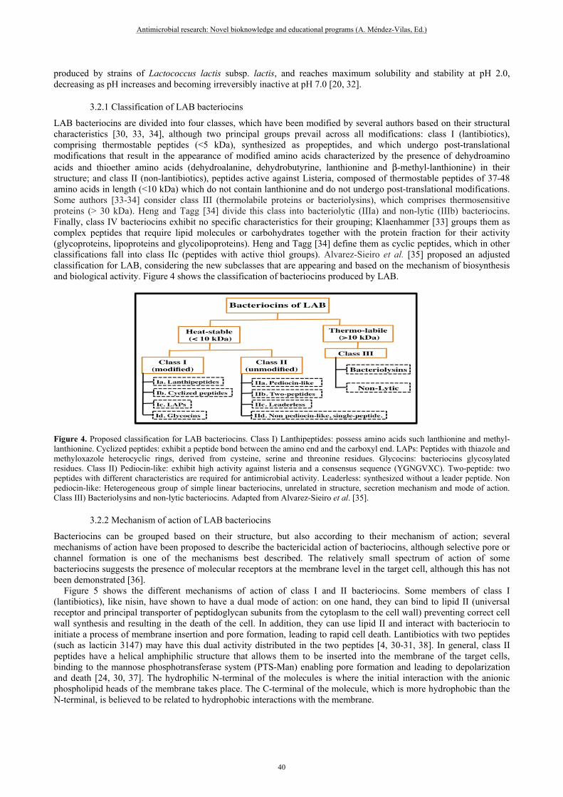
3.2.1 Classification of LAB bacteriocins
LAB bacteriocins are divided into four classes, which have been modified by several authors based on their structural characteristics [30, 33, 34], although two principal groups prevail across all modifications: class I (lantibiotics), comprising thermostable peptides (<5 kDa), synthesized as propeptides, and which undergo post-translational modifications that result in the appearance of modified amino acids characterized by the presence of dehydroamino acids and thioether amino acids (dehydroalanine, dehydrobutyrine, lanthionine and β-methyl-lanthionine) in their structure; and class II (non-lantibiotics), peptides active against Listeria, composed of thermostable peptides of 37-48 amino acids in length (<10 kDa) which do not contain lanthionine and do not undergo post-translational modifications. Some authors [33-34] consider class III (thermolabile proteins or bacteriolysins), which comprises thermosensitive proteins (> 30 kDa). Heng and Tagg [34] divide this class into bacteriolytic (IIIa) and non-lytic (IIIb) bacteriocins. Finally, class IV bacteriocins exhibit no specific characteristics for their grouping; Klaenhammer [33] groups them as complex peptides that require lipid molecules or carbohydrates together with the protein fraction for their activity (glycoproteins, lipoproteins and glycolipoproteins). Heng and Tagg [34] define them as cyclic peptides, which in other classifications fall into class IIc (peptides with active thiol groups). Alvarez-Sieiro et al. [35] proposed an adjusted classification for LAB, considering the new subclasses that are appearing and based on the mechanism of biosynthesis and biological activity. Figure 4 shows the classification of bacteriocins produced by LAB.
Figure 4. Proposed classification for LAB bacteriocins. Class I) Lanthipeptides: possess amino acids such lanthionine and methyl-lanthionine. Cyclized peptides: exhibit a peptide bond between the amino end and the carboxyl end. LAPs: Peptides with thiazole and methyloxazole heterocyclic rings, derived from cysteine, serine and threonine residues. Glycocins: bacteriocins glycosylated residues. Class II) Pediocin-like: exhibit high activity against listeria and a consensus sequence (YGNGVXC). Two-peptide: two peptides with different characteristics are required for antimicrobial activity. Leaderless: synthesized without a leader peptide. Non pediocin-like: Heterogeneous group of simple linear bacteriocins, unrelated in structure, secretion mechanism and mode of action. Class III) Bacteriolysins and non-lytic bacteriocins. Adapted from Alvarez-Sieiro et al. [35].
3.2.2 Mechanism of action of LAB bacteriocins
Bacteriocins can be grouped based on their structure, but also according to their mechanism of action; several mechanisms of action have been proposed to describe the bactericidal action of bacteriocins, although selective pore or channel formation is one of the mechanisms best described. The relatively small spectrum of action of some bacteriocins suggests the presence of molecular receptors at the membrane level in the target cell, although this has not been demonstrated [36]. Figure 5 shows the different mechanisms of action of class I and II bacteriocins. Some members of class I (lantibiotics), like nisin, have shown to have a dual mode of action: on one hand, they can bind to lipid II (universal receptor and principal transporter of peptidoglycan subunits from the cytoplasm to the cell wall) preventing correct cell wall synthesis and resulting in the death of the cell. In addition, they can use lipid II and interact with bacteriocin to initiate a process of membrane insertion and pore formation, leading to rapid cell death. Lantibiotics with two peptides (such as lacticin 3147) may have this dual activity distributed in the two peptides [4, 30-31, 38]. In general, class II peptides have a helical amphiphilic structure that allows them to be inserted into the membrane of the target cells, binding to the mannose phosphotransferase system (PTS-Man) enabling pore formation and leading to depolarization and death [24, 30, 37]. The hydrophilic N-terminal of the molecules is where the initial interaction with the anionic phospholipid heads of the membrane takes place. The C-terminal of the molecule, which is more hydrophobic than the N-terminal, is believed to be related to hydrophobic interactions with the membrane.
Antimicrobial research: Novel bioknowledge and educational programs (A. Méndez-Vilas, Ed.)
40
_____________________________________________________________________________

Bacteriocin classes I and II may share similar mechanisms of action. Apparently, peptides bind to the cytoplasmic membrane through electrostatic bonds with negatively charged phospholipids, then are inserted into the membrane with a potential-dependent reorientation (which is influenced by pH and phospholipid composition). Bacteriocin monomers form pore-forming protein aggregates, with consequent ion efflux (mainly potassium and magnesium), loss of proton-motive force, and leakage of ATP and amino acids. The proton-motive force plays a central role in ATP synthesis, in active transport and bacterial movement, thus its loss inhibits the synthesis of macromolecules (DNA, RNA, proteins) and energy production, resulting in cell death [19, 24, 30].
Figure 5. Mechanism of action of class I and II bacteriocins produced by lactic acid bacteria. GlcNac=N-acetylglucosamine; MurNac= N-acetylmuramic acid.
Adapted from: Cotter et al. [24]; Alvarez-Cisneros et al. [38]. LAB bacteriocins are characterized by having different inhibition spectra, although they are produced by the same genus, thus they are classified into three groups accordingly. The first includes bacteriocins with a narrow inhibitory spectrum, capable of inhibiting the growth of bacteria belonging to the same genus (diplococci, lactocin 27, lacticin B or helveticin J); The second is composed of bacteriocins with an intermediate inhibitory spectrum; they also inhibit other genera of LAB and other Gram-positive bacteria (lactacin F, lacticin 481, plantaricin C and plantaricins S and T); the third contains bacteriocins with a broad inhibitory spectrum, which act against a large number of Gram-positive bacteria, including foodborne pathogens, such as nisin A and Z, pediocin PA-1/AcH, leucocin S and enterocin L50 [20, 30, 38].
3.2.3 Biosynthesis of LAB bacteriocins
Bacteriocin synthesis is generally produced when bacteria are under stress, and depends on the ecosystem, pH, oxidation-reduction potential, amount of nutrients, growth phase, temperature and available oxygen [32]. Due to their protein nature, bacteriocins are synthesized in processes that involve transcription and translation. Genes coding for the bacteriocin are usually organized into groups of operons located in the chromosome or in plasmids. [38]. The lantibiotic operon (nisin) consists of 11 chromosomally localized genes (nisABTCIPRKFEG), which encode proteins involved in the post-translational modification of nisin: the nisA gene encodes the protein responsible for prepeptide synthesis; the nisB and nisC genes produce the proteins that carry out post-translational modifications; the nisP gene encodes an extracellular protease to separate the leader peptide from the mature nisin; the nisT gene is an ABC translocator that secretes the prenisin molecule to the outside of the bacteria and encodes the proteins associated with prepeptide immunity and secondary transport; the nisI, nisF, nisE, nisG genes encode proteins associated with immunity and secondary transport and, finally, nirR and nisK genes encode proteins responsible for regulation and expression [35, 38-39]. So far, genetically characterized class IIa bacteriocins comprise one or four genes in an operon; pediocin PA-1, for example, contains the four genes necessary for bacteriocin production (papA precursor of Ped A-1 and papB immunity peptide) and exportation (papC membrane-binding protein for secretion and papD ABC transporter) in the same operon [40]. Other bacteriocins in the same class have genes distributed through several operons, where each operon has at least two genes. Enterocin A, for example, needs seven genes to be synthesized (entAIFKRTD), all of which are organized into three operons: structural (entA-structural gene of the prebacteriocin and entI-immunity gene), regulatory (entF-induction gene, entKR-regulatory genes) and transport (entTD-genes encoding the ABC secretion system of the bacteriocin [35, 38, 41].
3.2.4 Regulation of LAB bacteriocin synthesis
Antimicrobial research: Novel bioknowledge and educational programs (A. Méndez-Vilas, Ed.)
41
_____________________________________________________________________________

The regulation of bacteriocin synthesis is mediated by two signal transduction systems consisting of two or three components. Factors that activate these systems include the presence of other competing bacteria, temperature or pH stress, and a quorum sensing mechanism [42]. Examples of regulation mediated by two-component systems include numerous lantibiotics. For example, nisin acts as an inducer of its own synthesis through a two-component system consisting of nisR and nisK (which translate the signal by a phosphorylation/dephosphorylation mechanism). Transcription of the nisin genes is led by three promoters preceding nisA, nisR and nisF. While transcription of nisRK is constitutive, the nisA and nisF promoters are induced proportionally to the concentration of nisin in the medium. Once the nisin precursors are processed and modified to obtain the mature peptide, this is exported to the cell exterior as a prepeptide (nisin bound to the leader peptide), and finally, on the outside of the bacteria, the nisin is separated from the leader peptide by proteolysis and the active product is obtained [42-44]. This nisin molecule may act as an inducer of more molecules or stop production, depending on the concentration of nisin in the environment where the producing strain is found. In these systems, therefore, bacteriocins have a dual function, since they possess antimicrobial activity and act as a signal molecule inducing their own synthesis [45-46]. Figure 6 shows the mechanism of synthesis and regulation of nisin.
Figure 6. Synthesis and regulation of nisin by quorum sensing. Adapted from Chatterjee et al. [42].Cheigh and Pyun [43]; Mierau and Kleerebezem [44].
An example of the three-component quorum sensing system is the regulation of the synthesis of enterocin A produced by Enterococcus faecium, which includes a Histidine Protein Kinase (HPK), located in the cytoplasmic membrane, which receives and transduces extracellular signals; a cytoplasmic response regulator (RR), which mediates an adaptive response that is generally a change in gene expression, and an induction factor (IF), the presence of which is detected by the HPK protein [38, 41]. As discussed earlier, the majority of bacteriocins are synthesized as a biologically inactive prepeptide carrying an N-terminal leader peptide. Furthermore, the induction prepeptide is synthesized, also undergoing maturation and secretion processes. Once secreted, it interacts with a membrane receptor that contains a histidine kinase domain which phosphorylates to a cytoplasmic response regulator protein; this, in turn, activates the transcription of the operons involved in the bacteriocin synthesis [38, 41]. Other examples of this kind of regulation include several members of class II, such as sakacin P and sakacin A produced by Lactobacillus sakei. Figure 7 shows the synthesis and regulation mechanism of enterocin.
Figure 7. Schematic presentation of the biosynthesis of enterocin A. (1) Proteins involved in the synthesis of enterocin A (EntA); (2) the immunity protein (EntI) protects cell of Ent A; (3) processing, transport and secretion of EntA and inducer peptide (EntF) through the ABC transporter (EntT) and its accessory protein (EntD); (4) regulation of synthesis of EntA by a regulatory system of three components: an induction factor (EntF), the histidine kinase sensor protein (EntK) and the regulatory protein response (EntR) the presence of EntF in the extracellular medium results in phosphorylation of EntK, which transfers its phosphate group to EntR to activate the transcription of operons involved in the synthesis of EntA [38].
Antimicrobial research: Novel bioknowledge and educational programs (A. Méndez-Vilas, Ed.)
42
_____________________________________________________________________________

3.3 Bacillus bacteriocins
The genus Bacillus is capable of producing a large quantity of antimicrobial peptides (APs), which have been characterized, many of them having different applications [47]. Among these APs are glycopeptides, lipopeptides, cyclic peptides and bacteriocins [5, 48]. Bacteriocins produced by Bacillus sp. exhibit a broader antimicrobial spectrum than the APs produced by the majority of lactic acid bacteria [49]. Some examples of these are subtilin from Bacillus subtilis; coagulin from Bacillus coagulans; bacthuricin F4, thuricin 17, entomycin 9 and tochicin from Bacillus thuringiensis; cerein 7 from Bacillus cereus, among others. In addition, this type of bacteriocin has a greater potential for use as a substitute for antibiotics, for example, mersacidin (1.8 kDa) is a tetracyclic peptide that belongs to the lantibiotics group and exhibits bactericidal activity against methicillin-resistant Staphylococcus aureus (comparable to the effect of vancomycin or teicoplanin), without developing resistance [5]. Different species of Bacillus produce bacteriocins with different mechanisms of action, for example: tochicin, lichenin, thuricin 439 and thuricin exhibit bactericidal effects. Meanwhile, cerein 8A has several mechanisms of action including the formation of vesicles in protoplasm, pore formation and the complete disintegration of cells [50]. Abriouel et al. [47] classified Bacillus bacteriocins into three categories: class I (peptides with post-translational modifications), class II (non-modified peptides), and class III (large proteins). Subtilin is a class I bacteriocin, but its characteristic structure could put it into a unique class of bacteriocins, since it is a cyclic anionic peptide of 25 amino acids with a unique post-translational structure that includes three cross-linked sulfide bonds between its cysteine residues, the α-carbons of two phenylalanines and one threonine residue [5]. Regarding the biosynthesis of these bacteriocins, the gene transcription and translation mechanism is similar to class I and II of LAB antimicrobial peptides. Arias et al. [51] characterized a bacteriocin of the group of lantibiotics produced by Bacillus amyloliquefaciens GA, called amylolysin, which is encoded in an operon that contains a structural gene (am/A), genes responsible for modifications (am/M), transport (am/T), regulation (am/KR) and immunity (am/FE) It can be said that bacteriocins produced by the genus Bacillus are ideal therapeutic tools, due to their ample specificity and their activity against several pathogens; however, more studies are needed to establish whether their use is safe.
4. Conclusion
The study of NRPS and RPS antimicrobial peptides is of great interest, whether for the pharmaceutical or food industry, due to their different applications as preservatives or antibiotics. Although ribosomal peptides produced by LAB are on the rise due to their mechanisms of action, low resistance and GRAS category, bacteriocins produced by the genus Bacillus are increasingly studied because of their properties as antibiotics and broad spectrum preservatives. Finally, knowing the characteristics of antimicrobial peptides produced by bacteria enables the proposal of purification methods and possible industrial applications.
References [1] Organización Mundial de la Salud (OMS). Fact Sheet: Antibiotic resistance; 2015 [cited 2017 Jan 25]. Available from:
http://www.who.int/mediacentre/factsheets/antibiotic-resistance/en/. [2] Paiva, E. A. y Breukink, E. Antimicrobial Peptides Produced by Microorganisms. In: Hiemstra, P.S. y Zaat, S.A.J, editors.
Antimicrobial Peptides and Innate Immunity, Springer Basel, 2013. p.53-95. [3] Bahar, A. A. y Ren, D. Review: Antimicrobial Peptides, Pharmaceuticals, Frontiers in Microbiology. 2013; 6:1543-1575. [4] Tonarelli, G. y Simonetta, A. Péptidos antimicrobianos de organismos procariotas y eucariotas como agentes terapéuticos y
conservantes de alimentos, Revista FABICIB. 2013; 17: 137-177. [5] Sumi, C. D., Yang, B. W., Yeo, I. y Tae, H. Antimicrobial peptides of the genus Bacillus: a new era for antibiotics. Canadian
Journal of Microbiology, 2015; 61: 93-103. [6] Arnison, P. G., Bibb, M. J., Bierbaum, G., Bowers, A. A., Bugni, T. S., Bulaj, G., et al. Ribosomally synthesized and post-
translationally modified peptide natural products: overview and recommendations for a universal nomenclature. Natural Product Reports, 2013; 30: 108–160.
[7] Hancock, R.E.W. y Chapple, D.S. Peptide Antibiotics. Antimicrobial Agents and Chemotherapy, 1999; 43: 1317–1323. [8] Schwarzer, D., Finking. R. y Mohamed A. Nonribosomal peptides: from genes to products, Natural Product Reports, 2003; 2:
275–287. [9] Winn, M., Fyans, J. K., Zhuo, Y., y Micklefield, J. Recent advances in engineering nonribosomal peptide assembly lines. Natural
product reports, (2016); 33(2): 317-347. [10] Kries, H., Niquille, D. L., y Hilvert, D. A subdomain swap strategy for reengineering nonribosomal peptides. Chemistry &
biology, 2015; 22(5): 640-648. [11] Yoshida, Y., Matsuo, M., Oogai, Y., Kato, F., Nakamura, N., Sugai, M. Y Komatsuzawa, H. Bacitracin sensing and resistance in
Staphylococcus aureus. FEMS Microbiology Letters, 2011; 320: 33-3. [12] Sang, Y. y Blecha, F. Antimicrobial peptides and bacteriocins: alternatives to traditional antibiotics. Animal Health
Research Reviews, 2008; 9: 227–235.
Antimicrobial research: Novel bioknowledge and educational programs (A. Méndez-Vilas, Ed.)
43
_____________________________________________________________________________

[13] Papagianni, M. Ribosomally synthesized peptides with antimicrobial properties: biosynthesis, structure, function,and applications. Biotechnology Advances, 2003; 21: 465–499.
[14] Flaherty, R.A., Freed, S.D. y Shaun W.L. The Wide World of Ribosomally Encoded Bacterial Peptides. PLOS Pathogens, 2014; 10: e100422
[15] Yang, S.C, Lin, C.H, Sung, C.T. y Fang, J.Y. Antibacterial activities of bacteriocins: application in foods and pharmaceuticals. Frontiers in Microbiology, 2014; 5: 241.
[16] Subramanian, S. y Smith, D.L. Bacteriocins from the rhizosphere microbiome from an agriculture perspective. Frontiers in Plant Science, 2015; 6: 909.
[17] Prudêncio, C. V., Dos Santos, M.T. y Dantas, M.C. Strategies for the use of bacteriocins in Gram-negative bacteria: relevance in food microbiology. Journal of Food Science Technology, 2015; 52: 5408–5417.
[18] Okereke, A. y Montville, T.J. Bacteriocin inhibition of Clostridium botulinum spores by lactic acid bacteria. Journal of Food Protection, 1991; 55: 349–353.
[19] Vázquez, M.S.M., Suárez, M.H., Zapata, B.S. Utilización de sustancias antimicrobianas producidas por bacterias ácido lácticas en la conservación de la carne. Revista Chilena de Nutrición, 2009; 36: 64-71.
[20] Chen, H. y Hoover, D.G. Bacteriocins and their food applications. Comprehensive Reviews in Food Science and Food Technology, 2003; 2: 82-99.
[21] Gillor, O., Kirkup, B.C., Riley, M.A. Colicins and microcins: the next generation antimicrobials. Advances in Applied Microbiology, 2004; 54: 129-146.
[22] Dobson, A., Cotter, P.D., Paul Ross, R., y Hill, C. Bacteriocin Production: a Probiotic Trait? Applied and Environmental Microbiology, 2012; 78: 1-6.
[23] Casaburi, A., Di Martino, V., Ferranti, P., Picariello, L. y Villani, F. Technological properties and bacteriocins production by Lactobacillus curvatus 54M16 and its use as starter culture for fermented sausage manufacture. Food Control, 2016; 59: 31-45.
[24] Cotter, P.D., Paul Ross, R., y Hill, C. Bacteriocins: a viable alternative to antibiotics? Nature Reviews Microbiology, 2013; 11: 95-105.
[25] Pons, A. M., Lanneluc, I., Cottenceau, G., y Sable, S. New developments in non post-translationally modified microcins. Biochimie, 2002; 84: 531–537.
[26] Duquesne, S., Petit, V., Peduzzi, J. y Rebuffat, S. Structural and functional diversity of microcins, gene-encoded antibacterialpeptides from enterobacteria. Journal of Molecular Microbiology and Biotechnology, 2007; 13: 200-209.
[27] Hubert, Lobos, Brevis y Padilla. Purification and characterization of the bacteriocin PsVP-10 produced by Pseudomonas sp. Journal of Applied Microbiology, 1998; 84: 910–913.
[28] Padilla. C., Lobos, O., Brevis, P., Abaca, P. y Hubert, E. Effects of the bacteriocin PsVP-10 produced by Pseudomonas sp. on sensitive bacterial strains. Revista Latinoamericana de Microbiología, 2002; 44: 19-23.
[29] Padilla, C., Lobos, O., Hubert, E., Poblete, F., Navarro, A. y Nuñez, L. In vitro antibacterial activity of the peptide PsVP-10 against Streptococcus mutans and Streptococcus sobrinus with and without glycocalyx. International Journal of Antimicrobial Agents, 2006; 27: 212-216.
[30] Cotter, P.D., Hill, C. y Ross, R. P. Bacteriocins: developing innate immunity for food. Nature Microbiology Reviews, 2005; 3: 777-788.
[31] Perez, R.H., Zendo, T. y Sonomoto, K. Novel bacteriocins from lactic acid bacteria (LAB): various structures and applications. Microbial Cell Factories, 2014; 13: S3.
[32] Cheftel, C. (High-pressure, microbial inactivation and food preservation. Food Science and Technology International, 1995; 1: 75-90.
[33] Klaenhammer, T. R.. Genetics of bacteriocins produced by lactic acid bacteria. FEMS Microbiology Reviews, 1993; 12: 39-86. [34] Heng, N.C.K. y Tagg, J.R. Class distinction for bacteriocins. Nature Reviews Microbiology, 2006; 4: 1-2. [35] Alvarez-Sieiro, P., Montalbán-López, M., Mu, D., y Kuipers, O. P. Bacteriocins of lactic acid bacteria: extending the
family. Applied microbiology and biotechnology, 2016; 100: 2939-2951. [36] Van Belkum, M.J. y Stiles, M.E. Non lantibiotics antibacterial peptides from lactic acid bacteria. Natural Product Reports,
2000; 17: 323–335. [37] Cintas L. M., Casaus, P., Fernandez, M.F., Hernandez, P. E. Comparative antimicrobial activity of enterocin L50, pediocin PA-
1, nisin A and lactocin S against spoilage and foodborne pathogenic bacteria. Food Microbiology, 2000; 15: 289–298. [38]Álvarez-Cisneros, Y.M., Sáinz Espuñes, T.R., Wacher, C., Fernandez, F.J. y Ponce-Alquicira, E. Enterocins: Bacteriocins with
applications in the food industry. Chapter in: Science against microbial pathogens: communicating current research and technological advances. Editores A. Mendez Vilas, Editorial Formatex Research Center 2, 2011; 1330-1341.
[39] Xie L., Wilfred A. y Van Der Donk. Post-translational during lantibiotic biosynthesis. Chemistry and Biology, 2004; 8: 498-507. [40] Papagianni, M. y Anastasiadou, S. Pediocins: The bacteriocins of Pediococci Sources, production, properties and applications.
Microbial Cell Factories, 2009; 8: 3. [41]Djamel, D., Fimland, G., Hechard, Y., McMullen, L.M. y Prévost, H. The continuing story of Class IIa bacteriocins.
Microbiology Molecular Biology Reviews, 2006; 70: 564–582. [42] Chatterjee C., Paul M., Xie L., van der Donk W. (). Biosynthesis and mode of action of lantibiotics. Chemical Reviews, 2005;
105: 633-683. [43] Cheigh, C. I., y Pyun, Y. R. Nisin biosynthesis and its properties. Biotechnology letters, 2005; 27,:1641-1648. [44] Mierau, I., y Kleerebezem, M. 10 years of the nisin-controlled gene expression system (NICE) in Lactococcus lactis. Applied
microbiology and biotechnology, 2005; 68: 705-717. [45] Hanan, T., Hilmi, A., Nikkila, K.K, Runar R. y Saris, P.E. Nisin induction without nisin secretion. Microbiology, 2006; 152:
1489–1496. [46] López, J. E., Ochoa Z. A., Anaya, L. J. L., Martínez, T.M., y Medina, M.E. Bacteriocinas de bacterias Gram positivas: una
fuente potencial de nuevos tratamientos biomédicos. Revista Mexicana de ciencias farmacéuticas, 2008; 39: 49-57.
Antimicrobial research: Novel bioknowledge and educational programs (A. Méndez-Vilas, Ed.)
44
_____________________________________________________________________________

[47] Abriouel, H., Franz, C.M., Omar, N.B. y Gálvez, A. Diversity and applications of Bacillus bacteriocins. FEMS Microbiology Reviews, 2011; 35: 201–232.
[48] Xie, J., Zhang, R., Shang, C., y Guo, Y. Isolation and characterization of a bacteriocin produced by an isolated Bacillus subtilis LFB112 that exhibits antimicrobial activity against domestic animal pathogens. African Journal of Biotechnology, 2009; 8: 5611–5619.
[49] Wang, G., Manns, D.C., Churey, J.J., y Worobo, R.W. () Development of a homologous expression system development and the systematic site- directed mutagenesis analysis of thurincin H, a bacteriocin produced by Bacillus thuringiensis SF361. Applied and Environmental Microbiology, 2014; 80: 3576–3584.
[50] Bizani, D., Motta, A.S., Morrissy, J.A., Terra, R.M., Souto, A.A., y Brandeii, A. Antibacterial activity of cerein 8A, a bacteriocin-like peptide produced by Bacillus cereus. International Microbiology, 2005; 8: 125–131.
[51] Arias, A.A., Ongena, M., Devreese, B., Terrak, M., Joris, B., y Fickers, P. Characterization of amylolysin, a novel lantibiotic from Bacillus amyloliquefaciens GA1. PLoS One, 2013; 8: e83037.
Antimicrobial research: Novel bioknowledge and educational programs (A. Méndez-Vilas, Ed.)
45
_____________________________________________________________________________