antigen translocation machineries in adaptive immunity · pdf fileantigen translocation...
TRANSCRIPT

Review
Peter U. Mayer
0022-2836/© 2014 Elsevi
Antigen Translocation Machineries inAdaptive Immunity and ViralImmune Evasion
hofer1 and Robert Tampé
1, 21 - Institute of Biochemistry, Biocenter, Goethe University Frankfurt, Max-von-Laue-Strasse 9, 60438 Frankfurt am Main, Germany2 - Cluster of Excellence Macromolecular Complexes, Goethe University Frankfurt, Max-von-Laue-Strasse 9, 60438 Frankfurt amMain, Germany
Correspondence to Robert Tampé: Institute ofBiochemistry, Biocenter,GoetheUniversity Frankfurt,Max-von-Laue-Strasse9, 60438 Frankfurt am Main, Germany. [email protected]://dx.doi.org/10.1016/j.jmb.2014.09.006Edited by D. F. Jacob
Abstract
Protein homeostasis results in a steady supply of peptides, which are further degraded to fuel proteinsynthesis or metabolic needs of the cell. In higher vertebrates, a small fraction of the resulting peptidome,however, is translocated into the endoplasmic reticulum by the transporter associated with antigen processing(TAP). Antigenic peptides are guided to major histocompatibility complex class I (MHC I) molecules and arefinally displayed on the cell surface, where they mount an adaptive immune response against viral infected ormalignantly transformed cells. Here, we review the structural organization and the molecular mechanism of thisspecialized antigen translocon.Wediscusshow theATP-binding cassette (ABC) transporter TAPcommunicatesand cooperates within the multi-component peptide loading machinery, mediating the proper assembly andediting of kinetically stable peptide/MHC I complexes. In light of its important role within the MHC I antigenprocessing pathway, TAP is a prime target for viral immune evasion strategies, and we summarize how thisantigen translocation machinery is sabotaged by viral factors. Finally, we compare TAP with other ABC systemsthat facilitate peptide translocation.
© 2014 Elsevier Ltd. All rights reserved.
Introduction
The proteome of eukaryotic cells is dynamicallyshaped by a fine-tuned balance between the de novosynthesis of proteins and their degradation. Theturnover of intracellular proteins is a specific andhighly regulated process, in which most proteins aredegraded via two pathways, the autophagy-lysosomesystem and the ubiquitin-proteasome system [1]. Theresulting peptides are short-lived intermediates be-cause they are immediately processed by a variety ofcytosolic peptidases that degrade such peptideswithin seconds [2]. Hence, these peptides serve asa steady supply of amino acids, which are used eitheras an energy source during starvation or for de novosynthesis of proteins, thereby closing the cycle.Besides serving as fuel for cellular processes, small
peptides have acquired a broad range of sophisticat-ed functions during evolution. For instance, plant
er Ltd. All rights reserved.
peptides are involved in the defense against infectionby pathogens and in the regulation of growth anddevelopment [3]. Moreover, various peptides areutilized as signaling molecules. The yeast matingpheromones a-factor and α-factor are small signalingpeptides, which are subjected to multiple rounds ofposttranslational modification and proteolytic cleav-age prior to their secretion [4]. Host defense peptides,also known as antimicrobial peptides, which arefound in all phyla of life, were originally described toexhibit antimicrobial activity and also have immunemodulatory properties including anti-infective, anti-inflammatory and wound-healing activities [5,6].Moreover, small peptidesplaya key role in the adaptiveimmune system of jawed vertebrates, where they arepresented on the cell surface for clonal recognitionand expansion of lymphocytes or for the recognitionand subsequent elimination of infected or malignantlytransformed cells.
J. Mol. Biol. (2015) 427, 1102–1118

1103Review: Antigen Translocation Machineries
Elaborate translocation machineries regulate thecorrect compartmentalization of bioactive peptides,thereby mediating peptide transport from their site ofproduction to the particular compartment, where theyfulfill their physiological functions (Fig. 1). Manypeptides, for example, the yeast α-factor matingpheromone [4], are sorted via the “classical” secretorypathway and released into the external milieu. Inaddition, certain bioactive peptides require special-ized translocation machineries for their proper trans-port. Here, we summarize the current view on howpeptides that serve as antigens in adaptive immunityare transported across the endoplasmic reticulum(ER) membrane via a specialized and unique class oftranslocation machineries, the ATP-binding cassette(ABC) transport systems.
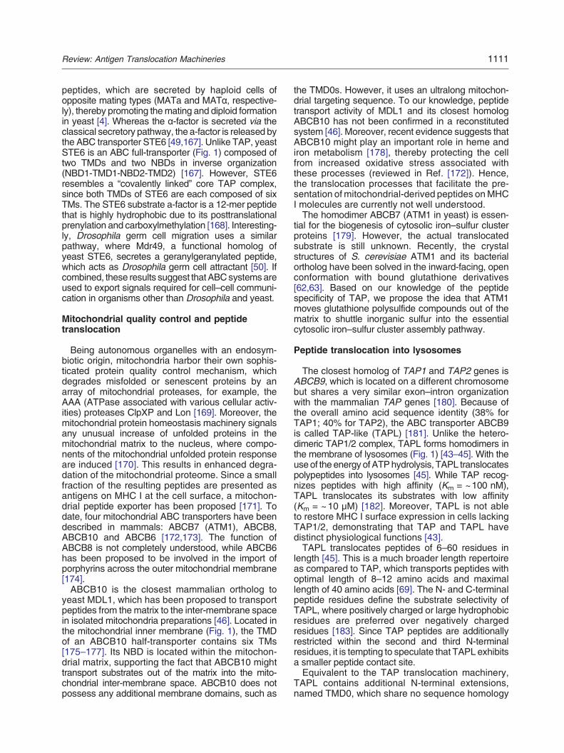
Fig. 1. Intracellular peptide trafficking and antigen processinacross cell membranes, for example, the plasma membraneand the ER. Peptides derived from the ubiquitin-proteasometranslocated by TAP1/2 into the ER lumen, where they are trimmolecules. De novo synthesized MHC I initially assembles wPLC, composed of TAP1/2, tapasin, ERp57, calreticulin, β2m acomponents catalyzes peptide loading onto MHC I. Stable pepvia the Golgi network to the plasma membrane, where thtranslocated into lysosomes by the homodimeric TAPL compyeast homolog of ABCB10, is proposed to be involved in the excontrol mechanisms into the inter-membrane space. The ABsecretes prenylated peptides for cell–cell communication.
Scanning Debris—The Role of Peptides inthe MHC I Antigen Presentation Pathway
Organismsare under continuousassault by viruses,bacteria, fungi or parasites. Hence, mutual survivalrelies on a fine-tuned balance between pathogenreplication and the clearance of pathogens by the hostimmune system. In addition, malignantly transformedcells attack the body from within. Cancer or infectedcells are eliminated by CD8+ cytotoxic T lymphocytes(CTLs) that recognize antigenic peptide epitopes incomplex with major histocompatibility complex class I(MHC I) molecules on the surface of the threatenedcell (Fig. 1). Themajority of these peptides are derivedfrom proteasomal degradation in the cytosol [7]. The
g. Various ABC transporters mediate peptide translocationand intracellular membranes of mitochondria, lysosomespathway are either degraded by cytosolic peptidases ormed by ERAAP proteases and further loaded onto MHC Iith the chaperones BiP and calnexin. Subsequently, thendMHC I, is formed. Functional cooperation between PLCtide/MHC I complexes dissociate from the PLC and trafficey are monitored by CTLs. Alternatively, peptides arelex for putative loading of MHC II molecules. MDL1, theport of peptides derived frommitochondrial protein qualityC full-transporter STE6 of yeast (or Mdr49 of Drosophila)

1104 Review: Antigen Translocation Machineries
proteome of a living cell is subjected to steadyconversion to peptides and amino acids by theubiquitin-proteasome and autophagy-lysosomalpathways. Designated for destruction, this “cellulardebris” includes, but is not limited to, senescentproteins, short-lived (and hence temporally regu-lated) proteins, viral products in infected cells,mutated proteins in malignant cells and substratesof cellular quality control mechanisms, such asthe ER-associated degradation (ERAD) [8] or co-translational quality control pathways [9].The proteasome is a large multimeric protease that
is functionally and structurally divided into two mainmodules. The proteolytic core is formed by acylindrical 20S particle, which is gated by regulatoryparticles (caps) that bind to the ends of the coreparticle [10]. The best-characterized cap is the 19Sregulatory particle, which recognizes ubiquitin tagson substrate proteins. ATP hydrolysis by the 19Scap drives substrate unfolding and translocation intothe core of the complex, where it is proteolyticallydegraded [11]. Two specialized subtypes of protea-somes are known in higher eukaryotes: the thymo-proteasome and the immunoproteasome [12].Thymoproteasome-mediated production of self-peptides is required for the development of animmune-competent and self-protective repertoire ofCD8+ T lymphocytes during their differentiation fromlymphoid progenitor cells to mature T lymphocytesin the thymus [13]. In contrast, the immunoprotea-some optimizes the quality and quantity of generat-ed peptides for presentation on MHC I molecules[14] due to an altered cleavage pattern caused bystructural and functional substitutions in the 20Sproteolytic core particle [15].Proteasome-generated peptides are further
trimmed by cytosolic peptidases, which ultimatelydegrade these peptides to aminoacidswithin seconds[2]. However, a small subset of the peptidome escapeselimination and is transported from the cytosol into theER lumen in an energy-consuming reaction via aspecific translocon, the transporter associated withantigen processing (TAP). Embedded in a macromo-lecular and dynamic peptide-loading complex (PLC),TAP delivers the peptides onto MHC I molecules asfinal acceptors (Fig. 1). During this process, ERlumenal aminopeptidases, ERAAP1 and ERAAP2(ER aminopeptidases associated with antigen pro-cessing), can further trim the N termini of the peptidesuntil they optimally fit into the MHC I binding pocket[16–18]. In addition, peptide loading onto MHC Imolecules is catalyzed by the PLC-resident chaper-one tapasin (Tsn), the oxidoreductase ERp57 andlectin-like chaperone calreticulin (Crt) [19,20].Whether the majority of proteasome-generated
peptides displayed on MHC I molecules originatepreferentially either from mature functional proteinsor from newly synthesized proteins that are defec-tive and fail to pass co-translational quality control
mechanisms is still a matter of debate [21,22]. In anycase, kinetically stable peptide/MHC I complexesdissociate from the PLC and are subsequentlytransported to the cell surface, where the cargopeptides are presented to CD8+ CTLs. Peptidesoriginating from normal cellular proteins (self) areusually ignored by the CTLs (based on the previousnegative selection in the thymus), whereas peptidesfrom non-self-proteins, such as viral proteins or tumor-associated antigens, trigger an adaptive immuneresponse resulting in the elimination of the presentingtarget cell. Thus, by processing a small subset of thecellular peptidome, the MHC I antigen processingmachinery does not distinguish between self andnon-self, whereas a specific immune response isinitiated when CCLs recognize non-self-peptide epi-topes in complex with MHC I molecules at the cellsurface.
An ABC Transporter As Specific AntigenTranslocon
ABC transporters, which constitute one of thelargest superfamilies in all phyla of life, utilize theenergy released by binding and hydrolysis of ATP totransport a variety of substrates across membranes.A typical ABC transporter contains two nucleotide-binding domains (NBDs) that bind and hydrolyzeATP and two transmembrane domains (TMDs) thatfacilitate the transport of substrates across themembrane (Fig. 2a). This 2-fold architecture (twoNBDs connected to two TMDs) is conserved amongall ABC transporters identified so far. However, theirgene organization is diverse, and most commonvariations include one single open reading frame(ORF) that encodes all subunits (full-transporter),two ORFs each encoding a half-transporter (oneTMD is fused to one NBD) or four separate ORFseach encoding one single functional unit.Essentially, two types of ABC transport systems
exist [23]: First, ABC importers are mostly found inprokaryotes and are mainly involved in the uptakeof nutrients into the cell. The substrate is usuallycaptured by specific binding proteins, which thendeliver their cargo to the external face of thetransporter. Based on their TMD fold and architecture,these proteins are further divided into type I and type IIABC importers [24–26]. Second, ABC exportersextrude various compounds directly from the cytosol(or the inner leaflet of themembrane) out of the cell. Allmammalian ABC transporters, except the photore-ceptor transporter ABCA4 [27], are classified asexporters and further divided into five subfamilies,based on similarity in gene structure, order of thedomains and sequence homology in the NBD andTMD domains [28]. Two subfamilies, which includeABCE1 and ABCF1-3, are involved in the control ofprotein translation [29].

Fig. 2. The antigen translocation machinery TAP. (a) Structural organization of TAP1/2 illustrated by a homology modelbased on the crystal structure of Sav1866 (PDB ID 2ONJ) [35,37]. Each TAP subunit is composed of one TMD and oneNBD. The core TAP complex is essential and sufficient for peptide binding and translocation into the ER lumen. Colorcoding within the core TAP complex [189]: green for the Q-loop, cyan for the X-loop, orange for CH1 and magenta for CH2.The TMD0s are autonomous interaction platforms essential for the assembly of the PLC. (b) TM organization of the TAP1subunit. The elbow helix (EH) connects the TMD0 with the TMD of the core TAP subunit. Two coupling helixes areresponsible for allosteric communication between the TMDs and NBDs (CH1 in cis and trans; CH2 only in trans). (c) TwoasymmetricATPbinding sites of TAPare formed at theNBDdimer interface. ThedegenerateATPbindingpocket consists of amutated Walker B motif/H-switch in TAP1, as well as an altered C-loop in TAP2. (d) Model of the translocation cycle of TAP.ATP-loaded TAP rests in the inward-facing conformation (1). Peptide binding induces dimerization of the two NBDs, herebysandwiching the twoATPmoleculeswithin theNBD interface (2). The resulting conformational change into the outward-facingconformation induces peptide release into the ER lumen (3). After ATP hydrolysis (4), a conformational change switches TAPback into the inward-facing conformation (5). The cycle is closed by exchange of ADP for ATP (6).
1105Review: Antigen Translocation Machineries
The TMDs of an ABC transporter are responsiblefor the recognition and translocation of substratesacross membranes. The variety of these cargos isreflected by an intriguing diversity in the structuralorganization of the TMDs. On the other hand, theNBDs of ABC proteins are highly conserved anddimerize in a head-to-tail fashion, thereby enclosingtwo ATP molecules at the dimer interface. The NBDsharbor several consensusmotifs: theP-loop orWalkerA motif (GxxGxGKS/T, x represents any residue)and the Walker B motif followed by a conservedglutamate that serves as catalytic base for ATPhydrolysis (φφφφDE, φ represents a hydrophobic
residue) [30,31]. The Walker A and B motifs areresponsible for ATP binding and for the coordinationof the essential Mg2+ within the nucleotide bindingpocket. An additional component of the ATP bindingsite is the ABC signature motif LSGGQ (also knownas the C-loop). This hallmark of all ABC proteinsconnects both NBDs via the bound nucleotide[31–33]. Hence, the Walker A and B loops fromoneNBDand theC-loop of the other enclose one ATPmolecule. Other NBDmotifs are the H motif (or switchregion), which plays a role in ATP hydrolysis [34], andthe D-loop [35,36], which is involved in NBD–NBDcommunication [31]. The NBD–TMD communication

1106 Review: Antigen Translocation Machineries
is mediated by the X-loop, which appears conservedonly in ABC exporters [35,37], and by the Q-loop.Binding and hydrolysis of ATP is a crucial
requirement for substrate translocation by almostall ABC transporters. In our current model of thetranslocation cycle of ABC exporters, ATP andsubstrate binding induces tight dimerization of bothNBDs, thereby forcing the TMDs into the catalytic“outward-facing” conformation (see Fig. 2d). Subse-quent substrate translocation and release, ATPhydrolysis and disengagement of the NBDs returnthe transport complex to its resting “inward-facing”conformation. Hence, binding of ATP and dimerizationof the NBDs provide the power stroke for substratetransport. In the “progressive clamp” (or switch) model[38,39], the hydrolysis of both bound ATP moleculesinduces the dissociation of theNBDs, thereby resettingthe ABC protein for a new round of translocation[38–40]. In contrast, the “alternating site” (orconstant contact) model favors the constant occlu-sion of at least one of the nucleotide binding sites[41,42]. Hence, one site binds and hydrolyzes ATP,whereas the other site is empty or contains ADP/Pi.How such alternating events in the NBDs areintegrated into conformational changes of the TMDsremains an open question.ABC proteins transport chemically diverse sub-
strates across biological membranes including,but not limited to, inorganic ions, nutrients, drugs,antibiotics, vitamins, polysaccharides, lipids, aminoacids and proteins. In addition, certain transportersare involved in the intracellular trafficking and com-partmentalization of polypeptides across various cellmembranes (Fig. 1): TAP translocates antigenicpeptides into the ER, while the closely related ABCtransport complex TAPL (ABCB9) has been identifiedas a lysosomal polypeptide transporter [43–45]. Themitochondrial ABCsystemMDL1 (ABCB10 in human)[46] has been proposed to be involved in antigenpresentation of mitochondrial peptides [47,48], andspecialized ABC systems secrete lipid-modified pep-tides (pheromones) across the plasma membrane forcell–cell communication in yeast and Drosophila[49,50].
TAP, a Prime Example of a PeptideTranslocation Machinery
Recognizing antigenic epitopes on MHC I at thecell surface is the essential step in priming andexecuting of an adaptive immune response againstviral infections or malignancy. Cellular proteins,including virus- or tumor-associated gene products,are degraded predominantly via the ubiquitin-proteasomal pathway and are subsequently translo-cated into the ER lumen by TAP. The two TAP1(ABCB2) and TAP2 (ABCB3) subunits, each repre-senting an ABC half-transporter composed of one
TMD and one NBD, are assembled into a uniquepeptide translocation machine that shuffles peptidesinto the ER of nucleated cells from all higher (jawed)vertebrates. Each TMD comprises a core regionconsisting of six transmembrane helices (TMs) andan N-terminal region, known as TMD0, which iscomposed of four TMs (Fig. 2a and b). Together, the2 × 6 core TMs and the two NBDs form the core TAPtransport complex that is essential and sufficient forpeptide translocation [51]. The unique TMD0s of eachsubunit are autonomous interaction platforms that linkthe core TAP to the macromolecular PLC by a directinteraction with the ER-resident type I membraneglycoprotein tapasin [52,53].TAP is classified as an ABC exporter, and X-ray
structures of this ABC transporter family havebeen solved in recent years (summarized in Ref.[54]): the homodimeric ABC transporter Sav1866(Staphylococcus aureus) [35,55] was crystallizedin the outward-facing conformation, while the struc-tures of the heterodimeric ABC transporter TM287/TM288 (Thermotoga maritima) [56], the humanhomodimeric mitochondrial ABC transporter ABCB10[57] and P-glycoprotein homologs from mouse, Cae-norhabditis elegans and Cyanidioschyzon merolae[58–61] were solved in the inward-facing conforma-tion. Recently, the structure of the mitochondrialexporter ATM1 from Saccharomyces cerevisiae andits ortholog ofNovosphingobium aromaticivoranswasdetermined in the inward-facing, open conformationwith bound glutathione [62,63].Since the sequences of the core TAP subunits
(composed of the six core TMs and the NBD) aresignificantly similar to these crystallized ABC pro-teins, structural information for core TAP wasderived by homology modeling [37,64] and revealedseveral key elements of TAP (Fig. 2b): The “elbowhelix” (EH) as part of a cytosolic loop (CL) connectsthe TMD0 with the TMD of the core TAP transportsubunit. Two other cytosolic loops (CL1 and CL2)connect TM2 with TM3 and TM4 with TM5,respectively. Both loops are capped by short helicestermed “coupling helices” (CH1 and CH2), which areorientated in parallel relative to the membrane plane.CH1 and CH2 contact directly the NBDs, therebyforming the transition interface between the TMD andthe NBDs. CH2 interacts with the Q-loop of theopposite half-transporter in trans [65]. CH1 senses thenucleotide binding status during the catalytic cycle bycontacting the Q-loop of both NBDs [66] (in cis and intrans) in the outward-facing conformation, whereas itinteracts with the own NBD in cis in the inward-facingconformation [35,66,67].
Peptide specificity
TAP binds and transports peptides of a preferredlength from 8 to 16 amino acids [68]. However, longerpolypeptides up to 40 residues are translocated with

1107Review: Antigen Translocation Machineries
reduced efficiency [69,70]. Peptides are recognizedvia their flanking residues at the N and C termini.Blocking of these residues by chemical modificationsabolishes peptide binding and transport [70–72]. Atthe N terminus, hydrophobic residues at position 3and hydrophobic or charged residues at position 2 arehighly favored, while any aromatic or acidic residueat position 1 has a negative effect on translocation[73–76]. At the C terminus, basic or hydrophobicresidues are preferred, which is in line with therequirement of the peptide-binding groove of MHC Imolecules [73]. The sequence within the centralregion of the peptide is variable, and even residueswith bulky side chains or covalently attached probes(such as fluorophores or spin-probes) are accepted[73,77,78]. Notably, TAP recognizes peptides stereospecifically, since incorporation of D-amino acidssignificantly reduces peptide binding and transloca-tion [73,79].
Peptide binding pocket
Since the exact position and nature of the substratebinding pocket of TAP and most ABC exporters isstill unknown, several biochemical studieswere appliedto identify residues or regions that are involved inpeptide recognition and transport. Initial photo-cross--linking studies demonstrated that the CL2 betweenTM4 and TM5 and the linker region between TM6 andtheNBDsof both humanTAP1andTAP2are involvedin peptide translocation [80]. In addition, specificresidues within CL1 (Thr217 and Met218) and CL2(Ala374 and Arg380) of TAP2 were shown to mediatepeptide specificity [81,82]. By using a “Trojan Horse”approach, which linked a small chemical protease tothe peptide substrate, we showed Val288 within CL1of TAP1 to act as peptide sensor [83]. Located at themembrane/cytosol interface at the beginning of CL2 ofcore TAP2, Cys213 is crucial for peptide recognitionby providing directly or indirectly additional contactsites to orient the peptide in the binding pocket [78].Pulsed electron paramagnetic resonance (EPR)studies revealed that the distance between the NandC termini of TAP-bound peptides is approximately2.1 nm, regardless of the peptide length [84]. Hence,peptides are bound to TAP in an “extended kink”conformation, with both ends recognized by thebinding pocket, while the middle part kinks out of thebinding site. This provides both specificity via theanchor residues and diversity since the sequence inthe middle of the peptide can vary with respect tolength and chemical nature. Based on the experimen-tal restraints and electrostatic calculations, twobinding pockets in the inward-facing cavity thatmight accommodate the N terminus of the peptideand one positively charged pocket at the opposite siteof the cavity that might make contacts with the Cterminus of the boundpeptide have been proposed fora TAP homology model [64].
NBDs and the transport cycle
Peptide translocation by TAP strictly requires ATPhydrolysis, while binding of peptides to the translo-cation machinery is ATP independent [68,71,85].TAP, like many other ABC exporters, contains twononequivalent NBDs harboring one consensus (ca-nonic) and one nonconsensus (noncanonic) nucleo-tide binding site (Fig. 2c). The degenerative ATPbinding site consists of an atypical Walker B motif andH-switch in TAP1, as well as an altered C-loop inTAP2 that completes the ATP binding site trans[34,86,87]. Interestingly, both the canonic and non-canonic sites bind ADP and ATPwith similar affinities,and hydrolysis transition states were trapped bymetaloxides/fluorides in both sites [88–91]. However, muta-tions that compromise either binding or hydrolysis ofATP in the consensus site block peptide translocationby TAPcompletely, whereas comparablemutations inthe nonconsensus site are tolerated to various extents[86,91–94]. Hence, the functional role of the NBDasymmetry is not well understood, and structures ofTAP1–TAP2 NBD heterodimers are still lacking tostudy theexact arrangement and functions of residueswithin both sites.Recently, the structures of several ABC exporters
were solved in either the inward-facing conformation,with both NBDs disengaged, or the outward-facingconformation harboring dimerized and AMPPNP-bound (non-hydrolyzable) or ADP-bound NBDs [54].In addition as an intermediate form, the heterodimericputativemultidrug T.maritimaABC transporter TM287/TM288 was solved in an AMPPNP-bound inward-fa-cing but partly closed conformation,with bothNBDs stillcontacting each other [56]. As a result of this and otherbiochemical experiments, a working model for the TAPtranslocation cycle is proposed based on the progres-sive clamp model (Fig. 2d). TAP rests in the inward-fa-cing conformation. Since nucleotide and peptidebinding to TAP occur independently from each other,TAPmight already be pre-loadedwith ATP in its restingstate. Peptide binding induces dimerization of the twoNBDs, thereby sandwiching the two ATP moleculeswithin the NBD interface. Thus, maximal NBD closureis probably induced by the combination of peptide andATP binding [95]. The resulting conformational changeinto the outward-facing conformation releases thepeptide into the ER lumen. This triggers ATPhydrolysis in the canonic site, whichmay then inducesATP hydrolysis in the noncanonic binding site. ATPhydrolysis induces a conformational switch of TAPback into the inward-facing conformation. The cycle isclosed by exchange of ADP for ATP, which resets thetransporter to the resting state. Förster resonanceenergy transfer (FRET) studies in permeabilized cellsusing TAP1-CFP and TAP2-YFP fusion proteinsrevealed detectable distance changes betweenNBDs when cycling between the inward-facing andoutward-facing conformations [95], which supports

1108 Review: Antigen Translocation Machineries
the progressive clamp model rather than thealternating site model. Based on the measuredFRET efficiencies, three structurally distinct con-formational states of TAP were detected: Theinward-facing (resting) state, a partially closed con-formation, which is induced independently either viaADP-ATP exchange or peptide binding, and aconformation of maximal NBD closure, which de-mands the combination of peptide and ATP binding[95].
TMD0 as interaction hub to the PLC, a dynamicassembly line
The core TAP complex is necessary and sufficient toaccomplish peptide translocation. In addition, eachTAP subunit exhibits a unique N-terminal extension,which is composed of a bundle of four predicted TMs[51,96]. These autonomous domains, named TMD0s,do not possess any catalytic activity but are targeted tothe ER membrane, if expressed separately from coreTAP [97]. However, the TMD0s are required neither forthe targeting and integration of core TAP into the ERmembrane nor for the translocation of peptides [51,98–100]. The TMD0s act as interaction scaffolds, thusassuring the proper assembly of the PLC (Fig. 3a).Moreover, the TMD0s provide the necessary spatialproximity of peptide supply by core TAP and peptideconsumption by MHC I [98] but are not required for the
Fig. 3. Positive and negative modulation of TAP-dependentEach TMD0 of TAP serves as an autonomous interaction scaffoTAP/tapasin/MHC I ratio of 1:1:1 is shown; however, the comTAP-mediated peptide supply and the particular MHC I allele itrimming by the aminopeptidase ERAAP. Tapasin chaperoneswhich subsequently traffics via the Golgi apparatus to theTAP-mediated peptide translocation. Active regions of the inhibthe inhibition mechanism. ICP47 from HSV, which adopts a hblocks peptide binding to TAP. UL49.5 from BHV-1 arreststherefore blocking peptide binding to TAP. In addition, BHVcomplex. UL49.5 from EHV blocks ATP binding to TAP. The taATP binding to TAP. The glycoprotein US6 from HCMV inhibitpeptide binding is not affected. CPXV012 from CPXV inhibits peBy direct interaction with TAP, MK3 of murine γ-herpesvirus (M(P.a.) induces proteasomal degradation of TAP and MHC I.
peptide release into the ER lumen prior substratetranslocation. The TMD0s interact directly with theMHC I specific chaperone Tsn [97], a single spanningmembrane glycoprotein, which is a key component ofthe PLC [101]. Tsn connects TAP with MHC I but isalso covalently linked to the oxidoreductaseERp57 viaan intermolecular disulfide bridge in mammals but notin birds and fishes [102–104]. ERp57 is important forantigen processing [104,105] and the structuralintegrity of the PLC [106], whereas its redox activityis not required for PLC function [107,108]. Despite thefact that the TMD0s of TAP1 andTAP2 share less than17% sequence identity, both TMD0s are able tointeract independently with Tsn. Hence, the overallfold of the TMD0s, rather than a specific binding motif,is probably responsible for their interaction with Tsn.On the other hand, several key residues within Tsnhave been identified to be crucial for the TMD0TAP/Tsninteraction, including Leu410 of a leucine zipper-likemotif in human Tsn [109], Lys408 located in the TM ofhuman Tsn [110] and several residues (Phe397,Phe401, Gly405, Lys408 and Trp412) within mouseTsn [111].
Linking everything together—The PLCchaperone tapasin
In addition to its interaction with TAP, Tsn recruitsMHC Imolecules.Hence, theTMD0TAP/Tsn interaction
antigen processing. (a) Cooperation of TAP within the PLC.ld for PLC assembly via a direct interaction with tapasin. Aposition of the PLC dynamically adapts in response to thenvolved. Transported peptides are subjected to N-terminalthe formation of kinetically stable peptide/MHC I complex,cell surface. (b) Immune evasion strategies by blockingitory proteins are highlighted in red. Color coding indicateselix–loop–helix conformation upon membrane interaction,the transporter in a transport-incompetent conformation,-1 UL49.5 induces proteasomal degradation of the TAPil-anchored protein BNLF2a from EBV blocks peptide ands ATP binding to TAP via its ER lumenal domain, whereasptide transport activity of TAP by an unknown mechanism.HV) or the bacterial virulence factor Cif of P. aeruginosa

1109Review: Antigen Translocation Machineries
ensures a close proximity between the ER peptidesupplier TAP and the peptide acceptor MHC I. Thisspatial proximity between both proteins accumulatespeptides at high local concentration at MHC I mole-cules. This allows MHC I to scan a large number ofdifferent peptides until a high-affinity (immune domi-nant) epitope is selected. Suboptimally loaded MHC Imolecules are retained in the PLC, whereas peptide/MHC I complexes of high stability dissociate from thePLC and subsequently traffic to the cell surface. Suchpeptide/MHC I complexes present their antigens toCTLs for a relatively long time (up to several days),which is why their kinetic stability is a prerequisite for aneffective immune detection. With regard to efficientMHC I loading, peptide editing is an important taskof the PLC and Tsn plays a key role in this process[112–116]. Thus, Tsn might function as a chaperone,which acts on theMHC I peptide-binding cleft, therebyinducing amore open conformation. This promotes thedissociation of low-affinity peptides and the binding ofhigh-affinity peptides until an optimal peptide inducesthe closed conformation of the binding cleft. Thissubsequently triggers the dissociation of the peptide/MHC I complex from thePLC [112,114,115]. However,MHC I allele-specific differences have been observedwith regard to their dependence on Tsn for optimalpeptide loading [117] and their interaction with Tsn/TAP [118]. Moreover, peptide/MHC I complex stabilityis not entirely defined by the affinity of its peptide cargo[119,120] and further experiments are needed tounderstand the exact mechanistic basis of peptideediting.In addition to the role of Tsn in peptide editing and
MHC I loading, the interaction with Tsn stabilizes theTAP complex [121] and TAP expression levels aresignificantly reduced in the absence of Tsn [122–124].Interestingly, in addition to the Tsn binding sites of bothTMD0s, a third Tsn binding site has been describedexclusively within core TAP1 [125]. Since this thirdsite is only accessible in unassembled TAP1 chains(and hence in the absence of TAP2), binding of Tsnto this site is essential for TAP1 stability and, inparticular, for the subsequently heterodimerization ofthe transporter [126]. Besides, all three Tsn bindingsites cooperate to accomplish high TAP stability andproper assembly of the full transporter [126].Notably, avian TAP complexes harbor only one
single TMD0 at TAP2, which suggests that one Tsn/MHC I pair is sufficient to complete proper peptideloading and antigen presentation. Indeed, one Tsnwas shown to be essential and sufficient to promoteefficient antigen processing viaMHC I [98]. However,both TMD0s of TAP1 and TAP2 exhibit an indepen-dent Tsn binding site, which can be occupiedsimultaneously [51,97,98,125]. Since Tsn does notform homo-oligomers [127], a Tsn-to-TAP ratio of 2:1has been proposed, which was recently confirmed bysingle-molecule analysis [128].Within the humanPLC,Tsn/MHC I ratios of 2:1 and 2:2 were determined,
indicating that the composition of the PLC adapts inresponse to the TAP-mediated peptide supply andthe particular MHC I allele involved [128]. Also,structural rearrangements of the PLC occur duringpeptide transport and loading. During peptide translo-cation, the lateral diffusion of TAP-GFP in the ERmembrane decreases, whereas energy or peptidedepletion results in an increasedmobility of TAP [129].In summary, the antigen translocation machinery TAPis embedded into a multi-component assembly, thePLC, which dynamically adapts its composition andstoichiometry in response to the supplied peptiderepertoire and the specific MHC I allele engaged,hereby ensuring optimal antigen processing andpresentation.
Playing Hide and Seek—Viral ImmuneEvasion
With regard to its central role within the MHC Iantigen presentation pathway, the translocation ma-chinery TAP appears to be one of the prime targets forviral immune evasion. Hence, shutting down peptidesupply to the ER prevents efficient MHC I loading andsubsequent antigen presentation to CTLs, the latterbeing the prerequisite to mount an efficient cytotoxicimmune response against infected cells. Thus, byinhibiting TAP, nature plays “hide and seek” with apathogen and its host on a molecular level. As far aswe are aware, TAP is the only known ABC protein thatis “physiologically” inhibited by several viral proteins atvarious steps within its translocation cycle. Theseviruses have evolved sophisticated strategies thatspecifically target key functions of the peptide transportcycle. Five different viral proteins known to inhibit TAPfunction have been identified so far. Four are geneproducts encoded by the family of Herpesviridae,whereas one is encoded by Orthopoxvirus. Mecha-nistically, viral sabotage of TAP includes the inhibitionof peptide binding, the prevention of ATP binding orhydrolysis and the degradation of TAP via the ERADpathway (Fig. 3b). Therefore, it is not surprising thatthese viral proteins have proven to be powerful toolsto elucidate the structure, mechanism (reviewed inRef. [54]) and physiological function of TAP in differentantigen processing pathways [130–132].Certain immune evasins target TAP to attack the
PLC. By direct interaction with TAP, the E3ubiquitin ligase MK3 of murine γ-herpesvirus-68induces the degradation of TAP and MHC I byubiquitination of non-lysine residues [133,134], there-by marking these PLC components as substrates forthe derlin/p97-mediated ERAD pathway [135]. Nota-bly, the bacterial virulence factor Cif of Pseudomonasaeruginosa uses a similar strategy. Cif enhances theubiquitination of TAP1, thereby inducing the protea-somal degradation of the TAP complex [136]. Hence,Cif is the first identified bacterial immune evasin that

1110 Review: Antigen Translocation Machineries
directly targets TAP and hence the MHC I antigenpresentation pathway.The immediate-early gene product ICP47 of herpes
simplex virus (HSV) type 1 and type 2 functions asa competitive inhibitor of peptide binding to TAP,whereas ATP binding by the transporter remainsunaffected [137–140]. This small cytosolic protein(HSV-1 ICP47: 88 amino acids) binds to a site thatincludes the peptide-binding region of TAP withnanomolar affinity and induces a conformation thatis distinct from the peptide-bound state of thetransporter [141]. Hence, the interaction of ICP47with TAP is unlikely to mimic precisely that of thetransported substrate peptides [142]. The activedomain of ICP47 (residues 2–34) is sufficient to inhibitTAP [142,143]. This unstructured region adopts ahelix–loop–helix conformation upon membrane inter-action [144–146]. Thus, ICP47 is the only known TAPinhibitor that is not an integral membrane protein.However, contact with the ER bilayer induces itsactive conformation and concentrates the immuneevasion protein in close proximity to TAP.In contrast to ICP47, the ER-resident type I
membrane protein US6 from human cytomegalovi-rus (HCMV) blocks peptide translocation [147–149]by inhibiting ATP binding to TAP [150,151], whilepeptide binding remains unaffected. The active ERlumenal domain of US6 is essential and sufficient toblock TAP-mediated peptide translocation. Its inter-action with TAP in the ER blocks ATP binding to theTAP NBDs at the opposite side of the membrane[150,151]. Thus, US6 induces an allosteric commu-nication across the ER membrane and arrests thetransporter in a state unable to bind ATP via a long-range conformational effect. US6 binds to several ERlumenal loops of the heterodimeric TAP1/2 complexbut does not interact with singly expressed TAP1 orTAP2 subunits [152]. Remarkably, the active solubledomain of US6 was used as experimental tool tocharacterize how professional antigen-presentingcells, for example, dendritic cells, process andpresent extracellular antigens on MHC I molecules[130–132]. This pathway, named cross-presenta-tion [153,154], is crucial for the initiation of animmune response against antigens that are pre-sented by antigen-presenting cells without the needfor direct infection.The type I membrane protein, UL49.5 encoded
by the varicellovirus, a genus of the Herpesviridae,blocks TAP by various strategies, depending on thespecies of virus [155,156]. UL49.5 from bovineherpes virus 1 (BHV-1) interacts with core TAP andarrests the translocation complex in a transport-incompetent conformation, thereby blocking pep-tide binding to TAP [157]. A transport-inactiveconformation of TAP is also induced via interactionwith UL49.5 proteins from the equine herpes viruses(EHVs) (EHV-1 and EHV-4) or the pseudorabies virus.UL49.5 fromEHVblocksATPbinding toTAP,whereas
pseudorabies virus UL49.5 affects neither ATP norpeptide binding [156]. Interestingly, UL49.5 fromBHV-1 leads to the proteasomal degradation of theTAP complex [155], which is induced by theC-terminaltail of the viral immune evasin [158]. However, fullinhibition of TAPbyUL49.5 requires amulti-step actionof both the cytosolic C-terminal tip and the ER lumenalregion [158].BNLF2a from Epstein-Barr virus (EBV) blocks
peptide and ATP binding to TAP [159–161]. BNLF2abelongs to the group of tail-anchored proteins andexploits the Asna-1/WRB (Get3/1) machinery of themammalian host for posttranslational insertion intothe ER membrane [162,163]. BNLF2a binds directly tocore TAP and arrests the TAP complex in a transportincompetent conformation that also excludes binding ofHCMV US6 [163]. Hence, the inhibition mechanism ofBNLF2a is distinct from and mutually exclusive ofother viral TAP inhibitors. The cytosolic hydrophilicN-terminal domain of BNLF2a is essential but notsufficient for TAP inhibition and HLA class I down-regulation, whereas the hydrophobic C-terminal tailmediates the retention of BNLF2a in the ER mem-brane [162,163]. Despite several sequence polymor-phisms within BNLF2a proteins of different EBVisolates, the function of BNLF2a is conserved duringEBV evolution, thus highlighting the important contri-bution of BNLF2a-mediated immune evasion duringthe life cycle of the virus [164].CPXV012 encoded by cowpox virus (CPXV) is the
so far only identified viral TAP inhibitor that does notbelong to the family of Herpesviridae. CPXV012 wasshown to interact with the PLC, where it interfereswith antigen processing by inhibiting peptide trans-location into the ER lumen [165,166]. CPXV belongsto the Orthopoxvirus genus of the family Poxviridaethat also includes clinically relevant pathogens suchas the variola virus, which causes smallpox. CPXVcan infect and replicate in the cells of many differentmammalian species, including humans. CPXV012encodes a 69-amino-acid, ER-resident type II mem-brane protein that has a signal anchor sequence andexposes its C-terminal tail to the ER lumen [165]. Thissmall protein blocks TAP-mediated peptide translo-cation into the ER by a yet unknown mechanism.
Eukaryotic Peptide Translocation Machi-neries—Variations of a CommonScheme?
Peptide transporters that mediate cell–cellcommunication
Various peptides are used as signaling moleculesmediating cell–cell communication with excellentspatiotemporal control. The yeast mating phero-mones a-factor and α-factor are small signaling
1111Review: Antigen Translocation Machineries
peptides, which are secreted by haploid cells ofopposite mating types (MATa and MATα, respective-ly), thereby promoting themating and diploid formationin yeast [4]. Whereas the α-factor is secreted via theclassical secretory pathway, the a-factor is released bythe ABC transporter STE6 [49,167]. Unlike TAP, yeastSTE6 is an ABC full-transporter (Fig. 1) composed oftwo TMDs and two NBDs in inverse organization(NBD1-TMD1-NBD2-TMD2) [167]. However, STE6resembles a “covalently linked” core TAP complex,since both TMDs of STE6 are each composed of sixTMs. The STE6 substrate a-factor is a 12-mer peptidethat is highly hydrophobic due to its posttranslationalprenylation and carboxylmethylation [168]. Interesting-ly, Drosophila germ cell migration uses a similarpathway, where Mdr49, a functional homolog ofyeast STE6, secretes a geranylgeranylated peptide,which acts as Drosophila germ cell attractant [50]. Ifcombined, these results suggest thatABCsystemsareused to export signals required for cell–cell communi-cation in organisms other than Drosophila and yeast.
Mitochondrial quality control and peptidetranslocation
Being autonomous organelles with an endosym-biotic origin, mitochondria harbor their own sophis-ticated protein quality control mechanism, whichdegrades misfolded or senescent proteins by anarray of mitochondrial proteases, for example, theAAA (ATPase associated with various cellular activ-ities) proteases ClpXP and Lon [169]. Moreover, themitochondrial protein homeostasis machinery signalsany unusual increase of unfolded proteins in themitochondrial matrix to the nucleus, where compo-nents of the mitochondrial unfolded protein responseare induced [170]. This results in enhanced degra-dation of the mitochondrial proteome. Since a smallfraction of the resulting peptides are presented asantigens on MHC I at the cell surface, a mitochon-drial peptide exporter has been proposed [171]. Todate, four mitochondrial ABC transporters have beendescribed in mammals: ABCB7 (ATM1), ABCB8,ABCB10 and ABCB6 [172,173]. The function ofABCB8 is not completely understood, while ABCB6has been proposed to be involved in the import ofporphyrins across the outer mitochondrial membrane[174].ABCB10 is the closest mammalian ortholog to
yeast MDL1, which has been proposed to transportpeptides from the matrix to the inter-membrane spacein isolated mitochondria preparations [46]. Located inthe mitochondrial inner membrane (Fig. 1), the TMDof an ABCB10 half-transporter contains six TMs[175–177]. Its NBD is located within the mitochon-drial matrix, supporting the fact that ABCB10 mighttransport substrates out of the matrix into the mito-chondrial inter-membrane space. ABCB10 does notpossess any additional membrane domains, such as
the TMD0s. However, it uses an ultralong mitochon-drial targeting sequence. To our knowledge, peptidetransport activity of MDL1 and its closest homologABCB10 has not been confirmed in a reconstitutedsystem [46]. Moreover, recent evidence suggests thatABCB10 might play an important role in heme andiron metabolism [178], thereby protecting the cellfrom increased oxidative stress associated withthese processes (reviewed in Ref. [172]). Hence,the translocation processes that facilitate the pre-sentation of mitochondrial-derived peptides onMHCI molecules are currently not well understood.The homodimer ABCB7 (ATM1 in yeast) is essen-
tial for the biogenesis of cytosolic iron–sulfur clusterproteins [179]. However, the actual translocatedsubstrate is still unknown. Recently, the crystalstructures of S. cerevisiae ATM1 and its bacterialortholog have been solved in the inward-facing, openconformation with bound glutathione derivatives[62,63]. Based on our knowledge of the peptidespecificity of TAP, we propose the idea that ATM1moves glutathione polysulfide compounds out of thematrix to shuttle inorganic sulfur into the essentialcytosolic iron–sulfur cluster assembly pathway.
Peptide translocation into lysosomes
The closest homolog of TAP1 and TAP2 genes isABCB9, which is located on a different chromosomebut shares a very similar exon–intron organizationwith the mammalian TAP genes [180]. Because ofthe overall amino acid sequence identity (38% forTAP1; 40% for TAP2), the ABC transporter ABCB9is called TAP-like (TAPL) [181]. Unlike the hetero-dimeric TAP1/2 complex, TAPL forms homodimers inthe membrane of lysosomes (Fig. 1) [43–45]. With theuseof the energyof ATPhydrolysis, TAPL translocatespolypeptides into lysosomes [45]. While TAP recog-nizes peptides with high affinity (Km = ~100 nM),TAPL translocates its substrates with low affinity(Km = ~10 μM) [182]. Moreover, TAPL is not ableto restore MHC I surface expression in cells lackingTAP1/2, demonstrating that TAP and TAPL havedistinct physiological functions [43].TAPL translocates peptides of 6–60 residues in
length [45]. This is a much broader length repertoireas compared to TAP, which transports peptides withoptimal length of 8–12 amino acids and maximallength of 40 amino acids [69]. The N- and C-terminalpeptide residues define the substrate selectivity ofTAPL, where positively charged or large hydrophobicresidues are preferred over negatively chargedresidues [183]. Since TAP peptides are additionallyrestricted within the second and third N-terminalresidues, it is tempting to speculate that TAPL exhibitsa smaller peptide contact site.Equivalent to the TAP translocation machinery,
TAPL contains additional N-terminal extensions,named TMD0, which share no sequence homology

1112 Review: Antigen Translocation Machineries
to any other protein. In contrast to TAP, which doesnot rely on its TMD0s for proper ER insertion, theTMD0s of TAPL are essential for targeting of thetransporter to the lysosomal membrane but aredispensable for TAPL-mediated peptide transloca-tion [184]. Hence, TMD0-less TAPL is sufficient forpeptide translocation and therefore designated coreTAPL. An artificial core TAPL construct is mislocal-ized to the plasma membrane until the TMD0TAPL isco-expressed, which then induces lysosomal target-ing of the non-covalently assembled full translocationcomplex [184]. However, the exact lysosomal target-ing signal of the TMD0TAPL is still unknown. Asalready reported for the TMD0s of TAP, the equivalentTMD0TAPL forms an autonomous interaction platformthat mediates close contact to auxiliary factors.Recently, the lysosome-associated membrane pro-teins LAMP-1 and LAMP-2B have been reported tobind to the TMD0TAPL. This interaction stabilizes thelysosomal peptide translocation machinery againstdegradation but does not affect peptide transportactivity of TAPL or its subcellular localization [185].Thus, it might be possible that targeting of TAPL ismediated via interaction of its TMD0s with additionalproteins that escort the complex to the lysosome.In contrast to TAP, TAPL orthologs were identified
in organisms lacking an adaptive immune system,like jawless vertebrates, nematodes and plants[180]. In combination with the fact that the evolu-tionary rate of TAPL is significantly slower than thatof TAP [186], these data suggest that TAPL is acommon ancestor of all TAP genes, thus harboring amore general and conserved function. Notably, theTAPL homologs HAF-4 and HAF-9 of C. elegansform heterodimers in certain intestinal organelles[187].In the light of its important role within the MHC I
antigen presentation pathway, TAP is expressed in allnucleated cells [188]. In contrast, TAPL is expressedspecifically in various, but probably not all tissues, withincreased levels in the central nervous system, theheart and especially in the testis [44,181]. Interesting-ly, during the differentiation of monocytes to dendriticcells, TAPL expression is strongly up-regulated [43].These results suggest that TAPL facilitates thetranslocation of proteasomal products into the lyso-somes for loading of major histocompatibility complexclass II (MHC II) molecules (Fig. 1). This might be ofparticular importance for the selection of T cells in thethymus, where mainly dendritic cells present peptidesderived from cytosolic or nuclear proteins on MHC IImolecules. This would enhance the diversity of thepresented peptides [182], since TAPL substratesare generated by the cytosolic proteasome, whereasproteins from the autophagy pathways are degradedby lysosomal proteases, exhibiting a differentcleavage pattern. In addition to this role in antigenpresentation, TAPLmight clear peptides accumulatedfrom the cytosol in cells of high metabolic activity,
therefore preventing cell death via peptide-inducedapoptosis.In summary, eukaryotic ABC transporters that
translocate peptides are involved in a variety ofcellular processes including cell–cell communication,quality control and immune surveillance. Despitethese specialized functions, the basal peptide trans-location unit is always composed of 2 × 6 TMs andtwo NBDs. In addition to this core transport complex,certain translocation machineries have evolved addi-tional and autonomous domains that embed thetransporter into a multi-component protein machineryand that mediate the communication and cooperationwithin this complex.
Outlook
The mechanism of peptide transport of themajority of eukaryotic peptide translocation machin-eries is not well understood. In contrast, TAP is oneof the best-characterized mammalian ABC trans-porters and a tremendous amount of knowledge hasbeen gained in the past years about its substratespecificity, its allosteric coupling between ATPhydrolysis and peptide translocation and its func-tional role within the macromolecular MHC I PLC.However, a high-resolution structure of TAP is stilllacking, and numerous fundamental questionsremain.On a mechanistic level, the peptide transloca-
tion cycle of TAP is a hot topic of research. Theconformational dynamics during peptide transportis one of the central issues to be solved. Severalstructures of other ABC transporters in differentconformations have been reported recently, andexperimental breakthroughs in the purification andfunctional reconstitution of TAP have been made. Incombination with x-ray, single-particle cryo-electronmicroscopy, solid-state NMR, double electron–electron resonance EPR, single-molecule FRETand theoretical approaches, viral TAP inhibitorsmight be excellent tools to determine the eagerlyawaited three-dimensional structure and conforma-tional dynamics of the antigen translocation complex.In particular, they might help to unravel structuralintermediates of the translocation cycle.In addition to the mechanism of peptide translo-
cation, the unique autonomous interaction hubs ofthe transport complex are of particular interest. It willbe to decipher how they are structurally organizedand how they facilitate the spatial and functionalconnection with the PLC. Finally, the functionaldynamics within the PLC need to be elucidated. Newmicroscopic techniques, like single-molecule locali-zation microscopy, might be well suited to character-ize the distribution and mobility of several PLCcomponents and hence to reveal how the PLC isorganized and responses to cellular challenges.

1113Review: Antigen Translocation Machineries
Acknowledgments
We thank Christine Le Gal, Drs. Rupert Abele andSimon Trowitzsch for helpful comments on themanuscript. The German Research Foundation(SFB 807: Transport and Communication acrossBiological Membranes to R.T.) and the EuropeanDrug Initiative on Channels and Transporters (to R.T.)funded by the European Commission SeventhFramework Program supported this work.
Received 19 June 2014;Received in revised form 4 September 2014;
Accepted 5 September 2014Available online 16 September 2014
Keywords:antigen presentation;membrane protein;
peptide-loading complex;peptide transport;
viral immune evasion
Abbreviations used:ABC, ATP-binding cassette; BHV-1, bovine herpes
virus 1; CPXV, cowpox virus; CTL, cytotoxic Tlymphocyte; EBV, Epstein-Barr virus; EHV, equine herpesvirus; ER, endoplasmic reticulum; ERAD, ER-associated
degradation; FRET, Förster resonance energytransfer; HCMV, human cytomegalovirus; HSV, herpessimples virus; MHC I, major histocompatibility complex
class I; MHC II, major histocompatibility complexclass II; NBD, nucleotide-binding domain; ORF, openreading frame; PLC, peptide-loading complex; TAP,transporter associated with antigen processing; TM,transmembrane helix; TMD, transmembrane domain.
References
[1] Ciechanover A. Intracellular protein degradation: from avague idea through the lysosome and the ubiquitin-proteasome system and onto human diseases and drugtargeting. Neurodegener Dis 2012;10:7–22.
[2] Reits E, Griekspoor A, Neijssen J, Groothuis T, Jalink K, vanVeelen P, et al. Peptide diffusion, protection, and degradationin nuclear and cytoplasmic compartments before antigenpresentation by MHC class I. Immunity 2003;18:97–108.
[3] Marmiroli N, Maestri E. Plant peptides in defense andsignaling. Peptides 2014;56:30–44.
[4] Michaelis S, Barrowman J. Biogenesis of theSaccharomycescerevisiae pheromone a-factor, from yeast mating to humandisease. Microbiol Mol Biol Rev 2012;76:626–51.
[5] Hilchie AL, Wuerth K, Hancock RE. Immune modulation bymultifaceted cationic host defense (antimicrobial) peptides.Nat Chem Biol 2013;9:761–8.
[6] Silva PM, Goncalves S, Santos NC. Defensins: antifungallessons from eukaryotes. Front Microbiol 2014;5:1–17.
[7] Yewdell JW, Hill AB. Viral interference with antigenpresentation. Nat Immunol 2002;3:1019–25.
[8] Brodsky JL. Cleaning up: ER-associated degradation to therescue. Cell 2012;151:1163–7.
[9] Lykke-Andersen J, Bennett EJ. Protecting the proteome:eukaryotic cotranslational quality control pathways. J CellBiol 2014;204:467–76.
[10] Inobe T, Matouschek A. Paradigms of protein degradationby the proteasome. Curr Opin Struct Biol 2014;24:156–64.
[11] Bhattacharyya S, Yu H, Mim C, Matouschek A. Regulatedprotein turnover: snapshots of the proteasome in action. NatRev Mol Cell Biol 2014;15:122–33.
[12] Schmidt M, Finley D. Regulation of proteasome activity inhealth and disease. Biochim Biophys Acta 1843;2014:13–25.
[13] Nitta T, Murata S, Sasaki K, Fujii H, Ripen AM, Ishimaru N,et al. Thymoproteasome shapes immunocompetent reper-toire of CD8+ T cells. Immunity 2010;32:29–40.
[14] Basler M, Kirk CJ, Groettrup M. The immunoproteasome inantigen processing and other immunological functions. CurrOpin Immunol 2013;25:74–80.
[15] Huber EM, Basler M, Schwab R, Heinemeyer W, Kirk CJ,Groettrup M, et al. Immuno- and constitutive proteasomecrystal structures reveal differences in substrate andinhibitor specificity. Cell 2012;148:727–38.
[16] Saric T, Chang SC, Hattori A, York IA, Markant S, Rock KL,et al. An IFN-gamma-induced aminopeptidase in the ER,ERAP1, trims precursors to MHC class I-presentedpeptides. Nat Immunol 2002;3:1169–76.
[17] Saveanu L,Carroll O, LindoV, Del ValM, LopezD, LepelletierY, et al. Concerted peptide trimming by human ERAP1 andERAP2 aminopeptidase complexes in the endoplasmicreticulum. Nat Immunol 2005;6:689–97.
[18] Serwold T, Gonzalez F, Kim J, Jacob R, Shastri N. ERAAPcustomizes peptides for MHC class I molecules in theendoplasmic reticulum. Nature 2002;419:480–3.
[19] HulpkeS,TampéR.TheMHC I loadingcomplex: amultitaskingmachinery in adaptive immunity. Trends Biochem Sci 2013;38:412–20.
[20] Peaper DR, Cresswell P. Regulation of MHC class Iassembly and peptide binding. Annu Rev Cell Dev Biol2008;24:343–68.
[21] Rock KL, Farfan-Arribas DJ, Colbert JD, Goldberg AL. Re-examining class-I presentation and the DRiP hypothesis.Trends Immunol 2014;35:144–52.
[22] Bourdetsky D, Schmelzer CE, Admon A. The nature andextent of contributions by defective ribosome products tothe HLA peptidome. Proc Natl Acad Sci USA 2014;111:E1591–9.
[23] Hollenstein K, Dawson RJ, Locher KP. Structure andmechanism of ABC transporter proteins. Curr Opin StructBiol 2007;17:412–8.
[24] Locher KP. Review. Structure and mechanism of ATP-binding cassette transporters. Philos Trans R Soc Lond BBiol Sci 2009;364:239–45.
[25] Saier MH, Reddy VS, Tamang DG, Vastermark A. Thetransporter classification database. Nucleic Acids Res2014;42:D251–8.
[26] Rees DC, Johnson E, Lewinson O. ABC transporters: thepower to change. Nat Rev Mol Cell Biol 2009;10:218–27.
[27] Quazi F, LenevichS,MoldayRS. ABCA4 isanN-retinylidene-phosphatidylethanolamine and phosphatidylethanolamineimporter. Nat Commun 2012;3:925.

1114 Review: Antigen Translocation Machineries
[28] Dean M, Rzhetsky A, Allikmets R. The human ATP-bindingcassette (ABC) transporter superfamily. GenomeRes 2001;11:1156–66.
[29] Nürenberg E, Tampé R. Tying up loose ends: ribosomerecycling in eukaryotes and archaea. Trends Biochem Sci2013;38:64–74.
[30] Walker JE, Saraste M, Runswick MJ, Gay NJ. Distantlyrelated sequences in the α- and β-subunits of ATP synthase,myosin, kinases and other ATP-requiring enzymes and acommon nucleotide binding fold. EMBO J 1982;1:945–51.
[31] Smith PC, Karpowich N, Millen L, Moody JE, Rosen J,Thomas PJ, et al. ATP binding to the motor domain from anABC transporter drives formation of a nucleotide sandwichdimer. Mol Cell 2002;10:139–49.
[32] Fetsch EE, Davidson AL. Vanadate-catalyzed photoclea-vage of the signature motif of an ATP-binding cassette(ABC) transporter. Proc Natl Acad Sci USA 2002;99:9685–90.
[33] Hopfner KP, Karcher A, Shin DS, Craig L, Arthur LM,Carney JP, et al. Structural biology of Rad50 ATPase: ATP-driven conformational control in DNA double-strand breakrepair and the ABC-ATPase superfamily. Cell 2000;101:789–800.
[34] Zaitseva J, Jenewein S, Jumpertz T, Holland IB, Schmitt L.H662 is the linchpin of ATP hydrolysis in the nucleotide-binding domain of the ABC transporter HlyB. EMBO J 2005;24:1901–10.
[35] Dawson RJ, Locher KP. Structure of a bacterial multidrugABC transporter. Nature 2006;443:180–5.
[36] Locher KP, Lee AT, Rees DC. The E. coli BtuCD structure: aframework for ABC transporter architecture and mecha-nism. Science 2002;296:1091–8.
[37] Oancea G, O'Mara ML, Bennett WF, Tieleman DP, Abele R,Tampé R. Structural arrangement of the transmissioninterface in the antigen ABC transport complex TAP. ProcNatl Acad Sci USA 2009;106:5551–6.
[38] Janas E, Hofacker M, Chen M, Gompf S, van der Does C,Tampé R. The ATP hydrolysis cycle of the nucleotide-binding domain of the mitochondrial ATP-binding cassettetransporter Mdl1p. J Biol Chem 2003;278:26862–9.
[39] van der Does C, Tampé R. How do ABC transporters drivetransport? Biol Chem 2004;385:927–33.
[40] Abele R, Tampé R. The ABCs of immunology: structure andfunction of TAP, the transporter associated with antigenprocessing. Physiology (Bethesda) 2004;19:216–24.
[41] Senior AE, Gadsby DC. ATP hydrolysis cycles andmechanism in P-glycoprotein and CFTR. Semin CancerBiol 1997;8:143–50.
[42] Jones PM, George AM. Mechanism of the ABC transporterATPase domains: catalytic models and the biochemical andbiophysical record. Crit Rev Biochem Mol Biol 2013;48:39–50.
[43] Demirel O, Waibler Z, Kalinke U, Grünebach F, Appel S,Brossart P, et al. Identification of a lysosomal peptidetransport system induced during dendritic cell development.J Biol Chem 2007;282:37836–43.
[44] Zhang F, Zhang W, Liu L, Fisher CL, Hui D, Childs S, et al.Characterization of ABCB9, an ATP binding cassetteprotein associated with lysosomes. J Biol Chem 2000;275:23287–94.
[45] Wolters JC, Abele R, Tampé R. Selective and ATP-dependent translocation of peptides by the homodimericATP binding cassette transporter TAP-like (ABCB9). J BiolChem 2005;280:23631–6.
[46] Young L, Leonhard K, Tatsuta T, Trowsdale J, Langer T.Role of the ABC transporter Mdl1 in peptide export frommitochondria. Science 2001;291:2135–8.
[47] Haynes CM, Yang Y, Blais SP, Neubert TA, Ron D. Thematrix peptide exporter HAF-1 signals a mitochondrial UPRby activating the transcription factor ZC376.7 in C. elegans.Mol Cell 2010;37:529–40.
[48] Baker BM, Haynes CM. Mitochondrial protein quality controlduring biogenesis and aging. Trends Biochem Sci 2011;36:254–61.
[49] McGrath JP, Varshavsky A. The yeast STE6 gene encodesa homologue of the mammalian multidrug resistanceP-glycoprotein. Nature 1989;340:400–4.
[50] Ricardo S, Lehmann R. An ABC transporter controls exportof aDrosophilagermcell attractant. Science2009;323:943–6.
[51] Koch J, Guntrum R, Heintke S, Kyritsis C, Tampé R.Functional dissection of the transmembrane domains of thetransporter associated with antigen processing (TAP). J BiolChem 2004;279:10142–7.
[52] Sadasivan B, Lehner PJ, Ortmann B, Spies T, Cresswell P.Roles for calreticulin and a novel glycoprotein, tapasin, inthe interaction of MHC class-I molecules with TAP.Immunity 1996;5:103–14.
[53] Ortmann B, Androlewicz MJ, Cresswell P. MHC class I/beta2-microglobulin complexes associate with TAP transportersbefore peptide binding. Nature 1994;368:864–7.
[54] Seyffer F, Tampé R. ABC transporters in adaptive immunity.Biochim Biophys Acta 2014. http://dx.doi.org/10.1016/j.bbagen.2014.05.022.
[55] Dawson RJ, Locher KP. Structure of the multidrug ABCtransporter Sav 1866 from Staphylococcus aureus incomplex with AMP-PNP. FEBS Lett 2007;581:935–8.
[56] Hohl M, Briand C, Grütter MG, Seeger MA. Crystal structureof a heterodimeric ABC transporter in its inward-facingconformation. Nat Struct Mol Biol 2012;19:395–402.
[57] Shintre CA, Pike AC, Li Q, Kim JI, Barr AJ, Goubin S, et al.Structures of ABCB10, a human ATP-binding cassettetransporter in apo- and nucleotide-bound states. Proc NatlAcad Sci USA 2013;110:9710–5.
[58] Aller SG, Yu J, Ward A, Weng Y, Chittaboina S, Zhuo R,et al. Structure of P-glycoprotein reveals a molecular basisfor poly-specific drug binding. Science 2009;323:1718–22.
[59] Jin MS, Oldham ML, Zhang Q, Chen J. Crystal structure ofthe multidrug transporter P-glycoprotein from Caenorhabditiselegans. Nature 2012;490:566–9.
[60] Kodan A, Yamaguchi T, Nakatsu T, Sakiyama K, HipolitoCJ, Fujioka A, et al. Structural basis for gating mechanismsof a eukaryotic P-glycoprotein homolog. Proc Natl Acad SciUSA 2014;111:4049–54.
[61] Ward AB, Szewczyk P, Grimard V, Lee CW, Martinez L,Doshi R, et al. Structures of P-glycoprotein reveal itsconformational flexibility and an epitope on the nucleotide-binding domain. Proc Natl Acad Sci USA 2013;110:13386–91.
[62] Srinivasan V, Pierik AJ, Lill R. Crystal structures ofnucleotide-free and glutathione-bound mitochondrial ABCtransporter Atm1. Science 2014;343:1137–40.
[63] Lee JY, Yang JG, Zhitnitsky D, Lewinson O, Rees DC.Structural basis for heavy metal detoxification by an Atm1-type ABC exporter. Science 2014;343:1133–6.
[64] Corradi V, Singh G, Tieleman DP. The human transporterassociated with antigen processing: molecular models todescribe peptide binding competent states. J Biol Chem2012;287:28099–111.

1115Review: Antigen Translocation Machineries
[65] Serohijos AW, Hegedus T, Aleksandrov AA, He L, Cui L,Dokholyan NV, et al. Phenylalanine-508 mediates acytoplasmic-membrane domain contact in the CFTR 3Dstructure crucial to assembly and channel function. ProcNatl Acad Sci USA 2008;105:3256–61.
[66] Dalmas O, Orelle C, Foucher AE, Geourjon C, Crouzy S, DiPietro A, et al. The Q-loop disengages from the firstintracellular loop during the catalytic cycle of the multidrugABC transporter BmrA. J Biol Chem 2005;280:36857–64.
[67] Ward A, Reyes CL, Yu J, Roth CB, Chang G. Flexibility inthe ABC transporter MsbA: alternating access with a twist.Proc Natl Acad Sci USA 2007;104:19005–10.
[68] van Endert PM, Tampé R, Meyer TH, Tisch R, Bach JF,McDevitt HO. A sequential model for peptide binding andtransport by the transporters associated with antigenprocessing. Immunity 1994;1:491–500.
[69] Koopmann JO, Post M, Neefjes JJ, Hämmerling GJ,Momburg F. Translocation of long peptides by transportersassociated with antigen processing (TAP). Eur J Immunol1996;26:1720–8.
[70] Androlewicz MJ, Cresswell P. Human transporters associ-ated with antigen processing possess a promiscuouspeptide-binding site. Immunity 1994;1:7–14.
[71] Uebel S, Meyer TH, Kraas W, Kienle S, Jung G, WiesmüllerKH, et al. Requirements for peptide binding to the humantransporter associated with antigen processing revealed bypeptide scans and complex peptide libraries. J Biol Chem1995;270:18512–6.
[72] Schumacher TN, Kantesaria DV, Heemels MT, Ashton-Rickardt PG, Shepherd JC, Früh K, et al. Peptide length andsequence specificity of the mouse TAP1/TAP2 translocator.J Exp Med 1994;179:533–40.
[73] Uebel S, Kraas W, Kienle S, Wiesmüller KH, Jung G,Tampé R. Recognition principle of the TAP transporterdisclosed by combinatorial peptide libraries. Proc Natl AcadSci USA 1997;94:8976–81.
[74] Neisig A, Roelse J, Sijts AJA, Ossendorp F, FeltkampMCW, Kast WM, et al. Major differences in transporterassociated with antigen presentation (TAP)-dependenttranslocation of MHC class I-presentable peptides and theeffect of flanking sequences. J Immunol 1995;154:1273–9.
[75] Momburg F, Roelse J, Howard JC, Butcher GW,Hämmerling GJ, Neefjes JJ. Selectivity of MHC-encodedpeptide transporters from human, mouse and rat. Nature1994;367:648–51.
[76] Heemels MT, Ploegh HL. Substrate specificity of allelicvariants of the TAP peptide transporter. Immunity 1994;1:775–84.
[77] Gorbulev S, Abele R, Tampé R. Allosteric crosstalkbetween peptide-binding, transport, and ATP hydrolysis ofthe ABC transporter TAP. Proc Natl Acad Sci USA 2001;98:3732–7.
[78] Baldauf C, Schrodt S, Herget M, Koch J, Tampé R. Singleresidue within the antigen translocation complex TAPcontrols the epitope repertoire by stabilizing a receptiveconformation. Proc Natl Acad Sci USA 2010;107:9135–40.
[79] Grommé M, van der Valk R, Sliedregt K, Vernie L, LiskampR, Hämmerling GJ, et al. The rational design of TAPinhibitors using peptide substrate modifications and pepti-domimetics. Eur J Immunol 1997;27:898–904.
[80] Nijenhuis M, Hämmerling GJ. Multiple regions of thetransporter associated with antigen processing (TAP)contribute to its peptide binding site. J Immunol 1996;157:5467–77.
[81] Momburg F, Armandola EA, Post M, Hämmerling GJ.Residues in TAP2 peptide transporters controlling substratespecificity. J Immunol 1996;156:1756–63.
[82] Armandola EA, Momburg F, Nijenhuis M, Bulbuc N, Früh K,Hämmerling GJ. A point mutation in the human transporterassociated with antigen processing (TAP2) alters thepeptide transport specificity. Eur J Immunol 1996;26:1748–55.
[83] Herget M, Oancea G, Schrodt S, Karas M, Tampé R, AbeleR. Mechanism of substrate sensing and signal transmissionwithin an ABC transporter: use of a Trojan horse strategy. JBiol Chem 2007;282:3871–80.
[84] Herget M, Baldauf C, Schölz C, Parcej D, Wiesmuller KH,Tampé R, et al. Conformation of peptides bound to thetransporter associated with antigen processing (TAP). ProcNatl Acad Sci USA 2011;108:1349–54.
[85] Neefjes JJ, Momburg F, Hämmerling GJ. Selective andATP-dependent translocation of peptides by the MHC-encoded transporter. Science 1993;261:769–71.
[86] Chen M, Abele R, Tampé R. Functional non-equivalence ofATP-binding cassette signature motifs in the transporterassociated with antigen processing (TAP). J Biol Chem2004;279:46073–81.
[87] Ernst R, Koch J, Horn C, Tampé R, Schmitt L. EngineeringATPase activity in the isolated ABC cassette of humanTAP1. J Biol Chem 2006;281:27471–80.
[88] Chen M, Abele R, Tampé R. Peptides induce ATPhydrolysis at both subunits of the transporter associatedwith antigen processing. J Biol Chem 2003;278:29686–92.
[89] Procko E, Gaudet R. Functionally important interactionsbetween the nucleotide-binding domains of an antigenicpeptide transporter. Biochemistry 2008;47:5699–708.
[90] Lapinski PE, Raghuraman G, Raghavan M. Nucleotideinteractions with membrane-bound transporter associatedwith antigen processing proteins. J Biol Chem 2003;278:8229–37.
[91] Karttunen JT, Lehner PJ, Gupta SS, Hewitt EW, CresswellP. Distinct functions and cooperative interaction of thesubunits of the transporter associated with antigen pro-cessing (TAP). Proc Natl Acad Sci USA 2001;98:7431–6.
[92] Saveanu L, Daniel S, van Endert PM. Distinct functions ofthe ATP binding cassettes of transporters associated withantigen processing: a mutational analysis of Walker A and Bsequences. J Biol Chem 2001;276:22107–13.
[93] Lapinski PE, Neubig RR, Raghavan M. Walker A lysinemutations of TAP1 and TAP2 interfere with peptidetranslocation but not peptide binding. J Biol Chem 2001;276:7526–33.
[94] Perria CL, Rajamanickam V, Lapinski PE, Raghavan M.Catalytic site modifications of TAP1 and TAP2 and theirfunctional consequences. J Biol Chem 2006;281:39839–51.
[95] Geng J, Sivaramakrishnan S, Raghavan M. Analyses ofconformational states of the transporter associated withantigen processing (TAP) protein in a native cellularmembrane environment. J Biol Chem 2013;288:37039–47.
[96] Schrodt S, Koch J, Tampé R. Membrane topology of thetransporter associated with antigen processing (TAP1)within an assembled functional peptide-loading complex. JBiol Chem 2006;281:6455–62.
[97] Hulpke S, Tomioka M, Kremmer E, Ueda K, Abele R,Tampé R. Direct evidence that the N-terminal extensions ofthe TAP complex act as autonomous interaction scaffoldsfor the assembly of the MHC I peptide-loading complex. CellMol Life Sci 2012;69:3317–27.

1116 Review: Antigen Translocation Machineries
[98] Hulpke S, Baldauf C, Tampé R. Molecular architecture ofthe MHC I peptide-loading complex: one tapasin molecule isessential and sufficient for antigen processing. FASEB J2012;26:5071–80.
[99] Koch J, Guntrum R, Tampé R. Exploring the minimalfunctional unit of the transporter associated with antigenprocessing. FEBS Lett 2005;579:4413–6.
[100] Procko E, RaghuramanG,Wiley DC, RaghavanM, GaudetR. Identification of domain boundaries within the N-terminiof TAP1 and TAP2 and their importance in tapasin bindingand tapasin-mediated increase in peptide loading of MHCclass I. Immunol Cell Biol 2005;83:475–82.
[101] Ortmann B, Copeman J, Lehner PJ, Sadasivan B, HerbergJA, Grandea AG, et al. A critical role for tapasin in theassembly and function of multimeric MHC class I-TAPcomplexes. Science 1997;277:1306–9.
[102] Dong G, Wearsch PA, Peaper DR, Cresswell P, ReinischKM. Insights into MHC class I peptide loading from thestructure of the tapasin-ERp57 thiol oxidoreductase hetero-dimer. Immunity 2009;30:21–32.
[103] Peaper DR, Wearsch PA, Cresswell P. Tapasin and ERp57form a stable disulfide-linked dimer within the MHC class Ipeptide-loading complex. EMBO J 2005;24:3613–23.
[104] Dick TP, Bangia N, Peaper DR, Cresswell P. Disulfide bondisomerization and the assembly of MHC class I-peptidecomplexes. Immunity 2002;16:87–98.
[105] Garbi N, Tanaka S, Momburg F, Hämmerling GJ. Impairedassembly of the major histocompatibility complex class Ipeptide-loading complex in mice deficient in the oxidore-ductase ERp57. Nat Immunol 2006;7:93–102.
[106] Stepensky D, Bangia N, Cresswell P. Aggregate formationby ERp57-deficient MHC class I peptide-loading com-plexes. Traffic 2007;8:1530–42.
[107] Peaper DR, Cresswell P. The redox activity of ERp57 is notessential for its functions in MHC class I peptide loading.Proc Natl Acad Sci USA 2008;105:10477–82.
[108] Zhang Y, Kozlov G, Pocanschi CL, Brockmeier U, IrelandBS, Maattanen P, et al. ERp57 does not require interactionswith calnexin and calreticulin to promote assembly of classI histocompatibility molecules, and it enhances peptideloading independently of its redox activity. J Biol Chem2009;284:10160–73.
[109] Tan P, Kropshofer H, Mandelboim O, Bulbuc N,Hämmerling GJ, Momburg F. Recruitment of MHC classI molecules by tapasin into the transporter associatedwith antigen processing-associated complex is essentialfor optimal peptide loading. J Immunol 2002;168:1950–60.
[110] Petersen JL, Hickman-Miller HD, McIlhaney MM, VargasSE, Purcell AW, Hildebrand WH, et al. A charged aminoacid residue in the transmembrane/cytoplasmic region oftapasin influences MHC class I assembly and maturation. JImmunol 2005;174:962–9.
[111] Papadopoulos M, Momburg F. Multiple residues in thetransmembrane helix and connecting peptide of mousetapasin stabilize the transporter associated with theantigen-processing TAP2 subunit. J Biol Chem 2007;282:9401–10.
[112] Chen M, Bouvier M. Analysis of interactions in a tapasin/class I complex provides amechanism for peptide selection.EMBO J 2007;26:1681–90.
[113] Williams AP, Peh CA, Purcell AW, McCluskey J, Elliott T.Optimization of the MHC class I peptide cargo is dependenton tapasin. Immunity 2002;16:509–20.
[114] Praveen PV, Yaneva R, Kalbacher H, Springer S. Tapasinedits peptides on MHC class I molecules by acceleratingpeptide exchange. Eur J Immunol 2010;40:214–24.
[115] Wearsch PA, Cresswell P. Selective loading of high-affinitypeptides onto major histocompatibility complex class Imolecules by the tapasin-ERp57 heterodimer. Nat Immunol2007;8:873–81.
[116] Howarth M, Williams A, Tolstrup AB, Elliott T. Tapasinenhances MHC class I peptide presentation according topeptide half-life. Proc Natl Acad Sci USA 2004;101:11737–42.
[117] Peh CA, Burrows SR, Barnden M, Khanna R, Cresswell P,Moss DJ, et al. HLA-B27-restricted antigen presentation inthe absence of tapasin reveals polymorphism in mecha-nisms of HLA class I peptide loading. Immunity 1998;8:531–42.
[118] Neisig A, Wubbolts R, Zang XS, Melief C, Neefjes J. Allele-specific differences in the interaction of MHC class-Imolecules with transporters associated with antigen-pro-cessing. J Immunol 1996;156:3196–206.
[119] Zarling AL, Luckey CJ, Marto JA, White FM, Brame CJ,Evans AM, et al. Tapasin is a facilitator, not an editor, ofclass I MHC peptide binding. J Immunol 2003;171:5287–95.
[120] Roder G, Geironson L, Rasmussen M, Harndahl M, Buus S,Paulsson K. Tapasin discriminates peptide-human leuko-cyte antigen-A*02:01 complexes formed with naturalligands. J Biol Chem 2011;286:20547–57.
[121] Raghuraman G, Lapinski PE, Raghavan M. Tapasininteracts with the membrane-spanning domains of bothTAP subunits and enhances the structural stability of TAP1x TAP2 Complexes. J Biol Chem 2002;277:41786–94.
[122] Lehner PJ, Surman MJ, Cresswell P. Soluble tapasinrestores MHC class I expression and function in thetapasin-negative cell line.220. Immunity 1998;8:221–31.
[123] Bangia N, Lehner PJ, Hughes EA, Surman M, Cresswell P.The N-terminal region of tapasin is required to stabilize theMHC class I loading complex. Eur J Immunol 1999;29:1858–70.
[124] Garbi N, Tiwari N, Momburg F, Hämmerling GJ. A major rolefor tapasin as a stabilizer of the TAP peptide transporter andconsequences for MHC class I expression. Eur J Immunol2003;33:264–73.
[125] Leonhardt RM, Keusekotten K, Bekpen C, Knittler MR.Critical role for the tapasin-docking site of TAP2 in thefunctional integrity of the MHC class I-peptide-loadingcomplex. J Immunol 2005;175:5104–14.
[126] Leonhardt RM, Abrahimi P, Mitchell SM, Cresswell P. Threetapasin docking sites in TAP cooperate to facilitatetransporter stabilization and heterodimerization. J Immunol2014;192:2480–94.
[127] Bangia N, Cresswell P. Stoichiometric tapasin interactionsin the catalysis of major histocompatibility complex class Imolecule assembly. Immunology 2005;114:346–53.
[128] Panter MS, Jain A, Leonhardt RM, Ha T, Cresswell P.Dynamics of major histocompatibility complex class Iassociation with the human peptide-loading complex. JBiol Chem 2012;287:31172–84.
[129] Reits EAJ, Vos JC, Grommé M, Neefjes J. The majorsubstrates for TAP in vivo are derived from newly synthesizedproteins. Nature 2000;404:774–8.
[130] Ackerman AL, Kyritsis C, Tampé R, Cresswell P. Earlyphagosomes in dendritic cells form a cellular compartmentsufficient for cross presentation of exogenous antigens.Proc Natl Acad Sci USA 2003;100:12889–94.

1117Review: Antigen Translocation Machineries
[131] Ackerman AL, Giodini A, Cresswell P. A role for theendoplasmic reticulum protein retrotranslocation machineryduring crosspresentation by dendritic cells. Immunity 2006;25:607–17.
[132] Burgdorf S, Schölz C, Kautz A, Tampé R, Kurts C. Spatialand mechanistic separation of cross-presentation andendogenous antigen presentation. Nat Immunol 2008;9:558–66.
[133] Boname JM, May JS, Stevenson PG. The murine gamma-herpesvirus-68 MK3 protein causes TAP degradationindependent of MHC class I heavy chain degradation. EurJ Immunol 2005;35:171–9.
[134] Herr RA, Harris J, Fang S, Wang X, Hansen TH. Role of theRING-CH domain of viral ligase mK3 in ubiquitination ofnon-lysine and lysine MHC I residues. Traffic 2009;10:1301–17.
[135] Wang X, Ye Y, Lencer W, Hansen TH. The viral E3 ubiquitinligase mK3 uses the Derlin/p97 endoplasmic reticulum-associated degradation pathway to mediate down-regulationof major histocompatibility complex class I proteins. J BiolChem 2006;281:8636–44.
[136] Bomberger JM, Ely KH, Bangia N, Ye S, Green KA, GreenWR, et al. Pseudomonas aeruginosa Cif protein enhancesthe ubiquitination and proteasomal degradation of thetransporter associated with antigen processing (TAP) andreduces major histocompatibility complex (MHC) class Iantigen presentation. J Biol Chem 2014;289:152–62.
[137] Ahn K, Meyer TH, Uebel S, Sempé P, Djaballah H, Yang Y,et al. Molecular mechanism and species specificity of TAPinhibition by herpes simplex virus ICP47. EMBO J 1996;15:3247–55.
[138] Tomazin R, Hill AB, Jugovic P, York I, van Endert P, PloeghHL, et al. Stable binding of the herpes simplex virus ICP47protein to the peptide binding site of TAP. EMBO J 1996;15:3256–66.
[139] Früh K, Ahn K, Djaballah H, Sempé P, van Endert PM,Tampé R, et al. A viral inhibitor of peptide transporters forantigen presentation. Nature 1995;375:415–8.
[140] Hill A, Jugovic P, York I, Russ G, Bennink J, Yewdell J, et al.Herpes simplex virus turns off the TAP to evade hostimmunity. Nature 1995;375:411–5.
[141] Lacaille VG, Androlewicz MJ. Herpes simplex virus inhibitorICP47 destabilizes the transporter associated with antigenprocessing (TAP) heterodimer. J Biol Chem 1998;273:17386–90.
[142] Galocha B, Hill A, Barnett BC, Dolan A, Raimondi A, CookRF, et al. The active site of ICP47, a herpes simplex virus-encoded inhibitor of the major histocompatibility complex(MHC)-encoded peptide transporter associated with antigenprocessing (TAP), maps to the NH2-terminal 35 residues.J Exp Med 1997;185:1565–72.
[143] Neumann L, Kraas W, Uebel S, Jung G, Tampé R. Theactive domain of the herpes simplex virus protein ICP47: apotent inhibitor of the transporter associated with antigenprocessing. J Mol Biol 1997;272:484–92.
[144] Aisenbrey C, Sizun C, Koch J, Herget M, Abele R,Bechinger B, et al. Structure and dynamics of membrane-associated ICP47, a viral inhibitor of the MHC I antigen-processing machinery. J Biol Chem 2006;281:30365–72.
[145] Beinert D, Neumann L, Uebel S, Tampé R. Structure of theviral TAP-inhibitor ICP47 induced by membrane associa-tion. Biochemistry 1997;36:4694–700.
[146] Pfänder R, Neumann L, Zweckstetter M, Seger C, Holak TA,Tampé R. Structure of the active domain of the herpes
simplex virus protein ICP47 in water/sodium dodecyl sulfatesolution determined by nuclear magnetic resonance spec-troscopy. Biochemistry 1999;38:13692–8.
[147] Ahn K, Gruhler A, Galocha B, Jones TR, Wiertz EJ, PloeghHL, et al. The ER-luminal domain of the HCMV glycoproteinUS6 inhibits peptide translocation by TAP. Immunity 1997;6:613–21.
[148] Hengel H, Koopmann JO, Flohr T, Muranyi W, Goulmy E,Hämmerling GJ, et al. A viral ER-resident glycoproteininactivates the MHC-encoded peptide transporter. Immunity1997;6:623–32.
[149] Lehner PJ, Karttunen JT, Wilkinson GW, Cresswell P. Thehuman cytomegalovirus US6 glycoprotein inhibits trans-porter associated with antigen processing-dependentpeptide translocation. Proc Natl Acad Sci USA 1997;94:6904–9.
[150] Hewitt EW, Gupta SS, Lehner PJ. The human cytomegalo-virus gene product US6 inhibits ATPbinding byTAP.EMBOJ2001;20:387–96.
[151] Kyritsis C, Gorbulev S, Hutschenreiter S, Pawlitschko K,Abele R, Tampé R. Molecular mechanism and structuralaspects of transporter associated with antigen processinginhibition by the cytomegalovirus protein US6. J Biol Chem2001;276:48031–9.
[152] Halenius A, Momburg F, Reinhard H, Bauer D, Lobigs M,Hengel H. Physical and functional interactions of thecytomegalovirus US6 glycoprotein with the transporterassociated with antigen processing. J Biol Chem 2006;281:5383–90.
[153] Nair-Gupta P, Blander JM. An updated view of theintracellular mechanisms regulating cross-presentation.Front Immunol 2013;4:401.
[154] Platzer B, Stout M, Fiebiger E. Antigen cross-presentationof immune complexes. Front Immunol 2014;5:140.
[155] Koppers-Lalic D, Reits EA, Ressing ME, Lipinska AD, AbeleR, Koch J, et al. Varicelloviruses avoid T cell recognition byUL49.5-mediated inactivation of the transporter associatedwith antigen processing. Proc Natl Acad Sci USA 2005;102:5144–9.
[156] Koppers-Lalic D, Verweij MC, Lipinska AD, Wang Y,Quinten E, Reits EA, et al. Varicellovirus UL 49.5 proteinsdifferentially affect the function of the transporter associatedwith antigen processing, TAP. PLoS Pathog 2008;4:e1000080.
[157] Verweij MC, Koppers-Lalic D, Loch S, Klauschies F, de laSalle H, Quinten E, et al. The varicellovirus UL49.5 proteinblocks the transporter associated with antigen processing(TAP) by inhibiting essential conformational transitions inthe 6 + 6 TAP core complex. J Immunol 2008;181:4894–907.
[158] Loch S, Klauschies F, Scholz C, Verweij MC, Wiertz EJ,Koch J, et al. Signaling of a varicelloviral factor across theendoplasmic reticulum membrane induces destruction ofthe peptide-loading complex and immune evasion. J BiolChem 2008;283:13428–36.
[159] Croft NP, Shannon-Lowe C, Bell AI, Horst D, Kremmer E,Ressing ME, et al. Stage-specific inhibition of MHC class Ipresentation by the Epstein-Barr virus BNLF2a proteinduring virus lytic cycle. PLoS Pathog 2009;5:e1000490.
[160] Horst D, van Leeuwen D, Croft NP, Garstka MA, Hislop AD,Kremmer E, et al. Specific targeting of the EBV lytic phaseprotein BNLF2a to the transporter associated with antigenprocessing results in impairment of HLA class I-restrictedantigen presentation. J Immunol 2009;182:2313–24.
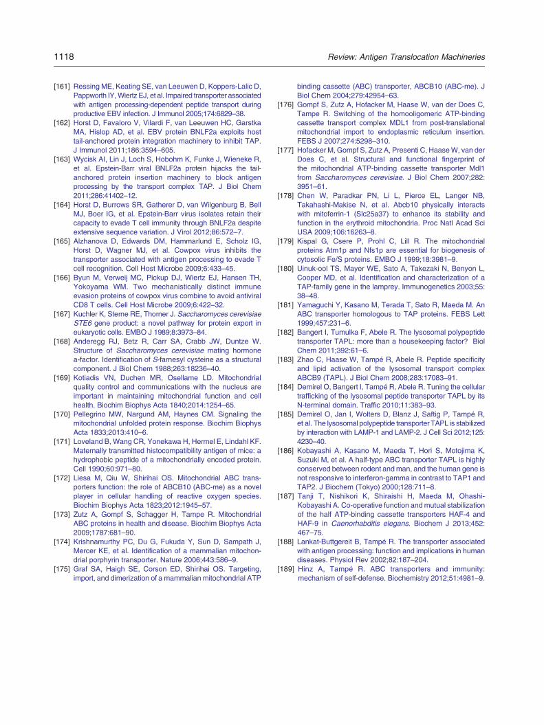
1118 Review: Antigen Translocation Machineries
[161] Ressing ME, Keating SE, van Leeuwen D, Koppers-Lalic D,Pappworth IY,Wiertz EJ, et al. Impaired transporter associatedwith antigen processing-dependent peptide transport duringproductive EBV infection. J Immunol 2005;174:6829–38.
[162] Horst D, Favaloro V, Vilardi F, van Leeuwen HC, GarstkaMA, Hislop AD, et al. EBV protein BNLF2a exploits hosttail-anchored protein integration machinery to inhibit TAP.J Immunol 2011;186:3594–605.
[163] Wycisk AI, Lin J, Loch S, Hobohm K, Funke J, Wieneke R,et al. Epstein-Barr viral BNLF2a protein hijacks the tail-anchored protein insertion machinery to block antigenprocessing by the transport complex TAP. J Biol Chem2011;286:41402–12.
[164] Horst D, Burrows SR, Gatherer D, van Wilgenburg B, BellMJ, Boer IG, et al. Epstein-Barr virus isolates retain theircapacity to evade T cell immunity through BNLF2a despiteextensive sequence variation. J Virol 2012;86:572–7.
[165] Alzhanova D, Edwards DM, Hammarlund E, Scholz IG,Horst D, Wagner MJ, et al. Cowpox virus inhibits thetransporter associated with antigen processing to evade Tcell recognition. Cell Host Microbe 2009;6:433–45.
[166] Byun M, Verweij MC, Pickup DJ, Wiertz EJ, Hansen TH,Yokoyama WM. Two mechanistically distinct immuneevasion proteins of cowpox virus combine to avoid antiviralCD8 T cells. Cell Host Microbe 2009;6:422–32.
[167] Kuchler K, Sterne RE, Thorner J. Saccharomyces cerevisiaeSTE6 gene product: a novel pathway for protein export ineukaryotic cells. EMBO J 1989;8:3973–84.
[168] Anderegg RJ, Betz R, Carr SA, Crabb JW, Duntze W.Structure of Saccharomyces cerevisiae mating hormonea-factor. Identification of S-farnesyl cysteine as a structuralcomponent. J Biol Chem 1988;263:18236–40.
[169] Kotiadis VN, Duchen MR, Osellame LD. Mitochondrialquality control and communications with the nucleus areimportant in maintaining mitochondrial function and cellhealth. Biochim Biophys Acta 1840;2014:1254–65.
[170] Pellegrino MW, Nargund AM, Haynes CM. Signaling themitochondrial unfolded protein response. Biochim BiophysActa 1833;2013:410–6.
[171] Loveland B, Wang CR, Yonekawa H, Hermel E, Lindahl KF.Maternally transmitted histocompatibility antigen of mice: ahydrophobic peptide of a mitochondrially encoded protein.Cell 1990;60:971–80.
[172] Liesa M, Qiu W, Shirihai OS. Mitochondrial ABC trans-porters function: the role of ABCB10 (ABC-me) as a novelplayer in cellular handling of reactive oxygen species.Biochim Biophys Acta 1823;2012:1945–57.
[173] Zutz A, Gompf S, Schagger H, Tampe R. MitochondrialABC proteins in health and disease. Biochim Biophys Acta2009;1787:681–90.
[174] Krishnamurthy PC, Du G, Fukuda Y, Sun D, Sampath J,Mercer KE, et al. Identification of a mammalian mitochon-drial porphyrin transporter. Nature 2006;443:586–9.
[175] Graf SA, Haigh SE, Corson ED, Shirihai OS. Targeting,import, and dimerization of a mammalian mitochondrial ATP
binding cassette (ABC) transporter, ABCB10 (ABC-me). JBiol Chem 2004;279:42954–63.
[176] Gompf S, Zutz A, Hofacker M, Haase W, van der Does C,Tampe R. Switching of the homooligomeric ATP-bindingcassette transport complex MDL1 from post-translationalmitochondrial import to endoplasmic reticulum insertion.FEBS J 2007;274:5298–310.
[177] Hofacker M, Gompf S, Zutz A, Presenti C, HaaseW, van derDoes C, et al. Structural and functional fingerprint ofthe mitochondrial ATP-binding cassette transporter Mdl1from Saccharomyces cerevisiae. J Biol Chem 2007;282:3951–61.
[178] Chen W, Paradkar PN, Li L, Pierce EL, Langer NB,Takahashi-Makise N, et al. Abcb10 physically interactswith mitoferrin-1 (Slc25a37) to enhance its stability andfunction in the erythroid mitochondria. Proc Natl Acad SciUSA 2009;106:16263–8.
[179] Kispal G, Csere P, Prohl C, Lill R. The mitochondrialproteins Atm1p and Nfs1p are essential for biogenesis ofcytosolic Fe/S proteins. EMBO J 1999;18:3981–9.
[180] Uinuk-ool TS, Mayer WE, Sato A, Takezaki N, Benyon L,Cooper MD, et al. Identification and characterization of aTAP-family gene in the lamprey. Immunogenetics 2003;55:38–48.
[181] Yamaguchi Y, Kasano M, Terada T, Sato R, Maeda M. AnABC transporter homologous to TAP proteins. FEBS Lett1999;457:231–6.
[182] Bangert I, Tumulka F, Abele R. The lysosomal polypeptidetransporter TAPL: more than a housekeeping factor? BiolChem 2011;392:61–6.
[183] Zhao C, Haase W, Tampé R, Abele R. Peptide specificityand lipid activation of the lysosomal transport complexABCB9 (TAPL). J Biol Chem 2008;283:17083–91.
[184] Demirel O, Bangert I, Tampé R, Abele R. Tuning the cellulartrafficking of the lysosomal peptide transporter TAPL by itsN-terminal domain. Traffic 2010;11:383–93.
[185] Demirel O, Jan I, Wolters D, Blanz J, Saftig P, Tampé R,et al. The lysosomal polypeptide transporter TAPL is stabilizedby interaction with LAMP-1 and LAMP-2. J Cell Sci 2012;125:4230–40.
[186] Kobayashi A, Kasano M, Maeda T, Hori S, Motojima K,Suzuki M, et al. A half-type ABC transporter TAPL is highlyconserved between rodent andman, and the human gene isnot responsive to interferon-gamma in contrast to TAP1 andTAP2. J Biochem (Tokyo) 2000;128:711–8.
[187] Tanji T, Nishikori K, Shiraishi H, Maeda M, Ohashi-Kobayashi A. Co-operative function andmutual stabilizationof the half ATP-binding cassette transporters HAF-4 andHAF-9 in Caenorhabditis elegans. Biochem J 2013;452:467–75.
[188] Lankat-Buttgereit B, Tampé R. The transporter associatedwith antigen processing: function and implications in humandiseases. Physiol Rev 2002;82:187–204.
[189] Hinz A, Tampé R. ABC transporters and immunity:mechanism of self-defense. Biochemistry 2012;51:4981–9.