anticancer effects of metformin on neuroendocrine tumor ... · anticancer effects of metformin on...
TRANSCRIPT

Anticancer effects of metformin on neuroendocrine tumor cells in vitro
George Vlotides,1* Ayse Tanyeri,1* Matilde Spampatti,1,2 Kathrin Zitzmann,1 Michael Chourdakis,3 Gerald Sp�ttl,1 Julian Maurer,1 Svenja N�lting,1 Burkhard G�ke,1 Christoph J. Auernhammer1
1Department of Internal Medicine II, Campus Grosshadern, University-Hospital, Ludwig-Maximilians-University of Munich, Munich, Germany; 2U.O.C. Gastroenterologia 2 Fondazione IRCCS Cá Granda Ospedale Maggiore Policlinico Cattedra di Gastroenterologia Universitá degli Studi di Milano, Italy; 3School of Medicine, Aristotle University of Thessaloniki, Thessaloniki, Greece
*Both authors contributed equally to this manuscript
ABSTRACTMetformin is a widely used oral antidiabetic drug with good tolerability. Recent studies suggest that it also possesses adjuvant potent anticancer properties in a variety of tumors. Neuroendocrine tumors (NETs) of the gastro-entero-pancreatic system (GEP) comprise a heterogeneous group of tumors with increasing incidence and limited effective therapeutic options. Here we report the antiproliferative effects of metformin in neuroendocrine tumor cells in vitro. Treatment of human pancreatic BON1, bronchopulmonary NCI-H727, and midgut GOT1 neuroendocrine tumor cells with increasing concentrations of metformin (0.1-10 mM) dose-dependently sup-pressed cell viability and cell counts. Metformin induced AMPK phosphorylation in pancreatic BON1 and midgut GOT1 but suppressed AMPK activity in bronchopulmonary NCI-H727. Thus, AMPK-dependent and AMPK-independent properties may be operative in NETs of different origin. Metformin suppressed mTORC1 signaling in all three tumor cell types, evidenced by suppression of 4EBP1, pP70S6K, and S6 phosphorylation, and was associated with compen-satory AKT activity. We observed induction of ERK phosphorylation in BON1 and NCI-H727 and inhibition of ERK in midgut GOT1 cells, while all three tumor cell types responded with induction of GSK3 phosphorylation. This suggests a central role for GSK3 in metformin-mediated signal transduction. Inhibition of cell proliferation by metformin was associated with apoptosis induction only in midgut GOT1, evidenced by increased subG0/1 fraction and PARP cleavage. These results suggest a potential role of metformin as a (adjuvant) therapeutic for patients with NETs.
Key words: AMPK, Cell viability, Metformin, mTOR, Neuroendocrine tumor
HORMONES
Address for correspondence:Christoph J. Auernhammer, Department of Internal Medicine II, University-Hospital Campus Grosshadern, Interdisciplinary Center of Neuroendocrine Tumours of the GastroEnteroPancreatic System (GEPNET-KUM), Ludwig-Maximilians-University of Munich, Marchioninistr. 15, 81377 Munich, Germany, Tel.: +49 89 4400 72520, Fax: +49 89 4400 75514, Homepage: http://www.klinikum.uni-muenchen.de/NET-Zentrum, E-mail: [email protected]
Received: 03-08-2013, Accepted: 20-03-2014
Research paper
G. Vlotides et Al
IntroductIon
Metformin (1.1-dimethylbiguanide hydrochloride) is the most frequently used antidiabetic medication with ca. 120 million prescriptions filled every year worldwide.1 This biguanide indirectly stimulates AMP-activated protein kinase (AMPK)2 with sub-sequent reduction of hepatic gluconeogenesis and glycogenolysis and an increase in glucose uptake in the muscle.3 AMPK activation also leads to suppression of the mammalian target of rapamycin (mTOR),4 a key regulator of cell proliferation in cancer cells. AMPK-independent mechanisms of action of metformin include induction of cell cycle arrest and reduction of insulin/IGF-1 signaling.4,5
Over the past few years, several epidemiological and preclinical/clinical studies suggest potent anti-cancer effects of metformin.4,5 Patients with diabetes mellitus type 2 treated with metformin appear to have a decreased risk for cancer development of the pan-creas,6-8 liver,9-11 lung,12 and breast.13 Additionally, in vitro studies have demonstrated inhibitory effects of metformin on cell proliferation in endometrial,14 ovar-ian,15 glioma,16 pancreatic cancer,17 and melanoma18 tumor cells. Thus, in addition to diabetes therapy, metformin may offer valuable anticancer effects, especially in endocrine-related cancers.19
Neuroendocrine tumors (NETs) of the gastro-enteropancreatic (GEP) system arise from diffuse neuroendocrine cells within the digestive system. Although considered rare, accounting only for ~2% of all gastrointestinal tumors,20,21 their prevalence has increased over the last two decades.20,21 Current systemic treatment options include biotherapy with somatostatin analogues and interferon-α, molecular targeted therapy with everolimus and sunitinib, cyto-toxic chemotherapy protocols, and peptide receptor-targeted therapy.22 However, most available therapies are efficacious for a limited period of time and rarely induce tumor remission.22 Despite the introduction of target-directed therapies such as the multiple tyrosine kinase inhibitor sunitinib23 and the mTOR inhibitor everolimus24,25 for a subset of progressive NET,22,26 there is a need for novel drugs for syndromic and tumor growth control for this heterogeneous tumor entity. As inhibition of mTORC1 signaling by evero-limus has already proven to be a promising target for antitumoral therapy in NETs23-25,27 and metformin
also inhibits mTORC1 signaling,28-30 we investigated potential effects of metformin on NETs.
MaterIals & Methods
Materials
DMEM/F12 media, penicillin, and streptomycin were purchased from Gibco/Invitrogen (Karlsruhe, Germany) and RPMI media was obtained from PAA (Pasching, Austria). Fetal bovine serum (FBS) and amphotericin B were acquired from Biochrom (Berlin, Germany). Metformin was purchased from Sigma-Aldrich (St. Louis, USA), IGF1 from Amersham Pharmacia Biotech Europe, and rapamycin from Biomol (Hamburg, Germany).
Cell cultures
All human neuroendocrine cell lines were received and cultured as recently described.31 Briefly, pan-creatic neuroendocrine BON1 tumor cells32 (kindly provided by R. Göke, Marburg) were cultured in DMEM/F12 (1:1) medium supplemented with 10% FBS, 10 mg/ml penicillin/streptomycin and 4 mg/ml amphotericin B. Human midgut carcinoid GOT1 cells33 (kindly provided by Prof. Ola Nilsson, Sahlg-renska University Hospital Göteborg, Sweden) and human bronchopulmonary neuroendocrine NCI-H727 tumor cells (purchased from ATCC, Manassas, VA) were both cultured in RPMI medium supplemented with 10% FBS, 10 mg/ml penicillin/streptomycin and 4 mg/ml amphotericin B. Additional supplements in GOT1 culture medium were 0.135 IU/ml insulin and 5mg/dl apo-transferrin.
Assessment of cell viability/cell numbers
Cell viability was assessed as recently described.31 Briefly, cells were seeded into 96-well plates at den-sities of 3000 (BON1), 50000 (GOT1) and 4000 (NCI-H727) cells per well, respectively, and grown for 24 hrs. The next day, the medium was replaced by serum rich medium (10% FBS) containing various concentrations of metformin (0.1 to 10 mM) and the cells were further incubated for the indicated time intervals. Cell viability expressed by metabolic activity was measured with Cell Titer 96 aqueous One Solu-tion Cell Proliferation Assay (Promega, Madison, WI, USA) according to the manufacturer’s instructions. Following 3 hrs of incubation with Cell Titer 96 solu-

Anticancer effects of metformin in Net
tion, absorbance at 492 nm was determined using an ELISA plate reader (Orion II, Berthold Detection Systems, Pforzheim, Germany). Cell counts were per-formed with an automated cell counter (CountessTM, Invitrogen, Germany).
Cell cycle analysis
Cell cycle distribution was analyzed using pro-pidium iodide staining and flow cytometry as described previously.31 Briefly, cells were washed with PBS and treated with 400 ml trypsin at 37°C for 4 minutes. After another wash cycle with PBS, the cells were resuspended in 350 µl prodidium iodide. Sub-G1 events and cell cycle distribution were measured in a fluorescence-activated cell sorter (BD Accuri C6 Analysis). Nuclei to the left of the G1-peak contain-ing hypodiploid DNA were considered apoptotic.
Protein Extraction and Western Blotting
Protein extraction and Western blotting were performed as described previously.31 Briefly, cells were lysed in 500 µl lysis buffer (MPER® Mammalian Protein Extraction Reagent; #78501F, containing HaltTM Protease & Phosphatase Inhibitor Cocktail; #78447; PIERCE, Rockford, USA). The lysates were centrifuged for 10 min at 4° C and 13,000g and supernatants were adjusted to equal protein loads and diluted 1:1 with SDS sample buffer. Samples were boiled for 5 min and separated on a SDS polyacryla-mide gel. Proteins were electrotransferred for 60 min onto PVDF membranes (Immobilone; Millipore, Eschborn, Germany) using a semi-dry Western-blot technique. After blocking in 2% non-fat dried milk, the membranes were incubated overnight in ap-propriate dilutions of antibodies against pAkt (Ser 473) (#4060), Akt (#2920), pERK (Thr202/Tyr204) 1/2 (#4370), pP70S6K (Thr389) (#9234), P70S6K (#9202), p4EBP1 (Ser65) (#9451), 4EBP1 (#9644), pS6 (Ser235/6 (#4858) and Ser240/4 (#5364)), S6 (#2317), pGSK3 (Ser21/9) (#9331), GSK3 (#9315), IGFR (#3027), EGFR (#4267), PARP (#9542) (all from Cell Signaling, Danvers, MA), Erk 1/2 (06-182; Millipore). After washing with PBS, the membranes were incubated with a peroxidase-conjugated second-ary antibody (1:25,000) for 2 hrs (anti-rabbit IgG; #7074 and anti-mouse IgG; #7076; Cell Signaling, Danvers, USA). The blots were washed and immersed in the chemiluminescent substrate SuperSignal West
Dura (Thermo Scientific, Rockford, USA) and ex-posed to Super RX X-ray film (FUJIFILM Corpora-tion, Tokyo, Japan).
Statistical analysis
For the statistical analyses the “Statistical Program for the Social Sciences-SPSS” software (v.16.0) was used. Results are expressed as mean ± SD of inde-pendently performed experiments. For proliferation assays and cell cycle analyses, comparisons were evaluated using the 2-tailed Student’s t test. Statistical significance was set at p <0.05, after a Bonferroni ad-justment to compensate for the multiple comparisons.
results
Metformin inhibits neuroendocrine cell viability
Treatment of human pancreatic neuroendocrine BON1 cells with metformin suppressed cell viability (Cell Titer Assay; Promega) in a dose-dependent manner (Figure 1A). Significant effects were ob-served at 144 and 216 hrs; at 144 hrs, treatment with metformin at 5 mM suppressed cell viability to 65 ± 3% (p=0.015) and at 10 mM to 55 ± 8% (n.s.). At 216 hrs, treatment at 5 mM suppressed cell viability to 65 ± 3% (p=0.010) and at 10 mM to 45 ± 5% (p=0.015). Cell counts performed with an automated cell counter revealed similar results (Figure 1B); significant effects were observed at 144 and 216 hrs; maximum suppression (to ~20% at 144 and 216 hrs) was observed at the highest metformin doses tested (5 to10 mM).
Metformin also suppressed cell viability in hu-man bronchopulmonary NCI-H727 cells in a dose-dependent manner (Figure 1C). Significant effects were observed at all time points tested (at 5-10 mM metformin). Maximal suppression levels were observed at 10 mM metformin with inhibition of cell viability to 55 ± 8% (p=0.007), 30 ± 10% (p=0.004) and 22 ± 15% (p=0.009) at 72, 144 and 216 hrs, respectively. Cell counts performed with an automated cell counter demonstrated similar results (Figure 1D).
Human midgut neuroendocrine GOT1 cells dem-onstrated high sensitivity to treatment with metformin (Figure 1E). Significant effects were observed at start-ing doses of 0.5 mM (at 72 hrs) and a dose-dependent decrease in cell viability was observed for all time

G. Vlotides et Al
points with a maximum suppression to ~10-20%.
Effects of metformin on AMPK phosphorylation in neuroendocrine tumor cells
Metformin is known to act via AMPK-dependent and -independent pathways.29,30,34 Treatment of hu-man neuroendocrine tumor cells with metformin had different effects on AMPK activity depending on the cell line used. In human pancreatic BON1 cells, treatment with 5-10 mM metformin for 48 hrs induced AMPK phosphorylation (Figure 2A). Midgut neuroendocrine GOT1 cells were highly responsive to metformin in terms of AMPK activation (Figure 2C); metformin doses as low as 0.1 mM induced AMPK
phosphorylation and positive effects were observed at all time points tested (including 2 hrs and 72 hrs, not shown). In contrast, bronchopulmonary NCI-H727 neuroendocrine cells responded with suppression of baseline AMPK phosphorylation after treatment with metformin (Figure 2B).
Effects of metformin on the PI3K/AKT/mTOR signaling pathway
Treatment of human pancreatic neuroendocrine BON1 cells with metformin, particularly at 48 hrs, dose-dependently induced AKT phosphorylation (Fig-ure 3A). Inhibition of the mTOR signaling pathway was confirmed by metformin-mediated suppression
Figure 1. Inhibition of neuroendocrine tumor cell viability by metformin. Human pancreatic BON1 (A, B), bronchopulmonary NCI-H727 (C, D) and midgut GOT1 (E) neuroendocrine tumor cells were treated with indicated concentrations of metformin for 72, 144 and 216 hrs, respectively. Cell viability based on metabolic activity was measured with Cell Titer 96 kit (Promega) (A, C and E) and cell counts were performed with an automated cell counter (B and D). Shown are the mean values ± SD of 3 independently performed experiments. *, p<0.05; **, p<0.01; ***, p<0.001 versus untreated control.

Anticancer effects of metformin in Net
of p4EBP1 (Figure 3A), pP70S6K and S6 phospho-rylation (Figure 3B). Metformin potently suppressed 4EBP1 and S6 (at Ser 235/6) phosphorylation at doses as low as 0.5 mM (Figure 3A and B), while only minor inhibitory effects on IGFR protein expression were observed.
In bronchopulmonary NCI-H727 neuroendocrine cells, metformin suppressed AKT phosphorylation at higher doses (1-10 mM), while at lower doses (0.1-0.5 mM) AKT phosphorylation was increased (at 48 hrs, Figure 3C). At starting doses of 0.5 to 1.0 mM, metformin suppressed phosphorylation of mTOR downstream targets 4EBP1, P70S6K and S6 (Figure 3C and D). No major effect was observed on IGFR protein expression.
Midgut neuroendocrine GOT1 cells (particularly at 48 hrs) also responded with decreased phosphoryla-tion of mTOR downstream signaling targets 4EBP1
Figure 2. Effect of metformin on AMPK phosphorylation in neuroendocrine tumor cells. Human pancreatic neuroendo-crine BON1 (upper panel; A), bronchopulmonary NCI-H727 (middle panel; B) and midgut GOT1 (lower panel; C) cells were treated with indicated concentrations of metformin for 24 and 48 hrs. Subsequently the expression of pAMPK, AMPK and β-actin loading control was evaluated by Western blot anal-ysis. A representative blot out of 3 independently performed experiments is shown.
Figure 3. Effect of metformin on PI3K/AKT/mTORC1 sig-naling in neuroendocrine. Human pancreatic BON1 (A, B), bronchopulmonary NCI-H727 (C, D) and midgut GOT1 (E, F) neuroendocrine tumor cells were treated with indicated concentrations of metformin for 24 and 48 hrs, respectively. Subsequently the expression of pAKT, AKT, pEBP1, EBP1, IGFR (A, C and E) as well as pP70S6K, P70S6K, pS6, S6 (B, D and F) and β-actin loading control was evaluated by Western blot analysis. A representative blot out of 3 independently per-formed experiments is shown.
G. Vlotides et Al
(Figure 3E), P70S6K and S6 (Figure 3F). Similar to BON1 cells, metformin dose-dependently increased AKT phosphorylation at 24 and 48 hrs (Figure 3E).
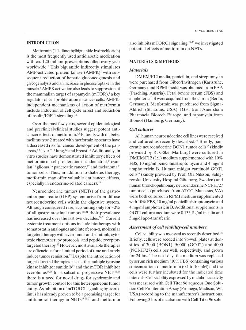
Specific treatment of neuroendocrine BON1 and NCI-H727 cells under serum-free conditions with IGF1 induced rapid (not shown) and sustained phosphorylation of the IGF receptor, as well as AKT (particularly in BON1 cells) and downstream mTOR S6 phosphorylation (Figure 4). In a dose-response curve employing a wide spectrum of metformin doses, even high metformin concentrations had no effect on IGF1-induced IGFR phosphorylation in both cell lines tested. In contrast to cells cultured in complete medium (Figure 3), under serum-free conditions (Fig-ure 4), we did not observe major effects of metformin on basal or IGF1-induced AKT phosphorylation, but metformin dose-dependently suppressed downstream mTOR signaling, evidenced by inhibition of basal and IGF1-induced S6 phosphorylation. This inhibition of basal and IGF1-induced S6 phosphorylation by met-formin was similar to inhibition of S6 phosphorylation by the established mTOR inhibitor rapamycin, used as a positive control (Figure 4).
Effects of metformin on GSK3 and ERK signaling
All three neuroendocrine tumor cell lines re-
sponded with a dose-dependent increase in GSK3 phosphorylation (at 24 and 48 hrs) at the lowest metformin doses tested (0.1 to 0.5 mM; Figure 5A-C). The effect of metformin on ERK phosphorylation differed between neuroendocrine cells of different origin. In pancreatic BON1 and bronchopulmonary NCI-H727 cells, metformin potently induced ERK phosphorylation peaking at 1 to 5 mM (Figure 5A-B). In contrast, GOT1 cells responded with a dose-dependent suppression of ERK phosphorylation in response to low starting metformin doses (particularly at 48 hrs, Figure 5C).
Effects of metformin on apoptosis in neuroendocrine tumor cells
In order to explore mechanisms for metformin-mediated inhibition of cell proliferation, we deter-mined the effect of metformin on sub-G0/1 distribu-tion in neuroendocrine tumor cells. Treatment with metformin did not increase the percentage of BON1 and NCI-H727 cells in sub-G0/1 (data not shown). Failure of apoptosis induction by metformin in these cells was confirmed by grossly unaffected levels of cleaved PARP (not shown).
In contrast to BON1 and NCI-H727 cells, in mid-gut neuroendocrine GOT1 cells metformin induced apoptosis, as demonstrated by increased percentage of cells in sub-G0/1 (Figure 5D, left panel) and induc-tion of PARP cleavage (particularly at 48 hrs, Figure 5D, right panel).
Effects of metformin on cell cycle distribution in neuroendocrine tumor cells
Treatment of unsynchronized (culture in complete medium) pancreatic neuroendocrine BON1 cells with metformin doses 1 mM for 72 hrs reduced the percent-age of cells in S phase from 16% to 10% (p=0.013) and increased cells in G0/1 phase from 57% to 65% (p=0.013 at 0.5 mM) (Figure 6A).
Similarly to the observed effects with BON1 cells, metformin dose-dependently decreased NCI-H727 cell proliferation, evidenced by decreased entry into S phase (from 13% to min. 8% at 1 mM; p=0.001) and G2/M phase (from 27% to 20% at 10 mM, p<0.002) and increased percentage of cells in G0/1 (from 51% to 63% at 5mM, p<0.006) at 72 hrs (Figure 6B).
In contrast, midgut neuroendocrine GOT1 cells
Figure 4. Effect of metformin and rapamycin on IGF1-induced signaling in neuroendocrine tumor cells. Human pancreatic BON1 (left panel) and bronchopulmonary NCI-H727 (right panel) neuroendocrine tumor cells were pretreated with indi-cated concentrations of metformin or rapamycin for 1 hr prior to induction with IGF1 for 24 hrs. The expression of pIGFR, IGFR, pAKT, AKT, pS6, S6 and β-actin loading control was evaluated by Western blot analysis. A representative blot out of 3 independently performed experiments is shown.

Anticancer effects of metformin in Net
Figure 5. Effect of metformin on GSK3 / ERK1/2 signaling and apoptosis induction in neuroendocrine tumor cells. Human pancre-atic BON1 (A), bronchopulmonary NCI-H727 (B) and midgut GOT1 (C) neuroendocrine tumor cells were treated with indicated concentrations of metformin for 24 and 48 hrs, respectively. Subsequently the expression of pGSK3, GSK3, pERK1/2, ERK1/2, EGFR and β-actin loading control was evaluated by Western blot analysis. A representative blot out of 3 independently performed experiments is shown. Human midgut neuroendocrine GOT1 cells cultured in complete medium were treated with the indicated concentrations of metformin (Figure 5D). After 72 hrs the proportion of cells in subG0/1 phase was examined by flow cytometry (left panel). Demonstrated are the mean values ± SD of 2 independently performed experiments in duplicates (N=4); *, p< 0.05; **, p<0.01 versus untreated control. The expression of total / cleaved PARP and β-actin loading control was evaluated by Western blot analysis (24 and 48 hrs; right panel). A representative blot out of 3 independently performed experiments is shown.
responded with a dose-dependent decrease of cells in all three phases of the cell cycle (Figure 6C), likely due to strong induction of apoptosis (Figure 5D).
dIscussIon
Current systemic treatment options of neuroen-docrine tumors include biotherapy with somatosta-tin analogues and interferon-α, molecular targeted therapy with everolimus and sunitinib, cytotoxic che-motherapy protocols, and peptide receptor-targeted
therapy.22,26,35 Target-directed therapies such as the antiangiogenic multi-tyrosine kinase inhibitor suni-tinib23 and the mTOR inhibitor everolimus24,25 have increased the therapeutic spectrum for a subset of progressive NET.22,26 However, novel therapeutics are needed for sporadic NETs and particularly for NETs associated with hereditary syndromes such as MEN1 characterized by early onset and multiplicity of lesions.36,37 Although the widely used antidiabetic drug metformin appears to exert potent anticancer effects in multiple types of cancer,29,30,34 potential

G. Vlotides et Al
effects on NET have not been explored so far. As inhibition of the mTORC1 signaling by everolimus has already been proven a promising target for an-titumoral therapy in NETs23-25,27 and metformin also inhibits mTORC1 signaling,28-30 potential effects of metformin on NETs were investigated.
In addition to indirectly-reached insulin-dependent effects of metformin (positive effects of lowered insulin levels on cancer), metformin appears to act through AMPK-dependent and AMPK-independent pathways.29,30,34 Metformin leads to tumor suppres-sor liver kinase B1 (LKB1)-mediated activation of AMPK activity. This in turn suppresses mTORC1 signaling and protein synthesis through activation of tuberous sclerosis complex 2 (TSC2) or inhibition of
raptor.29,30 In addition to AMPK-dependent reduction of mTORC1 signaling, metformin-mediated AMPK activation results in p53 phosphorylation and p53-mediated cell cycle arrest or apoptosis, depending on the p53 mutational status and cell type-dependent p53 signaling.29,30
AMPK-independent effects of metformin include Rag GTPase-mediated inhibition of mTORC1, re-duced production of tumor necrosis factor alpha (TNFα) and p53/p21-mediated inhibition of cyclin D1, and retinoblastoma-protein (pRb) and G1 cell cycle arrest.29,30 The complexity of metformin signaling is also highlighted by the fact that, depending on the cancer cell type, inhibition of cell proliferation may be associated with G0/1, G2/M or even S phase arrest.34
In this study, the effect of metformin was examined on cell proliferation / viability in three human NET cell lines from pancreatic (BON1), bronchopulmonary (NCI-H727), and midgut (GOT1) origin. Treatment with increasing metformin concentrations (0.1 to 10 mM) dose-dependently suppressed cell proliferation / viability in all three cell lines. To further explore the mechanism of metformin action in NET, we examined the effect of metformin on AMPK phosphorylation. Pancreatic BON1 and particularly midgut GOT1 neuroendocrine tumor cells reacted with increased AMPK phosphorylation in response to metformin treatment, indicating that the observed antiprolifera-tive effects were at least in part mediated through an AMPK-dependent mechanism. In contrast, broncho-pulmonary NCI-H727 cells responded to metformin with decreased AMPK phosphorylation, suggesting involvement of AMPK-independent signaling.
Based on the central role of the PI3K/AKT/mTORC1 pathway for neuroendocrine tumor cell growth22,26 and demonstrated (AMPK-dependent and -independent) suppression by metformin in cancer,29,30 we examined the effect of metformin on neuroendocrine mTORC1 downstream targets. Treatment with increasing doses of metformin sup-pressed baseline phosphorylation of 4EBP1, P70S6K and S6 in all three neuroendocrine cell lines tested, suggesting that metformin-mediated inhibition of cell proliferation is mediated by inhibition of mTORC1 signaling. In NCI-H727 cells, lower metformin doses induced whereas higher doses suppressed AKT activ-ity, while BON1 and GOT1 cells responded with a
Figure 6. Effect of metformin on cell cycle distribution of neuroendocrine tumor cells. Human pancreatic BON1 (A), bronchopulmonary NCI-H727 (B) and midgut GOT1 (C) neuroendocrine tumor cells were treated with the indicated concentrations of metformin for 72 hrs. Demonstrated are the mean values ± SD of 3 independently performed experiments in duplicates (n=6); *, p< 0.05; **, p<0.01; ***, p< 0.001 ver-sus untreated control.
Anticancer effects of metformin in Net
dose-dependent increase in AKT phosphorylation at all time points tested. This argues in favor of cell type-dependent compensatory AKT activation in response to mTORC1 inhibition by metformin, similar to compensatory effects on AKT signaling as observed in response to everolimus.38,39
The complexity of metformin signaling in neu-roendocrine cells of different origin is further high-lighted by differences observed on ERK activity. In pancreatic BON1 and bronchopulmonary NCI-H727 cells, metformin potently induced ERK activity, while in midgut GOT1 cells it suppressed ERK phospho-rylation. Failure of compensatory ERK activation in GOT1 cells could partially account for the more potent antiproliferative effect of metformin observed in these cells. It is likely that ERK (and AKT) activa-tion in response to treatment with metformin reflects compensatory mechanisms of the tumor cell machin-ery responding to inhibition of mTORC1 signaling. This indicates that the effectiveness of combined treatments simultaneously blocking several signaling pathways may depend on the distinct neuroendocrine tumor signaling profile. This phenomenon appears to be cell type-specific, as treatment of PANC-1 or MiaPaCa-2 pancreatic cancer cells with metformin did not induce AKT and / or ERK activity (in contrast to rapamycin or active-site mTOR inhibitors).17 It is not clear whether these compensatory mechanisms in response to mTOR inhibition are a sign of clinical resistance39,40 or a sign of effectiveness of treatment.41 In any case, simultaneous inhibition of mTOR and AKT (or ERK) signaling could potentiate the inhibi-tory effect on cell proliferation in GEP NET.39
Although metformin exerted varying effects on ERK / AKT activity depending on the neuroendocrine cell type tested, suppression of mTORC1 signaling was associated with induction of GSK3 phosphoryla-tion in all three neuroendocrine cell lines. As GSK-3-mediated phosphorylation usually suppresses the activity of downstream targets involved in multiple intracellular signaling pathways, including cell prolif-eration and apoptosis,42-44 ERK-45 or AKT-dependent46 GSK3 phosphorylation appears critical for metformin signaling in neuroendocrine tumor cells.
In pancreatic BON1 and bronchopulmonary NCI-H727 neuroendocrine tumor cells, metformin-medi-ated inhibition of cell proliferation was not associated
with induction of apoptosis, as metformin did not increase the number of cells in sub-G0/1 nor induce PARP cleavage, but rather with alterations in cell cycle distribution with increased G0/1 and decreased S phase entry. In contrast, the metformin-mediated antiproliferative effect in midgut GOT1 cells was, at least in part, associated with apoptosis induction, evidenced by increased sub-G0/1 events and PARP cleavage. Induction of apoptosis in GOT1 cells may be associated with the observed inhibition (rather than compensatory activation) of ERK signaling (similar to findings in pheochromocytoma cell lines47) and explain the more potent antiproliferative effect of metformin on this neuroendocrine tumor cell line.
Previously reported data from preclinical studies employing metformin tested relatively high met-formin concentrations in vitro (1-40 mM (165-6600 mg/l);48-51) when compared to therapeutic plasma levels in human (2.8-15 µM (0.465-2.5 mg/l);30). In acute metformin overdose in humans, survivors had a median peak metformin level of 42 µg/ml vs 110 µg/ml in non-survivors,52 both concentrations ranging below 1 mM metformin. On the other hand, higher metformin concentrations may be necessary in vitro since cancer cells are cultured in complete medium containing extremely high amounts of growth fac-tors and glucose. Metformin may accumulate in tissues particularly of the gastrointestinal system at much higher concentrations after oral or intravenous administration.53 Further preclinical in vivo studies evaluating a wider spectrum of metformin dosages are needed to test the efficacy of metformin in neu-roendocrine tumors, while considering dose-related side effects, such as development of lactic acidosis. The first preliminary clinical data on the role of metformin in recurrence-free survival in patients with neuroendocrine tumors have been reported recently.54 In a retrospective analysis of 12 diabetic NET patients with metformin versus 24 non-diabetic NET patients without metformin, recurrence rate was lower by 8 versus 42% and recurrence-free survival was not at-tained compared to 86 months of the second group.54
This study demonstrates metformin-mediated inhibition of neuroendocrine tumor cell growth and signaling. Considering the good tolerability of the drug, the high rate of diabetes development of pa-tients with NET under somatostatin analogue and

G. Vlotides et Al
everolimus treatment, and the current lack of effective pharmacologic adjuvant therapies, further studies are needed to explore the potential use of metformin in patients harboring neuroendocrine tumors.
acknowledgMent
This work contains parts of the unpublished doc-toral theses of Ayse Tanyeri.
dIsclosure stateMent
CJA has received research contracts (Ipsen, Novartis), lecture honorarium (Ipsen, Novartis, Pfizer, Amgen) and advisory board honorarium (Novartis). This research did not receive any specific grant from any funding agency in the public, commercial or not-for-profit sector.
references 1. Ben sahra i, le Marchand-Brustel Y, tanti JF, Bost F,
2010 Metformin in cancer therapy: a new perspective for an old antidiabetic drug? Mol Cancer ther 9: 1092-1099.
2. Hawley sA, Ross FA, Chevtzoff C, et al, 2010 Use of cells expressing gamma subunit variants to identify diverse mechanisms of AMPK activation. Cell Metab 11: 554-565.
3. Zhou G, Myers R, li Y, et al, 2001 Role of AMP-activated protein kinase in mechanism of metformin action. J Clin invest 108: 1167-1174.
4. Jalving M, Gietema JA, lefrandt Jd, et al, 2010 Met-formin: taking away the candy for cancer? eur J Cancer 46: 2369-2380.
5. Micic d, Cvijovic G, trajkovic V, duntas lH, Polovina s, 2011 Metformin: its emerging role in oncology. Hormones (Athens) 10: 5-15.
6. li d, Yeung sC, Hassan MM, Konopleva M, Abbruzzese Jl, 2009 Antidiabetic therapies affect risk of pancreatic cancer. Gastroenterology 137: 482-488.
7. lee Ms, Hsu CC, Wahlqvist Ml, tsai HN, Chang YH, Huang YC, 2011 type 2 diabetes increases and metformin reduces total, colorectal, liver and pancreatic cancer incidences in taiwanese: a representative popu-lation prospective cohort study of 800,000 individuals. BMC Cancer 11: 20.
8. sadeghi N, Abbruzzese Jl, Yeung sC, Hassan M, li d, 2012 Metformin use is associated with better survival of diabetic patients with pancreatic cancer. Clin Cancer Res 18: 2905-2912.
9. donadon V, Balbi M, Mas Md, Casarin P, Zanette G, 2010 Metformin and reduced risk of hepatocellular carcinoma in diabetic patients with chronic liver disease.
liver int 30: 750-758. 10. Chen tM, lin CC, Huang Pt, Wen CF, 2011 Metformin
associated with lower mortality in diabetic patients with early stage hepatocellular carcinoma after radiofre-quency ablation. J Gastroenterol Hepatol 26: 858-865.
11. Zhang ZJ, Zheng ZJ, shi R, su Q, Jiang Q, Kip Ke, 2012 Metformin for liver cancer prevention in patients with type 2 diabetes: a systematic review and meta-analysis. J Clin endocrinol Metab 97: 2347-2353.
12. Mazzone PJ, Rai H, Beukemann M, Xu M, Jain A, sasidhar M, 2012 the effect of metformin and thia-zolidinedione use on lung cancer in diabetics. BMC Cancer 12:410.
13. Col NF, ochs l, springmann V, Aragaki AK, Chle-bowski Rt, 2012 Metformin and breast cancer risk: a meta-analysis and critical literature review. Breast Cancer Res treat 135: 639-646.
14. Cantrell lA, Zhou C, Mendivil A, Malloy KM, Gehrig PA, Bae-Jump Vl, 2010 Metformin is a potent inhibitor of endometrial cancer cell proliferation--implications for a novel treatment strategy. Gynecol oncol 116: 92-98.
15. Rattan R, Giri s, Hartmann lC, shridhar V, 2011 Metformin attenuates ovarian cancer cell growth in an AMP-kinase dispensable manner. J Cell Mol Med 15: 166-178.
16. isakovic A, Harhaji l, stevanovic d, et al, 2007 dual antiglioma action of metformin: cell cycle arrest and mitochondria-dependent apoptosis. Cell Mol life sci 64: 1290-1302.
17. soares HP, Ni Y, Kisfalvi K, sinnett-smith J, Rozengurt e, 2013 different Patterns of Akt and eRK Feedback Activation in Response to Rapamycin, Active-site mtoR inhibitors and Metformin in Pancreatic Cancer Cells. Plos one 8: e57289.
18. Janjetovic K, Harhaji-trajkovic l, Misirkic-Marjanovic M, et al, 2011 in vitro and in vivo anti-melanoma action of metformin. eur J Pharmacol 668: 373-382.
19. Brown KA, samarajeewa NU, simpson eR, 2012 endocrine-related cancers and the role of AMPK. Mol Cell endocrinol 366: 170-179.
20. Massironi s, sciola V, Peracchi M, Ciafardini C, spam-patti MP, Conte d, 2008 Neuroendocrine tumors of the gastro-entero-pancreatic system. World J Gastroenterol 14: 5377-5384.
21. Modlin iM, oberg K, Chung dC, et al, 2008 Gastroen-teropancreatic neuroendocrine tumours. lancet oncol 9: 61-72.
22. Pavel M, 2012 translation of Molecular Pathways into Clinical trials of Neuroendocrine tumors. Neuroendo-crinology 97: 99-112.
23. Raymond e, dahan l, Raoul Jl, et al, 2011 sunitinib malate for the treatment of pancreatic neuroendocrine tumors. N engl J Med 364: 501-513.
24. Yao JC, shah MH, ito t, et al, 2011 everolimus for advanced pancreatic neuroendocrine tumors. N engl J Med 364: 514-523.
Anticancer effects of metformin in Net
25. Pavel Me, Hainsworth Jd, Baudin e, et al, 2011 everoli-mus plus octreotide long-acting repeatable for the treat-ment of advanced neuroendocrine tumours associated with carcinoid syndrome (RAdiANt-2): a randomised, placebo-controlled, phase 3 study. lancet 378: 2005-2012.
26. Weber HC, 2013 Medical treatment of neuroendocrine tumours. Curr opin endocrinol diabetes obes 20: 27-31.
27. Zitzmann K, de toni eN, Brand s, et al, 2007 the novel mtoR inhibitor RAd001 (everolimus) induces antiproliferative effects in human pancreatic neuroen-docrine tumor cells. Neuroendocrinology 85: 54-60.
28. larsson o, Morita M, topisirovic i, et al, 2012 distinct perturbation of the translatome by the antidiabetic drug metformin. Proc Natl Acad sci U s A 109: 8977-8982.
29. emami Riedmaier A, Fisel P, Nies At, schaeffeler e, schwab M, 2013 Metformin and cancer: from the old medicine cabinet to pharmacological pitfalls and prospects. trends Pharmacol sci 34: 126-135.
30. dowling RJ, Niraula s, stambolic V, Goodwin PJ, 2012 Metformin in cancer: translational challenges. J Mol endocrinol 48: R31-43.
31. Zitzmann K, de toni e, von Ruden J, et al, 2011 the novel Raf inhibitor Raf265 decreases Bcl-2 levels and confers tRAil-sensitivity to neuroendocrine tumour cells. endocr Relat Cancer 18: 277-285.
32. evers BM, townsend CM Jr, Upp JR, et al, 1991 es-tablishment and characterization of a human carcinoid in nude mice and effect of various agents on tumor growth. Gastroenterology 101: 303-311.
33. Kölby l, Bernhardt P, Ahlman H, et al, 2001 A trans-plantable human carcinoid as model for somatostatin receptor-mediated and amine transporter-mediated radionuclide uptake. Am J Pathol 158: 745-755.
34. Bost F, sahra iB, le Marchand-Brustel Y, tanti JF, 2012 Metformin and cancer therapy. Curr opin oncol 24: 103-108.
35. Auernhammer CJ, Goke B, 2011 therapeutic strategies for advanced neuroendocrine carcinomas of jejunum/ileum and pancreatic origin. Gut 60: 1009-1021.
36. thakker RV, Newey PJ, Walls GV, et al, 2012 Clinical practice guidelines for multiple endocrine neoplasia type 1 (MeN1). J Clin endocrinol Metab 97: 2990-3011.
37. tonelli F, Giudici F, Fratini G, Brandi Ml, 2011 Pancre-atic endocrine tumors in multiple endocrine neoplasia type 1 syndrome: review of literature. endocr Pract 17 suppl 3: 33-40.
38. Moreno A, Akcakanat A, Munsell MF, soni A, Yao JC, Meric-Bernstam F, 2008 Antitumor activity of rapamy-cin and octreotide as single agents or in combination in neuroendocrine tumors. endocr Relar Cancer 15: 257-266.
39. Zitzmann K, Ruden J, Brand s, et al, 2010 Compensatory activation of Akt in response to mtoR and Raf inhibi-tors - a rationale for dual-targeted therapy approaches in neuroendocrine tumor disease. Cancer lett 295:
100-109. 40. svejda B, Kidd M, Kazberouk A, lawrence B, Pfrag-
ner R, Modlin iM, 2011 limitations in small intestinal neuroendocrine tumor therapy by mtor kinase inhibition reflect growth factor-mediated Pi3K feedback loop ac-tivation via eRK1/2 and AKt. Cancer 117: 4141-4154.
41. Meric-Bernstam F, Akcakanat A, Chen H, et al, 2012 PiK3CA/PteN mutations and Akt activation as markers of sensitivity to allosteric mtoR inhibitors. Clin Cancer Res 18: 1777-1789.
42. Woodgett JR, 1994 Regulation and functions of the glycogen synthase kinase-3 subfamily. semin Cancer Biol 5: 269-275.
43. Woodgett JR, 2001 Judging a protein by more than its name: GsK-3. sci stKe 2001:re12.
44. Ali A, Hoeflich KP, Woodgett JR, 2001 Glycogen syn-thase kinase-3: properties, functions, and regulation. Chem Rev 101: 2527-2540.
45. Rasola A, sciacovelli M, Pantic B, Bernardi P, 2010 signal transduction to the permeability transition pore. FeBs lett 584: 1989-1996.
46. Rayasam GV, tulasi VK, sodhi R, davis JA, Ray A, 2009 Glycogen synthase kinase 3: more than a name-sake. Br J Pharmacol 156: 885-898.
47. Nolting s, Garcia e, Alusi G, et al, 2012 Combined blockade of signalling pathways shows marked anti-tumour potential in phaeochromocytoma cell lines. J Mol endocrinol 49: 79-96.
48. Carvalho C, Correia s, santos Ms, seica R, oliveira CR, Moreira Pi, 2008 Metformin promotes isolated rat liver mitochondria impairment. Mol Cell Biochem 308: 75-83.
49. Capano M, Crompton M, 2006 Bax translocates to mitochondria of heart cells during simulated ischae-mia: involvement of AMP-activated and p38 mitogen-activated protein kinases. Biochem J 395: 57-64.
50. Gallo A, Ceolotto G, Pinton P, et al, 2005 Metformin prevents glucose-induced protein kinase C-beta2 activa-tion in human umbilical vein endothelial cells through an antioxidant mechanism. diabetes 54: 1123-1131.
51. Kefas BA, Cai Y, Kerckhofs K, et al, 2004 Metformin-induced stimulation of AMP-activated protein kinase in beta-cells impairs their glucose responsiveness and can lead to apoptosis. Biochem Pharmacol 68: 409-416.
52. dell’Aglio dM, Perino lJ, Kazzi Z, Abramson J, schwartz Md, Morgan BW, 2009 Acute metformin overdose: examining serum pH, lactate level, and met-formin concentrations in survivors versus nonsurvivors: a systematic review of the literature. Ann emerg Med 54: 818-823.
53. Wilcock C, Bailey CJ, 1994 Accumulation of metformin by tissues of the normal and diabetic mouse. Xenobiotica 24: 49-57.
54. Marciello F, del Preet, Marotta V, et al, 2014 Role of Metformin on Recurrence-Free survival (RFs) in neu-roendocrine tumors (Net). eNets 2014, Abstract M4.