antibody specific for the peptide-mhc complex: is it tcr ... · measured values of the free energy...
TRANSCRIPT
1
Antibody specific for the peptide-MHC complex: is it TCR-like? *)
Tatiana Mareeva, Tatiana Lebedeva, Nadia Anikeeva, Tim Manser and Yuri Sykulev
Department of Microbiology and Immunology and Kimmel Cancer Center, ThomasJefferson University, Philadelphia, PA19107
Address correspondence to:Yuri SykulevDepartment of Microbiology and ImmunologyKimmel Cancer Center, BLSB 650Thomas Jefferson UniversityPhiladelphia, PA 19107Phone: 215-503-4530Fax: 215-923-0249E-mail:[email protected]
Manuscript information:Abstract – 234 wordsText characters – 38,355 (with spaces)Tables - 2Figures - 4
Running Title: Comparison of TCR with TCR-like antibody
*)Abbreviations used: T cell receptor, TCR; B cell receptor, BCR; MHC-I, class I majorhistocompatibility complex protein; pMHC, complex of antigenic peptide with MHCprotein; MAb, monoclonal antibody; H, heavy chain; L, light chain; CTL, cytotoxic Tlymphocytes; VH, variable domain of antibody heavy chain; VL, variable domain ofantibody light chain;
JBC Papers in Press. Published on August 9, 2004 as Manuscript M407021200
Copyright 2004 by The American Society for Biochemistry and Molecular Biology, Inc.
by guest on February 9, 2019http://w
ww
.jbc.org/D
ownloaded from

2
SUMMARY.
Antibodies recognizing peptide bound to an MHC protein usually have higheraffinity for the composite peptide-MHC (pMHC) ligand than T-cell receptors (TCR)with the same specificity. Since the solvent accessible peptide area constitutes only asmall portion of the contacting pMHC surface, we hypothesized that thecontribution of the MHC moiety to the TCR-pMHC complex stability is limited,ensuring a small increment of the binding energy delivered by the peptide to bedistinguishable by the TCR or the peptide-specific antibody. This suggests that thegain in affinity of the antibody-pMHC interaction can be achieved through anincrease in the on-rate without a significant change in the off-rate of the interaction.To test the hypothesis, we have analyzed the binding of an ovalbumin peptide(pOV8) and its variants, associated with soluble H-2Kb protein, to the 25-D1.16monoclonal antibody and compared it with the binding of the same pMHCcomplexes to the OT-1 TCR. This comparison revealed a substantially higher on-rate of the antibody-pMHC interaction as compared to the TCR-pMHC interaction.In contrast, both the antibody and the TCR-pMHC complexes exhibitedcomparably fast off-rates. Sequencing of the 25-D1.16 VH and VL genes showed thatthey have very few somatic mutations, mainly in framework regions. We proposethat the above features constitute a signature of the recognition of MHC-boundpeptide antigens by TCR and TCR-like antibodies and could explain why the latterare rarely produced in vivo.
by guest on February 9, 2019http://w
ww
.jbc.org/D
ownloaded from
3
INTRODUCTION.T-cell antigen-specific receptors (TCR) must recognize major histocompatibility
complex (MHC) protein and at the same time discriminate between many different
peptides bound to that MHC. About 80% of the solvent accessible area of the peptide is
buried in the MHC binding groove (1) limiting the amount of energy that a peptide can
contribute to the TCR-pMHC interaction. To ensure TCR specificity for the MHC bound
peptide, the amount of binding energy coming from the MHC moiety must be restricted,
allowing the peptide contribution of the composite ligand to be distinguishable by theTCR. In accord with this idea, it has been found that only two-three residues of an MHC
class I protein mediate critical TCR-MHC contacts (2,3). In addition, experimentally
measured values of the free energy for various TCR-pMHC interactions (4-8) are
significantly lower that the free energy of a typical protein-protein interaction (9,10).
However, it has been shown that the TCR intrinsic affinity can be significantly increased
without loss of its specificity (11). The increase in the TCR affinity was mainly due to
the on-rate, not off-rate, indicating that limited activation energy of the dissociation phase
of the TCR-pMHC reaction is required to preserve the peptide specificity. This led us to
suggest that, similar to the TCR with enhanced affinity, the apparent increase in the
binding energy of pMHC-specific antibodies is achieve through a faster on-rate without
significant change in the off-rate of the interaction.
To test this hypothesis we have analyzed binding of an ovalbumin peptide
SIINFEKL (pOV8) and its variants with known biological activities (12) in association
with soluble Kb protein to the pOV8-Kb-specific mAb 25-D1.16 (13) using biosensortechnology and compared the binding parameters with those previously measured for the
interaction of the OT-1 TCR with the same set of pMHC complexes (14,15). Comparison
of the equilibrium and kinetic constants of OT-1 TCR and 25-D1.16 antibody binding
revealed that an increase in intrinsic affinity of antibody interactions with peptide-Kb
complexes was mainly due to changes in on-rate, but not the off-rate. We have also found
very few somatic mutations in the VH and VL 25-D1.16 genes encoding this mAb, which
appeared to be very similar to the corresponding germ-line genes. Based on these data,
we suggest that the above features are essential characteristics of the recognition of short
peptides bound to MHC proteins by TCR and by TCR-like antibodies. This knowledge
by guest on February 9, 2019http://w
ww
.jbc.org/D
ownloaded from
4
improves our understanding of the specificity of recognition of pMHC ligands and may
be useful for designing pMHC-specific reagents. It may also help to understand why
pMHC-specific antibodies are usually selected from combinatorial libraries in vitro, but
are very rear elicited in vitro in response to immunization in vivo.
EXPERIMENTAL PROCEDURES
PeptidesPeptides were synthesized using F-moc chemistry by the Bio Synthesis
(Lewisville, TX). Purity of the peptides was confirmed by HPLC and mass spectrometric
analysis. Peptides were: SIINFEKL is chicken ovalbumin peptide 257-264 (pOV8) and
its variants RGYNYEKL (V-OVA), EIINFEKL (E1), SAINFEKL (A2), SIIRFEKL (R4),
SIINFEDL (D7); RGYVYQGL is vesicular stomatitis virus nucleocapsid protein (52-59)
peptide (VSV); FAPGNYPAL is Sendai virus nucleoprotein (324-332) peptide (SV9);
KVVRFDKL is chicken ovalbumin (55-62) peptide (KVDL).
Soluble pOV8-Kb complexes
The pRMHa-3 plasmids coding H-2Kb extracellular domain of the H-2Kb with
His6 tag at the C-terminal end and mouse b2-microglobulin (b2m) were kindly provided by
Anders Brunmark. These plasmids and a plasmid containing neomycin resistance gene
(Gibco) were co-transfected into Schneider cells (S2) by calcium-phosphate precipitation
and stable transfectants were selected as previously described (16). The cells were
expanded in Sf-900 II SFM serum free medium (Gibco) and grown to a density of 1.4-
2.0x10⁷/ml. The expression H-2Kb has been induced by 1 µM cupric sulphate for 72
hours and soluble H-2Kb molecules were isolated from the culture supernatant essentially
as described previously (8,17).
Soluble “empty” H-2Kb were loaded with peptides of interest. Typically, 50 µg ofpeptide in 5 µl DMSO was added to 1.2 mg of H2-Kb in 250 µl of phosphate buffered
saline, pH7.4 (PBS) and the reaction mixture was incubated at room temperature (22-
240C) overnight. The peptide-MHC complexes were stored in the presence of peptide
by guest on February 9, 2019http://w
ww
.jbc.org/D
ownloaded from
5
excess at 40C. Gel-filtration of the peptide-Kb complexes on Sephacryl S200 HiPrep
16/60 column (Pharmacia) did not reveal presence of aggregates in the samples.
Purification of 25-D.1.16 antibody and Fab fragmentMurine hybridoma 25-D1.16 secreting an IgG1k monoclonal antibody (MAb)
specific for pOV8-H-2Kb complex (13) was kindly provided by Drs. Germain and
Porgador. The hybridoma was grown in serum free high glucose DMEM/Ham’s F-12medium (1:1), supplemented by L-glutamine, sodium pyruvate, b-mercaptoethanol,
vitamins, essential and non-essential amino acids, L-ascorbic acid and SPITE (Sigma)
(18).
The monoclonal antibody 25-D1.16 was purified from culture supernatant by
affinity chromatography on Protein G-Agarose. Fab fragment was produced by papain
digestion and was purified on MonoQ anion exchange column (Pharmacia). Identity of
purified Fab fragment was confirmed by SDS-PAGE and by ELISA assay with soluble
pOV8-Kb ligand.
N-terminal sequencing of mAb 25-D.1.16 heavy and light chainsPartial N-terminal amino acid sequences of MAb 25-D1.16 heavy (H) and light
(L) chains were determined by Edman degradation on Applied Biosystems PROCISE-
cLC Sequencer. The H chain of the MAb contains glutamine at the amino terminus that
cyclizes to pyroglutamic acid (pGlu) precluding sequencing. To remove pGlu, 25-D1.16MAb was treated with pyroglutamate aminopeptidase from Pyrococcus furiosus (PGAP,
Takara Biotechnology) essentially as previously described (19).
8.5 mg Dithiothreitol (DTT) and 300 mg guanidine chloride (final concentration
about 3 M) were added to 0.5 ml of the antibody (2 mg/ml) in 0.35 M Tris-HCl, pH 8.5,
and incubated under argon at 600C for 90 min. After cooling to room temperature solid
sodium iodoacetate (28 mg) was added to the reaction and the mixture was incubated in
the dark for 45 min at room temperature. The reaction was terminated by addition of 5
mg of solid DTT. The reduced/carboxymethylated MAb 25-D1.16 was dialyzed against
100 mM sodium phosphate buffer, pH 8.0 containing 2 mM EDTA overnight at 40C and
the protein concentration was adjusted to 1 mg/ml. 5 µl of glycerol, 5 µl of PGAP stock
by guest on February 9, 2019http://w
ww
.jbc.org/D
ownloaded from
6
solution (0.2 U/ml) in 1X reaction buffer (50 mM sodium phosphate buffer, pH 7.0,
containing 10 mM DTT and 1mM EDTA) and 25 µl of 5X reaction buffer were added to
0.1 mg of reduced/carboxymethylated IgG and the mixture was incubated at 370C for 24
h. The H and L chains of deblocked 25-D1.16 MAb were separated by 12% SDS-PAGE
at reducing conditions, blotted onto PVDF membrane, and stained by Coomassie G-250.
The protein bands corresponding to H (50 kDa) and L (25 kDa) chain were excised, and
ten and thirty cycles of Edman sequencing were performed on each sample respectively.
Cloning and sequencing of VH and VL genesTotal RNA was extracted from 5x10⁶ hybridoma cells 25-D1.16 using the RNeasy
Mini kit (Qiagen). About 5 µg total RNA was reverse transcribed in a reaction volume of
20 µl using 200 U of Super Script II Rnase H- Reverse Transcriptase (Invitrogen) and 13
pmol Oligo (dT)18-primer according to the manufacturer`s protocol (Invitrogen). PCR
amplification was carried out using Taq DNA Polymerase (Eppendorf). The heavy chain
variable region (VH) gene was amplified with 5`- ATG GGA TGG AGC TGG ATC
TTT CTC -3` (HFOR) and 5`- CTC AAT TTT CTT GTC CAC CTT GGT
GC-3` (HBACK) primers, while the light chain variable region (VL) gene was
amplified with 5`- ATG AAG TTT CCT TCT CAA CTT CTG CTC -3` (LFOR)
and 5`-CTA ACA CTC ATT CCT GTT GAA GCT CTT GAC-3` (LBACK)
primers. The amplified fragments were inserted into the pGEM-T Easy vector (Promega)
and transformed into competent E.coli JM109 cells. Transformants were selected by a-
complementation screening, and plasmid DNA from several positive clones was purified.
Sequencing of cDNA was performed by BigDye terminator reaction on ABI PRISM 377DNA sequencer (Applied Biosystems).
Binding of peptide-Kb complexes to 25-D1.16 antibodyAnalysis of interaction of 25-D1.16 MAb with soluble peptide-Kb complexes was
performed on a Biacore“ 3000 biosensor (Biacore, AB). The antibody 25-D1.16 in 10 mM
acetic buffer pH 5.0 was covalently immobilized to the surface of Sensor Chip CM5 by
amine coupling to obtain a surface density of 100-400 resonance units (R.U.). In the
by guest on February 9, 2019http://w
ww
.jbc.org/D
ownloaded from
7
control flow cell polyclonal mouse antibodies were immobilized at the same level and
were used as a negative control.
Before experiments peptide-Kb complexes were transferred to HBS–EP buffer (10
mM HEPES, pH 7.4, 0.15M NaCl, 3 mM EDTA, 0.005% Surfactant P20)(Biacore, AB)
using Micro Bio-Spin 6 columns (Bio-Rad). Soluble peptide-Kb complexes were injected
into the flow cells at the concentration ranging from 50 to 3000 nM and flow rate 30 µl per
minute at 250C. Six different analyte concentrations were used. Binding of analyte
(peptide-Kb complexes) was measured by surface plasmon resonance (SPR) withassociation and dissociation time 1 and 2 minutes, respectively. Injection mode KINJECT,
optimized for kinetic measurements was utilized in all experiments. To regenerate the
surface 0.1 M glycine-HCl, pH 2.2 was applied for a contact time of 0.5 min at a flow rate
of 90 µl per min. The response curve of each analyte sample on the control surface was
subtracted from the corresponding experimental curve. Analysis of the binding kinetics
and calculations of the rate constants of association (kass) and dissociation (kdiss) were
performed BIAevaluation 4.1 software. No differences in calculated values of kass and kdiss
were observed in control experiments, in which reduced surface densities of immobilized
mAb 25-D1.16 were used showing that the interactions were not limited by mass transfer.
Values of kass and kdiss were determined as an average of at least 2 independent
measurements.
Thermodynamic analysis.The free energy of binding (Gibbs energy) was calculated from the value of the
equilibrium binding constant Keq:
∆G = -RT ln Keq (1)
where R is the universal gas constant (R = 1.987 cal/mol) and T is the temperature (K).
The difference in the free energy between OT-1 and 25-D1.16 binding to pOV8 and other
peptide-Kb complexes was determined as
∆∆G = ∆G(pOV8-Kb) - ∆G(peptide-Kb) (2)
by guest on February 9, 2019http://w
ww
.jbc.org/D
ownloaded from
8
The contribution of the association (∆G#ass) and dissociation (∆G#
diss) phases of the
interactions was determined from Eyring equation (20):
ln k = ln{(kB T k)/(h/2p)} - ∆G#/RT (3)
where k is the reaction rate constant of association or dissociation, kB = 3.3x10⁻²⁴ cal K-1
and h/2p = 1.586x10⁻³⁴ cal s are the Boltzmann and the Planck constants, respectively,
and k is the transmission coefficient. Because the k value is not known, actual values of
∆G#ass and ∆G#
diss is difficult to determine; here we assume that it is similar for both
reactions. Difference in energetic contributions of ∆G#ass and ∆G#
diss to the binding
energy between OT-1 and 25-D1.16 interactions with various peptide-Kb complexes was
calculated as
∆∆Gass = ∆G#ass(25-D1.16) - ∆G#
ass(OT-1) (4)
and
∆∆Gdiss = ∆G#diss(25-D1.16) - ∆G#
diss(OT-1) (5)
RESULTS
Analysis of VH and VL sequencesThe nucleotide sequences obtained for the V region genes encoding 25-D1.16
mAb V domains are shown in Fig. 1. Sequence analysis of the VH region (Fig. 1A)
shows that it shares 98.6% sequence identity with the germ-line gene J558.6. Comparison
of the two genes revealed four base pair changes: two are silent mutations located in the
framework regions (FR)-H1 and (FR)-H3, two others are located within the framework
region (FR)-H3, resulting in the replacement Thr-77 to Ala and Leu-82 to Val,
respectively. The CDR-H3 comprises 5 codons contributed by the D gene segment D-
SP2.7, used in reading frame 3, and the JH3 segment (Fig. 1B). No mutations are present
in the sequence contributed by D-SP2.7 and JH3. The alignment of the mature VH gene
and corresponding germline gene segments shows four nucleotide insertions at the V-D
by guest on February 9, 2019http://w
ww
.jbc.org/D
ownloaded from

9
junction and two nucleotide insertions at the D-J junction. This results in the appearance
of Lys-95 and Phe-100A.
Sequence analysis of the VL region showed that it is a k chain. The VL gene
segment of 25-D1.16 exhibits 98.2% homology with the AJ231273 germ-line sequence,
belonging to the Vk33 family (Fig. 1C) and is joined to the Jk5 segment (Fig. 1D). Five
base-pair differences with the germ-line sequence were found, two of which are silentmutations. The others result in amino acid replacements in FR-L1 (Met-4 to Val) and
FR-L3 (Gly-68 to Arg and Ser-74 to Ile). No mutations are present in the sequence
contributed by Jk5.
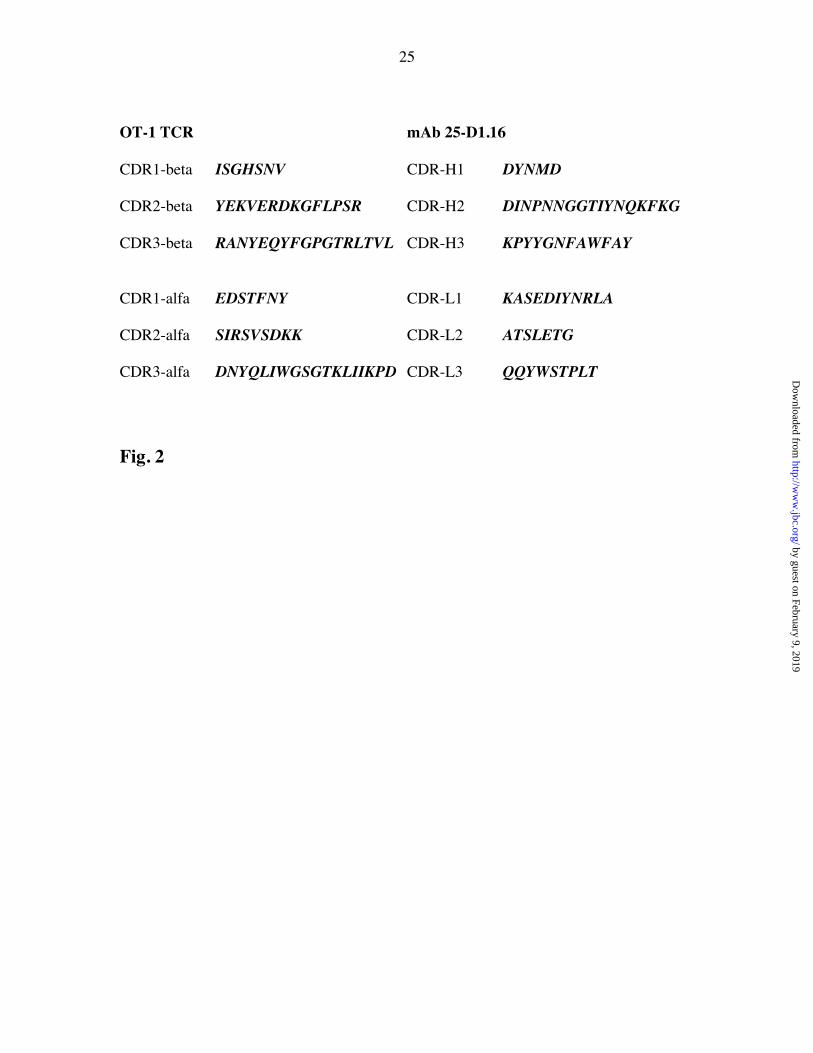
CDR regions of the 25-D1.16 mAb and the OT-1 TCRBased on X-ray crystallographic analysis, CDR regions of heavy and light chains
of Ig molecules can be described in term of known canonical structures (21,22). The
conformation of a particular canonical structure is determined by the length of the loop
and the nature of amino acid residues at key positions. This conformation is conserved
despite different positioning of the canonical loops relative to the framework regions.
The structure of hypervariable regions CDR-H1, CDR-H2, CDR-L1 and CDR-L2 of
mAb 25-D1.16 (Fig. 2) suggests that they belong to canonical classes 1, 2A, 2B, and 1
respectively (21). The CDR-H3 region that is expected to contact MHC-bound peptide ismuch more variable and is not included in the canonical-structure description (22), while
CDR-L3 belongs to canonical class 1.
The primary structures of the CDR regions of the OT-1 and the 25-D1.16 mAb
are shown in Fig. 2. They appear to be very different, indicating that CDRs with
divergent sequences may determine very similar specificities for pMHC binding (see
below).
Binding interactions of the 25-D1.16 mAb to various peptide-Kb complexesFig. 3 shows specific binding of soluble pOV8-Kb complex to the 25-D1.16 mAb
immobilized on a biosensor surface. The binding of two irrelevant peptide-Kb complexes
containing either Sendai virus peptide (SV9) or peptide from vesicular stomatitis virus
(VSV) was undetectable (not shown).
by guest on February 9, 2019http://w
ww
.jbc.org/D
ownloaded from
10
The values of kinetic and equilibrium (affinity) constants for the 25-D1.16 mAb
binding to pOV8-Kb and variant peptide-Kb complexes with defined biological activities
measured at 25oC are summarized in Table 1. Since the mAb was immobilized on the
biosensor surface, soluble monovalent pOV8-Kb complexes bound to the antibody
binding sites independently, allowing measurements of intrinsic parameters of the
interaction. This was confirmed by measuring binding of the antibody monovalent Fab
fragment to pOV8-Kb immobilized on the biosensor surface (data not shown).
Derivation of the kass and kdiss values using a 1:1 Langmuir binding modeldemonstrated a good correlation between the experimental and globally fitted data
(c2=0.05). The equilibrium binding constant Keq was calculated from the ratio kass/kdiss.
For a two-step reaction, Keq measured from the maximum (plateau) binding response is
expected to be different from the Keq value measured by the kass/kdiss ratio suggesting a
complex mechanism of the reaction (23,24). Very often, however, the intermediate
complex of the reaction is short-lived and the difference between the two Keq values is
not significant (25). We did not investigate this issue in detail here and utilized Keq
determined from the ratio kass/kdiss to compare it with corresponding data measured for the
binding of the same pMHC complexes to the OT-1 TCR (14).
Comparison of the specificity of the OT-1 TCR and the 25-D1.16 mAbTo compare the specificity of 25-D1.16 MAb and OT-1 TCR we analyzed
binding to peptide-Kb complexes whose biological activity (12,26) and the binding to the
OT-1 TCR (14,15) was previously determined. The value of kass of mAb 25-D1.16
binding to pOV8-Kb was 2.9x10⁴ M-1 s-1, an order of magnitude higher than kass for the
OT-1 TCR-pOV8-Kb interactions, while the difference in rate constant of dissociation for
the two reactions was only about 3 fold (Table 1). Thus, the gain in the free energy of 25-
D1.16-pOV8-Kb interaction is primarily determined by an increase in on-rate, but not by
a decrease in off-rate. This suggests that the activation energy of the dissociation phase of
the antibody-pMHC interaction is only slightly higher than that of the TCR-pMHC
interaction.The effect of amino acid substitutions in MHC-bound peptide on pOV8-Kb
recognition by the OT-1 TCR was similar to that for the 25-D1.16 mAb. For instance, the
by guest on February 9, 2019http://w
ww
.jbc.org/D
ownloaded from
11
R4-Kb complex, that induces positive selection of immature OT-1+ thymocytes (26) and
antagonizes cytolytic activity of mature OT-1 CTL (12), bound to the 25-D1.16 mAb
with lower affinity as it does to the OT-1 TCR (Table 1). In both cases, the loss of the
free binding energy was due to a faster off-rate, while changes in the on-rate were
marginal. Similarly, the Kb complex with V-OVA peptide that shares solvent accessible
amino acid residues with pOV8 and has similar to R4 biological activity (12,26) had a
faster rate of dissociation from the antibody and the TCR (Table 1). Interestingly, the free
energy of the V-OVA-Kb and pOV-8-Kb binding to the 25-D1.16 mAb was very similardue to a faster association rate of the V-OVA-Kb-25-D1.16 interaction. The most
profound effect resulted from substitution of a positively charged Lys for a negatively
charged Asp at P7 of the peptide (Table 1). The binding of the antibody to the D7-Kb
complex was not detectable. Although the OT-1-D7-Kb interaction has not been analyzed,
the D7 peptide failed to elicit a cytolytic response (12). Providing that D7 binds to Kb
with affinity similar to pOV8 and antagonizes responses of pOV8-specific OT-1 CTL
(12), the D7-Kb complex is apparently recognized by the TCR with a low affinity. Most
likely, the peptide contribution to the interaction is marginal, resulting in a very low free
energy of binding. The Ala substitution at P2 did not have any effect on the binding of
the A2-Kb complex to either the 25-D1.16 mAb or to the OT-1 TCR (Table 1).
Consistent with this, A2 peptide is an agonist for mature pOV8-specific CTL and causes
negative selection of immature T cells (27).
Substitution of the first Ser with negatively charged Glu in the E1-Kb complex,
that positively selects immature T cells (26) and functions as an agonist/antagonist (12),did not affect its binding to the 25-D1.16 MAb, but resulted in a faster off-rate of the
interaction with the OT-1 TCR (Table 1). Substitution of the Ser with positively charged
Arg (V-OVA) or Lys (KVDL) led to slightly higher rate constant of association of the
interaction with 25-D.1.16 MAb, while the interaction of the OT-1 TCR with V-OVA-Kb
was characterized by a faster off-rate and a slower on-rate. Although the binding of
KVDL-Kb complex to the OT-1 TCR was not measured, this complex did not induce any
detectable activity of pOV8-specific CTL (data not shown) indicating that KVDL-Kb
complex is not recognized by the TCR as opposed to the antibody. Thus, changes of the
by guest on February 9, 2019http://w
ww
.jbc.org/D
ownloaded from

12
first peptide amino acid influence the recognition of pOV8-Kb by the antibody and the
TCR in a different way.
The magnitude of the free energy changes (DDG) due to various peptide
substitutions was usually higher for OT-1 TCR binding than those for the 25-D1.16 mAb
binding, suggesting that the peptide energetic contribution to the TCR-pMHC interaction
was more significant than to the antibody-pMHC interaction (Table 1). Thus, in the latter
case the MHC moiety probably contributes more significantly, reflecting the 3-foldslower off-rate for the antibody-pOV8-Kb reaction. However, this increase in MHC
energetic contribution is still small enough to ensure sufficient peptide contribution and
the ability to distinguish the MHC bound peptide.
To further dissect the mechanism utilized by 25-D1.16 MAb to recognize Kb
bound peptides, we compared the energetic contributions of association and dissociation
phases of OT-1-peptide-Kb and 25-D1.16-peptide-Kb interactions exploiting the Eyring
transition-state theory that allows one to determine quasi-thermodynamic parameters of
the reaction association and dissociation phases from respective rate constants (20). The
DG#ass and DG#
diss values reflect the amount of energy contributed by the association and
dissociation phases to the free energy of interaction. The difference in these
contributions (Table 2) to the antibody and the TCR binding reflects changes in the
amount of energy coming from the association and the dissociation phases of 25-D1.16-
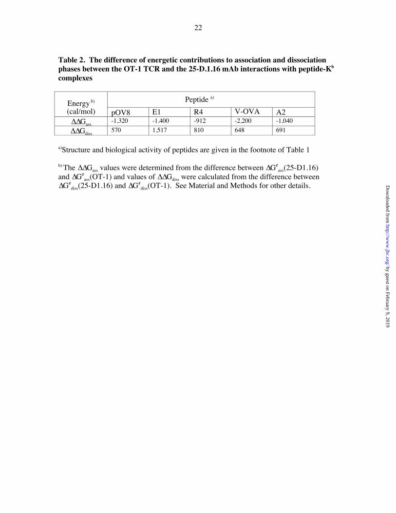
peptide-Kb interactions compared to corresponding OT-1-peptide-Kb interaction. Withonly one exception, the changes in the energetic contributions of the dissociation phase
(∆∆Gdiss) were smaller than those of the association phase (∆∆Gass). Mutation of Ser to
Glu (E1 peptide) had a different effect on the E1-Kb interaction with the TCR and the
antibody: it did not affect antibody binding, but did decrease the stability of the OT-1-E1-
Kb complex (Table 1). This explains an unusually high ∆∆Gdiss value for antibody
binding to E1-Kb (Table 2). These data provide evidence that the affinity gain for 25-
D1.16 to its natural ligand and other peptide-Kb complexes was mainly due to the lower
energy activation of the association phase, while changes in the energy of the dissociation
phase were limited to preserve the antibody specificity for the MHC-bound peptide.
by guest on February 9, 2019http://w
ww
.jbc.org/D
ownloaded from
13
DISCUSSIONAlthough recognition of pMHC complexes on the cell surface is a normal function of the
TCR, it has been shown that B cells can produce antibodies with TCR-like specificity in
vivo as well (13,28,29). In addition, the technology of phage display libraries (30,31) has
been successfully utilized to generate pMHC-specific antibodies (32-36). While these
antibodies are of great utility and can be used to study antigen-presentation as well as
detection and targeting of virus-infected and transformed cells, the mechanism, which
they utilize to specifically recognize associated with MHC peptide, is not unclear. In onestudy, Biddison et al. (34) performed alanine scanning of MHC and peptide amino acid
residues that make direct contacts with the TCR and compared the effect of these
substitutions on the recognition of these pMHC complexes by peptide-specific antibodies
and the TCR. They found that some amino acid substitutions have very similar effects on
recognition of pMHC variants by the TCR and the antibody, while others produce
different effects. This suggests that TCR and pMHC-specific antibodies use various
contacts on the pMHC surface to achieve the same specificity. This antibody, however,
was produced in vitro using combinatorial libraries. In addition, kinetics of the antibody
binding to the pMHC complex have not been measured. Thus, it is important to
investigate how the pMHC-specific antibodies derived in response to in vivo
immunization achieve specificities similar to the TCR.
In this study we have determined the intrinsic equilibrium and kinetic constants
for mAb 25-D1.16 binding to various peptide-Kb complexes with known biological
activity and compared these parameters with those for the interaction between the OT-1TCR and the same set of peptide-Kb complexes (Table 1). The comparison yielded two
major findings. The gain in affinity of the antibody for the peptide-Kb ligands was
mainly due to an increase in the rate constant of association; changes of the dissociation
rate constant were significantly smaller. Differences in the binding of pOV8 and its
variants associated with Kb to the antibody and to the OT-1 TCR were mainly due to
significant changes in on-rate, while changes in off-rate were moderate. This pattern of
changes suggests that the preservation of specificity for MHC-bound peptide limits
variation in the off-rate, but does not restrict changes in on-rate. In accord with these
findings, quasi-thermodynamic parameters of the association and dissociation phases
by guest on February 9, 2019http://w
ww
.jbc.org/D
ownloaded from
14
have shown that the gain in the free energy of 25-D1.16 binding is determined by a lower
energy barrier of the association phase, while energy changes of the dissociation phase
are similar for both the TCR and the antibody (Table 2 and Figure 4).
Kranz and colleagues produced soluble TCR encoded by mutated genes that bind
cognate pMHC ligand with intrinsic affinity of 3.6-6x10⁷ M-1 (11). The increase in the
TCR affinity relative to natural TCR was mainly due to an increase in the on-rate, not
off-rate, suggesting again that limited activation energy of the dissociation phase of the
TCR-pMHC reaction is required to preserve peptide specificity. While this TCR withenhanced affinity clearly discriminates cognate pMHC on the cell surface, T cell
hybridomas carrying this TCR respond not only to the cognate pMHC on target cells, but
also to target cells of the same haplotype that do not display foreign-like pMHC
complexes (11). This response is apparently mediated by a prohibitively high energetic
contribution of the MHC moiety, causing an autoimmune reaction. Thus, limited
energetic input by the MHC-TCR contact in recognition of syngeneic pMHC ligands is
an important condition by which the MHC restriction and the peptide specificity are met
and the reactivity against self pMHC is avoided. The same group has also investigated
the energy map of the interaction between the same TCR and its allogeneic pMHC ligand
(37). Site-directed mutagenesis of the TCR showed that CDR1 and CDR2 residues
contacting the MHC moiety collectively contribute more binding energy than CDR3
residues responsible for contact with the peptide. This indicates that the MHC energetic
contribution of the allogeneic ligand dominates, as opposed to that of the syngeneic
pMHC. Other analyses of the binding of some recombinant TCRs with enhanced affinityto a syngeneic or to an allogeneic pMHC revealed similar contributions of the MHC and
the peptide moieties (38). However, in this particular system the recognition of
allogeneic ligand is strongly peptide-dependent (39,40). In the addition, the peptide
induces conformational changes in the MHC protein that are detectable by antibodies
(41,42). In these circumstances, the peptide may contribute significantly to the amount of
binding energy delivered by the allogeneic MHC indirectly through conformational
changes of the MHC moiety. This may also explain why the affinity of the TCR
recognizing this allogeneic pMHC could be increased by significant changes of the
interaction’s off-rate (43). Not surprisingly T cells expressing this high-affinity TCR did
by guest on February 9, 2019http://w
ww
.jbc.org/D
ownloaded from

15
not respond to target cells bearing irrelevant pMHC complexes (43). In another example,
the equilibrium binding constant for a TCR interaction with class II invariant chain
associated peptide (CLIP) bound to I-Ab MHC class II protein (44) was measured to be
1.6x10⁷ M-1 (Teyton and Rudensky, personal communication). Since CLIP peptide is
thought to cause conformational changes of MHC class II molecules (45), this TCR may
recognize a unique conformation of the MHC bound to CLIP peptide, and a substantial
energetic contribution of the MHC moiety may account for high TCR affinity in this
case.The increase of the kass value reflects more precise docking of interacting proteins
and a higher probability of productive protein-protein encounters. Usually, kass depends
on long-range electrostatic interactions (25,46). These interactions generate an “energy
funnel” between the two molecules, facilitating proper orientation that increases the
probability of specific complex formation (47). Higher stability and lower flexibility of
the interacting surfaces (48) are thought to facilitate long-range electrostatic interactions
(49). The latter may not necessarily become short-term interactions contributing to the
stability of the complex measured by the reaction off-rate. Most likely, enhanced long-
range interactions lead to an increase of the on-rate of 25-D1.16 antibody binding to
pOV8-Kb, but do not change the balance of energetic contributions made by the peptide
and the MHC, ensuring the peptide specificity of the antibody.
Analysis of the primary structure of the 25-D1.16 V genes encoding VH and VL
domains have very few mutations, all are in the framework regions, while CDR regions
appear to be in germ-line configuration. This suggests that the VH and VL genes of 25-D1.16 have not completed affinity maturation. It has been suggested that initially affinity
maturation of antibodies is driven by increased on-rate (50) and this process may be
mediated by mutations in framework regions. Subsequent somatic mutations in CDR
regions lead to lower off-rate of antibody binding and further increase of the binding
affinity (50). Davis and colleagues found that further maturation of an antibody specific
for moth cytochrome C (MCC) peptide in association with I-Ek MHC class II led to a
higher affinity antibody that could no longer discriminate bound to the I-Ek MCC peptide
from other self-I-Ek complexes on the surface of live cells (51). Thus, it appears that the
limited affinity of pMHC-specific antibodies and the absence of extensive somatic
by guest on February 9, 2019http://w
ww
.jbc.org/D
ownloaded from
16
mutations in the CDR regions of these antibodies may be linked and are similar to those
properties of TCR whose genes are not a subject for somatic mutations. Normally, a B
cell response to protein antigens is driven to produce IgG antibody with high affinity,
through extensive somatic hypermutation and clonal selection manifested in the germinal
center. The antibody studied here does not appear to fall into this conventional category,
providing a possible explanation for the rare occurrence of pMHC-specific antibodies
elicited in vivo.
ACKNOLEDGEMENTWe are grateful to Drs. Angel Porgador and Ronald Germain for providing hybridoma
25-D1.16. We also thank Dr. Steve Jameson for critical reading of the manuscript. This
work was supported by an NIH research grants AI43254 and AI39966 to Y.S.
REFERENCES1. Rudolph, M. G., and Wilson, I. A. (2002) Curr Opin Immunol 14, 52-65
2. Baker, B. M., Turner, R. V., Gagnon, S. J., Wiley, D. C., and Biddison, W. E.
(2001) J Exp Med 193, 551-562
3. Wang, Z., Turner, R., Baker, B. M., and Biddison, W. E. (2002) J Immunol 169,
3146-3154
4. Willcox, B., Gao, G., Wyer, J., Ladbury, J., Bell, J., Jakobsen, B., and van derMerwe, P. (1999) Immunity 10, 357-365
5. Boniface, J. J., Reich, Z., Lyons, D. S., and Davis, M. M. (1999) Proc Natl Acad
Sci U S A 96, 11446-11451
6. Manning, T. C., and Kranz, D. M. (1999) Immunol Today 20, 417-422.
7. Garcia, K. C., Radu, C. G., Ho, J., Ober, R. J., and Ward, E. S. (2001) Proc Natl
Acad Sci U S A 98, 6818-6823.
8. Anikeeva, N., Lebedeva, T., Krogsgaard, M., Tetin, S. Y., Martinez-Hackert, E.,
Kalams, S. A., Davis, M. M., and Sykulev, Y. (2003a) Biochemistry 42, 4709-
4716
9. Janin, J., and Chothia, C. (1978) Biochemistry 17, 2943-2948
10. Stites, W. E. (1997) Chem. Rev 97, 1233-1250
by guest on February 9, 2019http://w
ww
.jbc.org/D
ownloaded from

17
11. Holler, P. D., Chlewicki, L. K., and Kranz, D. M. (2003) Nat Immunol 4, 55-62
12. Jameson, S. C., Carbone, F. R., and Bevan, M. J. (1993) J. Exp. Med. 177, 1541-
1550
13. Porgador, A., Yewdell, J. W., Deng, Y., Bennink, J. R., and Germain, R. N.
(1997) Immunity 6, 715-726.
14. Alam, S. M., Travers, P. J., Wung, J. L., Nasholds, W., Redpath, S., Jameson, S.
C., and Gascoigne, N. R. (1996) Nature 381, 616-620
15. Alam, S., Davies, G., Lin, C., Zal, T., Nasholds, W., Jameson, S., Hogquist, K.,Gascoigne, N., and Travers, P. (1999) Immunity 10, 227-237
16. Brunmark, A., and Jackson, M. (1998) in MHC (Fernandez, N., and Butcher, G.,
eds) Vol. 2, pp. 53-78, Oxford University Press, Oxford
17. Anikeeva, N., Lebedeva, T., Sumaroka, M., Kalams, S. A., and Sykulev, Y.
(2003b) J. Immunol. Meth. 277, 75-86
18. Markvicheva, E. A., Kuptsova, S. V., Mareeva, T. Y., Vikhrov, A. A., Dugina, T.
N., Strukova, S. M., Belokon, Y. N., Kochetkov, K. A., Baranova, E. N., Zubov,
V. P., Poncelet, D., and Rumsh, L. D. (2000) Appl. Biochem.Biotechnol. 88, 145-
157
19. Mozdzanowski, J., Bongers, J., and Anumula, K. (1998) Anal Biochem 260, 183-
187
20. Eisenberg, D., and Crothers, D. (1979) Physical chemistry with applications to the
life sciences, The Benjamin/Cummings Publishing Company, Menlo Park,
California21. Al-Lazikani, B., Lesk, A. M., and Chothia, C. (1997) J Mol Biol 273, 927-948
22. Morea, V., Tramontano, A., Rustici, M., Chothia, C., and Lesk, A. M. (1998) J
Mol Biol 275, 269-294
23. Wu, L. C., Tuot, D. S., Lyons, D. S., Garcia, K. C., and Davis, M. M. (2002)
Nature 418, 552-556.
24. Gakamsky, D. M., Luescher, I. F., and Pecht, I. (2004) Proc Natl Acad Sci U S A
101, 9063-9066
25. Schreiber, G. (2002) Curr Opin Struct Biol 12, 41-47.
by guest on February 9, 2019http://w
ww
.jbc.org/D
ownloaded from
18
26. Hogquist, K. A., Jameson, S. C., Heath, W. R., Howard, J. L., Bevan, M. J., and
Carbone, F. R. (1994) Cell 76, 17-27.
27. Hogquist, K. A., Tomlinson, A. J., Kieper, W. C., McGargill, M. A., Hart, M. C.,
Naylor, S., and Jameson, S. C. (1997) Immunity 6, 389-399.
28. Aharoni, R., Teitelbaum, D., Arnon, R., and Puri, J. (1991) Nature 351, 147-150
29. Murphy, D. B., Rath, S., Pizzo, E., Rudensky, A. Y., George, A., Larson, J. K.,
and Janeway, C. A., Jr. (1992) J Immunol 148, 3483-3491
30. Clackson, T., Hoogenboom, H. R., Griffiths, A. D., and Winter, G. (1991) Nature
352, 624-628
31. Nissim, A., Hoogenboom, H. R., Tomlinson, I. M., Flynn, G., Midgley, C., Lane,
D., and Winter, G. (1994) Embo J 13, 692-698
32. Andersen, P. S., Stryhn, A., Hansen, B. E., Fugger, L., Engberg, J., and Buus, S.
(1996) Proc. Natl. Acad. Sci. USA 93, 1820-1824
33. Denkberg, G., Cohen, C. J., Lev, A., Chames, P., Hoogenboom, H. R., and Reiter,
Y. (2002) Proc Natl Acad Sci U S A 99, 9421-9426.
34. Biddison, W. E., Turner, R. V., Gagnon, S. J., Lev, A., Cohen, C. J., and Reiter,
Y. (2003) J Immunol 171, 3064-3074
35. Lev, A., Denkberg, G., Cohen, C. J., Tzukerman, M., Skorecki, K. L., Chames, P.,
Hoogenboom, H. R., and Reiter, Y. (2002) Cancer Res 62, 3184-3194
36. Cohen, C. J., Denkberg, G., Lev, A., Epel, M., and Reiter, Y. (2003) J Mol
Recognit 16, 324-332
37. Manning, T. C., Schlueter, C. J., Brodnicki, T. C., Parke, E. A., Speir, J. A.,Garcia, K. C., Teyton, L., Wilson, I. A., and Kranz, D. M. (1998) Immunity 8,
413-425
38. Lee, P. U., Churchill, H. R., Daniels, M., Jameson, S. C., and Kranz, D. M. (2000)
J Exp Med 191, 1355-1364
39. Udaka, K., Wiesmuller, K.-H., Kienle, S., Jung, G., and Walden, P. (1996) J.
Immunol. 157, 670-678
40. Sykulev, Y., Brunmark, A., Jackson, M., Cohen, R. J., Peterson, P. A., and Eisen,
H. N. (1994a) Immunity 1, 15-22
by guest on February 9, 2019http://w
ww
.jbc.org/D
ownloaded from
19
41. Al-Ramadi, B. K., Jelonek, M. T., Boyd, L. F., Margulies, D. H., and Bothwell,
A. L. (1995) J. Immunol. 155, 662-673
42. Robinson, R. A., and Lee, D. R. (1996) J. Immunol. 156, 4266-4273
43. Holler, P. D., Lim, A. R., Cho, B. K., Rund, L. A., and Kranz, D. M. (2001) J Exp
Med 194, 1043-1052
44. Zhu, Y., Rudensky, A. Y., Corper, A. L., Teyton, L., and Wilson, I. A. (2003) J
Mol Biol 326, 1157-1174
45. Zarutskie, J. A., Busch, R., Zavala-Ruiz, Z., Rushe, M., Mellins, E. D., and Stern,L. J. (2001) Proc Natl Acad Sci U S A 98, 12450-12455
46. England, P., Bregegere, F., and Bedouelle, H. (1997) Biochemistry 36, 164-172.
47. Zhang, C., Chen, J., and DeLisi, C. (1999) Proteins 34, 255-267
48. Shusta, E. V., Holler, P. D., Kieke, M. C., Kranz, D. M., and Wittrup, K. D.
(2000) Nat Biotechnol 18, 754-759
49. Sinha, N., Mohan, S., Lipschultz, C. A., and Smith-Gill, S. J. (2002) Biophys J 83,
2946-2968
50. Foote, J., and Milstein, C. (1991) Nature 352, 530-532
51. Reay, P. A., Matsui, K., Haase, K., Wulfing, C., Chien, Y. H., and Davis, M. M.
(2000) J Immunol 164, 5626-5634.
52. Kabat, E. A., Wu, T. T., Perry, H. M., Gottesman, K. S., and Feoller, C. (1991)
Sequences of Proteins of immunological Interest, 5th Ed., US Department of
Health and Human Services, NIH, Bethesda, MD
FIGURE LEGENDS
Figure 1. Nucleotide and encoded amino acid sequences for the variable domaingenes of 25-D1.16The amino acid residue numbering and the CDR limits are according to Kabat (52).
Differences with germline sequences and the corresponding encoded amino acid
substitutions are also shown.
A: VH domain compared with the VH germline J558.6 gene (Accession number
AF303837).
by guest on February 9, 2019http://w
ww
.jbc.org/D
ownloaded from
20
B: CDR-H3 and FR-H4 compared with the DSP2.7 and JH3 segments. (Accession
Numbers J00438 and V00770)
C: VL domain compared with the germline Vk33 gene (Accession number
AJ231273).
D: FR-L4 compared with the Jk5 germline gene. (Accession number V00777)
Figure 2. Primary structure of hypervariable regions of the 25-D1.16 mAb and theOT-1 TCR
Figure 3. Interaction of soluble pOV8-Kb with 25-D1.16 immobilized on abiosensor surfaceA . Sensograms showing the binding of soluble pOV8-Kb complex at indicated
concentrations to immobilized mAb 25-D1.16 (400 R.U. immobilized). For other
details see Material and Methods.
B. Residual data distribution for the association and dissociation phases, after global
curve fitting to the 1:1 bimolecular (Lengmuir) interaction model (c2=0.05)
Figure 4. Free-energy changes in the interactions of the pOV8-Kb complex with the25-D1.16 mAb and OT-1 TCRActivation free-energy of the association phase (DG#
ass) of the antibody binding is
significantly higher than that of TCR binding, while activation free-energy of the
dissociation phase (DG#diss) is similar for both interactions.
by guest on February 9, 2019http://w
ww
.jbc.org/D
ownloaded from
21
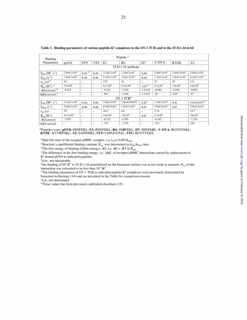
Table 1. Binding parameters of various peptide-Kb complexes to the OT-1 TCR and to the 25-D.1.16 mAb
a)Peptides were: pOV8: SIINFEKL; E1: EIINFEKL; R4: SIIRFEKL: D7: SIINFEDL; V-OVA: RGYNYEKL; KVDL: KVVRFDKL; A2: SAINFEKL; SV9: FAPGNYPAL; VSV: RGYVYQGL
b)Half-life time of the receptor-pMHC complex, i.e. t1/2= 0.693/kdissc)Reaction’s equilibrium binding constant, Keq, was determined as kass/kdiss ratiod)The free energy of binding (Gibbs energy), ∆G, i.e. ∆G = -RT ln Keqe)The difference in the free binding energy, i.e. ∆∆G, of receptor-pMHC interactions caused by replacement ofKb-bound pOV8 to indicated peptidesf)n.m., not measurableg)the binding of D7-Kb to 25-D.1.16 immobilized on the biosensor surface was to too weak to measure; Keq of thisinteraction was estimated to be less than 104 M-1
h)The binding parameters of OT-1 TCR to indicated peptide-Kb complexes were previously determined bybiosensor technology (14) and are presented in the Table for comparison reasonsi)n.d., not determinedk)These values has been previously published elsewhere (15)
Binding Peptide a)
Parameters pOV8 SV9 VSV E1 R4 D7 V-OVA KVDL A225.D-1.16 antibody
kass (M-1 s-1) 2.9±0.1x10⁴ n.m.f) n.m. 3.2±0.3x10⁴ 1.2±0.2x10⁴ n.m. 5.4±0.1x10⁴ 3.4±0.3x10⁴ 2.8±0.1x10⁴
kdiss (s-1) 7.6±0.3x10⁻³ n.m. n.m. 5.2±0.1x10⁻³ 3.8±0.3x10⁻² n.m. 1.3±0.1x10⁻² 1.8±0.2x10⁻² 6.2±0.1x10⁻³
t1/2 (s) b) 91 - - 133 18 - 53 39 112Keq (M-1) c) 3.9x10⁶ - - 6.1x10⁶ 0.32x10⁶ <104 g) 4.1x10⁶ 1.9x10⁶ 4.6x10⁶
∆G(cal/mol) d) -8,955 - - -9,218 -7,479 <-5,434 -8,984 -8,530 -9,052∆∆G(cal/mol) e) - - - 263 -1,476 >-3,521 29 -425 97
OT-1 TCRh)
kass (M-1 s-1) 3.1±0.1x10³ n.m. n.m. 3.0±0.3x10³ 2.56±0.04x10³ n.d.i) 1.3±0.3x10³ n.d. 4.8±0.8x10³ k)
kdiss (s-1) 2.0±0.1x10⁻² n.m. n.m. 6.8±0.4x10⁻² 1.5±0.1x10⁻¹ n.d. 3.9±0.5x10⁻² n.d. 2.0±0.3x10⁻²
t1/2 (s) 35 - - 10.2 4.6 - 17.8 34.7Keq (M-1) 15.5x10⁴ - - 4.4x10⁴ 1.8x10⁴ n.d. 3.3x10⁴ 24x104
∆G(cal/mol) -7,055 - - -6,312 -5,785 - -6,142 -7,314∆∆G(cal/mol) - - - -743 -1,270 - -913 259
by guest on February 9, 2019http://w
ww
.jbc.org/D
ownloaded from
22
Table 2. The difference of energetic contributions to association and dissociationphases between the OT-1 TCR and the 25-D.1.16 mAb interactions with peptide-Kb
complexes
a)Structure and biological activity of peptides are given in the footnote of Table 1
b) The ∆∆Gass values were determined from the difference between ∆G#ass(25-D1.16)
and ∆G#ass(OT-1) and values of ∆∆Gdiss were calculated from the difference between
∆G#diss(25-D1.16) and ∆G#
diss(OT-1). See Material and Methods for other details.
Energy b) Peptide a)
(cal/mol) pOV8 E1 R4 V-OVA A2∆∆Gass -1,320 -1,400 -912 -2,200 -1,040∆∆Gdiss 570 1,517 810 648 691
by guest on February 9, 2019http://w
ww
.jbc.org/D
ownloaded from

23
AVH 25-D1.16
J558.6(AF303837)
VH 25-D1.16
J558.6
VH 25-D1.16
J558.6
VH 25-D1.16
J558.6
1 2 3 4 5 6 7 8 9 10 11 12 13 14 15 16 17 18 19 20 21 22 23 24 25 26 E V L L Q Q S G P E L V K P G A S V K I P C K A S GGAG GTC CTG CTG CAA CAG TCT GGA CCT GAG CTG GTG AAG CCT GGG GCT TCA GTG AAG ATA CCC TGT AAG GCT TCT GGA--- --- --- --- --- --- --- --- --- --- --- --- --- --- --- --- --- --- --- --- --- --C --- --- --- --- * * * * * * * * * * * * * * * * * * * * * * * * * *
--------CDR1------- -------------27 28 29 30 31 32 33 34 35 36 37 38 39 40 41 42 43 44 45 46 47 48 49 50 51 52 Y T F T D Y N M D W V K Q S H G K S L E W I G D I NTAC ACA TTC ACT GAC TAC AAC ATG GAC TGG GTG AAG CAG AGC CAT GGA AAG AGC CTT GAG TGG ATT GGA GAT ATT AAT--- --- --- --- --- --- --- --- --- --- --- --- --- --- --- --- --- --- --- --- --- --- --- --- --- --- * * * * * * * * * * * * * * * * * * * * * * * * * *
-----------------CDR2---------------------------------- A 53 54 55 56 57 58 59 60 61 62 63 64 65 66 67 68 69 70 71 72 73 74 75 76 77 P N N G G T I Y N Q K F K G K A T L T V D K S S S ACCT AAC AAT GGT GGT ACT ATC TAC AAC CAG AAG TTC AAG GGC AAG GCC ACA TTG ACT GTA GAC AAG TCT TCC AGC GCA--- --- --- --- --- --- --- --- --- --- --- --- --- --- --- --- --- --- --- --- --- --- --C --- --- A-- * * * * * * * * * * * * * * * * * * * * * * * * * T
---------------CDR3-----78 79 80 81 82 A B C 83 84 85 86 87 88 89 90 91 92 93 94 95 96 97 98 99 100 A Y M E V R S L T S E D T A V Y Y C A R K P Y Y G NGCC TAC ATG GAG GTC CGC AGC CTG ACA TCT GAG GAC ACT GCA GTC TAT TAC TGT GCA AGA AAA CCC TAC TAT GGT AAC--- --- --- --- C-- --- --- --- --- --- --- --- --- --- --- --- --- --- --- --- * * * * L * * * * * * * * * * * * * * *
----------------------- A B C D 101 102 103 104 105 106 107 108 109 110 111 112 113 F A W F A Y W G Q G T L V T V S ATTC GCC TGG TTT GCT TAC TGG GGC CAA GGG ACT CTG GTC ACT GTC TCT GCA
B
VH 25-D1.16
DSP2.7(J00438)JH3 (V00770)
---------------CDR3----------------------------93 94 95 96 97 98 99 100 A B C D 101 102 103 104 105 106 107 108 109 110 111 112 113 A R K P Y Y G N F A W F A Y W G Q G T L V T V S AGCA AGA AAA CCC TAC TAT GGT AAC TTC GCC TGG TTT GCT TAC TGG GGC CAA GGG ACT CTG GTC ACT GTC TCT GCA -- --- --- --- --- - --- --- --- --- --- --- --- --- --- --- --- --- --- --- --- --- * * * * * * * * * * * * * * * * * * * * *
C
VL 25-D1.16
VL kappa33(AJ231273)
----------- 1 2 3 4 5 6 7 8 9 10 11 12 13 14 15 16 17 18 19 20 21 22 23 24 25 26 D I Q V T Q S S S S F S V S L G D R V T I T C K A SGAC ATC CAG GTG ACA CAA TCT TCA TCC TCC TTT TCT GTT TCT CTA GGA GAC AGA GTC ACC ATT ACT TGC AAG GCA AGT--- --- --- A-- --- --- --- --- --- --- --- --- --A --- --- --- --- --- --- --- --- --- --- --- --- --- * * * M * * * * * * * * * * * * * * * * * * * * * *
--------CDR1------------------- ------------27 28 29 30 31 32 33 34 35 36 37 38 39 40 41 42 43 44 45 46 47 48 49 50 51 52 E D I Y N R L A W Y Q Q K P G N A P R L L I S G A TGAG GAC ATA TAT AAT CGT TTA GCC TGG TAT CAG CAG AAA CCA GGA AAT GCT CCT AGG CTC TTA ATA TCT GGT GCA ACC--- --- --- --- --- --G --- --- --- --- --- --- --- --- --- --- --- --- --- --- --- --- --- --- --- --- * * * * * * * * * * * * * * * * * * * * * * * * * *
--CDR2---------53 54 55 56 57 58 59 60 61 62 63 64 65 66 67 68 69 70 71 72 73 74 75 76 77 78 S L E T G V P S R F S G S G S R K D Y T L I I T S LAGT TTG GAA ACT GGG GTT CCT TCA AGA TTC AGT GGC AGT GGA TCT AGA AAG GAT TAC ACT CTC ATC ATT ACC AGT CTT--- --- --- --- --- --- --- --- --- --- --- --- --- --- --- G-- --- --- --- --- --- -G- --- --- --- --- * * * * * * * * * * * * * * * G * * * * * S * * * *
-----------------CDR3--------------79 80 81 82 83 84 85 86 87 88 89 90 91 92 93 94 95 96 97 98 99 100 101 102 103 104 Q T E D V A T Y Y C Q Q Y W S T P L T F G A G T K LCAG ACT GAA GAT GTT GCT ACT TAT TAC TGT CAA CAG TAT TGG AGT ACT CCT CTC ACG TTC GGT GCT GGG ACC AAG CTG--- --- --- --- --- --- --- --- --- --- --- --- --- --- --- --- --- * * * * * * * * * * * * * * * * *
105 106 107 108 E L K RGAG CTG AAA CGG
DVL 25-D1.16
JK5 (V00777)
96 97 98 99 100 101 102 103 104 105 106 107 108 L T F G A G T K L E L K RCTC ACG TTC GGT GCT GGG ACC AAG CTG GAG CTG AAA CGG--- --- --- --- --- --- --- --- --- --- --- --- -- * * * * * * * * * * * * *
by guest on February 9, 2019http://w
ww
.jbc.org/D
ownloaded from
25
OT-1 TCR mAb 25-D1.16
CDR1-beta ISGHSNV CDR-H1 DYNMD
CDR2-beta YEKVERDKGFLPSR CDR-H2 DINPNNGGTIYNQKFKG
CDR3-beta RANYEQYFGPGTRLTVL CDR-H3 KPYYGNFAWFAY
CDR1-alfa EDSTFNY CDR-L1 KASEDIYNRLA
CDR2-alfa SIRSVSDKK CDR-L2 ATSLETG
CDR3-alfa DNYQLIWGSGTKLIIKPD CDR-L3 QQYWSTPLT
Fig. 2
by guest on February 9, 2019http://w
ww
.jbc.org/D
ownloaded from

26
Fig. 3
Time (s)0 50 100 150 200
Res
pons
e, R
U
0
30
60
90
Res
idua
l 0.8
- 0.8
0
RU
Time (s)0 50 100 150 200
450 nM300 nM200 nM150 nM 50 nM
A
B
by guest on February 9, 2019http://w
ww
.jbc.org/D
ownloaded from
27
mAb 25-D1.16 TCR OT-1
∆G#ass
∆G#diss
∆G
Ab + pM
Ab~pM#
Ab–pM
∆G#ass
∆G#diss
∆GTCR + pM
TCR~pM#
TCR–pM
G
Fig. 4
by guest on February 9, 2019http://w
ww
.jbc.org/D
ownloaded from

Tatiana Mareeva, Tatiana Lebedeva, Nadia Anikeeva, Tim Manser and Yuri SykulevAntibody specific for the peptide-MHC complex: is it TCR-like?
published online August 9, 2004J. Biol. Chem.
10.1074/jbc.M407021200Access the most updated version of this article at doi:
Alerts:
When a correction for this article is posted•
When this article is cited•
to choose from all of JBC's e-mail alertsClick here
by guest on February 9, 2019http://w
ww
.jbc.org/D
ownloaded from