antibiotics: classification and mechanisms of action … · antibiotics: classification and...
TRANSCRIPT

IJAMBR 4 (2016) 90-101 ISSN 2053-1818
Antibiotics: Classification and mechanisms of action with emphasis on molecular perspectives
Ebimieowei Etebu1* and Ibemologi Arikekpar2
1Molecular Microbiology Unit, Department of Biological Sciences, Faculty of Science, Niger Delta University,
Amassoma, Wilberforce Island, Nigeria. 2Department of Medical Laboratory Sciences, Faculty of Basic Medical Sciences, College of Health Sciences,
Niger Delta University, Amassoma, Wilberforce Island, Nigeria.
Article History ABSTRACT Received 19 September, 2016 Received in revised form 09 October, 2016 Accepted 13 October, 2016 Keywords: Antibiotics, Bacteria, Molecular biology, Protein synthesis, Ribosomes. Article Type: Review
Man and the microbial world have co-existed since time immemorial. Whilst some bacteria are able to strike a symbiotic balance with man; providing a protective and stabilizing effect on the body as resident microbes, pathogenic bacteria invade and grow in man’s tissues causing diseases and damaging the body, sometimes leading to death. Man’s search for a cure to her; many bacterial adversaries led to the discovery and use of antibiotics in the 1940’s. Whilst antagonizing disease causing bacteria, antibiotics are known to cause harmful effects on the normal and useful microbiota of the human biological system. The use of antibiotics is therefore, hinged on the overall intended benefit, taking into consideration the attendant negative side effects. Proper characterization and adequate understanding of the mode of action of antibiotics is therefore an indispensable necessity required to safeguard man’s healthcare delivery system. Recent molecular biological approaches have greatly contributed to understanding how antibiotics antagonize bacteria. Hence in this paper, the classification of antibiotics and their mode of action are reviewed with emphasis on molecular perspectives.
©2016 BluePen Journals Ltd. All rights reserved
INTRODUCTION The term antibiotic was coined from the word „antibiosis‟ which literally means „against life‟. In the past, antibiotics were considered to be organic compounds produced by one microorganism which are toxic to other microorganisms (Russell, 2004). As a result of this notion, an antibiotic was originally, broadly defined as a substance, produced by one microorganism (Denyer et al., 2004), or of biological origin (Schlegel, 2003) which at low concentrations can inhibit the growth of, or are lethal to other microorganisms (Russell, 2004). However, this definition has been modified in modern times, to include antimicrobials that are also produced partly or wholly
*Correspondence author. E-mail: [email protected]. Tel: +234 (0) 802-982-9015.
through synthetic means. Whilst some antibiotics are able to completely kill other
bacteria, some are only able to inhibit their growth. Those that kill bacteria are termed bactericidal while those that inhibit bacterial growth are termed bacteriostatic (Walsh, 2003). Although antibiotic generally refers to antibacterial, antibiotic compounds are differentiated as antibacterials, antifungals and antivirals to reflect the group of microorganisms they antagonize (Brooks et al., 2004; Russell, 2004).
Penicillin was the first antibiotic discovered in September 1928 by an English Bacteriologist, late Sir Alexander Fleming who accidentally obtained the antibiotic from a soil inhabiting fungus Penicillium notatum but its discovery was first reported in 1929 (Aminov, 2010), and clinical trials first conducted on humans in 1940 (Russell, 2004; Schlegel, 2003).

Figure 1. Chemical structure of a beta-lactam ring (Tidwell, 2008).
Figure 2. Chemical structure of beta-lactam structure. Core structure of penicillins (top) and cephalosporins (bottom) (Holten and Onusko, 2000).
The discovery and development of the first significant antibiotic “penicillin” in 1920‟s, and subsequent introduction into man‟s health care system in the 1940‟s has continued to transform the management and fight against bacterial infections (White and Cox, 2013). However, antibiotics are not totally selective in their antibacterial activity. Whilst antagonizing disease causing bacteria, they also antagonize the normal and useful microbiota that we all have and need in our systems as those in the gastrointestinal tract (Walsh, 2003). Prescription and administration of any given antibiotics is
Int. J. Appl. Microbiol. Biotechnol. Res. 91 therefore predicated on the overall intended benefit, taking into consideration the attendant side effects. For this reason, it is pertinent to understand the mechanism of action of every identified antibiotic before introduction into our health care delivery system, and recent molecular biological approaches have played very significant roles to elucidate our understanding in this regard.
Hence this paper aimed to review the classification of antibiotics and their mode of action with emphasis on molecular perspectives. CLASSIFICATION OF ANTIBIOTICS There are several ways of classifying antibiotics but the most common classification schemes are based on their molecular structures, mode of action and spectrum of activity (Calderon and Sabundayo, 2007). Others include route of administration (injectable, oral and topical). Antibiotics within the same structural class will generally show similar pattern of effectiveness, toxicity and allergic-potential side effects.
Some common classes of antibiotics based on chemical or molecular structures include Beta-lactams, Macrolides, Tetracyclines, Quinolones, Aminoglycosides, Sulphonamides, Glycopeptides and Oxazolidinones (van Hoek et al., 2011; Frank and Tacconelli, 2012; Adzitey, 2015). Beta-lactams Members of this class of antibiotics contain a 3-carbon and 1-nitrogen ring that is highly reactive (Figures 1 and 2). They interfere with proteins essential for synthesis of bacterial cell wall, and in the process either kills or inhibits their growth. More succinctly, certain bacterial enzymes termed penicillin-binding protein (PBP) are responsible for cross linking peptide units during synthesis of peptidoglycan. Members of beta-lactam antibiotics are able to bind themselves to these PBP enzymes, and in the process, they interfere with the synthesis of peptidoglycan resulting to lysis and cell death (Heesemann, 1993). The most prominent representatives of the beta-lactam class include Penicillins, Cephalosporins, Monobactams and Carbapenems. Penicillins The first antibiotic, penicillin, which was first discovered and reported in 1929 by Alexander Fleming was later found to be among several other antibiotic compounds called the penicillins. (McGeer et al., 2001). Penicillins

Etebu and Arikekpar 92
Figure 3. Structure of Cephalosporins (Pegler and Healy, 2007).
are involved in a class of diverse group of compounds, most of which end in the suffix -cillin. They are beta-lactam compounds containing a nucleus of 6-animopenicillanic acid (lactam plus thiazolidine) ring and other ring side chains (Zahner and Maas, 1972).
Members of Penicillin class include Penicillin G, Penicillin V, Oxacillin (dicloxacillin), Methicillin, Nafcillin, Ampicillin, Amoxicillin, Carbenicilin, Piperacillin, Mezlocillin and Ticarcillin (Boundless, 2016). Penicillin G was the first to be produced amongst this group of antibiotics, and in fact of all antibiotics. Although penicillin G was discovered by Alexander Fleming in the 1920‟s, it took the efforts of several other workers such as Ernst Chain, Edward Abraham, Norman Heatley and Howard Florey in 1945 to understand the cultural requirements of the fungus and its clinical effectiveness. Furthermore, although Penicillin G was originally discovered and isolated from the fungus P. notatum by Alexander Flemming, a close relative Penicilliun chrysogenum is the preferred choice of source. Also, producing the antibiotics through biochemical microbial fermentation more cost effective as compared to synthesizing it from raw materials (Talaro and Chess, 2008). There is no gainsaying that the discovery of this drug heralded the introduction of antibiotics into our health care delivery system. Sadly, however, Penicillin G has a narrow spectrum; only Gram positive bacteria (streptococci) and some Gram-negative bacteria such as Treponema pallidum causative agent for syphilis, and meningococci are sensitive to it (Talaro and Chess, 2008).
As with every biological interaction systems where living systems seek to protect itself from attack, certain bacteria are able to counter the activity of antibiotics by encoding enzymes. In view of this, some antibiotics such as ampicillin, carbenicillin and amoxicillin have been developed semi-synthetically with different side-chains. These side chains confer on the antibiotics the ability to evade the degradative capacity of certain enzymes
produced by certain bacterial strains as well as facilitating the movement of antibiotics across the outer membrane of such bacterial cell walls. This double-pronged capability increases their spectrum of activity against Gram-negative bacteria. In particular, some penicillins such as Augmentin are produced in combination with non-antibiotic compound that are able to inhibit the activity of bacterial penicillinase enzyme. Augmentin is actually a drug comprising amoxicillin (antibiotic) and clavulanic acid a non-antibiotic compound. Clavulanic acid is able to inhibit beta-lactamase enzyme thereby prolonging the antibacterial activity of the amoxicillin component of Augmentin even amongst penicillinase producing bacteria (Poirel et al., 2005). Cephalosporin Members of this group of antibiotics are similar to penicillin in their structure and mode of action. They form part of the most commonly prescribed and administered antibiotics; more succinctly, they account for one-third of all antibiotics prescribed and administered by the National Health Scheme in the United Kingdom (Talaro and Chess, 2008). The first known member of this group of antibiotics was first isolated by Guiseppe Brotzu in 1945 from the fungus Cephalosporium acremonium. Although the drug was first isolated by Guiseppe Brotzu, it was Edward Abraham who got the credit to patent it having been able to extract the compound. Cephalosporins contain 7-aminocephalosporanic acid nucleus and side chain containing 3,6-dihydro-2 H-1,3- thiazane rings (Figure 3). Cephalosporins are used in the treatment of bacterial infections and diseases arising from Penicillinase-producing, Methicillin-susceptible Staphylococci and Streptococci, Proteus mirabilis, some Escherichia coli, Klebsiella pneumonia, Haemophilus influenza, Enterobacter aerogenes and some Neisseria (Pegler and Healy, 2007).
They are subdivided into generations (1st-5
th) in
accordance to their target organism but later versions are increasingly more effective against Gram-negative pathogens. Cephalosporins have a variety of side chains that enable them get attach to different penicillin-binding proteins (PBPs), to circumvent blood brain barrier, resist breakdown by penicillinase producing bacterial strains and ionize to facilitate entry into Gram-negative bacterial cells (Abraham, 1987). Monobactams The discovery of this class of antibiotics was first reported by Skyes and co-workers. The antibiotic was obtained from the bacterium Chromobacterium violaceum. They are part of beta-lactam compounds but unlike most other

Figure 4. Structure of Monobactam (Bonner and Sykes, 1984).
Figure 5. Structure of Carbapenem (Papp-Wallace et al., 2011).
beta-lactams, the beta-lactam ring of monobactams stand alone and is not fused to another ring (Figure 4) (Bonner and Sykes, 1984; Sykes and Bonner, 1985). Aztreonam is the only commercially available monobactam antibiotic, with a narrow spectrum of activity. Aztreonam is active only against aerobic Gram-negative bacteria such as Neisseria and Pseudomonas; used for treating pneumonia, septicemia and urinary tract infections caused by these groups of bacteria. The monobactams are not effective against Gram-positive bacteria or anaerobes. They are used as injectables and inhalers (Sykes et al., 1981). Carbapenems This class of antibiotics, represented in Figure 5, was discovered out of necessity in 1976. Prior to this time in the late 1960‟s the effectiveness of penicillin was greatly
Int. J. Appl. Microbiol. Biotechnol. Res. 93 threatened owing to the emergence of beta-lactamase in bacteria. Bacterial beta-lactamases conferred resistance on bacteria against penicillin (Papp-Wallace et al., 2011). This seemingly ugly scenario led scientists to embark on a massive search for beta-lactamase inhibitors. Their efforts yielded result in 1976 when olivanic acids, produced by a Gram-positive bacterium Streptomyces clavuligerus, was noted to inhibit beta-lactamase (Brown et al., 1976; Butterworth et al., 1979). Unfortunately, these acids were chemically unstable and could not easily penetrate the bacterial cell. These setbacks slowed down further works on the olivanic acids (Reading and Farmer, 1984), but interestingly, shortly afterwards, two superior beta-lactamase inhibitors were discovered. These were clavulanic acid obtained also from S. clavuligerus (Brown et al., 1976), and thienamycin isolated from Streptomyces cattleya (Kropp et al., 1976). Thienamycin is reportedly considered to be the first “carbapenem” and serves as a standard for every other carbapenem (Papp-Wallace et al., 2011). A good number of other carbapenems have also been identified (Cassidy et al, 1981; Kobayashi et al., 1982).
Carbapenems occupy a very important place in our fight against bacterial infections. This is because they are able to resist the hydrolytic action of beta-lactamase enzyme. Among the several hundreds of known beta-lactams, carbapenems possess the broadest spectrum of activity and greatest potency against Gram-positive and Gram-negative bacteria. As a result, they are often called “antibiotics of last resort” and are administered when patients with infections become gravely ill or are suspected of harboring resistant bacteria (Torres et al., 2007). Examples of carbapenem are:
i. Imipenem – a broad spectrum effective against aerobic and anaerobic pathogens, usually taken orally and active in low concentrations, with minimal allergy side effects;
ii. Meropenem – a broad spectrum effective against non-fermentative Gram-negative bacilli particularly against acquired infections;
iii. Ertapenem – a broad spectrum with limited activity against non-fermentative Gram-negative bacilli (Brink et al., 2004).
Sadly, emergence of bacterial pathogens resistant to this life saving class of antibiotics has been reported. More worrisome is the fact that bacterial resistance to carbapenems is on the increase globally (Livermore et al., 2011; Patel and Bonomo, 2011) and is fast becoming an international concern (Papp-Wallace et al., 2011).
Macrolides
The first antibiotic belonging to this class was first discovered and isolated in 1952 by J. M. McGuire as a

Etebu and Arikekpar 94
Figure 6. Structure of Macrolide (Hamilton-Miller, 1973).
Figure 7. Structure of Tetracycline (Chopra and Roberts, 2001).
metabolic product of a soil inhabiting fungus Saccharopolyspora erythraea. This fungus was formerly known as Streptomyces erythraeus belonging to the genus Saccharopolyspora of actinomycete bacteria (Moore, 2015).
Macrolides are characterized by 14-, 15-, or 16- membered macrocyclic lactose rings with unusual deoxy sugars L-cladinose and D-desosamine attached (Figure 6). They have a wider spectrum of antibiotic activity than Penicillins and are often administered to patients allergic to penicillin (Moore, 2015).
Macrolides either kill or inhibit microorganisms by effectively inhibiting bacterial protein synthesis. They do so by binding to bacterial ribosome, and in the process, prevent the addition of amino acid to polypeptide chains during protein synthesis. Macrolides tend to build up in the body because the liver is able to recycle it into the bile. They also have the capacity to cause inflammation.
As a result, clinicians usually recommend administering low doses. Although, Macrolides are generally broad spectrum, some bacterial species such as Streptococcus pneumoniae have resistance against the antibiotics. Example of members includes Erythromycin, Azithromycin and Clarithromycin (Hamilton-Miller, 1973). Tetracyclines Tetracycline was discovered in 1945 from a soil bacterium of the genus Streptomyces by Benjamin Duggar (Sanchez et al., 2004). The first member of this class was chlorotetracycline (Aureomycin). Members of this class have four (4) hydrocarbon rings (Figure 7) and they are known by name with the suffix „–cycline‟.
Historically, members of this class of antibiotics are grouped into different generations based on the method of synthesis. Those obtained by biosynthesis are said to be First generation. Members include Tetracycline, Chlortetecycline, Oxytetracycline and Demeclocycline. Members such as Doxycycline, Lymecycline, Meclo cycline, Methacycline, Minocycline, and Rolitetracycline are considered Second generation because they are derivatives of semi-synthesis. Those obtained from total synthesis such as Tigecycline are considered to be Third generation (Fuoco, 2012).
Their target of antimicrobial activity in bacteria is the ribosome. They disrupt the addition of amino acids to polypeptide chains during protein synthesis in this bacterial organelle (Medical News Today, 2015). Patients are advised to take tetracyclines at least two hours before or after meals for better absorption. All tetracyclines are recommended for patients above eight (8) years because the drugs have shown to cause teeth discoloration among patients below this age can be used in treating malaria, elephantiasis, amoebic parasites and rickettisia (Sanchez et al., 2004).
In the past, antibiotics belonging to this class were very much the envy of numerous Clinicians owing to their wide antimicrobial spectrum but this is no longer the case because numerous bacteria are now able to resist them (Chopra and Roberts, 2001).
Quinolones
This class of antibiotics was first discovered as nalidixic acid by Scientists involved in search of antimalarial drugs. Nalidixic acid was discovered as an impurity during the development of quinine in the early sixties. They are able to interfere with DNA replication and transcription in bacteria. Two major groups of compounds have been developed from the basic molecule: quinolones and naphthyridones which include cinoxacin, norfloxacin, ofloxacin, ciproxacin, temafloxacin,

Figure 8. Structure of Aminoglycoside (Streptomycin) (Mingeot-Leclercq et al., 1999).
sparfloxacin, nalidixic acid, enoxacin etc. (Domagala, 1994). Their structure generally consists of two rings but recent generations of quinolones possess an added ring structure which enables them to extend their spectrum of antimicrobial activity to some bacteria, particularly anaerobic bacteria that were hitherto resistant to quinolone.
Since its discovery in the early 1960‟s, several modifications have been made to its parent structure and this has led to the development and synthesis of many derivatives with tested antibiotic potency. The nomenclature of members of this class of antibiotics is complex (Domagala, 1994) but members are often known by the suffix–oxacin, such as floxacin, ciprofloxacin and levofloxacin.
Modifications in the basic structure of quinolones are reported to have improved their bioavailability and increased both their spectrum of activity and potency; enhancing their performance in the treatment of various forms of illnesses such as urinary, systemic and respiratory tract infections. Notwithstanding these notable feats, there still exist safety concerns with some members of this class of antibiotics which has led to the withdrawal of grepafloxacin, sparfloxacin, temafloxacin, trovafloxacin etc., all belonging to the class quinolones, from the market (Domagala, 1994). Although a good deal of progress is being made in terms of in vitro studies and pharmacodynamics, knowledge of the dynamics of toxicity amongst some of this class of antibiotics is yet inconclusive. Aminoglycosides
The first drug to be discovered among members of this class of antibiotics was streptomycin, first isolated in 1943 (Mahajan and Balachandran, 2012). Streptomycin has been greatly used against Mycobacterium tuberculosis, the causal agent of tuberculosis among
Int. J. Appl. Microbiol. Biotechnol. Res. 95 humans. The aminoglycosides are compounds of usually 3-amino sugars connected by glycosidic bonds (Figure 8). They are obtained from soil Actimomycetes.
Aminoglycoside have a broad spectrum of antibacterial activity. They are able to inhibit the protein synthesis in bacteria by binding to one of the ribosomal subunits (Peterson, 2008), and are effective against aerobic Gram-negative rods and certain Gram-positive bacteria. The oldest known aminoglycoside, as earlier inferred is Streptomycin which has been used severally in treating bubonic plague, tularemia and tuberculosis (Talaro and Chess, 2008). Notwithstanding its effectiveness against a wide array of infections, streptomycin was found to be highly toxic. This unfortunate feature of the drug necessitated the need to search for new members of aminoglycosides that would still be effective against bacteria but less toxic to humans. The search was fruitful with the discoveries of antibiotics such as Gentamicin, Neomycin, Tobramycin and Amikacin. Gentamicin is less toxic and is widely used for infections caused by Gram-negative rods (Escherichia, Pseudomonas, Shigella and Salmonella). Tobramycin, in particular, is used in treating Pseudomonas infections in cystic fibrosis patients (Gilbert, 2000).
Sulphonamides Sulphonamides are reportedly, the first group of antibiotics used in therapeutic medicine, and they still play very important role in medicine and veterinary practice (Eyssen et al., 1971). Sulphonamides inhibit both Gram-positive and Gram-negative bacteria such as Nocardia, E. coli, Klebsiella, Salmonella, Shigella and Enterobacter, Chlamydia trachomatis and some Protozoa, and are widely used in the treatment of various infections including tonsillitis, septicemia, meningococcal meningitis, bacillary dysentery and some urinary tract infections (Eyssen et al., 1971). Studies have shown that Sulphonamides are also able to impede cancerous cell agents (Stawinski et al., 2013; Xu et al., 2014). The original antibacterial sulphonamide (also spelt sulfonamide by some Workers), are synthetic antimicrobial agents that contain the sulphonamide group (Figure 9) (Henry, 1943). Sulphonamides are generally thought to be bacteriostatic rather than bactericidal. However, Henry (1943) in his thorough early work opined that sulphonamides may become bactericidal if their concentration is sufficiently high or if the presence of any sulfonamide concentration is accompanied by other environmental conditions unfavourable to bacteria. Such unfavourable conditions would include poor cultural conditions, adverse temperature, antibodies, toxic proteolytic product etc.
Although sulphonamides are adjudged good and effective in treating various diseases and infections, they
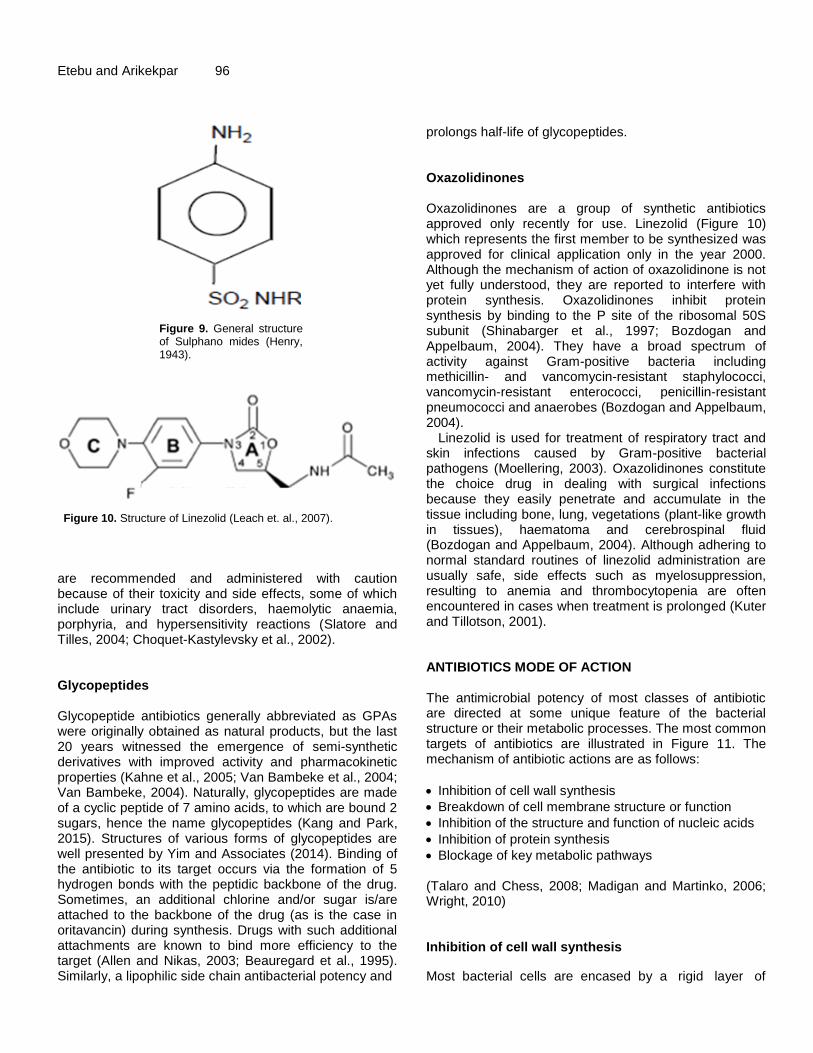
Etebu and Arikekpar 96
Figure 9. General structure of Sulphano mides (Henry, 1943).
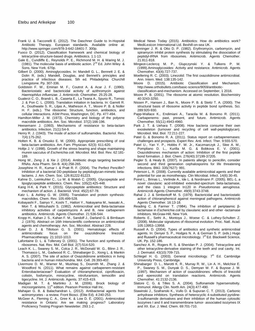
Figure 10. Structure of Linezolid (Leach et. al., 2007).
are recommended and administered with caution because of their toxicity and side effects, some of which include urinary tract disorders, haemolytic anaemia, porphyria, and hypersensitivity reactions (Slatore and Tilles, 2004; Choquet-Kastylevsky et al., 2002). Glycopeptides Glycopeptide antibiotics generally abbreviated as GPAs were originally obtained as natural products, but the last 20 years witnessed the emergence of semi-synthetic derivatives with improved activity and pharmacokinetic properties (Kahne et al., 2005; Van Bambeke et al., 2004; Van Bambeke, 2004). Naturally, glycopeptides are made of a cyclic peptide of 7 amino acids, to which are bound 2 sugars, hence the name glycopeptides (Kang and Park, 2015). Structures of various forms of glycopeptides are well presented by Yim and Associates (2014). Binding of the antibiotic to its target occurs via the formation of 5 hydrogen bonds with the peptidic backbone of the drug. Sometimes, an additional chlorine and/or sugar is/are attached to the backbone of the drug (as is the case in oritavancin) during synthesis. Drugs with such additional attachments are known to bind more efficiency to the target (Allen and Nikas, 2003; Beauregard et al., 1995). Similarly, a lipophilic side chain antibacterial potency and
prolongs half-life of glycopeptides. Oxazolidinones Oxazolidinones are a group of synthetic antibiotics approved only recently for use. Linezolid (Figure 10) which represents the first member to be synthesized was approved for clinical application only in the year 2000. Although the mechanism of action of oxazolidinone is not yet fully understood, they are reported to interfere with protein synthesis. Oxazolidinones inhibit protein synthesis by binding to the P site of the ribosomal 50S subunit (Shinabarger et al., 1997; Bozdogan and Appelbaum, 2004). They have a broad spectrum of activity against Gram-positive bacteria including methicillin- and vancomycin-resistant staphylococci, vancomycin-resistant enterococci, penicillin-resistant pneumococci and anaerobes (Bozdogan and Appelbaum, 2004).
Linezolid is used for treatment of respiratory tract and skin infections caused by Gram-positive bacterial pathogens (Moellering, 2003). Oxazolidinones constitute the choice drug in dealing with surgical infections because they easily penetrate and accumulate in the tissue including bone, lung, vegetations (plant-like growth in tissues), haematoma and cerebrospinal fluid (Bozdogan and Appelbaum, 2004). Although adhering to normal standard routines of linezolid administration are usually safe, side effects such as myelosuppression, resulting to anemia and thrombocytopenia are often encountered in cases when treatment is prolonged (Kuter and Tillotson, 2001). ANTIBIOTICS MODE OF ACTION The antimicrobial potency of most classes of antibiotic are directed at some unique feature of the bacterial structure or their metabolic processes. The most common targets of antibiotics are illustrated in Figure 11. The mechanism of antibiotic actions are as follows:
Inhibition of cell wall synthesis
Breakdown of cell membrane structure or function
Inhibition of the structure and function of nucleic acids
Inhibition of protein synthesis
Blockage of key metabolic pathways (Talaro and Chess, 2008; Madigan and Martinko, 2006; Wright, 2010) Inhibition of cell wall synthesis
Most bacterial cells are encased by a rigid layer of

Int. J. Appl. Microbiol. Biotechnol. Res. 97
Figure 11. Antibiotic target sites (Madigan and Martinko, 2006).
peptidoglycan (PG), also called murein in older sources) which both protect the cells in the face of prevailing osmotic pressure consistent with the often-harsh environment and conditions under which they exist. Peptidoglycan has a degree of cross-linking peptide bonds called β-(1-4) –N– acetyl Hexosamine (Bugg and Walsh, 1992; Holtje, 1998). To stay alive, bacteria must of necessity synthesize peptidoglycan; they do this by the activity of PBPs which are transglycosylases and transpeptidases. These two enzymes play very pivotal roles by adding disaccharide pentapeptides to extend the glycan strands of existing peptidoglycan molecule and also cross-link strands of immature peptidoglycan units (Park and Uehara, 2008). Drugs like penicillins, carbapenems and cephalosporins are able to block the cross-linking of peptidoglycan units by inhibiting the peptide bond formation catalyzed by PBPs (Josephine et al., 2004).
Most antibiotics belonging to the glycopeptide class of antibiotics (for example, vancomycin) are able to inhibit bacterial growth by inhibiting the synthesis of PG. They inhibit the synthesis of PG by binding themselves to PG units, as well as blocking transglycosylase and transpeptidase activity (Kahne et al., 2005).
Breakdown of the cell membrane structure or function The classes of antibiotics that damage cell membranes of bacteria are specific in each microbial group based on the differences in the types of lipids in their cell membranes. For example, Daptomycin depolarizes calcium-dependent membrane, and that leads to the cessation of macromolecular synthesis and disruption of the cellular membrane in bacteria (Alborn et al., 1991). The polymyxins cause disintegration of bacterial cell membrane by effectively binding to the lipid moiety of the lipopolysaccharide in the bacterial cell (Falagas et al., 2010). Inhibition of nuclei acid synthesis The metabolic pathways that result in synthesis of nucleic acids are very essential; disruption of nucleic acid synthesis is inimical to both the survival and posterity of bacterial cells. Antibiotics interfere with nuclei acid synthesis by blocking replication or stopping transcription. DNA replication involves the unwinding of the traditional

Etebu and Arikekpar 98 double helix structure, a process facilitated by the helicase enzymes (Gale et al., 1981). The quinolones group of antibiotics, for example, do interfere with the functionality of the helicase enzyme thereby disrupts the enzyme from playing its function of unwinding DNA. This antibiotic action of the quinolones ultimately truncates the process of DNA replication and repair amongst susceptible bacteria (Chen et al., 1996). Antibiotics whose mode of action is inhibition of nucleic acid synthesis also target topoisomerase II and topoisomerase IV of bacteria. Disrupting the activities of these enzymes in bacteria adversely affects RNA polymerase which in turn prevents RNA synthesis. Quinolones that inhibit bacterial nucleic acid synthesis in this way do not interact with mammalian RNA polymerase, making them specifically antagonistic to Gram-positive bacteria and some Gram-negative bacteria. Inhibition of protein synthesis Living things including bacteria are defined by the amount and type of proteins they are composed of, and continually produce. Proteins are responsible for the structural composition, metabolic and physiological processes, and response to adverse conditions, amongst other roles. However, the type and amount of proteins produced by a bacterium at any given time is dependent on information contained in yet another very important biomolecule – Deoxyribonucleic acid (DNA). DNA determines the type of protein a bacterial cell produces through certain information it harbors within itself. The information is a set of genetic codes called codons, handed down to an identical biomolecule – Ribonucleic acid (RNA), specifically messenger RNA (mRNA). Transfer RNA (tRNA), a similar biomolecule is also formed under the directive of DNA. This biomolecule together with mRNA travels to the ribosomes – the factory for protein synthesis in a living cell. The tRNA then deciphers the codons contained in the mRNA and facilitates the translation of the sequence of codons to a sequence of amino acids which are the building blocks of proteins (Etebu, 2013).
The translation of mRNA into proteins occurs over three sequential phases (initiation, elongation and termination) involving the ribosome and a host of cytoplasmic accessory factors (Gualerzi et al., 2000). Ribosomes are made up of RNA and proteins, and are generally called RIBONUCLEOPROTEINS. The RNA component is what is referred to as Ribosomal RNA (rRNA), and comprises two subunits, one small subunit (SSU) and the other large subunit (LSU). These two subunits are usually described in terms of their sedimentation coefficients (that is, their rate of sedimentation is an ultracentrifuge), and are measured in
Svedberg units (symbols) termed the 30S and 50S, respectively (Nissen et al., 2000).
Bacterial possess 5S, 16S and 23S genes on their rRNA (Moore, 2001). The 16S rRNA gene resides as a single RNA gene in their SSU (16S) whilst the other two rRNA genes (23S and 5S) occur on the LSU of the bacterial ribosome (Lafontaine and Tollervey, 2001). There is huge difference between prokaryotic and eukaryotic rRNA, and this feat has greatly enabled Scientists to develop antibiotics that would target rRNA of a wide spectrum of pathogenic bacteria (Hong et al., 2014).
Given the importance of proteins in the metabolic and life processes of all living organisms, whatever disrupts the process of its synthesis in a bacterial cell would ultimately incapacitate the cell; inhibit its growth or even kill it completely. Drugs that inhibit protein synthesis are among the broadest classes of antibiotics and can be divided into two subclasses: the 50S inhibitors and 30S inhibitors.
Antibiotics such as erythromycin, clindamycin, lincomycin, chloramphenicol, linezolid etc. have been shown to be among the 50S ribosome inhibitors (Douthwaite, 1992; Katz and Ashley, 2005). In general terms, antibiotics that inhibit 50S ribosome do so by physically blocking either the initiation phase of protein translation or the elongation phase of protein synthesis where the incoming amino acid is linked up with the growing nascent peptide chain (Patel et al., 2001; Vannuffel and Cocito, 1996; Menninger and Otto, 1982). Examples of antibiotic that block initiation of protein translation are members of Oxazolidinones (Patel et al., 2001) whilst macrolides such as lincosamide and streptogramin block protein synthesis by inhibiting the elongation phase of mRNA translation (Vannuffel and Cocito, 1996; Menninger and Otto, 1982). These latter groups of antibiotics are therefore reportedly ineffective when elongation has progressed beyond a critical length (Tenson et al., 2003).
The 30S ribosome-inhibitors principally work by blocking the access of aminoacyl-tRNAs to the ribosome. Examples of antibiotics that function in this manner include the tetracycline, streptomycin, spectinomycin, etc. (Hong et al., 2014; Chopra and Roberts, 2001). It is worthy to note that some earlier works have shown that tetracycline also inhibits some proteins at the 50S ribosomes (Epe and Woolley, 1984).
Among ribosome inhibitors, the naturally-derived aminoglycoside subclass is the only one that is broadly bactericidal. Macrolides, streptogramins, spectinomycin, tetracyclines and chloramphenicol are typically bacteriostatic. However, some of these ribosome inhibitory antibiotics that are typically bacteriostatic in action could be bactericidal under certain conditions relating to species- or treatment-specific fashion. For example, chloramphenicol known typically to be

bacteriostatic has been shown to effectively kill S. pneumoniae and Neisseria meningitidis (Rahal and Simberkoff, 1979), as well as H. influenza (Rahal and Simberkoff, 1979; Goldstein et al., 1990). This species-specific variability in ribosome inhibition or mediated cell death is potentially linked to sequence differences among bacterial species in the variable regions of the highly conserved ribosomal proteins and RNAs (Roberts et al., 2008). Blockage of key metabolic pathways Some antibiotics like sulphonamides and trimethoprim have been shown to mimic a substrate needed for cellular metabolism of bacteria. This deception cause bacterial enzymes to attach themselves to the antibiotic rather than the normal substrate. In particular, sulphonamides act like tetrahydrofolate which is required for the synthesis of folic acid in bacterial cells (Talaro and Chess, 2008). Folic acid is vital in the metabolism of nucleic acid and amino acids; for this reason, sulphonamides ultimately disrupt the production of nucleic acids (DNA and RNA) and amino acids, as they mimic substrates required for folic acid metabolism (Talaro and chess, 2008). CONCLUSION The continual discovery, development and introduction of antibiotics into our health care delivery system has no doubt immensely aided our fight against infectious diseases caused by bacteria and thus contributed to individual and societal well-being. However, the ever-developing emergence of bacteria resistant to virtually all known antibiotic is a cause of grave concern, and this phenomenon makes the search for new and more effective antibiotics to continue unabated. Although about 2,000 antibiotics have so far been discovered, only a few scores of them are currently used therapeutically (Schlegel, 2003), apparently owing to the attendant side effects of most of those discovered. Proper characterization and adequate understanding of the mode of action of antibiotics is key to safeguard man‟s health care delivery system. REFERENCES Abraham E. (1987). Cephalosporins. Drugs. 4(2):1-4. Adzitey F. (2015). Antibiotic classes and antibiotic susceptibility of
bacterial isolates from selected poultry; a mini review. World Vet. J. 5 (3):36-41.
Alborn W., Allen N. & Preston D. (1991). Deptomycin disrupts membrane potential in growing Staphylococcus aureus. Antimicrob. Agents Chemother. 31(7):1093-1099.
Allen N. E. & Nicas T. I. (2003). Mechanism of action of oritavancin and
Int. J. Appl. Microbiol. Biotechnol. Res. 99
relatedglycopeptide antibiotics. FEMS Microbiol. Rev. 26(5):511-532. Aminov R. I. (2010). A brief history of the antibiotic era: Lessons learned
and challenges for the future. Front Microbiol. 1(134):1-7. Beauregard D. A., Williams D. H., Gwynn M. N. & Knowles D. J. C.
(1995). Dimerization and membrane anchors in extracellular targeting of vancomycin group antibiotics. Antimicrob. Agents Chemother. 39(3):781-785.
Bonner D. P. & Sykes R. B. (1984). Structure activity relationships among the monobactams. J. Antimicrob. Chemother. 14:313-327.
Boundless (2016). Antibiotic Classifications. Boundless microbiology. https://www.boundless.com/microbiology/textbooks/boundless-microbiology-textbook/antimicrobial-drugs-13/overview-of-antimicrobial-therapy-153/antibiotic-classifications-775-4905/. Accessed September 13, 2016.
Bozdogan B. & Appelbaum P. C. (2004). Oxazolidinones: activity, mode of action, and mechanism of resistance. Int. J. Antimicrob. Agents. 23(2):113-119.
Brink A. J., Feldman C., Grolman D. C., Muckart D., Pretorius J., Richards G. A., Senekal M. & Sieling W. (2004). Appropriate use of the carbapenems. SAMJ. 94(10):857-861.
Brooks G. F., Butel J. S. & Morse S. A. (2004). Jawetz, Melnick and Adelberg‟s Medical Microbiology, 23
rd Edition. McGraw Hill
Companies, Singapore. Brown A. G., Butterworth D., Cole M., Hanscomb G., Hood J. D.,
Reading C. & Rolinson G. N. (1976). Naturally-occurring beta-lactamase inhibitors with antibacterial activity. J. Antibiot. (Tokyo). 29(6):668-669.
Bugg T. D. H. & Walsh C. T. (1992). Intracellular steps of bacterial cell wall peptidoglycan biosynthesis: Enzymology, antibiotics, and antibiotic resistance. Nat. Prod. Rep. 9:199-215.
Butterworth D., Cole M., Hanscomb G. & Rolinson G. N. (1979). Olivanic acids, a family of beta-lactam antibiotics with beta-lactamase inhibitory properties produced by Streptomyces species. Detection, properties and fermentation studies. J. Antibiot. (Tokyo). 32:287-294.
Calderon C. B. & Sabundayo B. P. (2007). Antimicrobial classifications: Drugs for bugs. In: Schwalbe R, Steele-Moore L & Goodwin AC (eds). Antimicrobial susceptibility testing protocols. CRC Press, Taylor and Frances group. ISBN 978-0-8247-4100-6.
Cassidy P. J., Albers-Schonberg G., Goegelman R. T., Miller T., Arison B., Stapley E. O. & Birnbaum J. (1981). Epithienamycins. II. Isolation and structure assignment. J. Antibiot. (Tokyo). 34:637-648.
Chen C. R., Malik M., Snyder M. & Drlica K. (1996). DNA gyrase and topoisomerase IV on the bacterial chromosome: quinolone – induced DNA cleavage. J. Mol. Biol. 258:627-637.
Chopra I. & Roberts M. (2001). Tetracycline antibiotics: Mode of action, applications, molecular biology, and epidemiology of bacterial resistance. Microbiol. Mol. Biol. Rev. 65(2):232-260.
Choquet-Kastylevsky G., Vial T. & Descotes J. (2002). Allergic adverse reactions to sulfonamides. Curr. Allergy Asthma Rep. 2(1):16-25.
Denyer S. P., Hodges N. A. & German S. P. (2004). Introduction to pharmaceutical microbiology. In: Denyer SP, Hodges NA & German SP (eds.) Hugo and Russell‟s Pharmaceutical Microbiology. 7
th Ed.
Blackwell Science, UK. Pp. 3-8. Domagala J. M. (1994). Structure-activity and structure-side-effect
relationships for the quinolone antibacterials. J. Antimicrob. Chemother. 33:685-706.
Douthwaite S. (1992). Interaction of the antibiotics clindamycin and lincomycin with Escherichia coli 23S ribosomal RNA. Nucleic Acids Res. 20:4717-4720.
Epe B. & Woolley P. (1984). The binding of 6-demethylchlortetracycline to 70S, 50S and 30S ribosomal particles: A quantitative study by fluorescence anisotropy. EMBO J. 3:121-126.
Etebu E. (2013). Potential panacea to the complexities of polymerase chain reaction (PCR). Adv. Life. Sci. Tech. 13:1-8.
Eyssen H. J., Van den Bosch J. F., Janssen G. A. & Vanderhaeghe H. (1971). Specific inhibition of cholesterol absorption by sulfaguanidine. Atherosclerosis. 14 (2):181-192.
Falagas M. E., Rafailidis P. I. & Matthaiou D. K. (2010). Resistance to polymyxins: Mechanisms, frequency and treatment options. Drug Resist. Update. 13:132-138.
Etebu and Arikekpar 100 Frank U. & Tacconelli E. (2012). The Daschner Guide to In-Hopsital
Antibiotic Therapy. European standards. Available online at: http://www.springer.com/978-3-642-18401-7. 300p.
Fuoco D. (2012). Classification framework and chemical biology of tetracycline-structure-based drugs. Antibiotics. 1:1-13.
Gale E., Cundliffe E., Reynolds P. E., Richmond M. H. & Waring M. J. (1981). The molecular basis of antibiotic action. 2
nd Ed. John Wiley &
Sons, New York. 670p. Gilbert D. (2000). Aminoglycosides. In: Mandell G. L., Bennett J. E. &
Dolin R, (eds.) Mandell, Douglas, and Bennett's principles and practice of infectious diseases. 5th ed. Philadelphia: Churchill Livingstone. Pp. 307-336.
Goldstein F. W., Emirian M. F., Coutrot A. & Acar J. F. (1990). Bacteriostatic and bactericidal activity of azithromycin against Haemophilus influenzae. J. Antimicrob. Chemother. 25:25-28.
Gualerzi C. O., Brandi L. B., Caserta E., La Teana A., Spurio R., Tomsic J. & Pon C. L. (2000). Translation initiation in bacteria. In: Garrett R. A., Douthwaite S. R., Liljas A., Matheson A. T., Moore P. B. & Noller H. F. (eds.). The ribosome: Structure, function, antibiotics, and cellular interactions. ASM Press, Washington, DC. Pp. 477-494.
Hamilton-Miller J. M. (1973). Chemistry and biology of the polyene macrolide antibiotics. Am. Soc. Microbiol. 37(2):166-196.
Heesemann J. (1993). Mechanisms of resistance to beta-lactam antibiotics. Infection. 21(1):S4-9.
Henry R. J. (1943). The mode of action of sulfonamides. Bacteriol. Rev. 7(4):175-262.
Holten K. B. & Onusko E. M. (2000). Appropriate prescribing of oral beta-lactam antibiotics. Am. Fam. Physician. 62(3): 611-620.
Holtje J. V. (1998). Growth of the stress bearing and shape maintaining murein sacculus of Escherichia coli. Microbiol. Mol. Biol. Rev. 62:181-189.
Hong W., Zeng J. & Xie J. (2014). Antibiotic drugs targeting bacterial RNAs. Acta Pharm. Sin B. 4(4):258-265.
Josephine H. R., Kumar I. & Pratt R. F. (2004). The Perfect Pencillin? Inhibition of a bacterial DD-peptidase by peptidoglycan-mimetic beta-lactams. J. Am. Chem. Soc. 126:81222-81223.
Kahne D., Leimkuhler C., Lu W. & Walsh C. (2005). Glycopeptide and lipoglycopeptide antibiotics. Chem. Rev. 105(2):425-448.
Kang H-K. & Park Y. (2015). Glycopeptide antibiotics: Structure and mechanism of action. J. Bacteriol. Virol. 45(2):67-78.
Katz L. & Ashley G. W. (2005). Translation and protein synthesis: macrolides. Chem. Rev. 105:499-528.
Kobayashi F., Sainyo Y., Koshi T., Hattori Y., Nakayama M., Iwasaki A., Mori T. & Mitsuhashi S. (1982). Antimicrobial and Beta-lactamase inhibitory activities of carpetimycins A and B, new carbapenem antibiotics. Antimicrob. Agents Chemother. 21:536-544.
Kropp H., Kahan J. S., Kahan F. M., Sandolf J., Darland G. & Birnbaum J. (1976). Abstract on 16th Interscientific conference on antimicrobial agents and chemotherapy. Am. Soc. Microbiol. Abstract 228.
Kuter D. J. & Tillotson G. S. (2001). Hematologic effects of antimicrobials: focus on the oxazolidinone linezolid. Pharmacotherapy. 21:1010-1013.
Lafontaine D. L. & Tollervey D. (2001). The function and synthesis of ribosomes. Nat. Rev. Mol. Cell Biol. 2(7):514-520.
Leach K. L., Swaney S. M., Colca J. R., McDonald W. G., Blinn J. R., Thomasco L. M., Gadwood R. C., Shinabarger D., Xiong L. & Mankin A. S. (2007). The site of action of Oxazolidinone antibiotics in living bacteria and in human mitochondria. Mol. Cell. 26:393-402.
Livermore D. M., Warner M., Mushtaq S., Doumith M., Zhang J. & Woodford N. (2011). What remains against carbapenem-resistant Enterobacteriaceae? Evaluation of chloramphenicol, ciprofloxacin, colistin, fosfomycin, minocycline, nitrofurantoin, temocillin and tigecycline. Int. J. Antimicrob. Agents. 37:415-419.
Madigan M. T. & Martinko J. M. (2006). Brock biology of microorganisms. 11
th edition. Pearson Prentice Hall Inc.
Mahajan G. B. & Balachandran L. (2012). Antibacterial agents from actinomycetes - a review. Front Biosci. (Elite Ed). 4:240-253.
McGeer A., Fleming C. A., Gree K. & Low D. E. (2001). Antimicrobial resistance in Ontario: Are we making progress? Laboratory Proficiency Testing Program Newsletter. 293:1-2.
Medical News Today (2015). Antibiotics: How do antibiotics work?
MediLexicon International Ltd. Bexhill-on-sea UK. Menninger J. R. & Otto D. P. (1982). Erythromycin, carbomycin, and
spiramycin inhibit protein synthesis by stimulating the dissociation of peptidyl-tRNA from ribosomes. Antimicrob. Agents Chemother. 21:811-818.
Mingeot-Leclercq M. P., Glupczynski Y. & Tulkens P. M. (1999).Aminoglycosides: Activity and resistance. Antimicrob. Agents Chemother. 43(4):727-737.
Moellering R. C. (2003). Linezolid: The first oxazolidinone antimicrobial. Ann. Intern. Med. 138:135-142.
Moore D. (2015). Antibiotic Classification and Mechanism. http://www.orthobullets.com/basic-science/9059/antibiotic-classification-and-mechanism. Accessed on September, 1 2016.
Moore P. B. (2001). The ribosome at atomic resolution. Biochemistry 40:3243-3250.
Nissen P., Hansen J., Ban N., Moore P. B. & Steitz T. A. (2000). The structural basis of ribosome activity in peptide bond synthesis. Sci. 289:920-930.
Papp-Wallace K., Endimiani A., Taracila M. & Bonomo R. (2011). Carbapenems: past, present, and future. Antimicrob. Agents Chemother. 55(11):4943-4960.
Park J. T. & Uehara T. (2008). How bacteria consume their own exoskeleton (turnover and recycling of cell wall-peptidoglycan). Microbiol. Mol. Biol. 72:211-227.
Patel G. & Bonomo R. A. (2011). Status report on carbapenemases: challenges and prospects. Expert Rev. Anti. Infect. Ther. 9:555-570.
Patel U., Yan Y. P., Hobbs F. W. Jr., Kaczmarczyk J., Slee A. M., Pompliano D. L., Kurilla M. G. & Bobkova E. V. (2001). Oxazolidinones mechanism of action: Inhibition of the first peptide bond formation. J. Biol. Chem. 276(40):37199-37205.
Pegler S. & Healy B. (2007). In patients allergic to penicillin, consider second and third generation cephalosporins for life threatening infections. BMJ. 335(7627): 991.
Peterson L. R. (2008). Currently available antimicrobial agents and their potential for use as monotherapy. Clin Microbial. Infect. 14(6):30-45.
Poirel L., Brinas L., Verlinde A., Ide L. & Nordmann P. (2005). BEL-1, a novel clavulanic acid-inhibited extended-spectrum beta-lactamase, and the class 1 integron In120 in Pseudomonas aeruginosa. Antimicrob Agents Chemother. 49(9):3743-3748.
Rahal J. J. & Simberkoff M. S. (1979). Bactericidal and bacteriostatic action of chloramphenicol against meningeal pathogens. Antimicrob Agents Chemother. 16:13-18.
Reading C. & Farmer T. (1984). The inhibition of periplasmic β-lactamase in Escherichia coli by clavulanic acid and other -lactamase inhibitors. McGraw-Hill, New York.
Roberts E., Sethi A., Montoya J., Woese C. & Luthey-Schulten Z. (2008). Molecular signatures of ribosomal evolution. Proc. Natl. Acad. 105:13953–13958.
Russell A. D. (2004). Types of antibiotics and synthetic antimicrobial agents. In: Denyer S. P., Hodges N. A. & German S. P. (eds.) Hugo and Russell‟s pharmaceutical microbiology. 7
th Ed. Blackwell Science,
UK. Pp. 152-186. Sanchez A. R., Rogers R. S. & Sheridan P. J. (2004). Tetracycline and
other tetracycline-derivative staining of the teeth and oral cavity. Int. J. Dermatol. 43(10):709-715.
Schlegel H. G. (2003). General microbiology. 7th Ed. Cambridge
University Press, Cambridge. Shinabarger D. L., Marotti K. R., Murray R. W., Lin A. H., Melchior E.
P., Swaney S. M., Dunyak D. S., Demyan W. F. & Buysse J. M. (1997). Mechanism of action of oxazolidinones: effects of linezolid and eperezolid on translation reactions. Antimicrob. Agents Chemother. 41:2132-2136.
Slatore C. G. & Tilles S. A. (2004). Sulfonamide hypersensitivity. Immunol. Allergy Clin. North Am. 24(3):477-490.
Stawinski J., Szafranski K., Vullo D. & Supuran C. T. (2013). Carbonic anhydrase inhibitors. Synthesis of heterocyclic 4-substituted pyridine-3-sulfonamide derivatives and their inhibition of the human cytosolic isozymes I and II and transmembrane tumor- associated isozymes IX and XII. Eur. J. Med. Chem. 69:701-710.

Sykes R. B. & Bonner D. P. (1985). Discovery and development of the
monobactams. Clin. Infect. Dis. 7(4):S579-S593. Sykes R. B., Cimarusti C. M., Bonner D. P., Bush K., Floyd D. M.,
Georgopapadakou N. H., Koster W. H., Liu W. C., Parker W. L., Principe P. A., Rathnum M. L., Slusarchyk W. A., Trejo W. H. & Wells J. S. (1981). Monocyclic β-lactam antibiotics produced by bacteria. Nature. 291:489-491.
Talaro K. P. & Chess B. (2008). Foundations in microbiology. 8th
Ed. McGraw Hill, New York.
Tenson T., Lovmar M. & Ehrenberg M. (2003). The mechanism of action of macrolides, lincosamides and streptogramin B reveals the nascent peptide exit path in the ribosome. J. Mol. Biol. 330(5):1005-1014.
Tidwell T. T. (2008). Hugo (Ugo) Schiff, Schiff bases, and a century of β-Lactam synthesis. Angew. Chem. Int. Ed. Engl. 47(6):1016-1020.
Torres J. A., Villegas M. V. & Quinn J. P. (2007). Current concepts in antibiotic-resistant gram-negative bacteria. Expert Rev. Anti. Infect. Ther. 5:833-843.
van Bambeke F. (2004). Glycopeptides in clinical development: pharmacological profile and clinical perspectives. Curr. Opin. Pharmacol. 4(5):471-478.
van Bambeke F., Van Laethem Y., Courvalin P. & Tulkens P. (2004). Glycopeptide antibiotics: From conventional molecules to new derivatives. Drugs. 64(9):913-936.
van Hoek A. H. A. M., Mevius D., Guerra B., Mullany P., Roberts A. P. & Aarts H. J. M. (2011). Acquired antibiotic resistance genes: An overview. Front. Microbiol. 2:203 doi: 10.3389/fmicb.2011.00203.
Vannuffel P. & Cocito C. (1996). Mechanism of action of streptogramins and macrolides. Drugs. 51(1):20-30.
Walsh C. (2003). Antibiotics: actions, origins, resistance. 1st Ed. ASM
Press, Washington, DC. 345p.
Int. J. Appl. Microbiol. Biotechnol. Res. 101 White D. & Cox E. (2013). Fighting the impact of antibiotic-resistance.
FDA Consumer health Information. www.fda.gov/downloads/.../UCM350090.pdf. Accessed September, 4, 2016.
Wright G. D. (2010). Q & A: Antibiotic resistance: Where does it come from and what can we do about it? BMC Biol. 8:123. http://doi.org/10.1186/1741-7007-8-123.
Xu F., Xu H., Wang X., Zhang L., Wen Q., Zhang Y. & Xu W. (2014). Discovery of N-(3-(7H-purin-6-yl)thio)-4-hydroxynaphthalen-1-yl)-sulfonamide derivatives as novel protein kinase and angiogenesis inhibitors for the treatment of cancer: synthesis and biological evaluation. Part III. Bioorg. Med. Chem. 22(4):1487-1495.
Yim G., Thaker M. N., Koteva K. & Wright G. (2014). Glycopeptide antibiotic biosynthesis. The Journal of Antibiotics. 67:31-41.
Zahner H. & Maas W. (1972). Biology of Antibiotics. Springer-Verlag, New York.