antibiotic susceptibility profiles and bacteriological risks...
TRANSCRIPT

American International Journal of Biology 1(1); July 2013 pp. 01-12 Bello et al.
© American Research Institute for Policy Development 1 www.aripd.org/aijb
Antibiotic Susceptibility Profiles and Bacteriological Risks Associated With Used Toothbrushes: A Case Study of Some Apparently Healthy University Students in
Southwestern Nigeria
O. O. Bello1*, A. Osho2, S.A. Bankole1 and T. K. Bello3
1Department of Microbiology, Olabisi Onabanjo University, P.M.B. 2002, Ago-Iwoye,Ogun State, Nigeria. 2Department of Biological Sciences, Redeemer’s University, Mowe, Ogun State. Nigeria. 3Department of Medical Microbiology and Parasitology, Olabisi Onabanjo University Teaching Hospital (OOUTH), Sagamu, Ogun State, Nigeria.
Abstract
Toothbrushes play a significant role in disease transmission and increase the risk of infection since they serve as reservoirs for microorganisms in healthy, oral-diseased and medically ill adults. Investigation was carried out on the antibiotic susceptibility profiles of bacteria isolated from used toothbrushes. Thirty toothbrushes used for at least 5 weeks by thirty University students were collected. Heads of the brushes were soaked in 10 ml of sterile tryptone soya broth (TSB) and agitated by vortex mixing. The bacterial suspension was serially diluted. Plate count agar, MacConkey agar and Mannitol salt agar media were used for the isolation of non-fastidious bacteria, coliforms and staphylococci, respectively, employing the spread plate technique. Biochemical characterization of isolates was carried out using standard methods. Survival ability of bacterial contaminants on the used toothbrushes was also investigated at the 24th hr, 72nd hr and 144th. The disk diffusion method was employed for the determination of the antimicrobial susceptibility profiles of the bacterial isolates. Seven genera of microorganisms were encountered and these include Staphylococcus, Escherichia, Klebsiella, Pseudomonas,
Lactobacillus, Leuconostoc and Proteus. Pseudomonas aeruginosa was most prevalent as shown by mean total plate count of 5.0 x 10²
CFU ml-1 while E. coli had the lowest prevalence (1.2 x 10² CFU ml-1). It was discovered that S. aureus, S. epidermidis, E. coli and Proteus sp all survived at 144th hr indicating high survival ability, while Lactobacillus sp only survived at 24th hr. There were variations in the susceptibility patterns of the isolates to the various antibiotics. It was determined that 62.5% of the isolates showed susceptibility; twenty percent (20%) of isolates were intermediately susceptible and the remaining 17.5% were resistant. It was concluded that most bacterial isolates from toothbrushes were susceptible to antibiotics but the percentage resistant should be of great concern as it poses high health risk and may generate the spread of antibiotic-resistant bacteria within the family and beyond. Organisms such as some members of the enterobacteriaceae which are not normally associated with oral flora isolated from used toothbrushes investigated in this study should also be of interest.
American International Journal of Biology 1(1); July 2013 pp. 01-12 Bello et al.
© American Research Institute for Policy Development 2 www.aripd.org/aijb
Key Words: Bacteria, toothbrush, risk, antibiotic, susceptibility, health.
Introduction and Literature Review
Toothbrushes play an essential role in oral hygiene and are commonly found in both community and hospital settings. Toothbrushes may play a significant role in disease transmission and increase the risk of infection since they can serve as a reservoir for microorganisms in healthy, oral-diseased and medically ill adults (Glass, 1992a; Downes et al., 2008). Contamination is the retention and survival of infectious organisms that occur on animate or inanimate objects. In healthy adults, contamination of toothbrushes occurs early after initial use and increases with repeated use (CDC, 2002). Toothbrushes can become contaminated from the oral cavity, environment, hands, aerosol contamination, and storage containers. Bacteria which attach to, accumulate, and survive on toothbrushes may be transmitted to the individual causing disease (Caudry et al., 1995; ADA, 2009).
The human oral cavity is colonized by a larger variety of bacteria flora than any other anatomic area. More than 700 species of bacteria have already been identified 400 of which were found in the periodontal pocket adjacent to teeth (Abraham et al., 1990). In the hospital setting, toothbrushes are commonly used for oral care by nurses. There is a need for standardized nursing guidelines to prevent toothbrush contamination, which may increase the risk of infections from potentially pathogenic microorganisms and is clinically relevant for assessing the risks and benefits of oral care and informing nursing practice (Bezirtzogloua et al., 2008). The toothbrush is used on a daily basis to clean the oral cavity, so it is a very important piece of equipment known for proper dental hygiene.
Sadly, toothbrushes are most commonly located near the bathroom sink, which is a good place to harvest hundreds of microorganisms. No matter how sanitized the bathroom is, the toothbrush will still be consistently exposed to the mouth which will inevitably result in bacterial growth on the toothbrush. A new toothbrush is usually not a favorable habitat for bacteria and fungi, but in some cases, toothbrushes are already slightly infected because there is not a regulation that states toothbrushes must be sold in a sterile package (Glass and Lare, 1986; Efstratiou et al., 2007). Typically, the presence of microbes on the toothbrush comes from brushing because the mouth is a hospitable niche to many kinds of microbes. Therefore, the bacteria will transfer from the inside of the mouth to the toothbrush (Kozai et al., 1989; Quirynen, 2003). In this way, the toothbrush is considered a niche for many microbes. The human body is constantly exposed to potentially harmful microbes. However, the body is normally able to defend itself against infections through a combination of passive and active mechanisms (Mehta et al., 2007). Intact skin and mucous membranes function as a passive barrier to bacteria and other organisms. When these barriers are challenged or breached, active mechanisms such as enzymes, digestive acids, tears, white blood cells and antibodies come into play to protect the body from disease. Although studies have shown that various microorganisms can grow on toothbrushes after use (Fernandes and Cesar, 2006; Devine, 2007), and other studies have examined various methods to reduce the level of these bacteria (Bunetel et al., 2000; Quirynen, 2003; Efstratiou et al., 2007), there is insufficient clinical evidence to support that bacterial growth on toothbrushes will lead to specific adverse oral or systemic health effects. In a vulnerable population such as critically ill adults, pathogenic contamination may increase the risk of infection and mortality.

American International Journal of Biology 1(1); July 2013 pp. 01-12 Bello et al.
© American Research Institute for Policy Development 3 www.aripd.org/aijb
Although some interventions such as chlorhexidine, toothpaste, mouthwash, and ultraviolet sanitizers reduce bacterial survival, oral hygiene practices in the hospital setting by nurses vary (Downes et al., 2006). Currently, there are no nursing guidelines related to toothbrush frequency of use, storage, and decontamination. In the hospital setting, the environment as a source of pathogenic bacteria is now a hot topic and the focus of many current infectious disease research studies. Surfaces in close contact with the patient such as bed frames, countertops, sinks, bedside tables, linens, and mattresses may act as fomites. Toothbrushes may come into contact with these surfaces prior to or after use thus increasing risk (Fernandes and Cesar, 2006). In clinical practice, Devine (2007) has observed that there is no standardized nursing protocol for the storage or replacement of toothbrushes and that some commonly observed nursing practices include storing the toothbrush in the bath basin with other bathing/personal supplies and linens, in a paper towel, in a plastic wrapper, on the bedside table, next to the sink, and in an oral rinse cup at the bedside. These practices may impact the contamination of toothbrushes. Toothbrushing plays an important everyday role for personal oral hygiene and effective plaque removal. Appropriate toothbrush care and maintenance are also important considerations for sound oral hygiene. The ADA recommends that consumers replace toothbrushes approximately every 3–4 months or sooner if the bristles become frayed with use. In recent years, scientists have studied whether toothbrushes may harbor microorganisms that could cause oral and/or systemic infection (ADA, 2009). The oral cavity is home to hundreds of different types of microorganisms (Mehta et al., 2007); therefore, it is not surprising that some of these microorganisms are transferred to a toothbrush during use.
It may also be possible for microorganisms that are present in the environment where the toothbrush is stored to establish themselves on the brush. Toothbrushes may even have bacteria on them right out of the box (Dabas, 2008), since they are not required to be sold in a sterile package. The toothbrush is not naturally favorable towards the growth of microbes, but can sustain bacterial life once they are transferred onto the toothbrush. Different modes of transfer are responsible for the bacteria on the toothbrush such as contact with the mouth, cross contamination, and the bacteria in the toilet community. Organisms that can survive for a certain amount of time on the toothbrush are diverse, ranging from fungus to bacteria to yeast. The environment of the toothbrush is affected by many conditions whether it is the architecture of the toothbrush itself regarding bristles or by adjusting the pH level. These conditions alter the population of bacteria on the toothbrush. While the toothbrush is not the ideal niche for a microbe, the toothbrush is capable of supporting microbial life (Downes et al., 2008). This study aims at investigating the antibiotic susceptibility profiles of bacteria isolated from used toothbrushes of apparently healthy University students in Ago-Iwoye, Southwestern Nigeria.
3.0 Materials and Methods
3.1 Collection of samples
In this study, thirty (30) toothbrushes from thirty different students of Olabisi Onabanjo University, Ago-Iwoye, Ogun State used for toothbrushing for at least 5 weeks were collected for the purpose of determining the microbial population on them
3.2 Isolation of organisms
Toothbrush of every person were rinsed in tap water and transported to the laboratory in sterile bag.
American International Journal of Biology 1(1); July 2013 pp. 01-12 Bello et al.
© American Research Institute for Policy Development 4 www.aripd.org/aijb
Handles of toothbrushes were cut off using a heat sterile scissors, heads of the brushes (containing the bristles) were then soaked in 10 ml of sterile tryptone soya broth (TSB) for 60 mins This was followed by vortex mixing for 1 min to dislodge suspected adherent bacteria. The bacterial suspension was serially diluted to obtain dilution factors of up to 10-3. The spread plate technique was employed. One mil (1 ml) each of the dilution factors was obtained using a sterile pipette and plated on plate count agar, MacConkey agar and Mannitol salt agar media for the isolation of non-fastidious bacteria, coliforms and staphylococci, respectively. Plates were incubated aerobically at 370C for 24- 48 h (Sammons et al., 2004). 3.3 Identification of isolates
Total viable counts of bacterial population were enumerated. Morphological characteristics of isolates were observed and Gramʼs staining was performed for each isolate.
A. Gram positive cocci of Manitol salt agar were further identified as Staphylococcus aureus and Staphylococcus epidermidis by several biochemical tests such as Catalase test (Collee et al., 1996), Oxidase test (Benson, 2002 ), Coagulase test (Collee et al.,1996), Carbohydrates fermentation test (Stukus,1996; Benson, 2002) and others
B. Gram negative bacilli on MacConkey plates were identified as follows:
a. Gram negative, non lactose fermenting, oxidase positive colonies were considered as Pseudomonas spp (Benson, 2002).
b. Gram negative, lactose fermenting, oxidase negative colonies were considered as Coliform spp.( (Collee et al., 1996)/
Survival of isolates on toothbrushes
Survival ability of bacterial contaminants on used toothbrushes was investigated.
Used toothbrushes kept in sterile polythene bag were re-subjected to microbiological assay to determine the natural survival ability of the bacterial contaminants after abandoning the toothbrushes for use for 24 hrs (one day), 72 hrs (three days) and 144 hrs (six days) (Sammons et al., 2004).
Antimicrobial Susceptibility Testing
The Kirby-Bauer (disk diffusion) method was used to determine the antimicrobial susceptibility profiles of the bacterial isolates. Antibiotic multidisks used consisted of Amoxycillin (Amx), Chloramphenicol (Chl), Ciprofloxacin (Cpx), Cloxacillin (Clo), Cotrimoxazole (Cot), Erythromycin (Ery), Gentamycin (Gen), Norfloxacin (Nfx), Rifampicin (Rfp), Streptomycin (Str) and Tetracycline (Tet). The medium used was Mueller Hinton (MH) agar. Pure cultures of organisms were enriched in nutrient broth and incubated at 370C to a turbidity of 0.5 Macfarland standards. The MH agar was inoculated by streaking using sterile cotton swab of each of the cultures. The antibiotic disks were applied using sterile forceps and sufficiently separated from each other in order to prevent overlapping of the zones of inhibition. The agar plates were left on the bench for 30minutes to allow for diffusion of the antibiotics and the plates were incubated inverted at 370C for 24 hours. Results were recorded by measuring the zone of inhibition and comparing with the NCCLS interpretive performance standard for antimicrobial disk susceptibility testing (NCCLS, 2004; Bello et al., 2013).
Results and Discussion
Table 1 showed the morphological and biochemical characteristics of bacterial isolates from used toothbrushes.

American International Journal of Biology 1(1); July 2013 pp. 01-12 Bello et al.
© American Research Institute for Policy Development 5 www.aripd.org/aijb
Seven (7) different genera of microorganisms were encountered in the study and these include Staphylococcus, Escherichia, Klebsiella,
Pseudomonas, Lactobacillus, Leuconostoc and Proteus. Two staphylococcal species – S. aureus and S. epidermidis were encountered (Table 1).
Table 1: Morphological and biochemical characteristics of bacterial isolates from used toothbrushes
Parameters S.
aureus S. epidermidis
E. coli Klebsiella sp
Pseudomonas aeruginosa
Lactobacillus sp
Leuconostoc sp
Proteus sp
Gram’s reaction
+ + - + - + + -
Catalase test + + + + + - - - Citrate test - + + + + NA - Oxidase test - - + + - - - Coagulase test
+ - - - - - NA -
Indole test - + - - - NA + Urease activity
+ - + NA NA NA +
Cellular morphology
Cocci straight rods
Rods Rods Cocci Rods rods
Growth on blood agar (colony)
creamy white
α-haemolysis
circular large white
Greenish Creamy NA NA
Growth on Mannitol salt agar
bright yellow
N/A N/A N/A N/A N/A N/A
Growth in MacConkey agar
N/A red/ pink
Mucoid Pale Pink NA Pale
Glucose A A A N/A A A/G A/G Lactose A A A N/A A - - Sucrose A A A N/A A - - Mannitol A A D N/A A A/G A/G Maltose A A N/A N/A A A/ G -
- (No growth), + (growth), N/A - Not applicable Percentage toothbrush contaminated with different bacterial species was shown in Table 2. Results showed that nineteen of thirty (63%) used toothbrushes investigated were contaminated with Pseudomonas aeruginosa making the organism the most prevalent in this study. Nine of thirty (30%) used toothbrushes were found to be contaminated with Staphylococcus aureus; eight of thirty (27%) were contaminated with Leuconostoc sp; seven of thirty (23%) were contaminated with
Lactobacillus sp. Other bacterial contaminants of used toothbrush include Staphylococcus epidermidis which contaminated six of thirty (20%) used toothbrushes; Proteus sp contaminated four of thirty (13.33%), Klebsiella sp also contaminated four of thirty (13.33%) and the least bacterial contaminant of used toothbrushes encountered in this study was Escherichia coli isolated from three of thirty (10%) of the toothbrushes investigated.
American International Journal of Biology 1(1); July 2013 pp. 01-12 Bello et al.
© American Research Institute for Policy Development 6 www.aripd.org/aijb
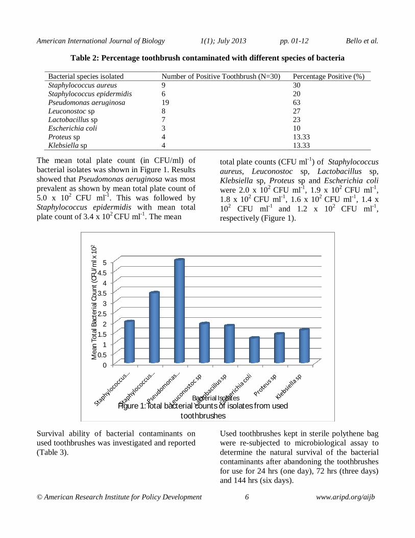
Table 2: Percentage toothbrush contaminated with different species of bacteria
Bacterial species isolated Number of Positive Toothbrush (N=30) Percentage Positive (%) Staphylococcus aureus 9 30 Staphylococcus epidermidis 6 20 Pseudomonas aeruginosa 19 63 Leuconostoc sp 8 27 Lactobacillus sp 7 23 Escherichia coli 3 10 Proteus sp 4 13.33 Klebsiella sp 4 13.33
The mean total plate count (in CFU/ml) of bacterial isolates was shown in Figure 1. Results showed that Pseudomonas aeruginosa was most prevalent as shown by mean total plate count of 5.0 x 10² CFU ml-1. This was followed by Staphylococcus epidermidis with mean total plate count of 3.4 x 10² CFU ml-1. The mean
total plate counts (CFU ml-1) of Staphylococcus aureus, Leuconostoc sp, Lactobacillus sp, Klebsiella sp, Proteus sp and Escherichia coli were 2.0 x 10² CFU ml-1, 1.9 x 10² CFU ml-1, 1.8 x 10² CFU ml-1, 1.6 x 10² CFU ml-1, 1.4 x 10² CFU ml-1 and 1.2 x 10² CFU ml-1, respectively (Figure 1).
Survival ability of bacterial contaminants on used toothbrushes was investigated and reported (Table 3).
Used toothbrushes kept in sterile polythene bag were re-subjected to microbiological assay to determine the natural survival of the bacterial contaminants after abandoning the toothbrushes for use for 24 hrs (one day), 72 hrs (three days) and 144 hrs (six days).
00.5
11.5
22.5
33.5
44.5
5
Mea
n To
tal B
acte
rial C
ount
(CFU
/ml x
102
Bacterial IsolatesFigure 1:Total bacterial counts of isolates from used
toothbrushes

American International Journal of Biology 1(1); July 2013 pp. 01-12 Bello et al.
© American Research Institute for Policy Development 7 www.aripd.org/aijb
It was discovered that Staphylococcus aureus, Staphylococcus epidermidis, Escherichia coli and Proteus sp all survived over a period of six days, though there were reductions in total plate counts but negligible. It was also found that Pseudomonas aeruginosa (which appeared as the most prevalent organism in this study), Leuconostoc sp and Klebsiella sp survived on toothbrushes for 72 hrs but were not isolated on the sixth: they did not survive on toothbrushes over a six-day period.
It was also interesting to find that Lactobacillus sp was isolated after one day but did not survive till 72th hour, and thus 144th hour. This established that use and re-use of toothbrushes over a long period of time is one of the major factors that contribute to the survival of bacterial contaminants on toothbrushes. This is because there could be the tendency that all bacterial contaminants are naturally eliminated on toothbrushes if not re-used over a considerably long period of time and if kept under aseptic conditions, since bacterial contaminants could not have possessed the ability to survive for that long on nutrient-free surface.
Table 3: Survival ability of bacterial isolates from used toothbrushes
Bacterial species isolated Mean Total Plate Count (CFU ml-1)
One day (24 hrs)
Three days (72 hrs)
Six days (144 hrs)
Staphylococcus aureus 2.0 x 10² + + + Staphylococcus epidermidis 3.4 x 10² + + + Pseudomonas aeruginosa 5.0 x 10² + + - Leuconostoc sp 1.9 x 10² + + - Lactobacillus sp 1.8 x 10² + - - Escherichia coli 1.2 x 10² + + + Proteus sp 1.4 x 10² + + + Klebsiella sp 1.6 x 10² + + -
Table 3 showed the antibiotic susceptibility patterns of bacterial isolates from used toothbrushes. There were variations in the susceptibility patterns of the isolates to the various antibiotics. Staphylococcus aureus was found to be susceptible to ciprofloxacin, erythromycin and norfloxacin but resistant to chloramphenicol and tetracycline. The organism was, however, intermediately susceptible to streptomycin, gentamycin, amoxycillin and cloxacillin (Table 4). Staphylococcus epidermidis was susceptible to ciprofloxacin, gentamycin, norfloxacin, streptomycin and tetracycline. It was found to be intermediately susceptible to chloramphenicol and erythromycin but resistant to amoxycillin, cloxacillin and cotrimoxazole.
Pseudomonas aeruginosa was susceptible to all but resistant to three antibiotics namely erythromycin, gentamycin and streptomycin. Similarly, Leuconostoc sp was found to be susceptible to all but intermediately susceptible to norfloxacin, stretomycin and tetracyclin. It was interesting to find that Lactobacillus sp showed susceptibility to all the antibiotics investigated in this study with inhibition zones ranging from 21 + 1.3 mm to 15 + 1.3 mm. Escherichia coli was susceptible to ciprofloxacin, erythromycin, gentamycin and streptomycin; it was intermediately susceptible to cloxacillin and resistant to amoxycillin, chloramphenicol, cotrimoxazole and tetracycline.

American International Journal of Biology 1(1); July 2013 pp. 01-12 Bello et al.
© American Research Institute for Policy Development 8 www.aripd.org/aijb
Proteus sp showed no resistance to any of the antibiotics. It showed susceptibility to amoxicillin, cloxacillin, ciprofloxacin, erythromycin, gentamycin, norfloxacin and streptomycin, and was intermediately susceptible to chloramphenicol, cotrimoxazole and tetracyclin. Klebsiella sp was susceptible to cloxacillin, cotrimoxazole, ciprofloxacin,
erythromycin, gentamycin, norfloxacin but resistant to chloramphenicol and tetracycline with no zone of inhibition at all. It was, however, intermediately susceptible to amoxycillin and streptomycin. Klebsiella sp showed no zone of inhibition to chloramphenicol and tetracycline, indicating their high level of resistance to the antibiotics (Table 3).
Table 4: Antibiotic susceptibility patterns of bacterial isolates from toothbrush Isolates Diameter of zones of inhibition (mm) to different antibiotics Amx
(30 µg) Chl (30 µg)
Clo (30 µg)
Cot (30 µg)
Cpx (10 µg)
Ery (30 µg)
Gen (10 µg)
Nfx (10 µg)
Str (30 µg)
Tet (25 µg)
Staphylococcus aureus
9.0+0.5 5.0 + 1.0 9.0+1.0 13+ 1.0 18+ 1.5 17 + 1.0 14+ 1.4 16 + 1.2 13+1.0 7.0+ 0.3
Staphylococcus epidermidis
8.0+0.1 11 + 0.3 6 + 0.2 8 + 0.3 17+ 1.0 12 + 0.5 17+ 0.5 15 + 0.9 17+1.2 16 + 1.0
Pseudomonas aeruginosa
21+1.5 19 + 1.5 26+ 2.0 19+ 1.4 20+ 1.5 6 + 0.2 2 + 0.0 15 + 1.0 5 + 0.2 15 + 1.2
Leuconostoc sp 18+1.0 17 + 1.5 21+ 2.0 15+ 1.8 18+ 1.0 22 + 2.0 16+ 1.5 11 + 1.0 13+1.0 10 + 0.8
Lactobacillus sp
17+1.2 16 + 1.2 15+ 1.0 16+ 0.9 16+ 1.2 19 + 2.0 21+ 1.3 18 + 1.0 15+0.8 15 + 1.3
Escherichia coli
6 + 0.2 2 + 0.0 14+ 1.0 5 + 0.2 16+ 1.2 17 + 1.4 21+ 2.0 19 + 2.0 20+1.8 3.0+ 0.1
Proteus sp 21+1.2 12 + 1.0 15+ 1.1 12+ 0.6 19+ 1.4 15+ 1.5 24+ 2.0 19 + 1.4 20+1.5 11 + 0.7
Klebsiella sp 14+1.0 0.0 17+ 1.8 18+ 1.5 20+ 2.0 16 + 1.5 23+ 1.8 18 + 1.8 13+1.0 0.0
Keys: Amoxycillin (Amx), Chloramphenicol (Chl), Ciprofloxacin (Cpx), Cloxacillin (Clo), Cotrimoxazole (Cot), Erythromycin (Ery), Gentamycin (Gen), Norfloxacin (Nfx), Rifampicin (Rfp), Streptomycin (Str) and Tetracycline (Tet)
< 8 = Resistant 9 to 14 = Intermediately susceptible
> 15 = Susceptible Figure 2 showed the percentage distributions of susceptibilty, intermediate susceptibilty and resistance of bacterial isolates from used toothbrushes.
It was determined that 62.5% of the isolates showed susceptibility to the various conventional antibiotics investigated; twenty percent (20%) of isolates were intermediately susceptible and the remaining 17.5 percent were resistant.

American International Journal of Biology 1(1); July 2013 pp. 01-12 Bello et al.
© American Research Institute for Policy Development 9 www.aripd.org/aijb
Organisms such as some members of the enterobacteriaceae which are not normally associated with oral flora have been isolated from used toothbrushes investigated in this study. So the infectious microorganisms remaining on the brush can reinfect our mouth again, some of them can even spread to the rest of our body and cause serious health problems, including heart disease, stroke, arthritis, haematogenous, bacterimia and chronic (Warren et al., 2001; Sammons et al., 2004). A single toothbrush can be the breeding ground for billions of bacteria (Abraham et al., 1990; Gabe-Mirkin, 2011). There are attempt to reduce bacterial survival time, deter colonization and inhibit biofilm formation by toothbrushes containing antibacterial agent have been developed and methods for sterilization of brushes devised (Caudry et al., 1995; Neal and Rippin, 2003).
Particular attention was paid to Staphylococci and Pseudomonas like organisms as both of these are opportunistic pathogens responsible for many nosocomial infections and because Pseudomonas species are also resistant to many disinfectants in toothpaste including triclosan (Warren et al., 2001). Glass (1992a) found that toothbrushes from both healthy patients and patients with oral disease contained potentially pathogenic bacteria and viruses such as Staphylococcus aureus, E. coli, Pseudomonas sp and herpes simplex virus. He also found toothbrushes contaminated with herpes simplex virus 1 in numbers sufficient to cause an infection in the patient (Glass, 1992b). Bunetel et al. (2000) found that toothbrushes used by patients with existing oral disease quickly became contaminated.
62.50%20%
17.50%
Figure 2: Percentage distributions of susceptibilty, intermediate susceptibilty and resistance of bacterial isolates from used toothbrushes to some conventional
antibiotics
% of bacterial isolates susceptible 50/80
% of bacterial isolates intermediately susceptible 16/80% of bacterial isolates resistant 14/80
American International Journal of Biology 1(1); July 2013 pp. 01-12 Bello et al.
© American Research Institute for Policy Development 10 www.aripd.org/aijb
This study also found a significant relationship between repeated use and bacterial retention on toothbrushes and that the oral cavity can be inoculated from a contaminated toothbrush. Several of the studies found that toothbrushes were contaminated before use (Glass and Lare, 1986; Glass and Jensen, 1994; Sato et al., 2005). Caudry et al. (1995) found that toothbrushes are heavily contaminated with normal use. Mehta et al. (2007) found that 70% of the toothbrushes in their study became heavily contaminated with pathogenic microorganisms after use. Studies by both Taji and Rogers (1998) and Glass (1992b) found extensive toothbrush contamination after use except in cases where an oral antiseptic, such as mouthwash, was used immediately prior to brushing. Verran and Leahy-Gilmartin (1996) found that toothbrushes supported many different bacteria and the amount of growth was varied.
Conclusion and Recommendation
It was concluded in this study that most bacterial isolates from used toothbrushes were susceptible to antibiotics but the percentage resistant should be of great concern as it poses high health risk and may generate the spread of antibiotic-resistant bacteria within the family and beyond.
Organisms such as some members of the enterobacteriaceae which are not normally associated with oral flora isolated from used toothbrushes investigated in this study should also be of interest. It is recommended in this study that toothbrush should not be shared. Sharing a toothbrush could result in an exchange of body fluids and/or microorganisms between the users of the toothbrush, placing the individuals involved at an increased risk for infections. This practice could be a particular concern for persons with compromised immune systems or existing infectious diseases (Bunetel et al., 2000). Toothbrushes should be thoroughly rinsed with tap water after brushing to remove any remaining toothpaste and debris. Toothbrush should be stored in an upright position if possible and allowed to air-dry until used again. If more than one brush is stored in the same holder or area, the brushes should be separated to prevent cross-contamination (Council on Scientific Affairs, 2011). Toothbrushes should not be routinely covered or stored in closed containers. A moist environment such as a closed container is more conducive for the growth of microorganisms than the open air. Toothbrushes should be replaced at least every 3–4 months. The bristles become frayed and worn with use and cleaning effectiveness will decrease (Quirynen, 2003). Children’s toothbrushes often need replacing more frequently than adult brushes (ADA, 2009)
American International Journal of Biology 1(1); July 2013 pp. 01-12 Bello et al.
© American Research Institute for Policy Development 11 www.aripd.org/aijb
References
Abraham, N. J., Ciricione, U. K. and Glass, R.
T. (1990): Dentists and dental hygienists’ attitudes toward toothbrush replacement and maintenance. Clinical Preventive Dentistry 12: 28—33.
ADA (2009): ADA statement on toothbrush care: cleaning, storage and replacement http://www.ada.org/1887.aspx.
Bello, O. O., Osho, A. and Bello, T.K. (2013): Microbial quality and antibiotic susceptibility profiles of bacterial isolates from borehole water used by some schools in Ijebu-Ode, Southwestern Nigeria. Scholars Academic Journal of Biosciences, 1(1):4-13
Benson, H.J. (2002): Microbiological applications.8th ed McGraw-Hill Higher Education Companies. U.S.A 152-177.
Bezirtzogloua, E., Gretoiub, S.M., Moldoveanus, M., Alexopoulosa, A., Lazard, V. and Nakoue, M. (2008): A quantatitative approach to the effectiveness of ozone against microbiota organisms colonizing toothbrushes . J. Dent. 36(8):600-5.
Bunetel, L., Tricot-Doleux, S., Agnani, G. and Bonnaure-Mallet, M. (2000): “In vitro evaluation of the retention of three species of pathogenic microorganisms by three different types of toothbrush,” Oral Microbiology and Immunology, 15: 313–316.
Caudry, S. D., Klitorinos, A. and Chan, E. C. S. (1995): Contaminated toothbrushes and their disinfection. Journal of the Canadian Dental Association 61: 511—516.
Collee, J.G., Fraser, A.G., Marmion, B.P. and Simmons, A. (1996): Practical medical Microbiology. 14th ed longman
Singapore publishers Ltd. Singapore.245-259.
Council on Scientific Affairs (2011): "ADA
Statement on Toothbrush Care: Cleaning, Storage and Replacement." American Dental Association.
Dabas, N. (2008): "A transcription factor regulatory cascade controls secreted aspartic protease expression in Candida albicans." Molecular Microbiology. 3:586-602.
Devine, D. (2007): "Inhibition of biofilms associated with dentures and toothbrushes by tetrasodium EDTA." Journal of Applied Microbiology 6: 2516-2524.
Downes, J., Samuel, H., Melanie, W. and William, W. (2008): "Prevotella histicola sp. nov., isolated from the human oral cavity." International Journal of Systematic and Evolutionary Microbiology 58: 1788-791.
Downes, J., Tor Hofstad, I. S. and William W. (2006): "Prevotella bergensis sp. nov., isolated from human infections." International Journal of Systematic and Evolutionary Microbiology 56: 609-12.
Efstratiou, M., Papaioannou, W., Nakou, M., Ktenas, E., Vrotsos, I. and Panis, V. (2007): "Contamination of a toothbrush with antibacterial properties by oral microorganisms." Journal of Dentistry 35: 331-37.
Fernandes, V. and Cesar, V. (2006): "Microbiology evaluation of toothbrushes." In Vitro Cellular and Developmental Biology Animal 42: 31.
Gabe-Mirkin, M.D. (2011): Chronic Strep infections and toothbrushes. http://www.drmirkin.com/morehealth/9073.ht ml . accssed october 1 2011
Glass, R. T. and Lare, M. M. (1986): “Toothbrush contamination: a potential health risk?” Quintessence International 17: 39–42.

American International Journal of Biology 1(1); July 2013 pp. 01-12 Bello et al.
© American Research Institute for Policy Development 12 www.aripd.org/aijb
Glass, R. T. (1992a): “The infected toothbrush,
the infected denture, and transmission of disease: a review,” Compendium, 13: 592–598.
Glass, R. T. (1992b): “Toothbrush types and retention of microorganisms: how to choose a biologically sound toothbrush,” Journal—Oklahoma Dental Association, 82: 26–28.
Glass, R. T. and Jensen, V. (1994): “The effectiveness of a u-v toothbrush sanitizing device in reducing the number of bacteria, yeasts and viruses on toothbrushes,” Journal—Oklahoma Dental Association, 84: 24–28.
Kozai, K, Iwai, T. and Miura, K. (1989): Residual contamination of toothbrushes by microorganisms Journal of Dentistry for Children 56, 210—214.
Mehta, A., Sequeira, P. S. and Bhat, G. (2007): “Bacterial contamination and decontamination of toothbrushes after use,” The New York State Dental Journal, 73: 20–22.
National Committee for Clinical Laboratory Standards (NCCLS) (2004): Performance standards for antimicrobial susceptibility testing. NCCLS approved standard M100-S14,Wayne, PA. USA, 2(2): 298 - 102
Neal, P. R. and Rippin, J. W. (2003): The efficacy of a toothbrush disinfectant spray — an in vitro study. Journal of Dentistry 31: 153—157.
Quirynen, M., De Soete, M., Pauwels, M.,
Gizani, S., Van Meerbeek, B. and van Steenberghe, D. (2003): “Can toothpaste or a toothbursh with antibacterial tufts prevent toothbrush contamination?” Journal of Periodontology 74: 312–322.
Sammons, R.L., Kaur, D. and Neal, P. (2004): Bacterial survival and biofilm formation on conventional and antibacterial toothbrushes. University of Birmingham school of dentristy, St chad ʼs Queensway,Birmingham B4 6NN, UK.1,123-130.
Sato, S., Pedrazzi, V., Guimarães Lara, E. H., Panzeri, H., De Albuquerque, R. F. and Ito, I. Y. (2005): “Antimicrobial spray for toothbrush disinfection: an in vivo evaluation,” Quintessence International, 36: 812–816.
Stukus, P.E. (1996): Investigating Microbiology: A Laboratory Manual for General Microbiology.1st ed Henry Holt and Company.U.S.A 147-237.
Taji, S. S. and Rogers, A. H. (1998): “The microbial contamination of toothbrushes. A pilot study,” Australian Dental Journal 43: 128–130.
Verran, J. and Leahy-Gilmartin, A.A. (1996): Investigations into the microbial contamination of toothbrushes. Microbios. 85(345): 231-8.
Warren, D. P., Goldschmidt, M. C., Thompson, M. B., Adler-Storthz, K. and Keene, H. J. (2001): “The effects of toothpastes on the residual microbial contamination of toothbrushes,” Journal of the American Dental Association 132: 1241–1245.