antibiotic associated diarrhea in horses
TRANSCRIPT

Antibiotic Associated Diarrhea in Horses
With special reference to Clostridium difficile
Agneta Gustafsson Department of Large Animal Clinical Sciences
Uppsala
Doctoral thesis Swedish University of Agricultural Sciences
Uppsala 2004
Acta Universitatis Agriculturae Sueciae Veterinaria 166 ISSN 1401-6257 ISBN 91-576-6652-0 © 2004 Agneta Gustafsson, Uppsala Tryck: SLU Service/Repro, Uppsala 2004
2

Abstract Gustafsson, A. 2004. Antibiotic associated diarrhea in horses, with special reference to Clostridium difficile. Doctor´s dissertation. ISSN 1401-6257, ISBN 91-576-6652-0 Antibiotic associated diarrhea is a serious illness in horses with high mortality. Affected horses are often hospitalized, with the disease appearing after a few days of antibiotic treatment. Another risk group for developing this acute diarrhea appears to be healthy mares with foals under treatment with oral erythromycin and rifampicin for Rhodococcus equi pneumonia. In humans, C. difficile is the agent most often implicated in antibiotic associated diarrhea. When the present work was initiated this organism was not reported as a pathogen in adult horses. The aim of this thesis was to evaluate the impact of antibiotic treatment on the equine intestinal microflora with focus on the risk for development of antibiotic associated diarrhea and its connection to C. difficile.
C. difficile and/or its cytotoxin was demonstrated in 40% of horses with acute colitis developing during antibiotic treatment but not found in other groups. All of these horses were treated with β-lactam antibiotics. The influence of different antibiotics was further evaluated in experimental studies. It was demonstrated that very low oral doses of erythromycin could induce acute colitis associated with C. difficile, thus, suggesting that the fatal colitis affecting the mares with foals under treatment for R. equi pneumonia was due to accidental ingestion of erythromycin. In contrast, very low oral doses of rifampicin and therapeutic doses of both oral and i.v. trimethoprim/sulfadiazine induced neither gastrointestinal disturbances nor major changes in the intestinal flora. In an oral infection model, C. difficile was cultured from faecal samples on significantly more sampling occasions from horses pre-treated with penicillin than from untreated horses indicating that penicillin treatment can predispose to establishment of C. difficile in the horse intestine.
This work demonstrates that antibiotic treatment is one of the most important risk factors for development of acute colitis in the horse. Antibiotics known to be associated with a high risk for development of colitis, such as erythromycin should be avoided. However even pencillin poses a risk. As C. difficile is associated with acute colitis in adult horses being treated with antibiotics, routine examination for C. difficile is recommended in cases of antibiotic associated diarrhea. Keywords: C.difficile, C. perfringens, Salmonella, toxin, penicillin, erythromycin, rifampicin, trimethoprim/sulfadiazine, experimental infection. Author’s address: Agneta Gustafsson, Department of Large Animal Clinical Sciences, Faculty of Veterinary Medicine, SLU, P.O. Box 7018, SE-750 07 Uppsala, Sweden. e-mail: [email protected]
3

To Axel and Nora
4

Contents Introduction 9 Background for studies 9 Acute colitis 9 Definition, nomenclature and frequency 9 Pathophysiological mechanisms of colitis 10 Clinical picture 12 Pathology 13 Aetiologies – an overview 14 Antibiotic associated diarrhea (AAD) 14 Clostridium difficile 17 Other aetiologies 20 General principles of treatment 22 The equine intestinal microflora 25 The normal intestinal microflora 25 Function of the intestinal microflora 26 Disruption of the intestinal microflora 26 Methods used for evaluation of the intestinal microflora 27 Aims of the study 29 Comments on Material and Methods 30 Animals and sampling 30 General study design 31 Bacterial culture and identification 32 C. difficile toxin assay 33 C.difficile inoculation (Paper IV) 34 Assay of antibiotics 34 Results and Discussion 36 C. difficile in association with AAD (Paper I, II) 36 Erythromycin/Rifampicin – impact on the equine intestinal microflora (Paper II) 39 Penicillin – impact on the intestinal microflora (Paper I, IV) 41 Experimental infection with C. difficile (Paper IV) 42 Trimethoprim/Sulfonamides – impact on the intestinal microflora (Paper III) 45 Oral administration 45 Clinical outcome and treatment of colitis (Paper II) 46 Concluding remarks 48 References 49 Acknowledgements 57
5

6

Appendix Papers I-IV This thesis is based on the following papers, which will be referred to by their Roman numerals: I Båverud, V., Gustafsson, A., Franklin, A., Lindholm, A. and Gunnarsson,
A. (1997). Clostridium difficile associated with acute colitis in mature horses treated with antibiotics. Equine Veterinary Journal 29, 279-284.
II Gustafsson, A., Båverud, V., Gunnarsson, A., Horn af Rantzien, M.,
Lindholm, A. and Franklin, A. (1997). The association of erythromycin ethylsuccinate with acute colitis in horses in Sweden. Equine Veterinary Journal 29, 314-318.
III Gustafsson, A., Båverud, V., Franklin, A., Gunnarsson, A., Ögren, G. and
Ingvast-Larsson, C. (1999). Repeated administration of trimethoprim / sulfadiazine in the horse – pharmacokinetics, plasma protein binding and influence on the intestinal microflora. Journal of Pharmacology and Therapeutics 22, 20-26.
IV Gustafsson, A., Båverud, V., Gunnarsson, A., Pringle, J. and Franklin, A.
Study of shedding of Clostridium difficile in horses treated with penicillin. Equine Veterinary Journal. In press.
Reprints are reproduced with permission of the journals concerned.
7

Abbreviations AAD antibiotic associated diarrhea CdAD Clostridium difficile associated diarrhea cfu colony forming units CR colonization resistance CPE Clostridium perfringens enterotoxin FAA fastidious anaerobe agar HPLC high pressure liquid chromatography MIC minimum inhibitory concentration NSAID non-steroidal anti-inflammatory drug PBS phosphate buffered saline PCR polymerase chain reaction PCV packed cell volume TCCFA taurocholate cycloserine cefoxitin and fructose agar TMP/SDZ trimethoprim/sulfadiazine t1/2β half-life of drug (h) VFA volatile fatty acids WBC white blood cells
8

Introduction Background for studies In the beginning of the 1990s, several equine clinics in Sweden and Norway experienced an increasing problem with horses while hospitalized, developing acute severe diarrhea (acute colitis) accompanied with high case fatality. Two groups of horses could be identified. The first group was adult horses being treated with antibiotics for reasons other than diarrhea. The second group appeared to be completely healthy mares whose foals were being treated for Rhodococcus equi (R. equi) pneumonia with erythromycin and rifampicin orally. At a meeting in 1992 in Lillehammer, Norway, various clinics presented a summary of their cases. The result of those discussions was a collaborative project between the Departments of Antibiotics and Bacteriology, National Veterinary Institute and the Department of Large Animal Clinical Sciences, Faculty of Veterinary Medicine with the aim to further study possible causes for this diarrhea syndrome, of which the underlying cause was obscure. Clostridium difficile (C. difficile) was by then well known in relation to antibiotic associated diarrhea (AAD) in humans, but not recognised as a pathogen in adult horses. Acute colitis Definition, nomenclature and frequency In adult horses, apart from the transient diarrhea associated with dietary changes, acute severe diarrhea (acute colitis) is relatively uncommon. However, acute colitis attracts much attention as the disease occurs suddenly, invariably with no premonitory signs, and has high mortality despite intensive and expensive treatment. The incidence of colitis can be increased in certain management conditions, especially in stressful environments, such as is presented at racing stables or equine hospitals (Rooney, Bryans & Doll, 1963; Wierup, 1977; Madewell et al., 1995).
Diarrhea in the adult horse rarely occurs without colonic dysfunction. The name colitis refers to inflammation of the large colon, and as the caecum is also often involved, typhlocolitis or even enterocolitis is also used interchangeably for colitis. Less affected sites of inflammation include the upper small intestine and the small colon. The syndrome and its severity was already described in the 19th century in Veterinary Notes at the Ontario Veterinary College by Smith (1885), who wrote that ‘dysentery is an affection of the mucous membrane, especially of the large intestines.’ In a well-recognised key report of acute colitis in the horse by Rooney and co-authors (1963), the name ‘colitis X’ was proposed, referring to a peracute fatal disease with unknown aetiology. Showing little progress, even today an aetiology is frequently not determined (Palmer, 1992a; Murray, 1997), as also reflected by the name ‘idiopathic colitis’ (Prescott et al., 1988; Staempfli, Townsend & Prescott, 1991). However, as several different causes actually have been identified, the name acute colitis or enterocolitis is currently preferred. In Sweden the disease became known especially among horse owners as Baron Gruff
9
Disease. This name stems from a very talented four-year thoroughbred trotter named Baron Gruff, which died from the disease in 1969, and shortly thereafter several other trotters died with similar signs (Bergsten & Lannek, 1970). Notably, Baron Gruff became even more famous after his death.
How often does this disease occur? In Table 1, data from some published studies are shown. Since principles of case selection differ greatly between studies their findings cannot be directly compared to each other. However, it is readily apparent that this is a worldwide problem with a high mortality. Shown in table 2 are the cases presented at the Norwegian-Swedish meeting in 1992, with the frequency of antibiotic treatment and general anaesthesia included. The cases presented there were remarkably similar and all of a severe nature. Of particular note was that the overwhelming majority of the horses had previously been given antibiotics, or were suspected to be exposed to accidental intake of antibiotics (mares with foals being treated orally with erythromycin and rifampicin), and that the mortality was very high (54-83%). Pathophysiologic mechanisms of colitis The pathophysiologic changes exhibited in acute colitis are very similar regardless of different aetiologies. The intestinal mucosa normally acts as a barrier between the intestinal content and the blood. This barrier is disrupted when the intestinal mucosa in damaged. There are various possible mechanisms for this damage. For infectious causes, there could be a primary infection with a pathogen solely responsible, or alternatively, changes in the composition of the intestinal flora that promote the growth of potentially pathogenic resident bacteria. The pathogens subsequently penetrate and invade the mucosa or produce cytotoxins that greatly alter the integrity of the mucosal surface (Roberts, 1990). Alternatively, as with any state of colic, especially those needing surgical treatment, impairment of intestinal oxygenation and motility can also cause mucosal devitalisation and disrupt this barrier.
With colitis a number of types of inflammatory cells and mediators are activated resulting in local inflammation. There is a loss of mucosal epithelium and occurrence of inflammatory infiltrates of the mucosa and submucosa of colon, caecum and ileum, leading to increased permeability of mucosal and submucosal capillaries and oedema formation. As an attendant phenomenon to this breakdown of the mucosal barrier, the large endotoxin pool that is normally restricted to the intestinal lumen (King & Gerring, 1988) gains access to the blood stream. The horse appears to be extremely sensitive to the effects of endotoxin (Burrows, 1981). Furthermore, the enteric nervous system is also intimately involved in fluid regulation. Normally, the adrenergic drive for absorption predominates over the cholinergic secretory drive. The inflammatory mediators of colitis also have an effect on these regulation mechanisms that can upset the normal balance. As a consequence, derangement of fluid, electrolyte and acid-base status occur, as well as toxemia (Roberts, 1990).
10

Table 1. Recently published studies on diarrhea in adult horses
Place Nr of cases/time period
Mortality (%) Reference
Southern England 66/2 yr 33 Mair et al., 1990
Guelph 47/12 yr 43 Staempfli, Townsend & Prescott, 1991
Sydney 86/6 yr 28 Stewart et al., 1995
Oslo 13/4 yr 54 Larsen, Dolvik & Teige, 1996
Sweden 39/4 yr 44 Study I, 1997
Guelph 71/4 yr 30 Sutton et al., 1998
Edinburgh 16/3 yr 38 McGorum, Dixon & Smith, 1998
Texas 122/7 yr 25 Cohen & Woods, 1999
Guelph 40/2 yr 15 Weese, 2000
Table 2. Diarrhea-cases in adult horses presented in Lillehammer 1992
Place Nr of cases/time period
Mortality (%) Antibiotics Anaesthesia
Skara 17*/4 yr 65 nr** nr**
Bjerke 23/3 yr 56 16 18
Oslo 13/4 yr 54 11 10
Helsingborg 6/1 yr 83 6 3
Umeå 1 100 1 1
Uppsala 19*/3 yr 68 15 10
* Including mares with foals treated for R. equi pneumonia with oral erythromycin and rifampicin, 10 cases in Skara and 4 cases in Uppsala. ** nr = not recorded.
11

Clinical picture Vaughan stated in 1973 that the subsequently described syndrome ‘Colitis X’ is basically severe shock. The main signs were also floridly described as far back as 300 years ago by our Swedish veterinary forefather Peter Hernquist. Manuscripts from the 18th century describe the clinical course as follows; ‘dysenteria appears with fever, strong anguish, anorexia, strong thirst, cramps, colic and watery diarrhea’ (author’s translation from old Swedish) (Dyrendahl, 1996). Diarrheic horses with different causative agents show a common set of clinical signs induced by hypovolemia and endotoxemia. Physical examination reveals mainly nonspecific findings, but can provide some important information suggesting a cause. Above all, assessing the clinical signs gives an impression of disease severity and thereby helps to guide treatment. The description of the clinical picture is similarly well documented in various textbooks and review papers (Murray, 1992; Palmer, 1992a; Murray, 1997; Cohen & Divers, 1998a; Divers, 2002).
The clinical course of the disease ranges from peracute with sudden death to a more prolonged stadium with varying clinical signs. Initial signs to appear often include the triad of anorexia, fever and severe depression. Diarrhea can coincide, or follow soon after, and can be abruptly projectile and watery, to initially cowpat in consistency which slowly develops to looser faeces. Variable degrees and signs of colic often accompany colitis, especially at the onset of disease (Rooney, Bryans & Doll, 1963). Elevated heart rate, occasionally arrhythmias and murmurs, weak peripheral pulse, prolonged capillary refill time, injected or purple and often dry mucous membranes can all be observed. Breathing frequency is also often elevated. These cardiopulmonary abnormalities s are believed to result from pain, dehydration and endotoxemia. The intestinal sounds vary greatly from increased, high-pitched sounds to complete absence of sounds. In the opinion of Cohen & Divers (1998a) (and of the author), horses with colitis that have scant faeces, severe tympany and colic have a poor prognosis. After several days, ventral and limb oedema frequently appear, likely due to hypoproteinemia resulting from protein exudation through the inflamed intestine. If the disease continues for some days, these horses loose weight remarkably rapidly due to a combination of anorexia, malabsorption of volatile fatty acids (VFA), protein wasting, and a hypermetabolic state where metabolic alterations result from excessive production and release of inflammatory mediators. These processes lead to protein calory deficit, and catabolism of both plasma proteins and body tissues occurs. Clearly, there are great differences in the spectrum of clinical signs and alterations between cases of colitis, where one horse can exhibit severe endotoxemia and dehydration but with no diarrhea and another horse profuse watery diarrhea in the almost complete absence of signs of endotoxemia.
Laboratory evaluation of horses with colitis most often includes assessment of white and red blood cell parameters, plasma protein fractions and fibrinogen, and serum electrolytes. The total white blood cells (WBC) and neutrophil counts initially decrease in most cases of acute colitis regardless of aetiology. The exception to this is larval cyathostomiasis, where neutrophilia without left shift is often observed (Giles, Urquhart & Longstaffe, 1985). The morphology of the
12

WBC can be used as a measure of the severity of the inflammatory response and degree of endotoxemia and help predict improvement or deterioration (Murray, 1997). Fibrinogen is often elevated with inflammation, whereas in selected severe peracute cases of endotoxemia with disseminated intravascular coagulation it can paradoxically be below normal values (Kirby & Rudloff, 2000). The packed cell volume (PCV) is elevated with dehydration and can be used as a rough guide for fluid therapy. However, it can also be elevated due to splenic contraction due to endotoxemia and pain. Normally, the total protein rises initially in horses with acute colitis but then subsequently decreases markedly as protein is lost though the inflamed intestine and also due to markedly increased protein catabolism over the following days of illness. Thus, to allow for an estimation of the degree of dehydration and protein loss it is necessary to also consider the hydration status clinically. Horses with diarrhea are also typically hyponatremic, hypochloremic and hypokalemic, due to excessive intestinal losses. Total calcium may also be low after some days of anorexia. Decreased renal function and leakage of liver associated enzymes may be detected on blood chemistry analysis, reflecting mainly hypoperfusion and endotoxemia. Diarrheic horses are also often acidotic, due to lactate elevation as a consequence of poor perfusion and intestinal loss of fluid relatively rich in sodium, potassium and bicarbonate ions. In a retrospective study of 47 horses with acute colitis by Staempfli, Townsend & Prescott (1991), base excess was the best predictor of death or survival, and was significantly more negative in the non-surviving category.
Severe sequelae to colitis occur. Of these laminitis probably is the most common and can be a key reason for euthanasia months after discharge (Staempfli, Townsend & Prescott, 1991). The prevalence of laminitis following colitis has been reported to be between 11.5 - 30% (Staempfli, Townsend & Prescott, 1991; Stewart et al., 1995; Cohen & Woods, 1999). It is puzzling that laminitis is seldom seen as sequela to colitis in Sweden but is otherwise a common disease in this country. Sound disease statistics however are lacking, and need to be performed to substantiate this clinical impression. Other medical complications include acute coagulopathy, thrombophlebitis, acute renal failure, chronic diarrhea and gastric ulcers. Pathology Macroscopically acute colitis often is characterised by retention of dark, non-clotting blood in the subcutaneous tissues and visceral organs, hyperaemia, petechiae or diffuse hemorrhages with marked congestion in the large intestine mucosa and muddy or watery, brown or dark red, foul-smelling content of large intestine. From the serosal surface the caecum and colon can have a cyanotic colour and frequently have oedema in the submucosa. Histologically all visceral organs are often congested with the most prominent changes in the large intestinal mucosa where fibrin thrombi are frequently noted. Furthermore, epithelial changes of the large intestinal mucosa range from exfoliation of the epithelial cells to hemorrhages, fibrinous exudation and neutrophilic infiltration (Rooney, Bryans & Doll, 1963; Umemura et al., 1982).
13
Aetiologies – an overview There are a number of both infectious and non-infectious causes of colitis in the horse. Of the infectious agents, C. difficile, Clostridium perfringens (C. perfringens) and Salmonella spp. appear to be key contributors. In association with these organisms, or even in their apparent absence, colitis can also occur as an adverse consequence of antibiotic administration, so called ‘antibiotic associated diarrhea’. However, in a substantial number of cases an etiological diagnose is not made due to failure to demonstrate a causative agent. Careful review of signalement and history, including age and intended use, feeding, deworming schedule, recent or ongoing medications as well as duration and character of the signs can provide important information concerning the cause and prognosis, based on the epizootiology of various causes of equine colitis. Furthermore, several risk factors in addition to antibiotic treatment may contribute to the development of disease, such as treatment with NSAIDs, administration of anthelmintics, and/or occurrence of stressful events such as transportation and racing, hospitalization, surgical treatment, respiratory disease, feeding alterations, starvation and even other less well-defined stressful events (Rooney, Bryans & Doll, 1963; Larsen, 1997). Antibiotic associated diarrhea (AAD) Prior antibiotic treatment is considered by many authors to be the most important factor for development of acute colitis in the horse. The association of antibiotic treatment with diarrhea cases has been described to be between 22-28% (Cohen & Woods, 1999; Sutton et al., 1998; Weese, 2000). Antibiotic therapy in horses is associated with disruption of the intestinal microflora, which can occasionally result in acute potentially life-threatening colitis. Although a causative relationship is difficult to prove, a presumptive diagnosis can be made when there is a history of initiation or cessation of antibiotic treatment coupled with the onset of an otherwise unexplained diarrhea. The equine intestinal microflora consists of hundreds of different bacterial species of which more than 99% are anaerobic (Jones, 2000). With antibiotic treatment the normal intestinal flora is disrupted and the intricate balance between the different bacterial species needed for colonization resistance (CR) may be greatly altered (Vollaard & Clasener, 1994). Further, the disturbance can lead to abnormal VFA production and disruption of normal secretory and absorptive patterns in the colon, facilitating the proliferation of potential pathogens and their expression of toxins in the intestine (Larsen, 1997; Murray, 1997). Even though the anaerobic flora is generally sensitive to many antibiotics, the degree of disturbance is, in part, dependent on the antibacterial spectrum of the drug. As well, the composition of the intestinal flora, in particular the presence of potential pathogens and their antibiotic susceptibility, may be key predisposing facets in the genesis of diarrhea. The pathogen most implicated in AAD is C. difficile, but C. perfringens, possibly other clostridia and Salmonella have also been associated with the syndrome. An additional factor in appearance of AAD can be nosocomial infection and selection of antimicrobial resistant bacterial strains in animal hospitals.
14
The underlying mechanisms that explain why some horses develop AAD while many other similarly treated animals remain unaffected are unclear. Of key importance is that the overall effect on the horse of the micro-floral disturbance may vary greatly according to feeding schedule, general health state, intestinal motility and secretion, tissue oxygenation, exercise, and especially various forms of stress (Larsen, 1997). The pathogenesis is likely complex and various factors probably have to exert their respective influences sequentially or simultaneously. Tetracyclines Different antibiotics have been associated with development of diarrhea, with the first documented being tetracycline. Severe diarrhea occurred in two of three horses after oxytetracycline was given i.v. in a single, excessive dose (27-40 mg/kg) for studying its uptake in bone. The antibiotic was given 48-72 h prior to surgery. The authors reproduced a similar clinical picture in three of four experimental horses not subjected to the stress of general anaesthesia and surgery (Andersson et al., 1971). With a concurrent stress of general anaesthesia, Cook (1973) reported three cases of severe diarrhea after oxytetracycline administration at therapeutic doses (4 mg/kg). Together with a similar case with fatal outcome, Baker & Leyland (1973) described profuse diarrhea and death in four horses after oxytetracyline was given as a single prophylactic measure in doses of 1-2 mg/kg. Owen (1975) reported that three of eight horses receiving oxytetracycline developed diarrhea and shed Salmonella. Exposure of four horses to tetracycline-contaminated sweet feed (analysis revealed a concentration of tetracycline of 10 mg/kg feed) resulted in acute colitis and subsequent death in one horse and milder diarrhea in the others (Keir, Staempfli & Crawford, 1999). Macrolides / Lincosamides Following these reports involving tetracyclines, other antibiotics came to light in association with diarrhea, most strikingly macrolides/lincosamides. Raisbeck, Holt & Osweiler (1981) described lincomycin-associated colitis due to feed contamination. All seven horses receiving accidental doses of less than 0.5 mg/kg for two days were affected. Prescott et al. (1988) experimentally induced acute colitis in five of seven ponies (for two of the ponies in combination with oral administration of intestinal content from horses previously diseased from colitis) by oral administration of lincomycin at a dose of 25 mg/kg q12h for three to seven treatments. The same investigators induced colitis with a single oral dose of lincomycin (25 mg/kg) in all eight ponies studied (two of which were subsequently gavaged with a suspension of Clostridium cadaveris) (Staempfli, Prescott & Brash, 1992). Båverud et al. (1998) reported colitis in 11 mares with foals treated for R. equi pneumonia with oral erythromycin and rifampicin. An accidental intake of erythromycin from the foal treatment was suspected. Other antibiotics In addition to the above findings, the presumed ‘safer’ commonly used antibiotics such as trimethoprim / sulfonamides and β-lactams (penicillin, ampicillin, cephalosporins), and other less commonly used antibiotics have also more recently been connected with AAD in the horse. In a comparison of the side effects of oral
15

treatment with trimethoprim/sulphadiazine (TMP/SDZ) and pivampicillin involving 200 horses, there were significantly more horses with diarrhea and loose faeces in the TMP/SDZ treated group (Ensink et al., 1996). In one retrospective study the most commonly administered antimicrobial prior to development of diarrhea was a trimethoprim/sulphonamide combination (Cohen & Woods, 1999). Even more recently, oral treatment with ciprofloxacin was associated with development of acute colitis in four horses (Weese et al., 2002). Furthermore, there has been anecdotal association between ceftiofur and AAD, and a higher incidence of diarrhea after ceftiofur treatment in connection with general anaesthesia and surgery (Foreman, 1998). One horse developed acute diarrhea after experimental treatment with oral bacitracin for four days (Collinder et al., 2000). In other studies of AAD the majority of horses had been given penicillin (Study I in this thesis; McGorum, Dixon & Smith, 1998; Weese, 2000). Which antibiotics are associated with higher risk? From the reports above, as in human medicine (Möllby; Nord & Aronsson, 1980; Aronsson, Möllby & Nord, 1982; Bartlett, 1990; Sullivan, Edlund & Nord, 2001), it can be concluded that most antibiotics are potentially able to disturb the intestinal microflora sufficiently to induce disease. However, the relative risk of developing AAD depends on which antibiotic is being administered and its route of administration. Traditionally, broad-spectrum antibiotics have been thought to be associated with a higher risk, both in horses and humans. However, according to current understanding in human medicine, antibiotics with a high activity against anaerobic bacteria are generally regarded to be of higher risk with respect to AAD, and more specifically Clostridium-associated diarrhea (Fekety et al., 1979; McFarland, Surawicz & Stamm, 1990). This seems also to be applicable in horses. Anaerobic bacteria are usually susceptible to lincosamides, macrolides, β-lactams and tetracyclines, which are the antibiotics mostly associated with acute colitis in horses. Antibiotics with less effect on anaerobic bacteria include trimethoprim/sulfonamides, fluoroquinolones and aminoglycosides, which are in the group of antibiotics presumed to be ‘safer’ with respect to AAD.
Antibiotics for oral administration and parenterally administered antibiotics that undergo enterohepatic circulation or excretion into the gut lumen are more likely to reach significant concentrations in the large intestine and thereby cause severe disturbance of the intestinal microflora (Jones, 2000). Oxytetracycline, lincomycin, erythromycin, rifampicin, and some cephalosporins are antibiotics that are incompletely absorbed from the intestine or are excreted from the liver mainly in the active form (Prescott & Baggot, 1993; Beard, 1998). The increased hazard with most of these antibiotics has been documented (Andersson et al., 1971; Raisbeck, Holt & Osweiler, 1981, Prescott et al., 1988; Staempfli, Prescott & Brash, 1992). Furthermore, factors such as possible enzymatic inactivation of the antibiotic and binding to intestinal material are also of importance in the degree of exposure that the intestinal flora undergoes (McKellar & Horspool, 1995; van Duijkeren et al., 1995a). However, the ecological outcome of antibiotic therapy is also dependent on individual variations of pharmacokinetics and composition of the normal microflora.
16

Clostridium difficile In humans, the pathogen most commonly implicated in AAD is C. difficile (Lyerly, Krivan & Wilkins, 1988; Kim et al., 1981; Tabaqchali & Jumaa, 1995, Thomas, Stevenson & Riley, 2003). This organism is a large Gram-positive rod that produces endospores and grows anaerobically. While C. difficile produces several hydrolytic enzymes and at least five toxins, but only enterotoxin A and cytotoxin B have been studied enough to confirm their role in enterocolitis (Borriello, 1998). The pathogenesis has not been studied specifically in horses, but in general terms spores are ingested, convert to vegetative forms in the distal ileum and may multiply to high numbers in the colon if the colonization resistance flora is disrupted (Jones, 2000). As C. difficile grows, toxins and enzymes that can damage tissue are produced. Enterotoxin A appears to act synergistically with cytotoxin B (Lyerly et al., 1985). The toxins bind to certain receptors of the colonic epithelium, causing damage to the cytoskeleton and disrupting cell-to-cell tight junctions. Toxin A also induces chemotaxis of neutrophils, enhancing the direct damage resulting in increased capillary permeability, inflammation, fluid accumulation and necrosis of the epithelium (Borriello, 1998). A horse can be infected with the spores, or possibly the vegetative bacteria, directly from a diarrheic horse shedding the bacteria, from the handling personnel, or from the environment. Alternatively, proliferation can originate from the horse’s own intestinal microflora, where a sparse population of C. difficile may, at least transiently, exist asymptomatically. Clinical signs of C. difficile associated diarrhea (CdAD) vary from moderate to severe and cannot be used to differentiate from other causes of acute colitis. However, Divers (2002) suggests that the tympanitic gas distension may be more common in CdAD than with other infectious diarrheal diseases in adult horses, and higher mortality has been reported in C. difficile toxin-positive horses than in C. difficile-negative horses (Magdesian et al., 1997; Weese, Staempfli & Prescott, 2001).
C. difficile was first isolated 1935 from human newborn infants (Hall & O’Toole, 1935). Recognition of its pathogenicity to humans appeared considerably later. In 1978, several authors associated C. difficile with AAD and pseudomembraneous colitis in man (Bartlett et al., 1978; George & Symonds, 1978; George, et al., 1978; Larson et al., 1978). In a study of an acute diarrhea syndrome in the Potomac River area, C. difficile was found in one diarrheic horse but also in two healthy horses, and therefore not considered to be primary determinant of the diarrhea syndrome (Ehrich et al., 1984). An association with enterocolitis in the horse was first described in foals (Jones, Adney & Shideler, 1987; Jones, Shideler & Cockerell, 1988; Jones et al., 1988). During the past decade, there have been several reports, including those subject of this thesis, of C. difficile in adult horses with acute colitis, mostly in association with antibiotic treatment (Perrin et al., 1993; Cosmetatos et al., 1994; Beier, Amtsberg & Peters, 1994; Madewell et al., 1995; Magdesian et al., 1997; Båverud et al., 1998; Teale & Taylor, 1998; Weese, 2000; Weese, Staempfli & Prescott, 2001; Båverud et al., 2003).
17

Nosocomial spread Another important factor with C. difficile is that in humans the infection is predominantly nosocomial. The bacteria or their spores have been isolated from the hospital environment and from the hands of staff (Kim et al., 1981; McFarland et al., 1989). Likewise, C. difficile has been isolated from the environment in equine departments of animal hospitals (Weese, Staempfli & Prescott, 2000; Båverud et al., 2003). Foals There are several reports on CdAD in foals, although the clinical picture differs somewhat from that of adult horses. CdAD in foals is often described to occur without prior antibiotic treatment (Jones, Adney & Shideler, 1987; Jones et al., 1988; Magdesian et al., 1999; Magdesian et al., 2002). However, Beier, Amtberg & Peters (1994) reported a C. difficile prevalence of 10.3% in 78 diarrheic foals, of which the majority were previously treated with antibiotics. Another reported difference in comparison with adults is that faecal samples from foals without diarrhea but treated with antibiotics were cultured positive for C. difficile in high numbers, with 44% under treatment with erythromycin or gentamicin in combination with rifampicin and 15% under treatment with penicillin and/or trimethoprim/sulfonamides (Båverud et al., 2003). Interestingly, asymptomatic carriers were also identified in 29% (16/56) of healthy foals younger than two weeks, whereas all older foals, with one exception, were culture negative (Båverud et al., 2003). In humans, healthy infants are also found to be asymptomatic carriers in a high frequency (Hall & O´Toole, 1935; Tullus et al., 1989). The fact that many foals were younger than two weeks in several of the above studies (Jones et al., 1987; Jones et al., 1988; Beier, Amtsberg & Peters, 1994; Magdesian et al., 2002) makes the association between diarrhea and positive cultures for C. difficile in those cases questionable. Groups of healthy untreated foals have been found negative for C. difficile (Beier, Amtsberg & Peters, 1994; Båverud et al., 1998; Weese, Staempfli & Prescott, 2001; Båverud et al., 2003), which corresponds to findings in adult horses. Diagnosis C. difficile infection in adult horses should be strongly suspected in cases of acute diarrhea in association with antibiotic treatment and hospitalization. Isolation of the bacterium or demonstration of its toxins from faecal samples is required for diagnosis of C. difficile infection in humans (Wilkins & Lyerly, 2003). To certify the isolation of C. difficile the faecal sample for analysis should be a substantial volume (20-30 ml), collected directly from the rectum to prevent contamination and packed in plastic bags or cans with excess air eliminated. Ideally the samples should be processed within a few hours to minimise influence on survival of vegetative bacteria, sporulation rate, and stability of toxins. If sample processing must be delayed, freezing at –70 ºC until assay is recommended (Jones, 2000).
When choosing diagnostic methods key aspects to be considered are that both nontoxigenic strains (Jang et al., 1997; Magdesian et al., 1997; Båverud et al., 2003) and strains with only toxin B genes (Magdesian et al., 2002) have been
18

Fig. 1. C. difficile colonies on TCCFA plate. Fig. 2. C. difficile toxin A test, pos. left, Photo: B. Ekberg. neg. right. Photo: V. Båverud.
Fig. 3 and 4. C. difficile cytotoxin B positive assay left and negative assay right. Photo V. Båverud.
19

isolated from equine samples. Furthermore, C. difficile toxin production can likely, as shown in humans (Karlsson, Burman & Åkerlund, 1999), be down regulated in the equine intestinal flora. There may also be a great variation in the degree of toxin production in equine isolates, as has been seen in human isolates (Lyerly, Krivan & Wilkins, 1988). The laboratory techniques used for diagnosing C. difficile include culture of faecal samples on selective agar media, demonstration of C. difficile toxin A and B in faecal samples by enzyme immunoassays or by tissue culture (mainly toxin B), and detection of toxin A and B genes by polymerase chain reaction (PCR) in faecal samples or in isolates. Of importance is that none of these tests is a stand-alone test, and that test result has to be considered in conjunction with every certain case (Wilkins & Lyerly, 2003). Other aetiologies Clostridium perfringens C. perfringens is classified into 5 types, A-E, based on their production of 4 major toxins (alpha, beta, epsilon and iota). Some strains of C. perfringens, mainly type A, also produce an enterotoxin, CPE. In 1977, Wierup reported an association between acute colitis and high counts of C. perfringens type A in faeces and intestinal contents of horses, whereas the organism only occurred in small numbers (102 colony forming units (cfu)/g) in healthy racehorses. He suggested from these findings that intestinal clostridiosis was an enterotoxaemia caused by C. perfringens type A. There is an isolated report of acute colitis and death in three ponies induced by i.v. injection of CPE (Ochoa & Kern, 1980). However, the disease described was not necessarily representative of acute colitis as changes could also have been solely due to severe shock. C. perfringens has further been demonstrated in other studies as a possible pathogen associated with severe colitis in horses (Andersson et al., 1971; Wierup & DiPietro, 1981; Donaldsson & Palmer, 1999; Herholz et al., 1999; Weese, Staempfli & Prescott, 2001), and also in association with AAD (Andersson et al., 1971; Herholtz et al., 1999; Weese, 2000). In humans, C. perfringens is mainly known as a cause of food poisoning, but there is mounting evidence that enterotoxigenic C. perfringens type A play a role in AAD (Borriello et al., 1984) as well as in sporadic cases of diarrhea distinct from food poisoning (Mpamugo, Donovan & Brett, 1995). The other C. perfringens, namely types B, C and D have also been associated with enterocolitis, but only sporadically in foals (Traub-Dargatz & Jones, 1993).
A new toxin produced by C. perfringens, β2-toxin, has recently been implicated in equine typhlocolitis (Herholz et al., 1999). That study included 25 horses with typhlocolitis, in which β2-toxigenic C. perfringens was detected in samples from 13 cases (52%) but not from 58 healthy controls.
Diagnosing C. perfringens as a cause of diarrhea in horses is difficult as the organism is readily found, though often only in low numbers, in healthy horses (Wierup, 1977; Wierup & DiPietro, 1981; Gautsch, 1990; Traub-Dargatz & Jones, 1993). After other causes of diarrhea have been ruled out, Divers (2002) suggests three diagnostic criteria for implicating C. perfringens as a cause of diarrhea; finding large numbers of the organism (>105 cfu/ml faeces) in the faecal sample, some evidence of sporulation, and presence of enterotoxin in the faeces. This
20

parallels the tests used in humans for diagnosing C. perfringens antibiotic associated diarrhea (Modi & Wilcox, 2001). The enterotoxin is demonstrated in faecal samples by enzyme immunoassays, tissue culture assay or alternatively the gene is detected by PCR on isolates. As noted above, the presence of β2-toxin may be even more relevant. The β2-toxigenic C. perfringens is demonstrated by detection of the gene by PCR (Herholtz et al., 1999).
Other Clostridium species that have been isolated from acute enterocolitis in adult horses are C. cadaveris, C. paraputrificum and several unidentified clostridia (Staempfli, Prescott & Brash, 1992; Staempfli et al., 1992), but their clinical significance at this time is questionable. Salmonella Salmonella is a Gram-negative bacterium with many different spp. of which all are considered pathogenic. The serotype with most serious consequences in horses is Salmonella Typhimurium. Of particular importance is that Salmonella infections are zoonotic, with reports of transmission to staff personnel at an equine hospital (Hanche-Olsen, S. personal communication, 1999). Salmonella is also frequently implicated in cases of AAD (Owen, 1975; Hird, Pappaioanou & Smith, 1984; Staempfli et al., 1992; Weese, 2000; Weese et al., 2002). Besides antibiotic treatment, other factors associated with horses developing salmonellosis or shedding of Salmonella include transportation, surgery, hospitalization, nasogastric intubation, colic and laminitis (Owen, Fullerton & Barnum, 1983; Hird, Pappaioanou & Smith, 1984; Kim et al., 2001). Based on related information from other species, it is suggested that horses under stress, such as simply being hospitalised, can be infected by doses of Salmonella that are 100-1000 times lower than those required to infect clinically normal non-stressed horses (Murray, 1997). The clinical picture of acute salmonellosis varies from mild signs of abdominal pain, depression and anorexia without diarrhea to peracute fatal colitis. Silent carriers have also frequently been identified. One study reported a frequency of 71.4% (50/70) of horses at slaughter, with fifteen different serotypes isolated (McCain & Powell, 1990). However, in reports from outbreaks in veterinary hospitals, spread of Salmonella is mostly found to be clonal (Bowen, 2002). In some studies of acute colitis, Salmonella was recovered from occasional horses at 7.8, 15 and 18.9% of the cases examined, respectively (Beier, Amtsberg & Peters, 1994; Cosmetatos et al., 1994; Magdesian et al., 1997), whereas in other studies of colitis no link with Salmonella has been found (Wierup, 1977; Larsen, Dolvik & Teige, 1996; McGorum, Dixon & Smith, 1998; Donaldson & Palmer, 1999).
For diagnosis Salmonella is cultured from faecal samples or rectal biopsies on selective agar plates directly or after enrichment. A key problem with the diagnosis by culture is that with a single culture the recovery of Salmonella is not assured, despite presence of the organism. PCR is a more rapid and sensitive test and less influenced by faecal consistency (Cohen et al., 1996). Based on studies and clinical experience, Cohen and Divers (1998a) recommend that a horse with diarrhea be considered negative if two faecal samples for PCR are negative or if five faecal cultures are negative. Further, rectal biopsy was reported nearly twice
21

as sensitive as faecal culture in detecting salmonellosis on a single culture (Palmer et al., 1985). Larval cyathostomiasis Infection with small strongyles of at least 40 different cyathostome genera called larval cyathostomiasis, characteristically results in clinical signs of chronic diarrhea and weight loss, but the diarrhea can also be acute and severe (Giles, Urquhart & Longstaffe, 1985). The syndrome most often occurs in late winter or early spring and there can be a history of incorrect deworming routines. Simultaneous maturation and release of inhibited larvae induce inflammation and damage of the large intestine mucosa. Clinical diagnosis can be difficult. Faecal egg count is of little value since larvae cause the damage, and the burden of egg producing adult parasites correlate poorly with the larval burden encysted in the mucosa or submucosa. Diagnosis can be made by microscopic evaluation of caecal or colonic biopsies, which is rarely collected from horses with colitis. Gentle scraping or biopsy from rectal mucosa can occasionally be diagnostically rewarding by demonstration of larvae (Cohen & Divers, 1998a). Response to treatment with anthelmintic may also be of diagnostic value and it is worthy of note that these horses rarely die acutely of colitis. Ehrlichial colitis Potomac horse fever is an infectious colitis caused by a rickettsial organism, Ehrlichia risticii. The disease was first reported in the area of Potomac River in Maryland, and is now enzootic in the United States USA (Palmer, 1992b). The disease is minimally contagious, seasonal, and risk factors such as stress and antibiotic treatment is not believed to contribute to the genesis of the disease. It has been reported in Europe (van der Kolk, Bernadina & Visser, 1991) but never found in Sweden. Other infectious causes and non-infectious causes Additional infectious agents that have been associated with diarrhea in adults include Aeromonas spp. (Hathcock et al., 1999), Mycobacterium avium, Aspergillus and Histoplasma spp. (Divers, 2002).
Acute diarrhea in the adult horse has also been associated with non-infectious causes, such as toxicity to phenylbutazone, intoxication by cantharidin, plants, and other compounds, carbohydrate overload, or dietary changes. Finally, conditions such as granulomatous enteritis, intestinal lymphosarcoma, peritonitis, anaphylaxis and stress can also manifest with diarrhea in the horses (Murray, 1997). General principles of treatment The description of principles of treatment for horses with acute colitis is well documented in different textbooks and review papers (Palmer, 1992a; Murray, 1997; Cohen & Divers, 1998b; Divers, 2002). Apart from some selected causes, the treatment is mainly supportive.
22

Fig. 5. Diarrheic horse on continous fluid treatment, note use of thoracic vein. Photo: B. Ekberg. Fluid and electrolyte therapy The most important treatment for horses with acute colitis is fluid replacement therapy to correct the fluid and electrolyte deficiencies. A balanced polyionic crystalloid solution is preferred. The need of fluid depends on volume replacement (which in litres is the percentage of dehydration x body weight in kg), the maintenance needs (which are 60-100 ml/kg and day) and estimation of ongoing losses (highly variable and difficult to estimate). A major challenge with fluid therapy is simply being able to deliver them (through one, or even several flow limiting venous catheters) at a sufficiently rapid rate to combat the ensuing shock. If perfusion is particularly compromised and shock is clearly apparent, initial treatment with hypertonic saline or colloid solutions is indicated. If hypertonic saline is given, it must be followed up with administration of large volumes of crystalloids, since fluid will be drawn into the intravascular space and otherwise the extravascular dehydration will be aggravated. If on the other hand colloids are selected, they have the advantage that the majority of the administered volume remains within the intravascular space for an extended period and helps draw fluids from other compartments into the intravascular space. Plasma can also be used as the ultimate ‘colloid’ as it, in additional to providing fluid support, contains critical coagulation factors that can be lost with the protein exudation through damaged intestine. Potassium should be added to the fluid since anorexia and the losses through diarrhea and urine can cause severe total body potassium deficit. If signs of hypocalcemia occur, such as diaphragmatic flutter, calcium borogluconate can be added. A mild to moderate acidemia can be corrected by the rehydration, but if more severe metabolic acidosis, correction of acid-base with
23

24
bicarbonate solution is preferable. Administration of fluids orally is seldom a feasible alternative as these horses can have altered intestinal motility and, on occasion, gastric reflux. Further, the uptake of fluid from the intestine can be reduced and there is an increased risk of bleeding with nasogastric tubing of these patients due to disturbance of the coagulation system. Anti-inflammatory therapy Treatment to help negate the effects of endotoxin/systemic inflammatory response should be routinely provided to all colitis cases. Non-steroidal anti-inflammatory drugs (NSAIDs) may be indicated both for analgesic and antiendotoxic effects, even though these drugs should be administered with caution because of the increased risk of toxic side effects due to concurrent hypoproteinemia and dehydration. A lower than label dose of flunixin meglumine, (0.25 mg/kg q6h) was shown to be equally effective compared to full label dose at reducing the eicosanoid production and lactic acidosis (Semrad et al., 1986). Antidiarrheic agents Reduction of diarrhea has been attempted pharmacologically on many fronts, including slowing of intestinal content transit time, altering the balance of absorption/secretion, administration of substances that may bind with or block effects of toxic entities in the intestinal lumen, and even attempts to introduce ‘healthy’ bacteria. While all of these modalities continue to be favoured in selected clinical settings, none as yet have any scientifically based evidence for benefit in the adult horse. Perhaps the most promising idea to date in this area is the use of biotherapy to support the intrinsic intestinal flora with use of different probiotics, as would be discussed further below. Antibiotic therapy Antibiotic therapy is generally controversial in horses with acute colitis, since in many cases antibiotics are actually associated with the development of disease. The general recommendation for horses with AAD is to immediately discontinue the antibiotic treatment. However, in severe cases where the risk for septicaemia is said to be high, antibiotic treatment may be indicated. Furthermore, when a clostridial organism is suspected or diagnosed to be a contributing factor of the diarrhea, treatment with antibiotics has been suggested. Metronidazole or vancomycin are drugs of choice for C. difficile associated diarrhea in humans, and the former has been reported effective in horses with colitis. In a study by McGorum, Dixon & Smith (1998), eight horses with colitis treated with metronidazole survived whereas five of seven not treated with metronidazole died. Vancomycin treatment of C. difficile associated diarrhea, alone or following metronidazole when the latter failed to produce a clinical response, resulted in resolution of diarrhea in 12 of 14 horses, whereas treatment with only metronidazole was successful in five of seven horses (Magdesian et al., 1997). Beneficial effects involving treatment of experimentally induced acute colitis with bacitracin have also been described (Staempfli et al., 1992). Interestingly, there are conflicting results concerning C. difficile and susceptibility to antibiotics. Whereas all C. difficile isolates tested were susceptible to metronidazole and
25
vancomycin in some studies (Weese, Staempfli & Prescott, 2001; Båverud et al., 2003), others have reported over 40% of equine C. difficile isolates to be metronidazole resistant (Jang et al., 1997). A high percentage of resistance to bacitracin by equine C. difficile isolates has also been reported (95 - 100%) (Jang et al., 1997; Weese, Staempfli & Prescott, 2001; Båverud et al., 2003), and therefore this drug does not appear appropriate for treating C. difficile confirmed cases. For Salmonella, antibiotic treatment probably has little or no positive effect in adult horses. Additional treatment The nursing and management of the colitis cases are important. Because horses with colitis have the highest rate of jugular thrombosis of any equine patient (Divers, 2002), the choice and site of the indwelling catheter is important. Polyurethane or silicone are least irritating materials and the thoracic vein could be preferred as site of the catheter (see Fig. 5), especially if jugular thrombosis has already occurred. As colitis is markedly catabolic, efforts should be made to increase feed intake by offering palatable feedings and other nutritional support. The equine intestinal microflora The normal intestinal microflora A newborn foal has a sterile gastrointestinal tract. Foals at the age of 4 days are already colonised by cellulolytic bacteria, which soon occur in the same number per ml of intestinal contents as in adult horses (Julliand, 1998). Though colonization of microorganisms starts immediately, the intestinal flora with its complex populations is not fully established until after weaning. This is reflected by the fact that a predominantly milk-fed foal is much less sensitive to the adverse effect of antibiotic treatment such as severe diarrhea (Båverud et al., 1998; Freestone, 2002). More than 500 bacterial species have been isolated from the intestines of humans in the colon and more than 99% are anaerobic (Kerr, 1991; Vollaard & Clasener, 1994). While detailed studies of the equine intestinal microflora are lacking, the same condition most probably exists in horses with a great variety of species and dominance of anaerobes (Jones, 2000). In a study on the equine gastrointestinal tract, a substantial culturable bacterial population was even found in the proximal duodenum (2.9 x 10⁶/g faeces) and the number progressively increased distally through the small intestine (jejunum, 29.0 x 10⁶ and ileum, 38.4 x 10⁶). The total bacterial counts were highest in the caecum (25.9 x 10⁸) then declined slightly in the colon (6.1 x 10⁸) confirming that these are the main sites for microbial colonization and fermentation. There were also differences in the pH, increasing along the small intestine, probably due to excretion of bicarbonate from pancreas and intestinal secretion, and then decreasing in the hindgut due to VFA production. Additionally, the presence and proportions of VFA varied in the different intestinal segments investigated (Mackie & Wilkins, 1988).
The cellulolytic bacteria identified in the equine large intestine are, amongst others, Ruminococcus flavefaciens and Fibrobacter spp. (Julliand et al., 1999). By

26
use of group-specific oligonucleotide probes the horse colonic microflora was recently quantitatively analysed. The predominant bacterial groups identified were Spirochaetaceae, the Cytophaga-Flexibacter-Bacteroides group, the Eubacterium rectale-Clostridium coccoides group and an unknown cluster of Clostridium spp. Each of these groups accounted for about 10 to 30% of the colonic microflora. Other bacterial groups identified were, amongst others, the Bacillus-Lactobacillus-Streptococcus group and Fibrobacter (Daly & Shirazi-Beechey, 2003). Function of the intestinal microflora Microbial digestion is the most important function of the equine intestinal microflora. In the large intestines microorganisms ferment cellulose into short-chained VFA, which are absorbed through the intestinal wall. Microbial digestion is influenced by the availability of substrate, retention-time of digesta, and anaerobic and pH conditions (Argenzio, 1975).
The normal intestinal microflora also acts as a barrier against colonization of exogenous potentially pathogenic bacteria and against overgrowth of already present opportunistic bacteria. This is referred to as colonization resistance (CR) (Vollaard & Clasener, 1994). There are different theories regarding the mechanisms of the protective function of the normal microflora, including bacterial competition for adhesion to receptors, steric hindrance of other receptors when bound, production of antibacterial products, lowering of the pH by VFA production and competition for substrates (Jones, 2000). Host factors are also important for CR. Different anatomical and physiological CR factors (intact mucosa, salivation, swallowing, secretion of immunoglobulin A, production of gastric acid, desquamation of cells of the mucous membranes and normal gastrointestinal motility) hinder bacterial adhesion to the mucosa and accelerate gastrointestinal transit time (Vollaard & Clasener, 1994). Disruption of the intestinal microflora Antibiotic administration is the most common and significant cause of disturbances in the human normal intestinal microflora (Nord, 1993). Experimental work in mice indicated that CR decreased to extremely low values during antibiotic treatment as a result of the suppression of the anaerobic fraction of the intestinal flora (van der Waaij et al., 1977). Disturbance of the colonization resistant microflora may allow selection and proliferation of potential pathogenic bacteria resistant to the antibiotic used. The pathogen could already be present within the large intestine or be acquired from the environment. A third possible mechanism is that CR is lowered by the antibiotic treatment, while at the same time the pathogen is also inhibited but not killed and thus colitis may develop after the treatment is discontinued and the pathogen has repopulated the colon. Spore formation might be important in this mechanism (Fekety et al., 1979).
Both episodic feeding and abrupt change of diet can markedly affect the microbial population (Goodson et al., 1985; Clarke, Roberts & Argenzio, 1990). It is possible that a similar course of events takes place in the equine intestines as those found in the bovine rumen. The pathophysiology of carbohydrate overload
27
can then be described by increased acid production that overwhelms buffer capacity, resulting in a shift of bacterial population to lactic-acid producers instead of VFA producers. The acid and hyperosmolality damage colonic mucosal barrier and endotoxins are absorbed. Mucosal mast cells respond to acid by releasing histamine, which causes increased capillary permeability and submucosal oedema formation (Clarke, Roberts & Argenzio, 1990).
In order to restore the balance when the microflora is disturbed, one theory has been to exchange it with normal flora. Earlier it was common to treat horses with chronic diarrhea with a suspension of normal horse faeces via nasogastric tubing. No valid evaluation of this treatment has been done, but these ideas are based on the belief of the importance of the normal anaerobic flora to maintain normal intestinal ecology. Successful treatment with rectal infusion of normal faeces in human patients with relapsing C. difficile enterocolitis has been reported (Schwan et al., 1984; Tvede & Rask-Madsen, 1989). Furthermore, a reduction of C. difficile and prevention of caecitis were observed in hamsters given caecal homogenates both orally and rectally (Wilson, Silva & Fekety, 1981). Enemas are not feasible to carry out in horses due to the anatomy of the gastrointestinal tract. Another method frequently used to achieve positive effect on the microflora is by giving probiotics, in the form of live microbial feed supplements. (e.g. Lactobacillus spp., coliforms, and yeasts) which could have a supportive effect on the intestinal flora. Still, proof of their efficacy in horses is lacking and possibly the effect, if any, is more of a preventive character rather than therapeutic, at least in such a severe condition as acute colitis in horses. In human patients, the incidence of antibiotic associated diarrhea has been reduced by treatment with the yeast Saccharomyces boulardii (Surawicz et al., 1989). As well, decreased recurrence rate of C. difficile infection has also been shown after treatment with the above noted yeast (Surawicz et al., 2000) and with Lactobacillus rhamnosus (Pochapin, 2000). Methods for examination of the intestinal microflora Little is known of the intestinal microbial population, the significance of specific changes and findings in the microflora and the complexity of interactions between the microbes and the host animal. Most of the information on the colonic microflora comes from studies of faecal samples, in which the microfloral composition may differ considerably (Kerr, 1991). Besides examination for specific or potential pathogens, investigation of the intestinal microflora has been directed to certain bacteria or groups of bacteria, of which changes have been judged to be indicative of disturbance of CR. In humans, impairment of CR may be indicated by an increase in the numbers of Gram-negative bacilli, enterococci and yeasts in faecal samples or by facilitation of colonization by a challenge strain (Vollaard & Clasener, 1994). However, the microfloral composition that provides CR is probably not directly comparable between different species.
In thesis work from 1977, Wierup established a method for examining of the equine intestinal microflora. The parameters investigated in this method were pH, the number of C. perfringens, α-Streptococcus, β-Streptococcus, Bacillus, moulds and coliform bacteria. Besides the main finding of high counts of C. perfringens in horses with diarrhea, no other parameter differed significantly between healthy

28
and sick horses, which was also shown in another report (Wierup & DiPietro, 1981). Similar results were observed in a later study of the correlation between faecal bacteriologic examination of the parameters described above and clinical signs. A correlation between high counts of clostridia and clinical signs of diarrhea or colic was found, but no correlation was present between counts of coliform bacteria and clinical signs (Wiberg, 1994).
The background for studying the number of intestinal coliforms was a correlation found between abnormal values of coliform counts and a disease causing certain skin changes (Månsson, 1957). Further, that study reported that horses with abnormal faecal consistency often had lower faecal coliform counts. Ronéus et al. (1993) determined counts of coliform bacteria in faecal samples before and after oral treatment with a commercial product containing Escherichia coli suspension, but did not find significant changes. Other authors have also used similar and some additional parameters in studies of the impact of antibiotic treatment on the intestinal microflora in horses (White & Prior, 1982; Horsepool, Taylor & McKellar, 1994). White & Prior (1982) noted appearance of C. perfringens type A, disappearance of Veillonella, and large increases in counts of Streptococcus spp., Bacteroides spp., and coliform bacteria after treatment with oral oxytetracycline. These changes were not observed after TMP/SDZ treatment. However, the study was performed on only three individuals in each group and different horses were used for the two antibiotics compared. The counts of Lactobacillus spp. were unaffected after both treatments.
Other methods for studies of the intestinal microflora, besides enumeration and isolation of microbes include investigation of capability, the enzymatic capacity of the microflora, and performance, for example production of short-chain fatty acids (Collinder, 2001). This work in horses is only in its infancy and the significance of findings remains to be further evaluated.
A great portion of humbleness has its place in evaluation of results from faecal bacteriologic examinations. It is important to have in mind that only a very small part of the intestinal microflora is examined and even isolation of a specific pathogen does not provide its definitive role in the pathophysiology (Roberts, 1990). C. perfringens, and even on occasion C. difficile and Salmonella may be cultured from healthy horses or from sick horses without being the causative agent. History and clinical signs must always be added to the interpretation of the bacteriological result. With more sensitive detection methods, such as PCR, it is possible that the isolation of these pathogens would be facilitated, as a healthy horse may house the bacteria in such a low number that it is below detection limit for culturing. In this thesis, however, conventional methods such as bacteriological culturing and toxin detection have been used to study changes in the intestinal microflora.

29
Aims of the study The principal aim of the investigations was to evaluate the impact of antibiotic treatment on the equine intestinal microflora with special interest in occurrence of C. difficile and the risk for development of antibiotic associated diarrhea in adult horses. The specific aims were as follows: ► To determine the impact of antibiotic treatment on the large intestinal
microflora in horses with and without colitis, in particular the association with C. difficile.
► To determine whether low doses of erythromycin and/or rifampicin could
induce acute colitis. ► To assess current dosage regimens for TMP/SDZ with focus on the
influence of the antibiotic combination on the intestinal microflora. ► To further assess the role of C. difficile as an enteric pathogen by
experimental oral infection. ► To assess if penicillin treatment affects the establishment of C. difficile.

30
Comments on Material and Methods Animals and sampling Horses In a case control study, Paper I, 208 mature horses of different breed, gender and age were sampled. The diseased horses were sampled at animal hospitals, animal clinics and in general practice, whereas the group of healthy horses, in total 140, also were sampled from riding schools, private stables, trotting camps and stud farms in order to broaden the material.
All animals used in the experimental studies, Papers II-IV, were owned by the Department of Large Animal Clinical Sciences. Altogether 22 horses were used, comprising 20 standardbreds, one Swedish Warmblood and one Icelandic horse. Different breeds were used by necessity, as there were some difficulties with accessibility of horses due to economic constraints. However, breed was not thought to affect the outcome of studies on the intestinal flora. Horses were of both genders, aged 4-16 years, weighing 375-558 kg, healthy on clinical examination and had normal routine haematology and blood biochemistry before the experimental studies. All horses were given their customary feed and were never starved. When studying the impact of antibiotic treatment on the intestinal flora it is considered important not to change the feed or to starve the horse as this may alter the composition of the flora (Argenzio, 1975; Goodson et al., 1985; Roberts, 1990). In Paper II and IV the horses were isolated due to an increased risk of faecal spreading of bacteria. Since the experimental studies involved some elements of risk to the horses, they were examined for vital signs (temperature, pulse, respiratory rate, mucous membranes), appetite and faecal consistency twice to four times daily through the experiments (Paper II, III and IV) because of the risk of inducing acute colitis, a syndrome that can develop peracutely.
All studies were approved by the Ethical Committee for Animal Experiments. Sampling Faecal samples were taken from the rectum, placed in plastic containers or thick plastic bags without excess air and processed, apart from one exception, within 4 h. In Paper I, group 2 and 3, comprising horses without signs of enteric disorders, a majority of the samples were processed within 8 h and a few within 24 h (not cultured). Portions of the samples were frozen at –20 ºC for analyses of C. difficile toxins, and, in Paper II, for assay of antibiotic concentrations.
Blood samples for analysis of antibiotic concentrations were collected from the jugular vein using vacutainer system (Paper II and III) or through a catheter for the intense sampling following completion of drug administration (Paper III). The samples were collected in glass tubes, refrigerated and centrifuged then held frozen (-70 ºC and –20 ºC) until assayed.

31
General study design Study I This study was designed as a case control study. Bacteriological examinations of faecal samples were performed on 208 horses classified into four categories. Group 1: horses that developed colitis during antibiotic treatment (n=25), group 2: horses without signs of enteric disorders (n=140), group 3: horses without signs of enteric disorders but treated with antibiotics (n=21) and group 4: horses with colitis but not treated with antibiotics (n=22). The antibiotic treatments were of different kinds and administered by different routes. The purposes were to study the impact of antibiotic treatment on the intestinal flora in mature horses with and without colitis and to observe whether antibiotic treatment favoured growth of specific pathogens, such as C. difficile. Study II Study II was designed to study the impact of very small oral doses of erythromycin and rifampicin on the intestinal flora in mature horses and to assess whether colitis could be induced by this treatment. Six horses were used, one of which was used on two occasions. Mixtures of erythromycin and rifampicin (Abboticin® 1.25 mg/kg, q8h and Rifadin® 0.25 mg/kg, q12h) were administered orally with syringes for 5 days to mimic accidental intake. One horse at a time was studied. As horse No. 1 developed severe colitis on the third day of antibiotic treatment, the impact of even lower dosages was evaluated in horse No. 2 (erythromycin 0.125 mg/kg and rifampicin 0.02 mg/kg). The following three horses were given only rifampicin and the last two horses were given only erythromycin in order to further evaluate which antibiotic causes the severe disturbances of the intestinal flora or if it has to be the combination of the two. Each horse served as its own control, with samples taken before onset of the antibiotic administration. Faecal and blood samples were henceforth taken for bacteriological examinations and determination of drug concentrations and the horses were checked for vital signs, appetite and faecal consistency. Study III The objective of this study was to assess dosage regimens of two formulations of trimethoprim/ sulfadiazine (TMP/SDZ) to horses with focus on the influence of this antibiotic combination on the intestinal flora and determination of the binding of TMP/SDZ to equine plasma proteins, the plasma concentrations and pharmacokinetic parameters for TMP/SDZ. Veterinary products with TMP/SDZ in Sweden are labelled for administration once daily but a pilot study showed that remarkably low antibiotic concentrations were obtained with recommended dosing (Ingvast-Larsson, C. personal communication, 1994). The combination of TMP/SDZ is one of the most widely used antibiotics for horses. TMP/SDZ has traditionally been considered potent to easily disturb the intestinal flora (Wilson et al., 1996) and there was a hesitation among clinicians in Sweden to increase dosing despite studies performed in other countries (Bertone et al., 1988; Brown, Gronwall & Castro, 1988, van Duijkeren et al., 1994) which lack mention of any

32
such adverse effects. TMP/SDZ was given orally and i.v. (Hippotrim® paste and Hippotrim® injection solution) for 5 days in labelled dosing (30 mg/kg p.o. and 15 mg/kg i.v.) but twice daily instead of once. The drug administration was performed on six horses, three horses at a time, beginning with the oral route. The horses were examined for vital signs, appetite and faecal consistency, and had faecal and blood samples taken for bacteriological examination and determination of drug concentrations. Two faecal samples from each horse were taken prior to antibiotic administration as time zero controls. Plasma concentrations and pharmacokinetic parameters of i.v. and oral administrations were measured and compared. Statistical calculations were performed using Wilcoxon´s signed rank test for paired samples. A non-parametric method was used as it gives a more representative result when using small numbers of animals in a study. Study IV The role of C. difficile as a potential enteric pathogen and the causal connection between C. difficile and prior antibiotic treatment were studied by an experimental oral infection model. In the first phase of the study a total of eight horses were administered C. difficile orally in broth and changes of vital signs, appetite and faecal consistency, and eventual excretion duration of C. difficile were monitored. After a period of at least four weeks had elapsed to allow stabilisation of the intestinal flora, the horses underwent a second inoculation of C. difficile after being pre-treated with penicillin procaine (Penovet®) intramuscularly at labelled dosing (20 mg/kg) q24h for 3 days. Faecal samples for bacteriological examinations were taken twice before inoculation, as time zero controls, then twice daily up to ten days post inoculation. In both studies experimental inoculation was performed on two horses at a time, with an additional horse as a control which underwent identical procedures as the experimental pair but was given only broth without C. difficile. The paired t-test was used for statistical calculations. Bacterial culture and identification Clostridium difficile (Paper I, II, III, IV) The samples were cultured on taurocholate cycloserine cefoxitin fructose agar (TCCFA), incubated anaerobically at 37 ºC and the plates were read after 48 and 96 h. This selective agar contains taurocholate, which facilitates outgrowth of spores, and cycloserine and cefoxitine, suppressing growth of other bacteria but not C. difficile (George et al., 1979; Wilson, Kennedy & Fekety, 1982). Without selective agar or spore selection methods like heat shock or use of alcohol, C. difficile is difficult to detect, as it grows slowly and is readily overgrown by other bacteria, thereof the name.
Initially, two other methods for culturing were used to further enhance the growth of C. difficile. Faecal material was inoculated in broth suspension and incubated anaerobically at 37 ºC for 48 h before spread onto TCCFA-plates.

33
Moreover, inoculation onto C. difficile-agar was also performed. These two methods did not improve the results of culturing and so were discontinued.
Identification of C. difficile was done based on typical growth and morphology. The colonies are 4-8 mm in diameter, flat and yellow with an irregular edge surrounded by a visible yellow margin. Additionally, colonies have a characteristic smell of horse stables and show yellow fluorescence under ultraviolet light. Further identification, as for other anaerobes was performed, including biochemical tests and gas-liquid chromatography (Holdeman, Cato & Moore, 1977). Other clostridia (Paper I, II, III) Faecal samples were cultured on fastidious anaerobe agar (FAA) with 5% defibrinated horse blood and incubated anaerobically at 37 ºC for 24 and 48 h (Holdeman, Cato & Moore, 1977) for growth of Clostridium spp. other than C. difficil,e with special focus on C. perfringens. Identification of C. perfringens was based on noting typical double-zoned hemolysis, Gram stain and positive lecitinase test. This culture was performed as a complement to the quantitative examination for confirmation of the diagnosis of C. perfringens. Only the portion of the samples in Paper I suitable for further processing was examined. Quantitative bacteriological and mould examination (Paper I, II, III) These studies started from a Swedish perspective where, since according to work by Wierup (1977) and Wierup and DiPietro (1981), a quantitative bacteriological examination has been in general use for demonstrating changes in the numbers of certain bacterial species, considered indicative of disturbances in the faecal microflora associated with diarrhea in horses. Culture of the samples must begin within 4 hours of collection in order not to avoid confounding influence of time on the relative quantities of the different bacteria being quantified. Due to this factor, not all samples in Paper I were examined. Counts of cfu/g faeces of lecitinase-positive clostridias, coliform bacteria, Bacillus spp. and moulds were performed. Faecal pH was also measured. Salmonella (Paper I, II, III) Approximately 3 g of faecal material was inoculated into selenite broth and incubated aerobically overnight at 37 ºC. The following day the broth was streaked on brilliant green agar and xylose-lysine-desoxycholate agar and incubated aerobically for 24 h at 37 °C. Colonies with morphology consistent with Salmonella were identified according to Kauffman and White. In Paper I, only samples from diarrheic horses were examined. C. difficile toxin assay In Paper I, II, III and IV, a tissue culture based assay was used for detection of C. difficile toxin B. Frozen samples were thawed at room temperature.

34
Approximately 1 ml of faeces was diluted in phosphate buffered saline (PBS), centrifuged and filtered through a sterile filter, and a 20 µL aliquot placed in wells with cell culture. Human diploid fibroblast cells were used for the main part of investigations. However, the initial studies were made at the Karolinska Institute, Stockholm, Sweden, with use of human embryonal intestinal cells. After incubation the effect of the faecal dilutions on the cells was assessed by microscopy, and a positive result was recorded when cytopathogenic effect was observed in at least 50% of the cells. This was confirmed by neutralisation with C. difficile antitoxin B. Both negative (PBS) and positive controls (a previous positive sample) were also included for each cell culture test for control of test performance. This method is considered reliable but time-consuming and costly.
For detection of C. difficile toxin A (Paper IV), a commercial immunoassay for direct qualitative detection (C. difficile toxin A test, Oxoid Ltd, Basingstoke, England) was performed. The sample was diluted, centrifuged and the supernatant added to the test unit, which contains monoclonal antibodies to C. difficile toxin A labelled with blue latex beads. Once bound the antigen-antibody complex travels through the test strip by capillary action to an area containing an immobilised line of anti C. difficile toxin A antibody. There the antigen-antibody-latex complex binds to the fixed antibody, which is shown by the formation of a blue line. This test procedure takes less than an hour, has a high specificity (97.8%) and sensitivity (90.4%) and is relatively inexpensive. C. difficile inoculation (Paper IV) For the experimental infections, an inoculum consisting of both spores and vegetative forms of C. difficile was used originating from a C.difficile strain isolated from a horse in study II and known to produce toxins A and B. For spore forms, colonies were selected from TCCFA-plates, placed in 250 ml of anaerobe broth (Båverud et al., 2003) and incubated for 48 h at 37 °C to promote growth, then for a further 5 days in room temperature. Sporulation occurs when the conditions for growth become unfavourable. For vegetative forms, additional colonies were selected, placed in broth and incubated in the same way for 48 h, then streaked on FAA-plates and incubated for another 4 days before being harvested in 10ml broth. The horses were sedated with detomidine and a nasogastric tube was passed into the stomach. A thin plastic tube with a syringe adaption on the outer end was placed inside the ordinary tube and the two bacterial broth cultures (250 ml and 10 ml with an estimated number of C. difficile of 107-108 cfu/ml) were administered into the stomach via a syringe. The tube was flushed with 1L of NaHCO3, pH 7, to increase pH in the stomach in order to assist bacterial survival. Assay of antibiotics Agar well diffusion (Paper II) To determine whether measurable concentrations of erythromycin and rifampicin were attained in serum and faeces after low dosing, erythromycin and rifampicin

35
in serum and faeces were assayed according to a modified agar well diffusion method (Chapin-Robertsson & Edberg, 1991). Two test organisms, each resistant to one of the antibiotics, were used. Standard dilutions of the antibiotics were prepared in serum and faeces dissolved in PBS. Samples, together with serum and faecal standards were set in duplicate on the same agar plate, and then incubated for 16h at 35 ºC prior to reading. The mean values of the inhibitory zone diameters were recorded. The sample concentrations were calculated from a log-linear plot of concentration versus zone-diameter of a range of standards. HPLC (high pressure liquid chromatography) (Paper III) The plasma concentrations of sulfadiazine (SDZ) and trimethoprim (TMP) were determined by normal phase HPLC. A modified method described by van Duijkeren (1994) was used. In order to precipitate the plasma proteins, acetonitrile was added to plasma samples and the supernatant was decanted after centrifugation. The acetonitrile was evaporated and the residue dissolved in mobile phase before being injected and analysed by the HPLC-system. The mobile phase is transferred to an UV-detector with wavelength of 289 nm, where both drugs have absorbance. The absorbance is proportional to drug concentration, which were established by comparison to standards. Plasma protein binding (Paper III) For determination of binding of SDZ and TMP to plasma proteins, equilibrium dialysis was used. Pooled plasma was dialysed until reaching a state of equilibrium against buffer with set concentrations of SDZ (100 µg/ml and 10 µg/ml) and of TMP (10 µg/ml and 1 µg/ml). Prior to the dialysis, pH was adjusted to 7.4 and the cells were gassed with CO2 and sealed to maintain constant pH. This step is particularly important since pH rises with handling of plasma due to loss of CO2 and as the plasma protein binding is dependent on pH (Brørs et al., 1983), and changes therein can affect results. The concentrations in plasma and buffer were determined from the dialysis cells. The fraction of free drug is the ratio of drug in buffer and total drug in plasma. Pharmacokinetic analysis (Paper III) The plasma concentration-time data after the final i.v. and oral administrations of TMP/SDZ were analysed using a computer programme, PCNONLIN, version 4.2. A two-compartment model was chosen for the i.v. administration. The goodness of fit was assessed by sum of residuals. The weighing factors for SDZ and TMP were chosen on basis of the lowest standard error of the estimates. The use of weighing factor is necessary when there are large differences in the concentration values. Without weighing factors, excessive emphasis is given the higher values and the curve will fit the lower values less well. A noncompartmental analysis was used for the concentration-time data obtained after oral administration.

36
Results and Discussion C. difficile in association with AAD (Paper I, II)
The impact of antibiotic treatment on the equine intestinal flora was demonstrated in study I, where 40% (10/25) of horses that developed acute colitis during antibiotic treatment proved positive for C. difficile by faecal culture and/or cytotoxin assay. C. difficile was not found in faecal samples from horses with colitis that were not treated with antibiotics, nor in any of the horses in the groups without signs of enteric disorders regardless presence or absence of antibiotic treatment. A similar prevalence (26-42%) of C. difficile among AAD cases diagnosed by positive culture and/or detection of toxins A and/or B is reported by others(Magdesian et al., 1997; Weese, 2000; Båverud et al., 2003). This is in agreement with studies in humans where C. difficile is believed to be the causative agent in 15-55% of AAD cases (Bartlett, 1990; McFarland, Surawicz & Stamm, 1990; Lyerly, 1995; Svenungsson, Lagergren & Lundberg, 2001; Wiström et al., 2001). There are also reports of C. difficile associated diarrhea (CdAD) in horses that lack clear documentation of concurrent antibiotic use, showing an association level of 14 to 25.4% (Donaldson and Palmer, 1999; Weese, Staempfli & Prescott, 2001). Cases of CdAD in the absence of antibiotic use are however a relatively minor proportion of diagnosed cases (Beier, Amtsberg & Peters, 1994; Cosmetatos et al., 1994; Magdesian et al., 1997; Båverud et al., 2003). This figure is consistent with findings in humans and laboratory animals (Svenungsson, Lagergren & Lundberg, 2001; Rehg & Lu, 1982). Furthermore, C. difficile appears to be present in very low numbers (Cosmetatos et al., 1994), or not at all, as shown in study I, (Donaldson & Palmer, 1999; Weese et al., 2001; Båverud et al., 2003), in the intestinal tract of healthy adult horses.
Thus, a strong association between C. difficile and AAD in horses was shown in this study, which also is reported elsewhere (Beier, Amtsberg & Peters, 1994; Cosmetatos et al., 1994; Madewell et al., 1995; Magdesian et al., 1997; Weese, 2000; Weese, Staempfli & Prescott, 2001; Båverud et al., 2003). This is in agreement with human medicine, where this association is clear (Kim et al., 1981; Bartlett, 1990; McFarland, Surawicz & Stamm, 1990; Tabaqchali & Jumaa, 1995; Wiström et al., 2001;). Additionally, C. difficile in man is mainly a nosocomial infection (Nolan et al., 1987; McFarland et al., 1989). This link to nosocomial infections in the horse, discussed by others (Cosmetatos et al., 1994; Madewell et al., 1995), also arises in study I, where eight of ten horses contracted C. difficile at an animal hospital or had recently been hospitalized. Furthermore, Madewell et al. (1985) reported an outbreak of C. difficile-associated diarrhea in nine horses in a veterinary medical teaching hospital, where ten horses developed diarrhea within two days, pointing strongly to nosocomial infection. All horses were treated previously with antibiotics. Otherwise, apparent outbreaks of C. difficile diarrhea in adult horses are rarely reported.
Outbreaks of acute colitis with unknown aetiology have been reported, with three cases from the same stable within nine days (Bergsten & Lannek, 1970), three cases at the same equine clinic in a week (Cook, 1973) and six cases in a
37
week at the Region Animal Hospital, Helsingborg (Table 2). Examination for C. difficile was not performed in those reports, and thus its involvement cannot be excluded, especially in the two latter cases, where all diseased horses were being administered antibiotics immediately prior to onset of colitis. Similar to Salmonella, C. difficile must be regarded as a contagious infection, and precautions taken to minimize spread of infection by isolation of all suspected cases. The results from study I also emphasize the need for routine examination of C. difficile and its toxins in horses with diarrhea, especially in combination with antibiotic treatment.
In assessing the role of C. difficile in AAD, a problem is interpretation of the presence of C. difficile without detection of toxin in the faeces. C. difficile was demonstrated by both culture and cytotoxin test in 4 horses, by culture in 4 horses and by cytotoxin test only in 2 horses (study I). In human patients with AAD, approximately 75-80% of C. difficile strains are toxin producing (Wilkins & Lyerly, 2003). Thus, both history and clinical signs have to be considered before a positive culture can be regarded as diagnostic. In study II, one horse proved positive on culture whereas cytotoxin was first detected 2 days later, followed by positive cultures and/or cytotoxin assays on different days. A reason for this could be that equine faeces are nonhomogeneous such that neither organism nor toxin may be present on a single occasion. Furthermore, Perrin et al. (1993) also reported delayed detection of both the organism and its cytotoxin. This emphasises that multiple samples should be taken on several consecutive days.
In nearly all cases of CdAD, some degree of disruption of the colonic microflora is required as a predisposing event to overgrowth of a toxigenic C. difficile strain. The susceptibility of C. difficile for different antibiotics is of importance, but perhaps more critical is the susceptibility of the CR flora, which consists of obligately anaerobic bacteria (van der Waaij et al., 1977; Fekety et al., 1979; Vollaard & Clasener, 1994). Fekety et al. (1979) observed that the disease could both be prevented and induced in the hamster with antibiotics to which the etiologic organisms were susceptible. A possible explanation for this dichotomy is that a susceptible C. difficile strain may be suppressed during antibiotic treatment, but after cessation of treatment when the antibiotic concentration subsides, C. difficile regains scope to grow before recovery of CR.
The precise role of antibiotic treatment in the development of acute colitis is difficult to evaluate. In a recent study Thomas et al. 2003) reviewed published studies of AAD in humans and found that only 2 of 48 had evidence of the causal role of the antibiotic. However, the majority of the studies found an association between antibiotic treatment and development of diarrhea. As it is difficult to demonstrate valid evidence for cause in a majority of cases, ‘association’ is often a more correct word to use instead of ‘cause’ or ‘induction’. Nonetheless, in study II it was concluded that erythromycin induced severe colitis. It is known that different antibiotics have varying effect on the intestinal microflora. As shown in Table 3, a variety of key factors for the most common and/or incriminated antibiotics are listed together with an estimate of the potential risk for disturbance of the intestinal microflora.

Table 3. Various antibiotics and presumed risk for disturbance of the intestinal microflora of horses1
Class of antibiotics Route of administration
Biliary excretion
Absorption with p.o. administration
Activity on anaerobic bacteria
C. difficile susceptibility (isolates from horses)
Potential for disturbance of the intestinal microflora
Penicillins: (penicillin, ampicillin)
i.v. / i.m. i.v. / i.m. / p.o.
none none
- moderate
moderate moderate
varying varying
low moderate
Cephalosporins (ceftiofur)
i.v. / i.m.
partly
-
moderate
resistant
moderate
Bacitracin not approved for use in horses (p.o.)
none
poor Gram-pos.moderate
resistant moderate?
Glycopeptides (vancomycin)
not approved for use in horses (i.v./ p.o.)
none
poor
Gram-pos high
sensitive
moderate?
Metronidazole p.o. yes good high sensitive lowTrimethoprim/ sulfonamides i.v. / p.o. partly good moderate varying low Aminoglycosides (gentamicin, amikacin,)
i.v. / i.m.
none
-
none
resistant
low
Macrolides (erythromycin)
p.o. / i.v.
yes
incomplete
high
varying
high
Lincosamides (lincomycin, clindamycin)
not approved for use in horses
yes
good
high
varying
high
Tetracyclines (oxytetracycline)
i.v.
partly
-
high
varying
high
Rifampicin p.o. partly good moderate varying moderateFluoroquinolones (enrofloxacin, ciprofloxacin)
not approved for use in horses (i.v. / p.o.)
partly good (enro-) poor (cipro-)
low NT low
1 Adapted from: Brumbaugh, 1987; Prescott & Baggot, 1993; Hardman et al., 1996; Beard, 1998; Weese, Staempfli & Prescott, 2001; Sullivan, Edlund & Nord, 2001, Båverud, 2002; Båverud et al., 2003; Papich, 2003. NT=not tested
38

39
Erythromycin/Rifampicin – impact on the equine intestinal microflora (Paper II) The key findings from the second study were that very low dosages of oral erythromycin given to four horses caused marked changes of the faecal flora and, in two of these cases induced severe acute colitis. C. difficile and/or its cytotoxin B were detected from faecal samples from one of the horses with colitis on various days from day 2-10 post initiation of antibiotic treatment. For the second horse that developed acute colitis, the only findings in the faecal samples were elevated counts of coliform bacteria, a finding observed in all horses given erythromycin. In another horse given a 10 fold lower dose, C. difficile and/or its cytotoxin B were detected on days 2-5 post-initiation of drug administration. The horse exhibited signs of uneasiness and anorexia on days 2 and 3. In the fourth horse, C. perfringens was isolated in high numbers (2 x 10⁵/g faeces) for several consecutive days. That horse had loose, foul smelling faeces days 5 and 6 post antibiotic administration. The dose of erythromycin given in this study was 20-200 times lower than the recommended dose, 25 mg/kg (Prescott, Hoover & Dohoo, 1983). The maximum levels of faecal erythromycin concentration measured for the horse given the lowest dose was 1.3 µg/g faeces, which is a concentration still sufficient to inhibit the growth of many Gram-positive and anaerobic bacteria (Wideman & Atkinson, 1991).
Even though the number of horses used in the experiment was limited due to ethical considerations, the findings clearly showed that erythromycin has strong potential to disturb the equine intestinal microflora, and that even a very low oral dose can induce acute colitis. This strongly supports the hypothesis that accidental ingestion of minute amounts of erythromycin by mares was sufficient to induce the fatal colitis affecting mares with foals being treated with oral erythromycin. Furthermore, C. difficile or its cytotoxin was identified in faecal samples from 5 of 11 mares that developed acute colitis following suspected accidental antibiotic intake from their foals, which were being treated for R. equi infections with erythromycin and rifampicin (Båverud et al., 1998). Although none of the mares tested had faecal concentrations of erythromycin above the detection limit in the assay (1.5 µg/g faeces), as discussed above, lower concentrations can cause disturbances. The hypothesis of an accidental intake was further supported with the findings that foals of diseased mares had consistently higher faecal concentrations of erythromycin (88.8-1651.0 µg/g faeces), compared with foals of healthy mares (3.7-26.3 µg/g faeces). Similarly, other cases with acute colitis after accidental intake of very low oral doses of lincomycin and tetracycline have also been described (Raisbeck, Holt & Osweiler, 1981; Keir, Staempfli & Crawford, 1999).
In Sweden, the oral erythromycin formulation ethylsuccinate is most commonly used for treatment of R. equi pneumonia in foals. Erythromycin-base enterotablets have also been used with the same severe side effects affecting the mares. In other countries the estolate formulation dominates. Both ethylsuccinate and estolate are esterified compounds, absorbed well as inactive esters from the duodenum and then undergo subsequent hydrolysis to the active free base. Erythromycin base is rapidly inactivated in the gastric acid environment if not chemically modified to
40
salts or esters, or made to enterotablets. The latter contains the erythromycin base in acid resistant coated corns and erythromycin base will first be liberated in the duodenum. At the time of publication of this study (1997), the referees suggested that the problem with mares developing acute colitis while their foals were being treated with erythromycin and rifampicin was limited to Sweden. Earlier pharmacokinetic studies on erythromycin including the ethylsuccinate with therapeutic doses to adult horses did not report any gastrointestinal side effects (Prescott, Hoover & Dohoo, 1983; Ewing et al., 1994). However, reports of these problems with mares have been published from other countries (Wilson, 1992; Cohen & Woods, 1999), as well as acknowledged, yet unpublished by foreign colleagues. The specific formulation used in Sweden (ethylsuccinate or enterotablets) was suggested as a possible factor, but according to the discussion above the effect of ethylsuccinate and estolate should be rather similar. A related hypothesis was that use of enterotablets is associated with a higher risk as the active substance is released in the gut lumen of duodenum and thereby a higher intestinal concentration could possibly be achieved. In the study by Båverud et al. (1998) 7 of 11 mares with colitis had foals treated with enterotablets and 4 with ethylsuccinate. Further, at Skara Animal Hospital, enterotablets were used earlier for R. equi treatment of foals, and their dams dominate the colitis cases reported from Skara (Table 2).
The fact that the problem with the mares at least appears to be of a more severe character in Sweden could rather be due to differences in the intestinal microflora, in particular presence or absence of potential pathogens and their antibiotic resistance patterns. The C. difficile strains isolated from the two horses were resistant to both erythromycin (minimum inhibitory concentration (MIC): >256 µg/ml) and rifampicin (MIC: >32 µg/ml). Recently, a German group (Baums et al., 2003) repeated study II by giving the same oral dose of erythromycin to four experimental horses. Following this they recovered a nontoxigenic, erythromycin resistant C. difficile strain. Evidently, low doses of erythromycin can promote colonization and select for erythromycin-resistant strains of C. difficile. Interestingly, in the study of Baums et al. (2003) all horses remained healthy, leading to suggestion that colonisation of the intestine with nontoxigenic C. difficile can lead to protection against a toxigenic C. difficile-associated enterocolitis. This is supported by Sambol et al. (2002) who found that intestinal colonisation by nontoxigenic C. difficile strains was highly effective in preventing CdAD in hamsters challenged with toxigenic strains, both after early and late challenge. In humans, prior asymptomatic colonization with either toxigenic or nontoxigenic C. difficile strains reduces the risk of subsequent CdAD (Shim et al., 1998). Therefore, future use of nontoxigenic C. difficile strains as probiotics might be an option for prevention of CdAD.
Another early hypothesis was that rifampicin and erythomycin potentiated each other’s effect on the microflora, but the present study proved that solely erythromycin was able to disturb the microflora enough to induce severe acute colitis. Further, the three horses given only rifampicin remained normal in all aspects and there were no abnormal changes in the parameters examined on faecal samples. The concentrations measured of rifampicin in faecal samples were 0.6-0.7 µg/g faeces. Rifampicin should have some potential to disturb the intestinal

41
microflora; the drug is given orally, undergoes biliary excretion and has moderate activity against anaerobes. However, it is rapidly absorbed and adverse effects of oral rifampicin are rarely reported (Beard, 1998). There are no reports describing rifampicin alone in association with AAD in horses, and it has been used safely at the University clinic, Uppsala, in adult horses in much higher doses without any reported side effects. Furthermore, rifampicin has not been described implicated in enterocolitis in humans (Fekety et al., 1979). It appears therefore that, despite its antibacterial spectrum and pharmacokinetic properties the risk of acquiring diarrhea after use of rifampicin is low. Penicillin – impact on the intestinal microflora (Paper I, IV) All horses positive for C. difficile in study I had been treated with β-lactam antibiotics, either solely (19 cases) or in combination with other antibiotics (Table 4). This is in accordance with McGorum, Dixon & Smith (1998) who found that 14 of 15 cases of AAD were treated with penicillin, of which 10 were given penicillin only. Weese (2000) reported that in cases of AAD, 17 of 40 were treated with penicillin, alone or in combination. As in human medicine (Aronsson, Möllby & Nord, 1982), the reason why penicillin is the antibiotic most commonly associated with acute colitis, at least in Sweden, is probably that it is by far the antibiotic most frequently used in horses. Intuitively, after parenteral administration the penicillin concentration in the large intestinal contents should be low. However, after a dose of 10 mg/kg of Penicillin G sodium i.v., caecal concentration of penicillin G of 0.6 µg/ml was measured, which should be enough to substantially affect the sensitive anaerobic flora (Horsepool & McKellar, 1995). Further, the growth of C. difficile would probably be preferentially favoured by this concentration since most strains of C. difficile have an MIC for penicillin of 1 µg/ml or more (Båverud et al., 2003). Table 4. Antibiotics used in 25 horses developing acute colitis (Paper I)
No. of cases Antibiotics
8 procaine benzylpenicillin
7 potassium benzylpenicillin
4 sodium ampicillin
2 procaine benzylpenicillin + dihydrostreptomycin sulphate
2 potassium benzylpenicillin + TMP/SDZ
1 procaine benzylpenicillin + metronidazole
1 sodium ampicillin + metronidazole

42
Experimental infection with C. difficile (Paper IV) After experimental oral infection, C. difficile was excreted in the faecal samples on significantly more sampling occasions when the horses were pre-treated with penicillin (p=0.04). This supports clinical experience that penicillin is the antibiotic most often incriminated in AAD in horses (Study I; Weese, 2000; McGorum, Dixon & Smith, 1998).
Neither C. difficile toxins A nor B were found in any of the samples throughout the study and none of the horses developed diarrhea. Possibly, the numbers of C. difficile in the large intestine of these horses were insufficient to produce detectable amounts of toxin. A correlation between the number of toxin-producing C. difficile present in the large intestine and the detection of toxin has been described (Greiß et al., 1996). Besides disruption of the protective flora and overgrowth of toxigenic strains, there are probably additional factors even more refractory to evaluation that may contribute to induction of colitis, such as other ongoing diseases and stress from hospitalisation or transportation. Therefore, the fact that colitis was not induced by the experimental challenge in the present study does not rule out the significance of C. difficile in AAD.
Experimental induction of clostridial enterocolitis has been difficult to accomplish in horses. Jones, Shideler & Cockerell (1988b) performed an experimental infection with C. difficile to newborn foals and managed to reproduce clinical disease in a small proportion of inoculated foals. Both the fragile immune system of newborns and the lack of an established intestinal bacterial flora make a comparison with adult horses difficult. Acute colitis in adult horses has not been experimentally induced with clostridia alone. An alteration of the intestinal microflora is also required (Traub-Dargatz & Jones, 1993). There are interesting parallels in laboratory animals. Experimental inoculation with C. difficile resulted in fatal entero-typhlitis in hamsters pre-treated with vancomycin, whereas animals not pre-treated with this antibiotic remained unaffected (Larsson, 1980). Thus, if an antibiotic, likely to severely disturb the intestinal flora and induce colitis, is used together with experimental challenge with clostridia, it is difficult to evaluate the effect exerted by the inoculated bacteria. Penicillin is not known to disrupt the microflora to the same degree as some other antibiotics, such as lincosamides, macrolides or tetracyclines. Moreover, since penicillin is the most commonly used antibiotic in horses and also the antibiotic mostly associated with AAD, the aim was primarily to study the impact of penicillin on proliferation and establishment of C. difficile in the horse intestine after experimental oral infection with C. difficile. Salmonella Paper IV, as accepted for publication, was focused entirely on C. difficile. However, the faecal samples were also examined for C. perfringens and Salmonella. Notably, two horses shed Salmonella Typhimurium phage type 40 following penicillin treatment and inoculation with C. difficile (Study B), horse # 7 on day 3 and horse #8 on days 3, 4, 5, and 8 post-inoculation with C. difficile.
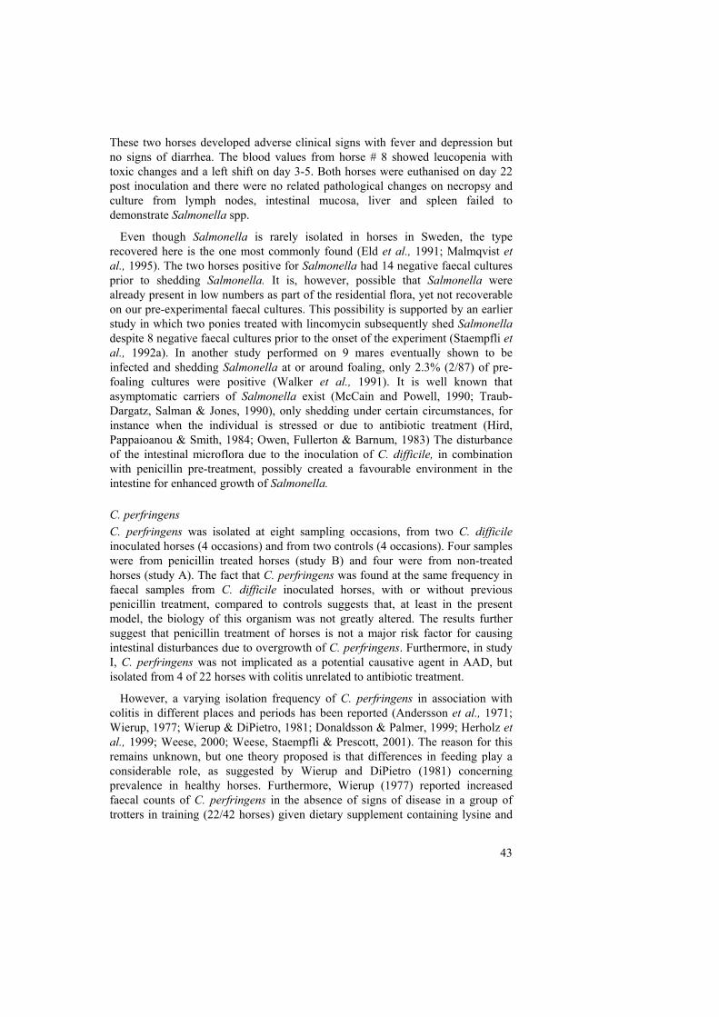
43
These two horses developed adverse clinical signs with fever and depression but no signs of diarrhea. The blood values from horse # 8 showed leucopenia with toxic changes and a left shift on day 3-5. Both horses were euthanised on day 22 post inoculation and there were no related pathological changes on necropsy and culture from lymph nodes, intestinal mucosa, liver and spleen failed to demonstrate Salmonella spp.
Even though Salmonella is rarely isolated in horses in Sweden, the type recovered here is the one most commonly found (Eld et al., 1991; Malmqvist et al., 1995). The two horses positive for Salmonella had 14 negative faecal cultures prior to shedding Salmonella. It is, however, possible that Salmonella were already present in low numbers as part of the residential flora, yet not recoverable on our pre-experimental faecal cultures. This possibility is supported by an earlier study in which two ponies treated with lincomycin subsequently shed Salmonella despite 8 negative faecal cultures prior to the onset of the experiment (Staempfli et al., 1992a). In another study performed on 9 mares eventually shown to be infected and shedding Salmonella at or around foaling, only 2.3% (2/87) of pre-foaling cultures were positive (Walker et al., 1991). It is well known that asymptomatic carriers of Salmonella exist (McCain and Powell, 1990; Traub-Dargatz, Salman & Jones, 1990), only shedding under certain circumstances, for instance when the individual is stressed or due to antibiotic treatment (Hird, Pappaioanou & Smith, 1984; Owen, Fullerton & Barnum, 1983) The disturbance of the intestinal microflora due to the inoculation of C. difficile, in combination with penicillin pre-treatment, possibly created a favourable environment in the intestine for enhanced growth of Salmonella. C. perfringens C. perfringens was isolated at eight sampling occasions, from two C. difficile inoculated horses (4 occasions) and from two controls (4 occasions). Four samples were from penicillin treated horses (study B) and four were from non-treated horses (study A). The fact that C. perfringens was found at the same frequency in faecal samples from C. difficile inoculated horses, with or without previous penicillin treatment, compared to controls suggests that, at least in the present model, the biology of this organism was not greatly altered. The results further suggest that penicillin treatment of horses is not a major risk factor for causing intestinal disturbances due to overgrowth of C. perfringens. Furthermore, in study I, C. perfringens was not implicated as a potential causative agent in AAD, but isolated from 4 of 22 horses with colitis unrelated to antibiotic treatment.
However, a varying isolation frequency of C. perfringens in association with colitis in different places and periods has been reported (Andersson et al., 1971; Wierup, 1977; Wierup & DiPietro, 1981; Donaldsson & Palmer, 1999; Herholz et al., 1999; Weese, 2000; Weese, Staempfli & Prescott, 2001). The reason for this remains unknown, but one theory proposed is that differences in feeding play a considerable role, as suggested by Wierup and DiPietro (1981) concerning prevalence in healthy horses. Furthermore, Wierup (1977) reported increased faecal counts of C. perfringens in the absence of signs of disease in a group of trotters in training (22/42 horses) given dietary supplement containing lysine and

44
methionine. Wierup & DiPietro (1981) also emphasized that a diagnosis cannot be based only on high C. perfringens counts, even if there is a clear association with high counts and disease. Leakage of plasma proteins into the lumen of the intestine during colitis can favour the growth of C. perfringens (Palmer, 1992a). Further, this author suggests that the significance of recovering C. perfringens may be more as a marker of massive protein leakage and a disrupted microflora rather than being a cause, an opinion shared by Nielsen & Vibe-Petersen (1979) and Larsen (1997). The latter author reported findings of C. perfingens from very few cases of colitis during 1987-1997 at the Norwegian College of Veterinary Medicine.
With regard to prevalence of C. perfringens toxins, variable results have been reported. Studies by Herholz et al. (1999), showed a high frequency (52%) of β2-toxigenic C. perfringens strains in horses with typhlocolitis, and reported that the majority of horses were treated with antibiotics and had a high mortality. The authors suggested that β2-toxigenic C. perfringens might be particularly fatal in combination with antibiotic treatment. Further, these authors isolated both toxigenic and nontoxigenic C. difficile as well as C. perfringens type A to a lesser degree, but no C. perfringens strains contained the gene for enterotoxin. In other studies CPE was detected in samples from 16 and 19% (9/57 and 9/47 respectively) of diarrheic adults but was absent in horses without gastrointestinal disease (57 and 47 respectively) (Donaldson & Palmer, 1999; Weese, Staempfli & Prescott, 2001). The latter author found no association between spore count and CPE.
Toxin assays for C. perfringens were not performed in the present studies. It appears that the role of C. perfringens as a pathogen in colitis in adult horses is even less clear than is the role of C. difficile. However, when consolidating earlier knowledge and recent reports it still appears that C. perfringens is of importance in the pathogenesis of colitis, but further investigations should be performed to understand more fully the role of both these organisms, including detection of toxins and toxigenicity of isolated strains. Isolation of more than one pathogen In the present study it was presumed that adverse clinical signs in horse # 7 and #8 originated from the Salmonella infection and not from inoculated C. difficile, as no toxins could be detected. However, simultaneous isolation of more than one pathogen from the same diseased horse are commonly reported (Wierup, 1977; Beier, Amtsberg & Peters, 1994; Cosmetatos et al., 1994; Madewell et al., 1995; Greiß et al., 1996; Madigan et al., 1997; Herholtz et al., 1999), and there are reports of both C. difficile and C. perfringens toxin detection from faecal samples of the same horse (Donaldson & Palmer, 1999; Weese, Staempfli & Prescott, 2001). The cause and effect in these cases are impossible to evaluate. For example, in the outbreak of C. difficile-associated diarrhea described by Madewell et al., (1995), Salmonella Krefeld was also isolated from one horse, and it was considered to be infected simultaneously with the two pathogens. On the other hand, Wierup (1977) suggested that the isolation of Salmonella together with C. perfringens in the intestinal content of one horse might be regarded as a secondary

45
finding. Further studies are clearly needed to better clarify the roles of different Clostridium spp. and Salmonella in acute colitis and AAD. Trimethoprim/Sulfonamides – impact on the intestinal microflora (Paper III) With given doses of TMP/SDZ the horses seemed unaffected and the faecal consistency appeared normal during the whole study. No major changes in the intestinal microflora were observed, which suggests a low risk of this drug combination concerning gastrointestinal disturbances, an opinion shared by White and Prior (1982). Furthermore, Wilson et al. (1996) showed that diarrhea was not associated with trimethoprim/sulphonamide treatment either in an extensive case control study or in a cohort study. In the present study, neither C. difficile, C. perfringens nor Salmonella were found in faecal samples following the i.v. and oral TMP/SDZ treatment. An initial approximate 10-fold reduction of the number of coliform bacteria was notable with no apparent difference between i.v. and oral administration. After completing the treatment, the numbers returned to normal. This was also the only change noted by White & Prior (1982) after TMP/SDZ administration.
It appears that reduction of counts of coliform bacteria does not reflect a severely disrupted intestinal microflora. In contrast, rapid increases in counts of coliform bacteria were noted after both erythromycin and rifampicin administration (study II). This has also been seen after oral administration of oxytetracycline (Andersson et al., 1971; White & Prior, 1982), clindamycin together with lincomycin (Prescott et al., 1988), and amikacin (Horspool, Taylor & McKellar, 1994), as well as in several horses with AAD in study I (not shown in the paper). Wierup (1977) did not state an upper limit of counts of coliform bacteria, but the present results suggest that an increase would better reflect a disruption of the flora than a decrease. This is in agreement with Voollard and Clasener (1994) who proposed that impairment of the CR flora in humans is indicated by an increase in the concentration of coliform bacteria. Furthermore, this increase may result in more endotoxin production and impairment of the mucosal barrier against endotoxin absorption (Jones, 2000). Oral adminstration Orally administered antibiotics that are well absorbed in the upper part of the small intestine, as the studied combination of TMP/SDZ studied, generally have minor impact on the large intestine microflora, whereas antibiotics that are poorly or incompletely absorbed can cause significant changes (Nord, 1993). It is reasonable to assume that the risk for disturbance of the microflora is generally increased with antibiotics used for oral treatment because higher concentrations of the drug in the large intestinal content normally should be achieved. However, this was not the experience in study I, where most of the cases reported with AAD occurred after parenteral use, a result also found by Weese, (2000). Certainly, the parenteral route is more commonly used in horses, with the exception of oral preparations of trimethoprim/sulfonamides.

46
Also of importance with oral administration is that time of feeding in relation to antibiotic administration affects the systemic availability of the drug, shown for some drugs to be lower with feeding before antibiotic administration compared to feeding after administration (Baggot, 1992; Bogan et al., 1984). Physical or chemical binding of the drug to the ingesta probably causes this (van Duijkeren et al., 1995a; 1995b; McKellar & Horspool, 1995). If oral antibiotics were given in connection with or just after feeding, a consequence of this binding could be subsequent release of the drug into the large intestine after fermentative digestion of the feedstuff and higher intestinal antibiotic concentrations.
In the present study the horses were not fasted and t1/2β were greater than in studies with the horses fasted prior to drug administration (van Duijkeren et al., 1994). Thus, it seems that the most appropriate time to administer the drug is before feeding. Ensink et al. (1996) found that previous lack of appetite was significantly correlated with a high incidence of diarrhea in horses receiving TMP/SDZ. All horses in the present study had normal appetite. In accordance with the above discussion great variation in plasma concentrations between individual horses were observed in study III. Others have also described this after oral administration of varying trimethoprim/sulphonamide combinations (Sigel et al., 1981; Morgan & White, 1983; Bogan et al., 1984; van Duijkeren et al., 1994; 1995a; 1995b). Further reasons for these variations could be individual differences in gastric emptying and to some extent, bile excretion with recirculation of the drugs or metabolism in the intestinal mucosa or the liver before reaching the systemic circulation (Baggot, 1992). Clinical outcome and treatment of colitis (Paper II) Diseased horses in the present study developed the dramatic and severe clinical changes on the third day of antibiotic treatment. Day three was also the mean debut day of diarrhea among the 25 horses in study I. The time after initiation of antibiotic treatment until onset of disease is typically reported to be only a few days (Andersson et al., 1971; Cook, 1973; Prescott et al., 1988; Staempfli, Prescott & Brash, 1992; Staempfli et al., 1992; Ensink et al., 1996). This shows that the disturbance of the intestinal microflora takes place in the first days of treatment, and that the risk for diarrhea thereafter is independent of duration of therapy. In humans, the median time for occurrence of symptoms is described to be longer, averaging 9 days after the start of treatment (Wiström et al., 2001). However, time of onset of disease can also be longer in horses, and even extend to beyond cessation of antibiotic administration (Magdesian et al., 1997; Weese, 2000).
One of the two diseased horses in study II was treated successfully with oral bacitracin, together with large amounts of i.v. fluids and flunixin meglumine and recovered within two days. Despite similar treatment, the clinical condition of the other horse gradually deteriorated with marked dehydration and typical signs of toxaemia. After 24 h, with decreased intestinal sounds and increased signs of colic, it was subjected to euthanasia for ethical reasons. Bacitracin treatment was initially presumed to have contributed to the rapid recovery in the first horse, but

47
paradoxically the C. difficile strain isolated from that case was resistant to bacitracin (MIC>256 µg/ml), as also reported by others (Jang et al., 1997; Weese, Staempfli & Prescott, 2001; Båverud et al., 2003). Mild CdAD may resolve when the offending antibiotic treatment is discontinued and the normal microflora is re-established. In severe cases where antibiotic treatment can be considered, metronidazole should be the first choice before vancomycin, due to concerns about spread of vancomycin-resistant bacteria and lower cost. Furthermore, two horses in study I acquired AAD despite inclusion of metronidazole in the previous treatment. This is in accordance with reports in human medicine. Neither metronidazole nor vancomycin were effective as prophylaxis of AAD when included in therapy (Wiström et al., 2001).

48
Concluding remarks The knowledge of how different antibiotics influence the intestinal microflora in horses is still limited. The work described in this thesis shows that C. difficile is associated with acute colitis in adult horses, following treatment with antibiotics. Most of the horses positive for C. difficile were treated with β-lactam antibiotics, alone or in combination with other antibiotics. Penicillin treatment predisposes the establishment of C. difficile in the horse intestine. Further it was demonstrated that very low oral doses of erythromycin could induce acute colitis associated with major changes of the intestinal microflora. Thus, it was considered most likely that the fatal colitis affecting the mares was due to accidental ingestion of erythromycin. In contrast, very low oral doses of rifampicin and therapeutic doses of both oral and i.v. TMP/SDZ were not associated with major changes in the intestinal flora and no evidence of gastrointestinal disturbances was observed.
Nevertheless, broad generalisations regarding the safety of any particular antibiotic used in equine practice should be avoided unless there are sufficient data to fully assess the risk. Our own choice of antibiotics in attempting to minimise risk of antibiotic associated diarrhea may not be the same as in other regions of the world, and clearly defined antibiotic usage guidelines in regard to risk of induction of diarrhoea in the horse, even if available, cannot be applied globally. This is likely because the intestinal flora varies with type of feeding the horse is given, and on the variable management and hygiene situation in different regions of the world. It appears that every time a horse is treated with antibiotics there is invariably a relative risk of colitis that has to be considered, and thus there are no completely safe antibiotics. As clinicians it is our responsibility to avoid unnecessary antibiotic treatment in the horse. Further, potential pre-disposing risk factors for inducing acute colitis, for example; stress, starvation, withholding roughage, abdominal surgery, intestinal stasis, hospitalisation, long transportation, presence of other disease/s and concurrent medications need to be carefully considered in each case when choosing to administer antibiotics. Ideally, antibiotics should be administered only when there is a known or highly suspected bacterial infection or when antibiotic prophylaxis is required. If there is a need for antibiotic treatment there should be a high likelihood of a successful therapeutic outcome. Antibiotics known to be associated with a high risk for development of colitis such as macrolides and lincosamides should be avoided. Furthermore, use of antibiotics that are poorly evaluated with regard to safety and effectiveness in horses clearly deviates from sound medical management principles.
Finally, as most clinicians are aware, there is an ever-increasing problem world-wide with development and spread of antibiotic-resistant bacteria, such as multi-resistant Salmonellae and C. difficile. Indiscriminate use of antibiotics only increases the potential for emergence and spread of these resistant bacteria that pose an increasing threat to both human and animal health.

49
References Agria Hästförsäkring. Stockholm. 1992. Diarrémöte i Lillehammer 920214-16. Andersson, G., Ekman, L., Mansson, I., Persson, S., Rubarth, S. & Tufvesson, G. 1971.
Lethal complications following administration of oxytetracycline in the horse. Nordisk veterinärmedicin 23, 9-22.
Argenzio, R.A. 1975. Functions of the equine large intestine and their interrelationship in disease. Cornell veterinarian 65, 303-330.
Aronsson, B., Möllby, R. & Nord, C.E. 1982. Clostridium difficile and antibiotic associated diarrhea in Sweden. Scandinavian journal of infectious diseases Suppl. 35, 53-58.
Baggot, J.D. 1992. Bioavailability and bioequivalence of veterinary drug dosage forms, with particular reference to horses: an overview. Journal of veterinary pharmacology and therapeutics 15, 160-173.
Baker, J.R. & Leyland, A. 1973. Diarrhoea in the horse associated with stress and tetracycline therapy. The Veterinary record 93, 583-584.
Bartlett, J.G. 1990. Clostridium difficile: clinical considerations. Review of infectious diseases 12 (Suppl. 2) 243-251.
Bartlett, J.G., Chang, T.W., Gurwith, M., Gorbach, S.L. & Onderdonk, A.B. 1978. Antibiotic-associated pseudomemranous colitis due to toxin-producing clostridia. New England journal of medicine 298, 531-534.
Baums, C., Bartmann, C., Jobst, D., Deegen, E., Amtsberg, G. & Goethe, R. 2003. Prevalence, selection and proposed importance of erythromycin-resistance among C. difficile and C. perfringens isolates from a German horse clinic. In Diagnosis, epidemiology and antibiotic resistance of the genus Clostridium. Second open meeting of the European CA QLK2-CT2001-01267, Parma, Italy.
Beard, L.A. 1998. Principles of antimicrobial therapy. In Equine internal medicine. (Eds, S.M. Read & W.M. Bayly) W.B. Saunders Company, Philadelphia, pp. 157-169.
Beier, R., Amtsberg, G. & Peters, M. 1994. Bakteriologische Untersuchungen zum Vorkommen und Bedeutung von Clostridium difficile beim Pferd. Pferdeheilkunde 10, 3-8.
Bergsten, G. & Lannek, N. 1970. Tarmkatarr hos häst. In Jordbrukets försäkringsbolag 1970, pp. 38-51.
Bertone, A.L., Jones, R.L. & McIlwraith, C.W. 1988. Serum and synovial fluid steady-state concentrations of trimethoprim and sulfadiazine in horses with experimentally induced infectious arthritis. American journal of veterinary research 49, 1681-1687.
Bogan, J.A., Galbraith, A., Baxter, P., Ali, N.M. & Marriner, S.E. 1984. Effect of feeding on the fate of orally administered phenylbutazone, trimethoprim and sulfadiazine in the horse. The Veterinary record 115, 599-600.
Borriello, S.P. 1998. Pathogenesis of Clostridium difficile infection. Journal of antimicrobial chemotherapy 41, Suppl. C, 13-19.
Borriello, S.P., Welch, A.R., Stringer, M.F., Larson, H.E., Barclay, F. & Bartholomew, B.A. 1984. Enterotoxigenic Clostridium perfringens: A possible cause of antibiotic-associated diarrhoea. The Lancet 1, 305-307.
Bowen, J. 2002. Commentary. Equine disease quarterly newsletter 11, 1. Brown, M.P., Gronwall, R. & Castro, L. 1988. Pharmacokinetics and body fluid and
endometrial concentrations of trimethoprim-sulfamethoxazole in mares. American journal of veterinary research 49, 918-922.
Brumbaugh, G. W. 1987. Rational selection of antimicrobial drugs for treatment of infections of horses. Veterinary clinics of North America. Equine practice 3, 191-220.
Brørs, O., Sager, G., Sandnes, D. & Jacobsen, S. 1983. Binding of theophylline in human serum determined by ultrafiltration and equilibrium dialysis. British journal of clinical pharmacology 15, 393-397.
Burrows, G.E. 1981. Endotoxaemia in the horse. Equine veterinary journal 13, 89-94.
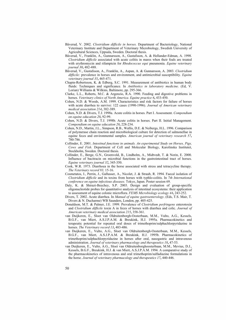
50
Båverud, V. 2002. Clostridium difficile in horses. Department of Bacteriology, National Veterinary Institute and Department of Veterinary Microbiology, Swedish University of Agricultural Sciences, Uppsala, Sweden. Doctoral thesis.
Båverud, V., Franklin, A., Gunnarsson, A., Gustafsson, A. & Hellander-Edman, A. 1998. Clostridium difficile associated with acute colitis in mares when their foals are treated with erythromycin and rifampicin for Rhodococcus equi pneumonia. Equine veterinary journal 30, 482-488.
Båverud, V., Gustafsson, A., Franklin, A., Aspan, A. & Gunnarsson, A. 2003. Clostridium difficile: prevalence in horses and environment, and antimicrobial susceptibility. Equine veterinary journal 35, 465-471.
Chapin-Robertsson, K. & Edberg, S.C. 1991. Measurement of antibiotics in human body fluids: Techniques and significance. In Antibiotics in laboratory medicine. (Ed, V. Lorian) Williams & Wilkins, Baltimore, pp. 295-366.
Clarke, L.L., Roberts, M.C. & Argenzio, R.A. 1990. Feeding and digestive problems in horses. Veterinary clinics of North America. Equine practice 6, 433-450.
Cohen, N.D. & Woods, A.M. 1999. Characteristics and risk factors for failure of horses with acute diarrhea to survive: 122 cases (1990-1996). Journal of American veterinary medical association 214, 382-389.
Cohen, N.D. & Divers, T.J. 1998a. Acute colitis in horses. Part I. Assessment. Compendium on equine education 20, 92-99.
Cohen, N.D. & Divers, T.J. 1998b. Acute colitis in horses. Part II. Initial Management. Compendium on equine education 20, 228-234.
Cohen, N.D., Martin, J.L., Simpson, R.B., Wallis, D.E. & Neibergs, H.L. 1996. Comparison of polymerase chain reaction and microbiological culture for detection of salmonellae in equine feces and environmental samples. American journal of veterinary research 57, 780-786.
Collinder, E. 2001. Intestinal functions in animals. An experimental Study on Horses, Pigs, Cows and Fish. Department of Cell and Molecular Biology, Karolinska Institutet, Stockholm, Sweden. Doctoral thesis.
Collinder, E., Berge, G.N., Groenvold, B., Lindholm, A., Midtvedt, T. & Norin, E. 2000. Influence of bacitracin on microbial functions in the gastrointestinal tract of horses. Equine veterinary journal 32, 345-350.
Cook, W.R. 1973. Diarrhoea in the horse associated with stress and tetracycline therapy. The Veterinary record 93, 15-16.
Cosmetatos, I., Perrin, J., Gallusser, A., Nicolet, J. & Straub, R. 1994. Faecal isolation of Clostridium difficile and its toxins from horses with typhlo-colitis. In 7th International conference on equine infectious diseases. Tokyo, Japan. Poster session 69.
Daly, K. & Shirazi-Beechey, S.P. 2003. Design and evaluation of group-specific oligonucleotide probes for quantitative analysis of intestinal ecosystems: their application to assessment of equine colonic microflora. FEMS Microbiology ecology 44, 243-252.
Divers, T. 2002. Acute diarrhea. In Manual of equine gastroenterology. (Eds, T.S. Mair, T. Divers & N. Ducharme) WB Saunders, London, pp. 405-425.
Donaldson, M.T. & Palmer, J.E. 1999. Prevalence of Clostridium perfringens enterotoxin and Clostridium difficile toxin A in feces of horses with diarrhea and colic. Journal of American veterinary medical association 215, 358-361.
van Duijkeren, E., Sloet van Oldruitenborgh-Oosterbaan, M.M., Vulto, A.G., Kessels, B.G.F., van Miert, A.S.J.P.A.M. & Breukink, H.J. 1995a. Pharmacokinetics and terapeutic potential for repeated oral doses of trimethoprim/sulphachlorpyridazine in horses. The Veterinary record 13, 483-486.
van Duijkeren, E., Vulto, A.G., Sloet van Oldruitenborgh-Oosterbaan, M.M., Kessels, B.G.F., van Miert, A.S.J.P.A.M. & Breukink, H.J. 1995b. Pharmacokinetics of trimethoprim/sulphachlorpyridazine in horses after oral, nasogastric and intravenous administration. Journal of veterinary pharmacology and therapeutics 18, 47-53.
van Duijkeren, E., Vulto, A.G., Sloet van Oldruitenborghoosterbaan, M.M., Mevius, D.J., Kessels, B.G.F., Breukink, H.J. & van Miert, A.S.J.P.A.M. 1994. A comparative study of the pharmacokinetics of intravenous and oral trimethoprim/sulfadiazine formulations in the horse. Journal of veterinary pharmacology and therapeutics 17, 440-446.
51
Dyrendahl, I. 1996. Peter Hernquists sjukdomslära – husdjurens inre sjukdomar. Kungliga skogs- och lantbruksakademien, Stockholm, pp. 130-131.
Ehrich, M., Perry, B.D., Troutt, F., Dellers, R.W. & Magnusson, R.A. 1984. Acute diarrhea in horses of the Potomac River area: Examination for clostridial toxins. Journal of American veterinary medical association 185, 433-435.
Eld, K., Gunnarsson, A., Holmberg, T., Hurvell, B. & Wierup, M. 1991. Salmonella isolated from animals and feedstuffs in Sweden during 1983-1987. Acta veterinaria scandinavica 32, 261-277.
Ensink, J.M., Klein, W.R., Barneveld, A., van Miert, A.S. & Vulto, A.G. 1996. Side effects of oral antimicrobial agents in the horse: a comparison of pivampicillin and trimethoprim/sulphadiazine. The Veterinary record 138, 253-256.
Ewing, P.J., Burrows, G., MacAllister, C. & Clarke, C. 1994. Comparison of oral erythromycin formulations in the horse using pharmacokinetic profiles. Journal of veterinary pharmacology and therapeutics 17, 17-23.
Fekety, R., Silva, J., Toshniwal, R., Allo, M., Armstrong, J., Browne, R., Ebright, J. & Rifkin, G. 1979. Antibiotic-associated colitis: effects of antibiotics on Clostridium difficile and the disease in hamsters. Review of infectious diseases 1, 386-397.
Foreman, J.H. 1998. Does ceftiofur cause diarrhea? In Proceedings of the 44th annual conference of American association equine practitioners. Vol. 44. Baltimore, Maryland, pp. 146-147.
Freestone, J.F. 2002. Diarrhea - other causes. In Manual of equine gastroenterology, Vol. 1. (Eds, TS. Mair, T. Divers & N. Ducharme) WB Saunders, London, pp. 507-508.
Gautsch, S. 1990. Bakteriologische Untersuchungen zum Vorkommen von enterotoxinbildenden Clostridium perfringens - Stämmen sowie von Clostridium difficile im Darmkanal von Pferden. Institut für Mikrobiologie und Tiersuchen und der Klinik für Pferde, Tiärztlichen Hochschule Hannofer, Hannofer, Germany. Doctoral thesis.
George, R.H. & Symonds, J.M. 1978. Identification of Clostridium difficile as a cause of pseudomembranous colitis. British medical journal 1, 695.
George, W.L., Sutter, V.L., Citron, D. & Finegold, S.M. 1979. Selective and differential medium for isolation of Clostridium difficile. Journal of clinical microbiology 9, 214-219.
George, W.L., Sutter, V.L., Goldstein, E.J., Ludwig, S.L. & Finegold, S.M. 1978. Aetiology of antimicrobial-agent-associated colitis. Lancet i, 802-803.
Giles, C.J., Urquhart, K.A. & Longstaffe, J.A. 1985. Larval cyathostomiasis (immature trichonema-induced enteropathy): A report of 15 clinical cases. Equine veterinary journal 17, 196-201.
Goodson, J., Tyznik, W.J., Dehority, B.A. & Cline, J.H. 1985. Effects of abrupt change from all hay to all concentrate on anaerobic bacterial numbers (grown on selective media), protozoal numbers and pH of the cecum. In Proceedings of the 9th equine nutrition and physiology symposium, pp. 52-63.
Greiß, C., Verspohl, J., Kropp, S., Rhode, J., Pohlenz, J., Scheidemann, W., Deegen, E. & Amtsberg, G. 1996. Die Zusammensetzung der Zäkalflora des Pferdes und ihre mögliche Bedeutung für die Entstehung der Typhlocolitis. Pferdeheilkunde 12, 725-736.
Hall, I.C. & O´Toole, E. 1935. Intestinal flora in newborn infants with a description of a new pathogenic anaerobe, Bacillus difficilis. American journal of diseases of children 49, 390-402.
Hardman, J.G., Limbird, L.E., Molinoff, 1996. Goodman & Gilman´s The pharmacological basis of therapeutics 9th edn. McGraw-Hill Companies, Inc. New York, pp. 1029-1153.
Hathcock, T.L., Schumacher, J., Wright, J.C. & Stringfellow, J. 1999. The prevalence of Aeromonas species in feces of horses with diarrhea. Journal of veterinary internal medicine 13, 357-360.
Herholz, C., Miserez, R., Nicolet, J., Frey, J., Popoff, M., Gibert, M., Gerber, H. & Straub, R. 1999. Prevalence of ß2-toxigenic Clostridium perfringens in horses with intestinal disorders. Journal of clinical microbiology 37, 358-361.
Hird, D.W., Pappaioanou, M. & Smith, B.P. 1984. Case-contol study of risk factors associated with isolation of Salmonella Saintpaul in hospitalized horses. American journal of epidemiology 120, 852-864.

52
Holdeman, L.V., Cato, E.P. & Moore, W.E.C. 1977. Anaerobe laboratory manual, 4th edn. Virginia Polytechnic Institute Anaerobe Laboratory, Blacksburg, Virginia.
Horspool, L.J.I. & McKellar, Q.A. 1995. Disposition of penicillin G sodium following intravenous and oral administration to Equidae. British veterinary journal 151, 401-411.
Horspool, L.J.I., Taylor, D.J. & McKellar, Q.A. 1994. Plasma disposition of amikacin and interactions with gastrointestinal microflora in Equidae following intravenous and oral administration. Journal of veterinary pharmacology and therapeutics 17, 291-298.
Jang, S.S., Hansen, L.M., Breher, J.E., Riley, D.A., Magdesian, K.G., Madigan, J.E., Tang, Y.J., Silva, J. Jr. & Hirsh, D.C. 1997. Antimicrobial susceptibilities of equine isolates of Clostridium difficile and molecular characterization of metronidazole-resistant strains. Clinical infectious diseases 25 Suppl 2, S266-S267.
Jones, R.L. 2000. Clostridial enterocolitis. Veterinary clinics of North America. Equine practice 16, 471-485.
Jones, R.L., Adney, W.S., Alexander, A.F., Shideler, R.K. & Traub-Dargatz, J.L. 1988. Hemorrhagic necrotizing enterocolitis associated with Clostridium difficile infection in four foals. Journal of American veterinary medical association 193, 76-79.
Jones, R.L., Shideler, R.K. & Cockerell, G.L. 1988. Association of Clostridium difficile with foal diarrhea. In Proc. 5th International conference on equine infectious diseases, Lexington, Kentucky. University Press of Kentucky, Lexington. pp. 236-240.
Jones, R.L., Adney, W.S. & Shideler, R.K. 1987. Isolation of Clostridium difficile and detection of cytotoxin in the feces of diarrheic foals in the absence of antimicrobial treatment. Journal of clinical microbiology 25, 1225-1227.
Julliand, V. 1998. Ecologie microbienne du système digestif des équidés: nouvelles approches: conséquences practiques. Journee de la recherche equine 27, 105-113.
Julliand, V., de Vaux, A., Millet, L. & Fonty, G. 1999. Identification of Ruminococcus flavefaciens as the predominant cellulolytic bacterial species of the equine caecum. Applied and environmental microbiology 65, 3738-3741.
Karlsson, S., Burman, L.G. & Åkerlund, T. 1999. Suppression of toxin production in Clostridium difficile VPI 10463 by amino acids. Microbiology 145, 1683-1693.
Keir, A.A., Staempfli, H.R. & Crawford, J. 1999. Outbreak of acute colitis on a horse farm associated with tetracycline-contaminated sweet feed. Canadian veterinary journal 40, 718-720.
Kerr, K.G. 1991. The gastrointestinal microflora: friends or foes? Journal of nutritional medicine 2, 39-44.
Kim, K.H., Fekety, F.R., Batts, D.H., Brown, D., Cudmore, M., Silva, J.J. & Waters, D. 1981. Isolation of Clostridium difficile from the environment and contact patients with antibiotic-associated colitis. Journal of infectious diseases 143, 42-50.
Kim, L.M., Morley, P.S., Traub-Dargatz, J.L., Salman, M.D. & Gentry-Weeks, C. 2001. Factors associated with Salmonella shedding among equine colic patients at a veterinary teaching hospital. Journal of American veterinary medical association 218, 740-748.
King, J.N. & Gerring, E.L. 1988. Detection of endotoxin in cases of equine colic. The Veterinary record 123, 269-271.
Kirby, R. & Rudloff, E. 2000. Acquired coagulopathy VI: Disseminated intravascular coagulation. In Schalm´s veterinary hematology, Vol. 5. (Eds, B.F. Feldman, J.G. Zinkl & N.C. Jain) Lippincott Williams & Wilkins, Philadelphia, pp. 581-587.
van der Kolk, J.H., Bernadina, W.E. & Visser, I.J. 1991. A horse seropositive for Ehrlichia risticii. Tijdschrift voor Diergeneeskunde 116, 69-72.
Larsen, J. 1997. Acute colitis in adult horses. A review with emphasis on aetiology and pathogenesis. The Veterinary quarterly 19, 72-80.
Larsen, J., Dolvik, N.I. & Teige, J., Jr. 1996. Acute post-treatment enterocolitis in 13 horses treated in a Norweigan surgical ward. Acta veterinaria scandinavica 37, 203-211.
Larson, H.E. 1980. The experimental pathogenesis of antibiotic related colitis. Scandandinavian journal of infectious diseases 22, 7-10.
Larson, H.E., Price, A.B., Honour, P. & Borriello, S.P. 1978. Clostridium difficile and the aetiology of pseudomembranous colitis. Lancet i, 1063-1066.
Lyerly, D.M. 1995. Clostridium difficile testing. Clinical microbiology newsletter 17, 17-22.

53
Lyerly, D.M., Krivan, H.C. & Wilkins, T.D. 1988. Clostridium difficile: Its disease and toxins. Clinical microbiology reviews 1, 1-18.
Lyerly D.M. Saum, K.E., MacDonald, D.K. & Wilkins, T.D. 1985. Effects of Clostridium difficile toxins given intragastrically to animals. Infection and immunity, 47, 349-352.
Mackie, R.I. & Wilkins, C.A. 1988. Enumeration of anaerobic bacterial microflora of the equine gastrointestinal tract. Applied and environmental microbiology 54, 2155-2160.
Madewell, B.R., Tang, Y.J., Jang, S., Madigan, J.E., Hirsh, D.C., Gumerlock, P.H. & Silva, J., Jr. 1995. Apparent outbreaks of Clostridium difficile-associated diarrhea in horses in a veterinary medical teaching hospital. Journal of veterinary diagnostic investigation 7, 343-346.
Magdesian, K.G., Hirsh, D.C., Jang, S.S., Hansen, L.M. & Madigan, J.E. 2002. Characterization of Clostridium difficile isolates from foals with diarrhea: 28 cases (1993-1997). Journal of American veterinary medical association 220, 67-73.
Magdesian, K.G., Hirsh, D.C., Jang, S.S. & Madigan, J.E. 1999. Characterisation of Clostridium difficile isolates from an outbreak of enteritis in neonatal foals. In Proceedings of the eighth international conference on equine infectious diseases, Dubai, 1998 (Eds, U. Wernery, J.F.Wade, J.A. Mumford & O.-R. Kaaden). R&W Publications, Limited, Newmarket, pp. 561-562.
Magdesian, K.G., Madigan, J.E., Hirsh, D., Jang, S., Tang, Y.J., Carpenter, T.E., Hansen, L.M. & Silva, J. Jr. 1997. Clostridium difficile and horses: a review. Reviews in medical microbiology 8 (Suppl 1), S46-S48.
Mair, T.S., de Westerlaken, L.V., Cripps, P.J. & Love, S. 1990. Diarrhoea in adult horses: A survey of clinical cases and an assessment of some prognostic indices. The Veterinary record 126, 479-481.
Malmqvist, M., Jacobsson, K.-G., Häggblom, P., Cerenius, F., Sjöland, L. & Gunnarsson, A. 1995. Salmonella isolation from animals and feedstuffs in Sweden during 1988-1992. Acta veterinaria scandinavica 36, 21-39.
McCain, C.S. & Powell, K.C. 1990. Asymptomatic salmonellosis in healthy adult horses. Journal of veterinary diagnostic investigation 2, 236-237.
McFarland, L.V., Mulligan, M.E., Kwok, R.Y.Y. & Stamm, W.E. 1989. Nosocomial acquisition of Clostridium difficile infection. New England journal of medicine 320, 204-210.
McFarland, L.V., Surawicz, C.M. & Stamm, W.E. 1990. Risk factors for Clostridium difficile carriage and C. difficile-associated diarrhea in a cohort of hospitalized patients. Journal of infectious diseases 162, 678-684.
McGorum, B.C., Dixon, P.M. & Smith, D.G.E. 1998. Use of metronidazole in equine acute idiopathic toxaemic colitis. The Veterinary record 142, 635-638.
McKellar, Q.A. & Horspool, L.J.I. 1995. Stability of penicillin G, ampicillin, amikacin and oxytetracycline and their interactions with food in in vitro simulated equine gastrointestinal contents. Research in veterinary science 58, 227-231.
Modi, N. & Wilcox, M.H. 2001. Evidence for antibiotic induced Clostridium perfringens diarrhoea. Journal of clinical pathology 54, 748-751.
Morgan, D.W.T. & White, G. 1983. Studies in horses dosed with trimethoprim and sulfadiazine. Vlaams Diergeneeskundig Tijdschrift 52, 88-94.
Mpamugo, O., Donovan, T. & Brett, M.M. 1995. Enterotoxigenic Clostridium perfringens as a cause of sporadic cases of diarrhoea. Journal of medical microbiology 43, 442-445.
Murray, M.J. 1992. Acute colitis. In Current therapy in equine medicine, Vol 3. (Ed, N.E. Robinson) W.B. Saunders Company, Philadelphia, pp. 244-250.
Murray, M.J. 1997. Acute colitis. In Current therapy in equine medicine, Vol. 4. (Ed, N.E. Robinson) W.B. Saunders Company, Philadelphia, pp. 197-203.
Månsson, I. 1957. The intestinal flora in horses with certain skin changes; with special reference to the coliform microbes. Acta pathologica et microbiologica scandinavica, Suppl. 119.
Möllby, R., Nord, C.-E. & Aronsson, B. 1980. Diagnosis of Clostridium difficile-associated enterocolitis in Sweden. Scandinavian journal of infectious diseases Suppl. 22, 30-36.
Nielsen, K. & Vibe-Petersen, G. 1979. Entero-colitis hos hest. En beskrivelse af 46 tilfaelde. Nordisk veterinärmedicin, 31, 376-381.

54
Nolan, N.P.M., Kelly, C.P., Humphreys, J.F.H., Cooney, C., O´Connor, R., Walsh, T.N., Weir, D.G. & Briain, D.S. 1987. An epidemic of pseudomembranous colitis: Importance of person to person spread. Gut 28, 1467-1473.
Nord, C.E. 1993. The effect of antimicrobial agents on the ecology of the human intestinal microflora. Veterinary microbiology, 35, 193-197.
Ochoa, R. & Kern, S.R. 1980. The effects of Clostridium perfringens type A enterotoxin in shetland ponies - clinical, morphologic and clinicopathologic changes. Veterinary pathology 17, 738-747.
Owen, R. ap R. 1975. Poststress diarrhea in the horse. The Veterinary record 96, 267-270. Owen, Rh. ap Rh., Fullerton, J. & Barnum, D.A. 1983. Effects of transportation, surgery,
and antibiotic therapy in ponies infected with Salmonella. American journal of veterinary research 44, 46-50.
Palmer, J.E. 1992a. Diarrhea. In Veterinary gastroenterology. (Ed, N.V. Anderson) Lea and Febiger, Philadelphia, pp. 634-654.
Palmer, J.E. 1992b. Potomac horse fever. In Current therapy in equine medicine, Vol. 3. (Ed, N.E. Robinson) Saunders, Philadelphia, pp. 250-253.
Palmer, J.E., Whitlock, R.H., Benson, C.E., Becht, J.L., Morris, D.D. & Acland, H.M. 1985. Comparison of rectal mucosal cultures and faecal cultures in detecting Salmonella infections in horses and cattle. American journal of veterinary research 46, 697-698.
Papich, M.G. 2003. Antimicrobial therapy for horses. In Current therapy in equine medicine, Vol. 5. (Ed, N.E. Robinson) Saunders, St Louis, pp. 6-11.
Perrin, J., Cosmetatos, I., Gallusser, A., Lobsiger, L., Straub, R. & Nicolet, J. 1993. Clostridium difficile associated with typhlocolitis in an adult horse. Journal of veterinary diagnostic investigation 5, 99-101.
Pochapin, M. 2000. The effect of probiotics on Clostridium difficile diarrhea. The American journal of gastroenterology 95, S11-S13.
Prescott, J.F. & Baggot, J.D. 1993. Antimicrobial therapy in veterinary medicine, 2nd edn. Iowa State University Press.
Prescott, J.F., Hoover, D.J. & Dohoo, I.R. 1983. Pharmacokinetics of erythromycin in foals and in adult horses. Journal of veterinary pharmacology and therapeutics 6, 67-74.
Prescott, J.F., Staempfli, H.R., Barker, I.K., Bettoni, R. & Delaney, K. 1988. A method for reproducing fatal idiopathic colitis (colitis X) in ponies and isolation of a clostridium as a possible agent. Equine veterinary journal 20, 417-420.
Raisbeck, M.F., Holt, G.F. & Osweiler, G.D. 1981. Lincomycin-associated colitis in horses. Journal of the American veterinary medical association 179, 362-363.
Rehg, J.E. & Lu, Y.-S. 1982. Clostridium difficile typhlitis in hamsters not associated with antibiotic therapy. Journal of the American veterinary medical association 181, 1422-1423.
Roberts, M.C. 1990. Acute equine colitis: experimental and clinical perspectives. Veterinary annual 30, 1-11.
Roneus, B., Roneus, N., Franklin, A. & Jonsson, P. 1993. Behandling med standardiserad kolikultur vid tarmflorerubbning hos hästar. Svensk veterinär tidning 45, 201-204.
Rooney, J.R., Bryans, J.T. & Doll, E.R. 1963. Colitis "X" of horses. Journal of the American veterinary medical association 142, 510-511.
Sambol, S.P., Merrigan, M.M., Tang, J.K., Johnson, S. & Gerding, D.N. 2002. Colonization for the prevention of Clostridium difficile disease in hamsters. Journal of infectious diseases 186, 1781-1789.
Schwan, A., Sjölin, S., Trottestam, U. & Aronsson, B. 1984. Relapsing Clostridium difficile enterocolitis cured by rectal infusion of normal faeces. Scandinavian journal of infectious diseases 16, 211-215.
Semrad, S.D., Moore, J.N., Hardee, G.E. & Hardee, M.M. 1986. Flunixine meglumine: Efficacy of reduced dosages in endotoxemia. In Equine colic research proceedings of the second symposium at the university of Georgia. (Ed, J.N. Moore, N.A. White & J.L. Becht). Veterinary learning systems Co., Lawrenceville, New Jersey, pp. 45-49.
Shim, J.K., Johnson, S., Samore, M.H., Blizz, D.Z. & Gerding, D.N. 1998. Primary symptomless colonisation by Clostridium difficile and decreased risk of subsequent diarrhoea. The Lancet 351, 633-636.
55
Sigel, C.W., Byars, T.D., Divers, T.J., Murch, O. & DeAngelis, D. 1981. Serum of trimethoprim and sulfadiazine following oral paste administration to the horse. American journal of veterinary research, 42, 2002-2005.
Smith, A. 1885. Diseases of the intestine. In Veterinary notes on the causes, symptoms and treatment of the diseases of domestic animals. J.A. Carveth & Co, Toronto, Canada, pp. 177-198.
Staempfli, H.R., Prescott, J.F. & Brash, M.L. 1992. Lincomycin-induced severe colitis in ponies: association with Clostridium cadaveris. Canadian journal of veterinary research 56, 168-169.
Staempfli, H.R., Prescott, J.F., Carman, R.J. & McCutcheon, L.J. 1992. Use of bacitracin in the prevention and treatment of experimentally-induced idiopathic colitis in horses. Canadian journal of veterinary research 56, 233-236.
Staempfli, H.R., Townsend, H.G.G. & Prescott, J.F. 1991. Prognostic features and clinical presentation of acute idiopathic enterocolitis in horses. Canadian veterinary journal 32, 232-237.
Stewart, M.C., Hodgson, J.L., Kim, H., Hutchins, D.R. & Hodgson, D.P. 1995. Acute febrile diarrhoea in horses. 86 cases (1986-1991). Australian veterinary journal 72, 41-44.
Sullivan, Å., Edlund, C. & Nord, C.-E. 2001. Effect of antimicrobial agents on the ecological balance of human microflora. The Lancet infectious diseases 1, 101-114.
Surawicz, C.M., Elmer, G.W., Speelman, P., McFarland, L.V., Chinn, J. & van Belle, G. 1989. Prevention of antibiotic-associated diarrhea by Saccharomyces boulardii: a prospective study. Gastroenterology 96, 981-988.
Surawicz, C.M., McFarland, L.V., Greenberg, R.N., Rubin, M., Fekety, R., Mulligan, M.E., Garcia, R.J., Brandmarker, S., Bowen, K., Borjal, D. & Elmer, G.W. 2000. The search for a better treatment for recurrent Clostridium difficile disease: use of high-dose vancomycin combined with Saccharomyces boulardii. Clinical infectious diseases 31, 1012-1017.
Sutton, G.A., Staempfli, H.R., Prescott, J.F. & Townsend, H.G.G. 1998. Efficacy of zinc bacitracin as an adjunct treatment of acute severe idiopathic colitis in the horse. In Proceedings of the eighth international conference on equine infectious diseases, Dubai, 1998 (Eds, U. Wernery, J.F. Wade, J.A. Mumford & O.-R. Kaaden). R&W Publications, Limited, Newmarket, pp. 191-195.
Svenungsson, B., Lagergren, Å. & Lundberg, A. 2001. Clostridium difficile cytotoxin B in adults with diarrhea: a comparison of patients treated or not treated with antibiotics prior to infection. Clinical microbiology and infection 7, 447-450.
Tabaqchali, S. & Jumaa, P. 1995. Diagnosis and management of Clostridium difficile infection. British medical journal 310, 1375-1380.
Teale, C.J. & Taylor, R.D. 1998. Clostridium difficile infection in a horse. The Veterinary record 142, 47.
Thomas, C., Stevenson, M. & Riley, T.V. 2003. Antibiotics and hospital-acqired Clostridium difficile-associated diarrhoea: A systematic review. Journal of antimicrobial chemotherapy 51, 1339-1350.
Traub-Dargatz, J.L. & Jones, R.L. 1993. Clostridia-associated enterocolitis in adult horses and foals. Veterinary clinics of North America. Equine practice 9, 411-421.
Traub-Dargatz, J.L., Salman, M.D. & Jones, R.L. 1990. Epidemiologic study of salmonellae shedding in the feces of horses and potential risk factors for development of the infection in hospitalized horses. Journal of American veterinary medical association 196, 1617-1622.
Tullus, K., Aronsson, B., Marcus, S. & Möllby, R. 1989. Intestinal colonization with Clostridium difficile in infants up to 18 months of age. European journal of clinical microbiology & infectious diseases 8, 390-393.
Tvede, M. & Rask-Madsen, J. 1989. Bacteriotherapy for chronic relapsing Clostridium difficile diarrhoea in six patients. Lancet 1, 1156-1160.
Umemura, T., Ohishi, H., Ikemoto, Y., Satoh, H. & Fujimoto, Y. 1982. Histopathology of colitis X in the horse. Japanese journal of veterinary sciences 44, 717-724.

56
Vaughan, J.T. 1973. The acute colitis syndrome. Colits X. Veterinary clinics of North America. Equine practice 3, 301-313.
Vollaard, E.J. & Clasener, H.A.L. 1994. Colonization resistance. Antimicrobial agents and chemotherapy 38, 409-414.
van der Waaij, D., Vossen, J.M., Korthals Altes, C. & Hartgrink, C. 1977. Reconventionalization following antibiotic decontamination in man and animals. The American journal of clinical nutrition 30, 1887-1895.
Walker, R.L., Madigan, J.E., Hird, D.W., Case, J.T., Villanueva, M.R. & Bogenrief, D.S. 1991. An outbreak of equine neonatal salmonellosis. Journal of veterinary diagnostic investigation 3, 223-227.
Weese, J.S. 2000. Antimicrobial-associated diarrhea in 40 horses: 1997-1999. In Clostridium difficile associated enterocolitis in adult horses and foals. Ontario Veterinary College, Guelph, Canada. DVSc thesis.
Weese, J.S., Kaese, H.J., Baird, J.D., Kenney, D.G. & Staempfli, H.R. 2002. Suspected ciprofloxacin-induced colitis in four horses. Equine veterinary education 14, 182-186.
Weese, J.S., Staempfli, H.R. & Prescott, J.F. 2000. Isolation of environmental Clostridium difficile from a veterinary teaching hospital. Journal of veterinary diagnostic investigation 12, 449-452.
Weese, J.S., Staempfli, H.R. & Prescott, J.F. 2001. A prospective study of the roles of Clostridium difficile and enterotoxigenic Clostridium perfringens in equine diarrhoea. Equine veterinary journal 33, 403-409.
White, G. & Prior, S.D. 1982. Comparative effects of oral administration of trimethoprim/sulfadiazine or oxytetracycline of the faecal flora of horses. The Veterinary record 111, 316-318.
Wiberg, Å. 1994. Eventuell korrelation mellan hästens fekala flora och sjukdomssymptom. Svensk veterinär tidning 46, 549-555.
Wiedeman, B. & Atkinson, A. 1991. Susceptibility to antibiotics: species incidence and trends. In Antibiotics in laboratory medicine. (Ed, V. Lorian) Williams and Wilkins, Baltimore, pp. 962-1208.
Wierup, M. 1977. Equine intestinal clostridiosis. Acta veterinaria scandinavica, (Suppl) 62, 1-182.
Wierup, M. & DiPietro, J.A. 1981. Bacteriologic examination of equine faecal flora as a diagnostic tool for equine intestinal clostridiosis. American journal of veterinary research 42, 2167-2169.
Wilkins, T.D. & Lyerly, D.M. 2003. Clostridium difficile: after 20 years still challenging. Journal of clinical microbiology 41, 531-534.
Wilson, D.A., MacFadden, K.E., Green, E.M., Crabill, M., Frankeny, R.L. & Thorne, J.G. 1996. Case control and historical cohort study of diarrhea associated with administration of trimethoprim-potentiated sulphonamides to horses and ponies. Journal of veterinary internal medicine 10, 258-264.
Wilson, K.H., Kennedy, M.J. & Fekety, F.R. 1982. Use of sodiumtaurocholate to enhance spore recovery on a medium selective for Clostridium difficile. Journal of clinical microbiology 15, 443-446.
Wilson, K.J., Silva, J. & Fekety, F.R. 1981. Suppression of Clostridium difficile by normal hamster cecal flora and prevention of antibiotic-associated cecitis. Infection and immunity 34, 626-628.
Wilson, W.D. 1992. Foal pneumonia: An overview. In Proceedings of the 38th annual convention of the American association of equine practitioners, (Ed, L.B. Caddel), Orlando, pp. 203-229.
Wiström, J., Norrby, S.R., Myhre, E.B., Eriksson, S., Granström, G., Lagergren, L., Englund, G., Nord, C.-E. & Svenungsson, B. 2001. Frequency of antibiotic-associated diarrhoea in 2462 antibiotic-treated hospitalized patients: a prospective study. Journal of antimicrobial chemotherapy 47, 43-50.

57
Acknowledgements This work was carried out at the Department of Large Animal Clinical Sciences, Faculty of Veterinary Medicine, Swedish University of Agricultural Sciences (SLU) and at the Departments of Bacteriology and Antibiotics, National Veterinary Institute (SVA), Uppsala. The studies were financed by ATG (the Swedish Horserace Totalizator Board), AGRIA Foundation for Research and Bayer AB. Many people have helped and supported me in different ways during the years I have been working with this thesis and I am grateful to all of you. However, I especially wish to express my warmest thanks to: Anders Franklin, my supervisor and head of the Department of Antibiotics, for so many things! For sharing your broad knowledge in the world of microbes and antibiotics and for all interesting and productive discussions we have had throughout the years. I am always in a good mood after our meetings as you are scientifically stringent and fun at the same time. For always being patient, kind and encouraging and giving me of your time no matter if you are in Brussels or not well and in bed. John Pringle, my main supervisor and professor in equine medicine, for constructive criticism - always spotting the weak parts, for friendship. Even though you came into this project at a later state you have contributed to my papers from the start both linguistically and with your clear-sightedness in research tasks. And if there is anything wrong with the English in this thesis – it’s Canadian… Anders Gunnarsson, my co-supervisor and head of the Department of Bacteriology, for many fruitful discussions and for letting me have access to facilities at your department. Viveca Båverud, co-author and friend, for everything we have shared during the journey, for all fun (and worries!) we have had both planning studies, doing the work and presenting the results abroad, complementing each other from beginning to the end of this project. Mats Forsberg, my co-supervisor and former head of our department, for always emphasizing the importance of science, for being uplifting and the person who actually made me take the step to finish this work. Carina Ingvast-Larsson, co-author, for cheerfully and pedagogically schooling me in the fields of pharmacology and pharmacokinetics. I have learnt a lot from you! Arne Lindholm, co-author and my former boss and professor, for leading me into this research area by bringing me instead of his wife to Lillehammer 1992.

58
Christina Greko, who has given me fantastic support during the final writing, being a sounding board whenever I felt too agonized for my supervisors. Both your technical help and scientific criticism has been invaluable! I am really looking forward to initiating those research projects we are discussing. Margareta Horn af Rantzien and Gunilla Ögren, my co-authors, for fruitful collaborations. Gull Holmström for excellent technical assistance, and for patiently teaching a clinician how to do laboratory work. Karin Thulin and Martina Andersson for superb assistance and care of the horses. Agneta Egenwall for statistical advices. My dear colleagues and friends at the equine medicine clinic, for heaps of support, cheering, fruitful comments constructive criticism and technical help. I consider myself privileged to work with you. Kia, being very close to my heart, checking out on me almost every day when I was writing, always encouraging me and sharing everything including our dark sides… We also are equally able to jump between totally different subjects, such as the format of a table, the significance of a heart murmur and a bad haircut – in milliseconds. Susy, for all your concern. You take care of me (and everybody else) in each ways. Thinking of how you managed to finish your thesis with 100 foals, a stud farm and a bunch of men to take care of, made my situation seem so surmountable… Johan, though different, my soul mate, for your never (?) seizing enthusiasm, for your clever visions and for sharing exactly the same kind of humour. Pia, sharp-sighted and honest, miss you in the clinic! Miia, so lovely organized and disorganized at the same time. Gittan and Peter, not to be forgotten. The technical staff at the horse clinic, for taking care of the horses and helping out with samples some weekends, for friendship and for all laughs we have shared; Kenneth, Anders (boys first…), Ylva (loudest first…), Jennie, Malin, Ulrika, and those who have left; Eva, Ingela, Lina, Cattis. ‘My’ former secretary, now friend and party organizer, Sussie. Björn Ekesten, head of our department, for giving me the opportunity to finish this work. All other colleagues at our department, at the horse clinic as well as upstairs, thanks for your support! Sune Persson and Staffan Rudberg, my first mentors in equine research and equine clinical work, respectively.

59
Anna Hellander-Edman, Jan Skidell and Bernt Green for delivering specimens. Colleagues in ’NORM-gruppen’ for interesting discussions concerning lege artis. My fine neighbours, for always giving my kids and me a helping hand. All old friends and all new friends, for lasting friendship and for friendship to last, I’m so happy I have you! ‘La Familia’ for your endless support and love, Ulla, Bengt, Lars and your families, mother also for running my family business for two weeks during my busy writing period with dinners, home-works, guitar-lessons, dance classes, house-cleaning and also filling my freezer, buying my Christmas presents and so much more…, father also for lending out mummy and understanding the importance of yourself staying at home and for always giving me wise advice regarding science in general and anxiety over science in particular. Last, but most important, Axel and Nora, for being the most wonderful…, the best part…, for reminding me of…, for everything but most of all simply for being you!