anterior temporal lobectomy
TRANSCRIPT

Anterior Temporal Lobectomy With AH
Dr. Apoorva Pandey04/18/23 1

Superior Surface
Inferior Surface
Medial Surface
Lateral Surface Calvarial Relations
04/18/23 2
ANATOMY

04/18/23 3

04/18/23 4

• The temporal lobe comprises A six-layered neocortex (with superior, middle, inferior, transverse, temporal,
and fusiform gyri)
• A three-layered archicortex that includes the hippocampus, the prepiriform area, the uncal semilunar gyrus, and the parahippocampus, a transitional region between the neocortex and the archicortex
04/18/23 5

HISTORICAL BACKGROUND
• 1940s and 1950
• Penfield and Jasper @Montreal Neurological Institute (MNI)
• Penfield and Baldwin – Anteriolateral Temporal lobectomy with Amygdalo-Hippocampectomy
• With the advent of neuroimaging, modifications to the technique reported by Penfield have been made to address specific pathology seen preoperatively.
04/18/23 6

IDENTIFICATION OF SURGICAL CANDIDATES: THE CONCEPT OF PHARMACORESISTANCE AND MEDICAL
INTRACTABILITY
• Indications for epilepsy surgery
• Focal epilepsy resistant to treatment
• Kwan and Brodie - three medications fail to control seizures, further success is unlikely
04/18/23 7

• Wiebe and associates - surgical therapy in combination with medical therapy was far superior to ongoing medical therapy alone
04/18/23 8

PREOPERATIVE EVALUATION
• History and Neurological Examination
• Video Electroencephalography Monitoring
04/18/23 9

Semiology and EEG Patterns in Mesial Temporal Lobe Epilepsy Due to Hippocampal Sclerosis or Mesial
Temporal Mass Lesions (Neoplasms or Cavernomas)
• A rising abdominal sensation aura that may be followed by mouth and hand automatisms and possible ictal contralateral hand dystonic posturing
• The presence of postictal speech difficulties may help in lateralizing the seizure onset zone to the dominant hemisphere for language.
04/18/23 10

Semiology and Electroencephalography Patternsin Neocortical Temporal Lobe Epilepsy Due toCortical Dysplasia or Temporal Mass Lesions
(Neoplasms or Cavernomas)
• The semiology of lateral neocortical temporal lobe seizures may be slightly different than that found in patients with mesial TLE because early motor phenomena are commonly seen
04/18/23 11

Imaging
• Magnetic resonance imaging
• Positron emission tomography (PET)
• PET scanning - TLE, the test is said to be 70% specific when hypometabolism
• Single-photon emission computed tomography [SPECT]
04/18/23 12

• Typical findings of mesial temporal sclerosis include atrophy of the affected hippocampus and increased
signal intensity on the FLAIR and T2 sequences.04/18/23 13

SURGICAL DECISION MAKING
• Localizing the epilepsy to the lateral or mesial temporal lobe
• Epileptic lesion / presence or absence of language or memory deficits
• Standard temporal lobectomy Anteromesial temporal lobectomy
04/18/23 14

Surgical Procedure
• The procedure does vary slightly for the language non dominant and language-dominant sides.
• Dominant side resection of the superior temporal gyrus >> language deficit.
• 3 to 4.5 cm >> The superior temporal gyrus from the anterior aspect of the middle fossa.
04/18/23 15

• On the nondominant side, Vein of Labbé 4.5 to 6 cm posterior to the temporal pole
• Risk for injuring the geniculocalcarine tract with resultant homonymous hemianopsia
04/18/23 16

Positioning the Patient
• Supine position
• Mayfield (three pin head holder) > 3 yrs
• A gel roll is placed under the ipsilateral shoulder head is turned to the contralateral side ( 30 degrees from the midline
• Neck is slightly extended by lowering the vertex approximately 15 degrees
04/18/23 17

• Zygoma the most prominent point on the midline
• The occiput can be tilted slightly toward the ipsilateral shoulder
• This head position places the base of the temporal fossa perpendicular to the horizontal plane.
• The lateral temporal surface in horizontal position
04/18/23 18

Scalp Incision
• Question mark–shaped scalp incision
• Incision of the temporal fascia, muscle, and periosteum is also completed sharply by cutting these layers parallel to the scalp incision
• Musculocutaneous
04/18/23 19

Craniotomy • Temporal Craniotomy
• Exposure of the lateral aspect of the temporal lobe from the base of the middle fossa to the sylvian fissure
• The spine of the sphenoid bone
04/18/23 20

04/18/23 21

• Dural opening should be created to maintain some blood flow into the dural flap
• Anteriorly and inferiorly
• VisualizationSylvian fissure / Temporal pole / base of the middle fossa
• Posterior limit of resection along the superior temporal gyrus is now measured with a Penfield dissector
04/18/23 22

• Lateral cortical resection is designed to allow access to the deeper mesial structures
• Assessment of the venous drainage pattern of the frontotemporal region
• Preserve all draining veins that connect to the sylvian venous system or to the vein of Labbé
04/18/23 23

• Resection line starts at the medial edge of the temporal pole and turns toward the middle temporal gyrus approximately 2 cm behind the temporal tip
Incision is marked with bipolar cautery before starting the neocortical resection
04/18/23 24

• Then cortex is subpially dissected from pia of the sylvian fissure anteriorly and from the superior temporal sulcus posteriorly
• Dissection is meticulously maintained sub pial to avoid injury to MCA branches
Subpial dissection is much more challenging in pediatric patients than adults because of the very thin and fragile nature of the pia
04/18/23 25
• The next critical step is finding the temporal horn
• The temporal horn starts approximately 3 cm behind the temporal tip,
• Average distance between the surface of superior temporal gyrus and the ventricle is approximately 31 to 34 mm
• T1 sulcus (superior temporal sulcus) directly brings the surgeon into the temporal horn
04/18/23 26

• If the temporal horn is not seen at this point the dissection is directed to the middle fossa floor and not medially.
• Then after reaching the subpial plane of the occipitotemporal (or fusiform) gyrus, the temporal horn is sought medially.
Two reasons for not being able to find the ventricle
are either placing the entry point of the
dissection too anteriorly or directing the dissection either too medially or too
laterally
04/18/23 27

Several other approaches to the temporal horn exist
• Follow the collateral sulcus• After completing the resection of the
anterolateral temporal lobe• Removal of the uncus is completed, its
posterior segment will open and expose the tip of the temporal horn
• Use of a neuronavigation system
The average distancefrom the depth of the
collateral sulcus to the temporal hornis 3 to 6 mm04/18/23 28

• The second cortical incision line starts from the most posterior extent of the first incision and is directed perpendicularly toward the floor of temporal fossa
Posterior line of the neocortical resection extends inferiorly traversing the superior, middle, inferior temporal, and fusiform
gyri, respectively, and ends at the collateral sulcus
04/18/23 29

• A third incision is directed to the collateral sulcus by cutting across the temporal stem and the white matter of the basal temporal lobe.
• This third incision disconnects the temporal neocortex from parahippocampus/hippocampus
• Completes the lateral neocortical temporal resection by dividing the collateral sulcus
04/18/23 30
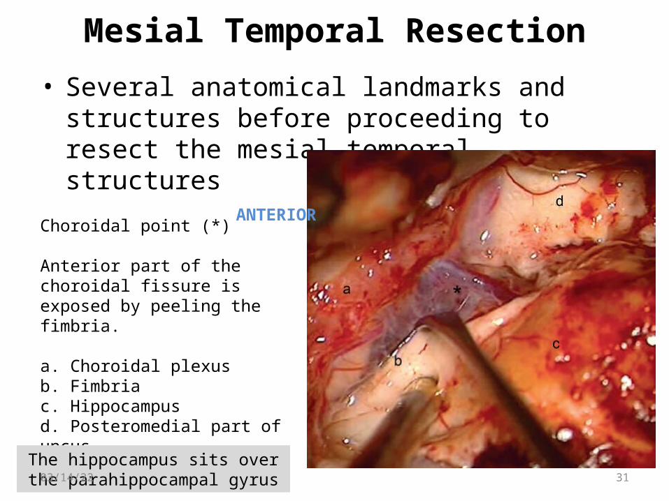
Mesial Temporal Resection
• Several anatomical landmarks and structures before proceeding to resect the mesial temporal structures
Choroidal point (*)
Anterior part of the choroidal fissure is exposed by peeling the fimbria.
a. Choroidal plexusb. Fimbriac. Hippocampusd. Posteromedial part of uncus
ANTERIOR
The hippocampus sits over the parahippocampal gyrus04/18/23 31

04/18/23 32

• The anterior portion of the hippocampal head blends into the posterior uncus and amygdala
The medial border of the hippocampus is lined by the choroid plexus over the
choroidal fissure and the choroidalpoint at the most anterior part.
The anterior choroidal artery (AChA) runs across the ambient and crural cisterns near the choroid plexus.
It pierces the arachnoid plane to supply the choroid plexus at the inferior choroidal point by giving rise to numerousbranches.
04/18/23 33

• The temporal horn is fully unroofed to expose the most anterior part of the temporal horn that includes the bulging amygdala, posterior uncus, amygdala– hippocampal junction, and posteriorly the head and body of the hippocampus.
Choroidal point (*) choroidal plexus .aFimbria .bvelum terminale .c stria terminalis .dhead of hippocampus (e)
04/18/23 34

• After locating the intraventricular landmarks, resection of mesial temporal structures starts with an incision on the lateral ventricular sulcus that is the demarcation line between the collateral eminence and hippocampus
a. collateral eminenceb. fimbria
04/18/23 35

• The ependyma of the lateral ventricular sulcus is coagulated posteroanteriorly as an entry point to the parahippocampal gyrus.
• Medial pial bank of the collateral sulcus is exposed by suctioning parahippocampus intra gyrally
• Completed along the collateral eminence, starting from hippocampus proper to the amygdala– hippocampal junction
04/18/23 36

04/18/23 37

• Entire hippocampus is subpially dissected between collateral eminence and fimbria.
The parahippocampus– hippocampus complex is subpially dissected by
peeling it from the collateral sulcus pia using the Penfield dissectors
04/18/23 38

• Then the dissection continues medially toward the tentorial edge until the pia along the medial border of the parahippocampus and hippocampal sulcus is seen.
• At this stage, the subiculum of the hippocampus is peeled off toward the hippocampal sulcus.
Fimbria is lifted with a dissector to expose the hippocampal sulcus and
hippocampal arteries
Further dissection and elevation of fimbria (a) exposes subiculum (b)
and hippocampal arteries extending into
hippocampal sulcus.
.Subiculum and hippocampal sulcus
are fully exposed, and hippocampal arteries
have been coagulated.
04/18/23 39

• The hippocampus proper can easily be retracted laterally into the cavity created by intragyral aspiration of the parahippocampus
• The hippocampal sulcus fans out at this junction between pes hippocampi, uncus and anterior end of the parahippocampus
This anatomy provides the surgeon with an excellent starting point for the dissection of hippocampal sulcus between fimbria,
inferior choroidal point, and choroidal fissure.04/18/23 40

Uncus Being taken out Residual part of amygdala
Tentorial edge / 3rd nerve / PCA04/18/23 41

Complications
• Intraoperative
• Immediate post-operative
• Longterm
04/18/23 42

04/18/23 43
• The most commonly reported complications visual field defects infection stroke manipulation or retraction hemiparesis third nerve palsy language disturbances.

• Superior quadrantanopsia - incidence of 35% to 50%
• 4% incidence of homonymous hemianopia
• Geniculocalcarine fibers (Meyer’s loop)
• Roof of the temporal horn of the lateral ventricle
04/18/23 44
Horizontal and vertical diplopias resulting from irritation to the third and fourth cranial nerves

• Transient dysphasia- dominant site temporal resections (resolves within a few weeks)
Retraction related / Disconnection of the medial lobe from the neocortex
• Although rare, third and fourth nerve palsies can be seen after AMTL
04/18/23 45

• One of the most devastating, although rare, complications in temporal lobe resection is hemiplegia.
• It is a well-recognized complication, with an incidence of 1 to 2%.5
• Manipulation hemiplegia and frequently is related to injuries of the AChA and PCA during the resection of the medial temporal structures.
04/18/23 46

• Preoperative and postoperative cognitive assessment is important to help counsel the patient on existing deficits and to predict and document the effects of temporal lobectomy on memory function.
• Preoperative risk factors for worsening memory function after surgery include
dominant TLE, lack of hippocampal sclerosis, normal baseline neuropsychological function, later age of onset of the epilepsy
04/18/23 47
MEMORY AND FUNCTIONAL DEFICIT

• Dominant temporal lobe surgery predisposes the patient to a decline in verbal memory and short-term memory functioning.
• The risks associated with nondominant temporal lobe surgery are less predictable
04/18/23 48

• Temporal lobe resection is effective surgical technique in the management of TLE with reported seizure control rates being 60 and 80%
• The best outcome was seen in the patients with temporal lobe neoplasms (88–92%) followed by the patients with gliosis (86%) and MTS (70%) in Benifla’s series.
04/18/23 49
Benifl a M, Otsubo H, Ochi A, et al. Temporal lobe surgery for intractable epilepsy in children: an analysis of outcomes in 126 children. Neurosurgery 2006;59(6):1203–1213, discussion 1213–1214
OUTCOME

• Thank You
04/18/23 50